Université de Montréal
Directed evolution of human dihydrofolate reductase:
towards a better understanding of binding
at the active site
par Elena Fossati
Département de biochimie Faculté de médecine
Mémoire présenté à la Faculté des études supérieures en vue de l’obtention du grade de
Maîtrise en biochimie
Novembre, 2008
Université de Montréal Faculté des études supérieures
Ce mémoire intitulé:
Directed evolution of human dihydrofolate reductase: towards a better understanding of binding at the active site
présentée par : Elena Fossati
a été évaluée par un jury composé des personnes suivantes :
Michel Bouvier, président-rapporteur Joelle N. Pelletier, directeur de recherche
iii
Résumé
La dihydrofolate réductase humaine (DHFRh) est une enzyme essentielle à la prolifération cellulaire, ce qui en fait une cible de choix pour le traitement de différents cancers. À cet effet, plusieurs inhibiteurs spécifiques de la DHFRh, les antifolates, ont été mis au point : le méthotrexate (MTX) et le pemetrexed (PMTX) en sont de bons exemples. Malgré l’efficacité clinique certaine de ces antifolates, le développement de nouveaux traitements s’avère nécessaire afin de réduire les effets secondaires liés à leur utilisation. Enfin, dans l’optique d’orienter la synthèse de nouveaux composés inhibiteurs des DHFRh, une meilleure connaissance des interactions entre les antifolates et leur enzyme cible est primordiale.
À l’aide de l’évolution dirigée, il a été possible d’identifier des mutants de la DHFRh pour lesquels l’affinité envers des antifolates cliniquement actifs se voyait modifiée. La mutagenèse dite de saturation a été utilisée afin de générer des banques de mutants présentant une diversité génétique au niveau des résidus du site actif de l’enzyme d’intérêt. De plus, une nouvelle méthode de criblage a été mise au point, laquelle s’est avérée efficace pour départager les mutations ayant entrainé une résistance aux antifolates et/ou un maintient de l’activité enzymatique envers son substrat natif, soient les phénotypes d’activité. La méthode de criblage consiste dans un premier temps en une sélection bactérienne à haut débit, puis dans un second temps en un criblage sur plaques permettant d’identifier les meilleurs candidats. Plusieurs mutants actifs de la DHFRh, résistants aux antifolates, ont ainsi pu être identifiés et caractérisés lors d’études de cinétique enzymatique (kcat et IC50). Sur la base de ces résultats cinétiques, de la modélisation moléculaire et des données structurales de la littérature, une étude structure-activité a été effectuée. En regardant quelles mutations ont les effets les plus significatif sur la liaison, nous avons commencé à construire un carte moléculaire des contacts impliqués dans la liaison des ligands. Enfin, des connaissances supplémentaires sur les propriétés spécifiques de liaison ont put être acquises en variant l’inhibiteur testé, permettant ainsi une meilleure compréhension du phénomène de discrimination du ligand.
Mots clés : dihydrofolate réductases humaine, méthotrexate, pemetrexed, résistance aux
médicaments, mutagenèse, évolution dirigée, criblage à haut débit, relation structure-fonction, cinétique enzymatique, modélisation moléculaire.
v
Abstract
Human dihydrofolate reductase (hDHFR) is an essential enzyme for cellular proliferation and it has long been the target of antifolate drugs for the treatment of various types of cancer. Despite the clinical effectiveness of current antifolate treatments, new drugs are required to reduce the side-effects associated with their use. An essential requirement for design of new antifolates is a better understanding of how these drugs interact with their targets.
We applied directed evolution to identify mutant hDHFR variants with modified binding to some clinically relevant antifolates. A saturation mutagenesis approach was used to create genetic diversity at active-site residues of hDHFR and a new, efficient screening strategy was developed to identify the amino acids that preserved native activity and/or conferred antifolate resistance. The screening method consists in a high-throughput first-tier bacterial selection coupled with a second-tier in vitro assay that allows for rapid detection of the best variants among the leads, according to user-defined parameters. Many active, antifolate-resistant mutants of hDHFR were identified. Moreover, the approach has proven efficient in rapidly assessing kinetic (kcat) and inhibition parameters of the hDHFR variants (IC50). Structure-function relationship analysis based on kinetic investigation, available structural and functional data as well as modeling were performed. By monitoring which mutations have the greatest effect on binding, we have begun to build a molecular picture of the contacts involved in drug binding. By varying the drugs we test against, we gain a better understanding of the specific binding properties that determine ligand discrimination.
Keywords : human dihydrofolate reductase, methotrexate, pemetrexed, drug-resistance,
saturation and combinatorial mutagenesis, directed evolution, high-throughput screening, structure-function relationship, enzyme kinetics, molecular modeling.
Table of contents
Chapter 1 - Introduction ... 16
Section 1.0 - Folate metabolism ... 16
Section 1.1 - Dihyrofolate reductase... 17
Section 1.1.1 - Reaction mechanism... 17
Section 1.1.2 - Vertebrate and bacterial DHFRs: differences and similarities ... 18
Section 1.1.3 - Human dihydrofolate reductase... 19
Section 1.2 – Antifolates... 20
Section 1.2.1 - Methotrexate... 21
Section 1.2.2 - Other antifolates ... 28
Section 1.3 – Drug discovery and structure-activity relationship analysis... 30
Section 1.4 – Presentation of the research project ... 31
Section 1.5 – Presentation of the experimental approach... 33
1.5.1 - Directed evolution ... 33
1.5.2 – Structure-function relationship analysis ... 35
Chapter 2 (Article 1) – Increasing methotrexate resistance by combination of active-site mutations in human dihydrofolate reductase... 47
Section 2.0 – Preface... 47
ABSTRACT... 50
INTRODUCTION ... 51
RESULTS ... 53
DISCUSSION ... 58
MATERIALS AND METHODS. ….. ………..63
REFERENCES………...79
ACKNOWLEDGMENTS ... 85
Chapter 3 (Article 2) – Two-tier bacterial and in vitro selection of active and methotrexate-resistant variants of human dihydrofolate reductase ... 87
vii
Section 3.0 – Preface... 87
ABSTRACT... 90
INTRODUCTION ... 90
MATERIALS AND METHODS... 92
RESULTS AND DISCUSSION ... 97
CONCLUSIONS... 107
REFERENCES ... 116
ACKNOWLEDGMENTS ... 118
Section 3.2 - Erratum corrigendum... 119
Chapter 4 – Binding of fragments of MTX to hDHFR... 120
4.0 - Preface ... 120
4.1 - Introduction ... 120
4.2 - Materials and methods... 121
4.2.1 Reagents and enzymes ... 121
4.2.2 Determination of binding parameters ... 122
4.3 Results and discussion ... 122
Chapter 5 – Docking for structure-function relationship analysis... 127
5.0 Preface... 127
5.1 Introduction... 127
5.2 Materials and Methods... 128
5.2.1 In silico automated docking of FOL, DHF, MTX and PMTX ... 128
5.3 Results and Discussion ... 129
5.3.1 Docking of MTX... 129
5.3.2 Docking of FOL... 130
5.3.3 Docking of DHF ... 130
5.3.4 Docking of PMTX ... 131
Chapter 6 – Conclusions and perspectives ... 138 References... 141
List of tables
Chapter 1
Table 1.1. Active-site interactions between hDHFR and folate or methotrexate……….37 Table 1.2. Mutations providing MTX-resistance in hDHFR…….………..…….38 Chapter 2
Table 2.1. Amino acids encoded at residues 31, 34 and 35 of the hDHFR ………...…..70 Table 2.2. Kinetic and inhibition constants for the selected MTX-resistant hDHFR
mutants.………..71
Table 2.3. EC50 MTX for CHO DUKX B11 cells transfected with MTX-resistant hDHFR mutants……….….…….72
Table 2.4. Comparison of MTX-resistant hDHFR mutated at positions F31 and F34….73 Chapter 3
Table 3.1. Reactivity (kcat) and MTX or PMTX resistance determined in 96-well plates using crude lysates of active hDHFR variants from library 115……….…109
Table 3.2. Kinetic and inhibition constants a of purified MTX or PMTX-resistant hDHFR mutants………...110
Chapter 4
Table 4.1. Inhibition constantsa of MTX and DAMPA for WT and selected MTX-resistant hDHFR mutants………...125
Chapter 5
ix
List of figures
Chapter 1
Figure 1.1. Folate-dependent metabolic reactions………..………..39
Figure 1.1a: Synthesis of thymidylate (dTMP) from deoxyuridine monophosphate (dUMP)………...……….39a Figure 1.1b: Role of the folate cofactor 10-formyl tetrahydrofolate (10-CHO-THF) in the synthesis of the inosine nucleus……… 39b Figure 1.2. Reaction catalyzed by dihydrofolate reductase (DHFR)………...…40
Figure 1.3. Sequence and structural comparison of human, murine and E. coli dihydrofolate reductase (DHFRs)………..41
Figure 1.4. Secondary structure of hDHFR……...………...42
Figure 1.5. Ligand binding at the active siteof WT hDHFR………....43
Figure 1.6. Binding of folate and methotrexate in the active site…...………..44
Figure 1.7. Structural representation of the DHFR substrate dihydrofolate (DHF) and different antifolates relevant to this study……….45
Figure 1.8. Acive-site residues of hDHFR targeted by mutagenesis………46
Chapter 2 Figure 2.1. Ligand binding at the active site of WT hDHFR………....74
Figure 2.2. Frequency of occurrence of the novel MTX-resistant mutants………...75
Figure 2.3. Relation between the number of hDHFR mutations and kcat/KMDHF or KiMTX relative to WT His6-hDHFR………76
Figure 2.4. Survival of CHO DUKX B11 cells transfected with selected mutants in presence of MTX………77
Figure 2.5. In silico comparison of MTX binding between (A) WT hDHFR (1U72) and (B) mutant AVH………78
Chapter 3
Figure 3.1. Structures of folate (1DRF.pdb) and MTX (1U72.pdb) bound to hDHFR
active site……….111
Figure 3.2. Flow-chart of the two-tier strategy to select mutated hDHFR library variants for catalytic activity or methotrexate (MTX) resistance………..112
Figure 3.3. Comparison of hDHFR mutations that allow for conservation of activity or MTX resistance on the basis of the two-tier selection strategy………...113
Figure 3.4. The two-tier selection results for library 115………...114
Figure 3.5. IC50MTX concentration-response curves……….115
Chapter 4 Figure 4.1. Comparison of binding of MTX and its fragment DAMPA in the double mutant F31R/Q35E (RFE) and the corresponding single mutants F31R and Q35E…...126
Chapter 5 Figure 5.1. Docking of MTX into WT hDHFR⋅NADPH………...134
Figure 5.2. Docking of FOL into WT hDHFR⋅NADPH……….135
Figure 5.3. Docking of DHF into WT hDHFR⋅NADPH……….…...136
xi
List of abbreviations
AICARFT Aminoimidazole carboxamide formyl transferase
Amp Ampicillin
DHF Dihydrofolate
DHFR Dihydrofolate reductase E.C. Enzyme commission number E. coli Escherichia coli
ecDHFR Escherichia coli dihydrofolate reductase FBP Folate binding protein
FOL Folate
FPGS Folypolygluatamate synthase
GARFT Glycinamide ribonucleotide formyl transferase hDHFR Human dihydrofolate reductase
IC50 Half maximal effective concentration for enzyme inhibition IPTG Isopropyl β-D-1-thiogalactopyranoside
kcat Catalytic constant (turnover number) kcat/KM Catalytic efficiency
KD Binding constant
Ki Inhibitor constant
KM Affinity constant (Michaelis-Menten constant)
koff Dissociation rate
kon Association rate
LB Luria-Bertani
NADP+ Oxidized nicotinamide adenine dinucleotide phosphate NADPH Reduced nicotinamide adenine dinucleotide phosphate
PDB Protein data bank
RCF Reduced folate carrier RMSD Root mean square deviation SAR Structure-activity relationship
THF Tetrahydrofolate
TMP Trimethoprim
TMTX Trimetrexate
xiii
Acknowledgements
After three years at the UdeM, here I am, finally finishing my Master degree, thinking about everything that has happened and about all the people I have met. Despite the sadness that comes with the choice of abandoning a project I know and I like for many new and still uncertain ones, I am excited and full of hope for my future. In fact, during my time at the UdeM I have grown as a scientist and as a person, and most of all I had the opportunity to meet a lot of interesting people and I feel I found another home in Québec.
First of all, I would like to thank my supervisor, Prof. Joelle Pelletier, for being such a dynamic and inspiring scientist. Joelle pushed me to my limits during my first and most difficult year, but she balanced this with compassion and patience, especially when my son arrived and when all the problems with the train de banlieue started! Her support, supervision and encouragement have been very precious, and her cakes and biscuits very appreciated!
Thanks to all the hDHFR gang. First of all to Jordan Volpato, for his scientific supervision and for making great Italian-like environment in the lab. Jordan is not only a brilliant scientist but an amazing communicator and…amazingly…he also knows the genetic code by heart, which may be useless, but it is certainly very impressive! Thanks to Lucie Poulin, who never missed either a sequencing gel or a chocolate cake! Lucie and I not only shared a bench but also a precious friendship. Thanks to Vanessa Guerrero for her good work and her flexibility around my ‘not so flexible’ working schedule…and for her desserts, obviously! Thanks also to Jonathan Blanchet and Mirja Krause.
Thanks to all the past and present members of the ‘coffee club’, first of all to Nicolas Chabot, the president! Thanks for les vins et fromages, les Ricards, les grains de chocolat, and the delicious ‘saltinbocca alla romana’ receipe (imagine a French guy who gives to an Italian an Italian receipe! I know, I should have kept this a secret!). If it is true that real friends are rare and precious…bhe…Nico is certainly one of them. He was always there to listen and cheer me up me when I was afraid, sad, or disappointed and to celebrate all the good moments. Thanks Nico! Thanks to Krista Morley, Audrey Nisole,
xv
Natalhie Campos Reales, Karine Caron and Rosine Pelletier for the dinners, laughs, long discussions, and for being good friends! Thanks to Christopher Clouthier for the discussion about science and life and for always making me laugh!
Thanks to all the other member of the Pelletier and the Keillor groups: Pierre-Yves De Wals (PY), Roberto Chica, Valerio Vinci, Claudio Gnaccarini, Natalia Kadnikova, Isabelle Roy, Julia Guy and Christophe Pardin. Thanks in particular to Nicolas Doucet, an extraordinary scientist with a deep love for knowledge, the ability of excel in everything he does and his natural gift of being a great pedagogue. Nicolas never hesistated to interrupt what he was doing to help or explain something to me. Thanks to Prof. Jeffrey Keillor, for being always very attentive, generous, and for being a great teacher.
Un ringraziamento speciale va ai miei genitori e a mio fratello Marco, per avermi continuamente incoraggiata nei miei progetti e per il loro amore incondizionato. Grazie di cuore anche ai miei nonni, Oreste e Ines, e ai miei zii, Rosa, Caludio e Mauro, per avere fatto razzia delle carte prepagate per chiamare il Nord America e per il loro affetto. Grazie anche a Sara, Maria, Cristina, Alessandra, Gonzalo, Andrea e Mattia, perche’ hanno saputo dimostrarmi il loro affetto e la loro amicizia nonostante la distanza e le mie a volte troppo sintetiche e-mails.
Thanks to Maxime, my fantastic son, whose smiles, hugs and ‘mama’ are worth more than any degree. Finally, a heartfelt thanks to Dominique, my husband, for his love, support and continuous encouragement, and without whom I probably would never had come to Quebec and all this would never had happened.
Chapter 1 - Introduction
Section 1.0 - Folate metabolism
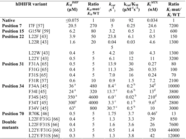
Folic acid derivatives are essential coenzymes required by all living organisms in the de novo synthesis of thymidylate and purines, the building blocks for nucleic acid synthesis (figure 1.1, 1.1a and 1.1b) [1]. Thus, these cofactors have a crucial role during cell proliferation. Folate derivatives, in the form of a series of tetrahydrofolate (THF) compounds, act as cofactors in a number of one-carbon-transfer reactions within these biosyntheses, and they are also involved in homocysteine methylation and glycine and serine interconversion (figure 1.1) [1]. THF is obligatorily produced from 7,8-dihydrofolate (DHF) by the enzyme 7,8-dihydrofolate reductase (DHFR) [2]. Enzymes depending on THF cofactors include thymidylate synthase (TS), glycinamide ribonucleotide formyl transferase (GARFT) and aminoimidazole carboxamide formyl transferase (AICARFT). TS uses the cofactor 5,10-methylene tetrahydrofolate to convert deoxyuridylate (dUMP) into thymidylate (dTMP)(figure 1.1a) [2]. GARFT and AICARFT add carbon 8 and 2, respectively to the ring structure of purine using N10
-formyl tetrahydrofolate as cofactor (figure 1.1b)[2].
Folates are mainly transported into cells by two energy-dependent carrier proteins: the reduced folate carrier (RFC) [3], which is bi-directional, and the folate binding protein (FBP) [4]. Intracellular concentration of folates is constantly maintained between 1 and 10 μM, compared to a plasma concentration of 10 to 30 nM [5]. This intracellular accumulation is mediated by polyglutamation. The enzyme folypolyglutamate synthase (FPGS) [1] adds 5 to 8 glutamate residues to the glutamate tail of folates via peptidic bonds. The enzyme γ-glutamyl hydrolase (GGH) [6], instead, removes terminal glutamate groups. Glutamation makes folates more polar and it can increase the affinity of folates for their target enzymes. The extent of polyglutamation of folates depends on their affinity for these two cytosolic enzymes.
17
Folate metabolism in cells is a dynamic process in which the levels of THF-cofactors and DHF vary with the intracellular activities. In resting cells (not in S-phase), DHFR activity is much higher than TS activity. This maintains cellular DHF at very low levels, in the range of the low nM [1, 7]. As the KMDHFfor DHFR is < 75 nM [8], only a fraction of DHFR activity (< 5%) is sufficient to sustain normal rates of THF synthesis [1]. Inhibition of folate-dependent enzymes in actively proliferating cells leads to arrest of the synthesis of DNA precursors. Thus, these enzymes are major drug targets for the treatment of cancer diseases [5], as well as fungal [9, 10], microbial [11] and parasitic [12] infections that are dependent on cellular proliferation.
Section 1.1 - Dihyrofolate reductase
Dihydrofolate reductase (tetrahydrofolate: NADP+ oxidoreductase; E.C.C. 1.5.1.3) is a cytosolic enzyme that catalyzes the NADPH-dependent reduction of 7,8-dihydrofolate (DHF) to 5,6,7,8-tetrahydrofolate (THF) in all living organisms (figure
1.2A) [1]. Vertebrate DHFRs also catalyse the reduction of folate (FOL) to DHF, at about
one tenth the rate of DHF reduction (figure 1.2A) [1].
DHFR has attracted the attention of protein chemists and of molecular biologists as a model in many structural, kinetic and mutagenic studies due not only to its clinical relevance as a pharmacological target, but also to its small size (18-22 kDa), stability and relative ease of producing the recombinant enzyme. In fact, eukaryotic DHFRs are small monomeric proteins which does not require any post-translational modification and thus, can be easily expressed in heterologous bacterial hosts for ease of manipulation [13]. The human dhfr gene, for example, was cloned and expressed in E. coli in 1988 [14], and since then, extensive investigations of this enzyme have been performed.
Section 1.1.1 - Reaction mechanism
For the reduction of DHF to THF the hydride is transferred from the C4 of NADPH to the C6 of DHF, and this transfer is critically dependent on the distance between the two carbon atoms (optimal distance 2.6 Å) and the relative orientation of the NADPH nicotinamide ring and the DHF pteridine ring (figure 1.2B) [15]. Different studies [16-18] suggest that protonation of N5 in the transition state, which either
immediately precedes or is concerted with hydride transfer to the C6, promotes hydride transfer by delocalizing a positive charge to C6. Residue 30 is the catalytic active-site residue, which mediates, through intervening water molecules, the proton transfer component of the reduction [15, 19]. A similar mechanism has been proposed for the DHFR-dependent reduction of FOL to DHF, where protonation of N8 could be promoted by formation of a H-bond with the backbone carbonyl of Ile7, as observed in the crystal of the binary complex of human DHFR with 5-deazafolate, a tight-binding inhibitor of DHFR very similar to DHF [18].
Section 1.1.2 - Vertebrate and bacterial DHFRs: differences and
similarities
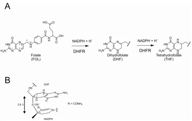
Vertebrate DHFRs are highly homologous (72-89% sequence identities for DHFRs from chicken, mouse, bovine and human), but only ~25% identity is observed between animal and bacterial sequences or between different species of bacteria (figure
1.3) [20]. Despite the low sequence identity between bacterial and vertebrate DHFRs,
structural analysis of DHFRs from different species has shown that the overall tertiary structures of these enzymes are very similar to one another (figure 1.3) [21, 22].
Notwithstanding the great structural similarity observed, this group of enzymes exhibits considerable species-to-species variability in sensitivity to different inhibitors [21]. E. coli DHFR (ecDHFR), for example, is 12000-fold more sensitive to the antifolate trimethoprim (TMP) than the human variant (hDHFR). Although active site residues are generally conserved, some differences exist and could partly explain this variability. Leu28 in E. coli DHFR, for example, which was reported to establish a major contact with the trimethoxybenzyl moiety of TMP [23], corresponds to Phe31 in hDHFR. However, mutation of Phe31 to Leu in hDHFR, did not increase TMP binding significantly, indicating that this residue was not the sole determinant of species selectivity of TMP [24]. Differences in active-site cavity were also reported to contribute to the observed differences in specificity [22, 25]. Despite extensive investigation, the structural basis for the various modes of binding in DHFR from different species still remains to be fully understood and it represents a crucial point for the design of
species-19
specific inhibitors [26]. Jordan Volpato, PhD student in the research group of Prof. Joelle Pelletier, has recently conducted a detailed review regarding this subject [27].
Section 1.1.3 - Human dihydrofolate reductase
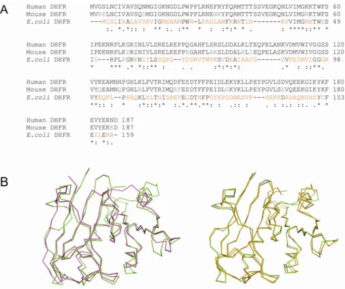
Human DHFR (hDHFR) is a monomeric enzyme of 186 amino acids (21544 Da). The polypeptide backbone of hDHFR is folded into an eight-stranded twisted β-sheet, consisting of 7 parallel strands and one anti-parallel strand leading to the carboxyl terminus. Five α-helices and loops provide connectivity within the sheets (figure 1.4) [28]. Residues 21-26 (sequence DLPWPP) form two turns of a polyproline helix (left handed, typeII) (figure 1.4) [28].
Section 1.1.3.1 – Folate binding site
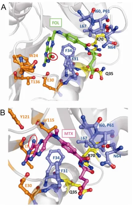
The structures of many complexes of hDHFR with cofactors, substrate or inhibitors have been determined both by X-ray diffraction methods [18, 22, 28, 29] and by NMR [30, 31]. Of the two substrates of hDHFR, only FOL has been crystallized in the binding cavity of the enzyme (in absence of the cofactor NADPH) (figure 1.5A) [18, 28]. FOL is a relatively stable molecule while DHF is readily oxidized to FOL, and therefore not suitable for protein co-cristallization [18]. The folate binding site is composed by residues in strands βA and βE, residues 61-70 and residues belonging to the left-handed polyproline helix and helices αB, αC [18].
The folate and folate-like molecules consist of polar pteridine and L-glutamate extremities, linked by a p-aminobenzoyl group (p-ABA) (figure 1.7). The active site of DHFR is a ~15 Å hydrophobic pocket in which the only polar side chain is the carbonyl of Glu30 [15]. The pteridine moiety of substrates and inhibitors binds nearly perpendicular to the benzoyl ring in the bottom of the hydrophobic pocket, with the benzoyl-glutamate side chain directed towards the surface of the protein. NADPH binds in an extended conformation with the reduced nicotinamide ring inserted into the active site pocket and the rest of the molecule along the surface of the protein. The nicotinamide ring is situated about 2.5 Å from the folate pteridine ring, with which it forms stacking interactions [18].
Table 1.1 lists the residues of hDHFR that directly interact with DHF. The
2-amino group, N3 and O4 of the pteridine moiety of folate interact specifically with residues Glu30, Thr136 and Trp24 by direct or water mediated H-bonds. Phe34 is within the van der Waals distance of the pteridine moiety. No specific interactions involve nitrogens N1, N5, N8 and N10 of folate. In DHF, which has a hydrogen atom attached to N8, a hydrogen bond between N8 and the carbonyl oxygen of Ile7 is likely to form, whereas an equivalent hydrogen bond to N8 of folate is likely to be formed only in the transition state [18].
The p-ABA moiety of folate is within van der Waals distance of the side chains of Phe31, Phe34, Ile60, Pro61 and Leu67. Moreover, the carbonyl oxygen of p-ABA forms a H-bond with the amide nitrogen of the Asn64 side chain [28].
The α-carboxylate portion of the glutamate fragment forms a salt bridge with the guanidinium of the conserved Arg70. Gln35 is in proximity to both the α-carboxylate and γ-carboxylate portions of DHF. The γ-carboxylate can form a H-bond mediated by a water molecule with the carbonyl oxygen of R28. However, this portion has a high B factor and the bond with R28 is not observed in all resolved structures, probably indicating that it is weak [18].
Section 1.2 – Antifolates
Antifolates constitute a large family of compounds which compete with folate derivatives for the binding to folate-dependent enzymes involved in nucleotide biosynthesis. Due to the role of their target enzymes in cellular proliferation, antifolates are used for the treatment of a broad range of proliferative diseases. Treatment of bacterial and parasitic infection is based on species-selectivities of some of these compounds. Trimethoprim (TMP), for example, is selective towards bacterial DHFRs [26] while pyrimethamine (PYR) towards malaria parasite DHFRs [26].
The importance of DHFR in bacterial, parasitic and cancer chemotherapy arises from its function in maintaining the pool of THF. Inhibition of DHFR leads to arrest of DHF recycling, and thus causes inhibition of cell growth and eventually cell death. It
21
should be noted that THF is regenerated in most one-carbon transfer reactions, with the exception of TS-catalyzed dTMP synthesis (figure 1.1). Therefore, in cells not actively synthesizing thymidylate and DNA, inhibition of DHFR does not result in any particular effect.
In 1947, pteroylaspartic acid, an antagonist of pteroyl glutamic acid (folic acid), was proved to interfere with folic acid metabolism and the normal growth of cells in in vivo test with both chicken and rats [32], and suggested that folic acid antagonists might be of value in patients with rapidly growing malignant disease. In 1948 the antifolate aminopterin (figure 1.7) was shown to be effective in children affected by acute leukemia at the terminal stage of the disease, marking the advent of cancer chemotherapy [33]. Soon after, aminopterin was replaced by the more effective and less toxic amethopterin (later named methotrexate (MTX), figure 1.7), which was approved by FDA in 1953. Since then, MTX has been the major antifolate used in cancer therapy [34].
Both aminopterin and MTX are strong competitive inhibitors of DHFR (for hDHFR, Ki aminopterin = 2 pM [35] and Ki MTX = 3.4 pM [13]).
Section 1.2.1 - Methotrexate
Methotrexate (MTX; 8-amino,10-methyl-pteryolglutamic acid) is a slow, tight binding, reversible inhibitor of the human enzyme dihydrofolate reductase (hDHFR) [13, 36]. The inhibition constant of hDHFR for MTX is of 3.4 pM [13, 36, 37]. MTX also inhibits human GARFT and human AICARFT, but the Ki values for the pentaglutamate forms of these enzymes (potency of DHFR inhibition depends on the polyglutamation status of the molecules) are of 2500 nM and 56 nM, respectively [5]. These kinetic data suggest that the main intracellular target is DHFR [5].
MTX shows high affinity for a wide range of DHFRs from different species (bacterial, parasitic, vertebrate). Due to its lack of specificity, it is mostly applied for the treatment of human proliferative diseases, both malignant (carcinoma, metastatic breast cancer, bladder cancer, lymphoma)[5] and not-malignant (rheumatoid arthritis [38], psoriasis [39] and graft versus host disease [40]). Despite the continuous discovery of
newer drugs, MTX often remains a component of the newer combination treatments [5, 34].
The kinetics of MTX binding to hDHFR was described extensively by Appleman et al. [13]. MTX binds rapidly and tightly (kon = 1.0 x 10⁻⁸ M-1 s-1; koff < 1 s-1; koff/kon = 210 pM), independently of the presence or absence of NADPH already bound to the enzyme [13]. The initial association of MTX to hDHFR is followed by some kind of conformational change of the complex, which increases the overall binding of 60-fold, leading to a Ki of 3.4 pM [13]. The authors also demonstrated that MTX polyglutamation, differently from what happens for TS, GARFT and AICARFT [5], does not affect binding to hDHFR. Like folate, MTX is transported into cells by both RFC and FBP [5, 41, 42].
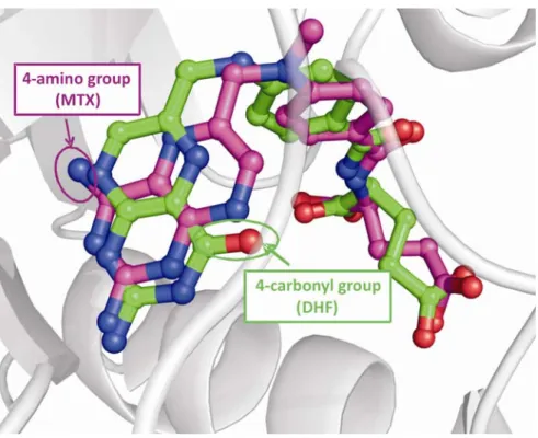
Binding of MTX to hDHFR has been studied using X-ray crystallography [22, 43] (table 1.1 and figure 1.5B). Despite its chemical and steric similarity with 4-oxo-folates (FOL, DHF, THF; figure 1.7), MTX binds in the active site with its pteridine moiety flipped 180° around the C6-C9 bond,relative to folate (figure 1.6). The main cause for
this flip is probably the presence of the 4-amino group instead of a 4-oxo group. Consequently, active-site residues form different contacts with the two molecules, and binding of the inhibitor MTX is 2000-fold stronger than binding of the substrate DHF.
Table 1.1 lists the residues that directly interact with DHF and/or MTX in the
hDHFR ternary complex. The hydrogen bonding network involving structural water, the conserved active site residues Thr136, Glu30, Trp24 and the pteridine moiety of the bound folate is maintained, but in the case of MTX it involves the N1 and N8 nitrogen and the 2-amino group [22]. The 4-amino group of MTX interacts with residues Ile7, Val115 and Tyr121 and with NADPH [22]. The backbone carbonyl groups of both residue 7 and 115 are within H-bonding distance with the 4-amino group of MTX [44]. Moreover, the side chains of these two residues are likely to form hydrophobic interactions with the inhibitor’s pteridine moiety [44]. Residue Phe34 is also within the van der Waals distance with the pteridine moiety.[45]
As in the case of folate, the p-ABA moiety of MTX forms van der Waals and hydrophobic interactions with the side chains of Phe31, Phe34, Ile60, Pro61 and Leu67
23
[22]. The carbonyl oxygen of p-ABA also forms a H-bond with the amide nitrogen of the Asn64 side chain [22].
The α-carboxylate portion of the glutamate moiety makes close contacts with the side chains of Arg70 (charge interaction) and Gln35. When the inhibitor does not occupy the active site, a subdomain shift brings Arg70 into contact with Gln35 [46].
Section 1.2.1.1 - Mechanism of resistance to MTX
Some type of neoplastic diseases are intrinsically resistant to MTX whereas those that are responsive can develop resistance following repeated treatments (acquired resistance) [42]. MTX was brought into clinical use and became an established component of many clinical regimens right after the demonstration of its effectiveness in the late 1940s [33, 47], when its mechanism of action was not clearly understood. Between the end of the 1970s and the beginning of the 1990s, a better understanding of metabolism, transport and kinetics of binding of MTX to DHFR helped to elucidate the basis of MTX resistance. Different mechanisms of acquired resistance that impair efficiency of MTX-treatment have been described [5, 41, 42]. Amplification of the dhfr gene [48] or DHFR over-expression [49] were frequently observed. Moreover, mammalian DHFR expression is regulated by binding of its own mRNA at the active site [50]. In fact, hDHFR binds specifically to its own mRNA and this interaction represents a mechanism of inhibition of hDHFR mRNA translation [50]. Binding of MTX dislodges the DHFR mRNA, rendering it available for translation and therefore increasing protein expression. The higher the level of DHFR, the higher is the level of free-drug necessary to suppress THF regeneration. The free drug level is a critical parameter in the interactions between MTX and both hDHFR and folylpolyglutamate synthetase (FPGS; see section 1.0 for a detailed description of the carrier proteins and enzymes involved in folate metabolism). Transport mechanisms regulate the influx and efflux of drugs across the cell membrane, and the net effect of these processes determines the level of free MTX achieved intracellularly. Alteration in the expression or mutations of the reduced folate carrier protein (RFC) [51] and of FPGS [52] determine reduction of cellular uptake or allow more rapid efflux of MTX [41], respectively. MTX polyglutamates accumulate at different rates in a variety of tumor cells and ultimately become the predominant species
and the form of antifolate bound to hDHFR, and this is an important determinant of the pharmacologic activity of this drug. Impaired polyglutamation can therefore be a determinant cause of resistance together with the other possible resistance mechanisms.
The above-mentioned MTX resistance mechanisms have all been observed either in tumours isolated from patients that relapsed during or following MTX-treatment, and ex vivo (by exposing cells isolated from patients to MTX in vitro) [48, 53-55]. An additional resistance mechanism is the occurrence of mutations in the dhfr gene, resulting in expression of hDHFR variants with reduced affinity for MTX. This mechanism of resistance was first observed in ex vivo studies, but it has never been identified as a cause of resistance in tumours isolated from patients who relapsed following MTX treatment. Even if it has been long proposed that mutations that confer resistance to MTX could occur in tumural cell of patients receiving a MTX-based therapy, and therefore contributing to their clinical relapse, a study performed by Spencer et al. described why this is unlikely to happen [56]. Briefly, the authors demonstrated that different mutations that occurred spontaneously and conferred MTX-resistance in ex vivo experiments, conferred only modest protection to MTX when transduced into cells using a retroviral vector. Moreover, accumulation of mutations that increase total MTX resistance seems unlikely because other mechanisms of resistance, as discussed above, prevail. Because it does not occur clinically, hDHFR MTX-resistant mutants represent a promising tool for gene theraphy, as it will be discussed in paragraph 1.2.1.3. Moreover, a second possible application would involve the use of these hDHFR MTX-resistant variants as selection markers for gene transfer into eukaryotic cells [57, 58].
Section 1.2.1.2 - Mutations of hDHFR that confer resistance to MTX
MTX-resistant mutants of DHFR from mammalian sources have been identified either in vivo, ex vivo (by exposing mammalian cells to MTX in vitro) or in vitro (created by mutagenesis).
The MTX-resistant DHFR G15W was the only variant isolated in vivo, from a MTX-resistant subline of murine leukemic cells implanted in mice. Subsequent in vitro characterization of both the mouse and the human G15W variants, demonstrated that
25
although effectively resistant (200-fold increase in KiMTX for mouse DHFR), the mutant was too unstable to be the primary cause of the observed resistance [59].
The first ex vivo studies reported in literature aimed to better understand the causes of emergence of MTX resistance. Mutation L22R [60] and F31W [61] were identified from MTX-resistant mouse cells, F31S from both hamster [62] and human [63] cells and F31W from both hamster [62] and murine [61] cells. In parallel, in vitro mutagenesis studies on E. coli, mouse and human DHFRs allowed characterization of the above-mentioned variants and identification of further mutations that confer resistance to MTX. All mutations characterized showed decreased affinity for MTX but also loss of catalytic activity, generally due to reduced DHF affinity. This result is readily rationalized by the fact that MTX and DHF make similar contacts with the enzyme, as illustrated in table 1.1 and figure 1.5.
Mutations of hDHFR that confer resistance to MTX and their kinetics and inhibition parameters are listed in table 1.2. Leu22 is a highly-conserved active site residue that establishes van der Waals contacts with the pterin ring of bound MTX and NADPH [35]. Variants substituted with Phe, Arg, Trp or Tyr at position 22 are all MTX-resistant and all exhibit a greater decrease in affinity for MTX than for DHF (table 1.2). Mutant L22F is the least resistant, presenting a moderate resistance and maintaining catalytic efficiency in the range of the native enzyme [43]. Decreased affinity for these mutants was associated with increased koff, and this may be due both to a decreased affinity between MTX and the active site or/and to an increased diffusion of MTX from the active site. When the crystal structures of hHDFR mutants L22R [43] and L22Y [43] in the ternary complexes with NADPH and MTX were compared with the wild-type hDHFR⋅NADPH⋅MTX ternary complex structure [64], a rational common explanation was difficult to postulate. In fact, while Arg22 appeared to have lost all close contacts with MTX by adopting a low probability conformation, Tyr22 showed the same contacts with MTX as the native Leu residue. Therefore, while in the case of L22R a loss of binding energy could partly explain the loss of affinity for MTX, such explanation was not valid in the case of the L22Y variant [43].
Phe31 and Phe34 belong to α-helix B and they both interact by van der Waals interactions with the pteridine extremity and the p-ABA moiety of MTX and DHF [22]. Replacements of residue Phe31 with the small amino acids Ala, Gly and Ser gave rise to moderately MTX-resistant mutants (70 to100-fold increase in KiMTX), with little loss of catalytic activity and DHF binding comparable to the native enzyme (table 1.2) [65]. The authors proposed that decreased MTX binding was due to the loss of interaction between the side chain of residue 31 and bound the MTX. Moreover, the absence of isomerisation of the initial complex, which increases the MTX binding of about 60-fold in the native enzyme, was proposed as further potential cause for the increase in KiMTX and for a little variation of KMDHF. The authors proposed that isomerisation of the enzyme·MTX·NADPH complex could depend on the motion of residue 31 [65], which occupies two alternative conformations in one crystal structure of folate complexed with hDHFR [18]. When a small residue replaces the bulky Phe, this conformational change does not occur and further stabilization of the complex cannot take place. Mutations at position 31 with the more bulky and hydrophobic amino acids Leu, Val and Thr did not significantly affect either catalysis or inhibitor binding [65]. Finally, mutation F31R conferred the highest degree of MTX-resistance at this position, again with a small effect on DHF binding, but with a 10-fold decrease in reactivity [57].
Residue Phe34 is strictly conserved in DHFRs from all species. Nakano et al. [45] mutated residue 34 to Ala, Ile, Ser, Thr and Val and observed an important increase of both the ternary KDMTX and KDDHF. Mutation at residue 34 mainly increased the KDMTX by decreasing kon and largely increasing koff. In the case of F34T the effect on KDDHF was even more significant than the effect on KDMTX. The effect on reactivity was minimal in all cases. However, the effect of the described mutations on DHF binding indicates that position 34 in human DHFRs is likely essential for substrate binding, and therefore less tolerant to mutations.
The backbone carbonyl of Ile7 is within H-bonding distance of the 4-amino group of the bound MTX and its side chain is likely to form hydrophobic interactions with the pteridine ring of the inhibitor [22]. Since the H-bonding interaction is not observed in the binary complex structure with bound FOL, mutations that disrupt this bond could potentially reduce the affinity to MTX while maintaining a native-like binding to the
27
substrate. However, the resolution of the structure of the binary wild-type hDHFR·5-deazafolate complex indicates that it is possible that the N8 of DHF is protonated, contrary to folate, and that it can form an H-bond with the backbone carbonyl of Ile7. If this hypothesis is valid, mutation at residue 7 could also significantly affect the DHF binding. The only mutation described for hDHFR at this position is I7F. This mutation yielded a very unstable enzyme with a 7000-fold increase in KiMTX and a 370-fold drop in the DHF binding, suggesting that this highly conserved residue plays a role in both substrate and inhibitor recognition [57]. The Val115 backbone carbonyl also forms an H-bond with bound MTX [22], and this interaction was never reported with either FOL [28] or 5-deazafolate [18]. Thillet et al. created mutant V115P of murine DHFR to disrupt the H-bonding between V115 and MTX. The mutation gave rise to a very unstable enzyme with no significant decrease in the MTX binding.[44]
Arg70 is a highly conserved residue that forms a salt bridge with the α-carboxylate portion of the glutamate of both DHF and MTX. Mutation R70K generated a stable enzyme with an increase of 4 orders of magnitude of the binary KDMTX (with respect to WT KiMTX) [46], and a 100-fold decrease in catalytic efficiency, due to a combined effect on both kcat and KMDHF. The authors proposed that the observed effect was due to the loss of protonation of Lys70. In fact, its pKa was lowered due to the hydrophobic environment in which the amino acid was buried. This result suggests that the loss of the salt-bridge with the glutamate moiety of MTX has an effect on both the ligand and the inhibitor binding. However, it is interesting to observe that the glutamate moiety does not appear to be essential to have inhibition. In fact, the antifolate trimetrexate (TMTX, figure 1.7), which does not possess the glutamate tail, also binds to hDHFR with a strong affinity (Ki = 13 pM) [35].
Finally, highly MTX-resistant hDHFRs were obtained by combining the L22 and F31 point mutants, which individually conferred a moderate MTX-resistance [66]. All double mutants tested had a higher KiMTX (from 800 to 44000-fold) and only a slightly reduced (~5-fold) KMDHF than the native enzyme [66]. Both double mutants L22F-F31G and L22F-F31S showed a synergistic effect on the MTX binding [66].
1.2.1.3 - MTX cytotoxictiy and gene therapy
The therapeutic utility of MTX is impaired not only by the emergence of resistance but also by its indiscriminate cytotoxicity towards normal proliferative tissues, such as the gastrointestinal tract and the bone marrow (myelosuppression). Despite its toxicity, the safety and cost-effectiveness of MTX guarantees that it will continue to be administered in cancer therapy world-wide for the foreseeable future [5].
Toxicity can be partially managed clinically by modification of dosage and scheduling [5]. However, a more effective and durable approach could be to render normal tissues MTX-resistant by introducing a drug-resistant DHFR gene [57, 58]. The principle of protection of cells by means of a transgene is also known under the name of gene therapy. To overcome MTX-induced myelosuppression, this would involve transplantation of bone marrow with haematopoietic stem cell (HSCs; progenitor cells) previously transduced with retro-viruses introducing a resistant DHFR variant [67].
MTX-resistant mammalian DHFRs are potential candidates for gene therapy and in fact, they have already been tested for MTX-protection both in murine and human cells lines [68-72]. Despite the fact that different problems (e.g. low efficiency of gene transfer, low long term expression, engraftment failure of ex-vivo manipulated cells, silencing of the transgene) have to be addressed before this approach becomes effective in clinical trials, the potential of this application remain undoubted [67].
Section 1.2.2 - Other antifolates
MTX is the principal antifolate in use, but resistance and toxicity are current important limitations of a treatment with this drug [5, 41]. Attempts to improve the effectiveness and to overcome the side-effects related to the MTX treatment have promoted a search for alternative antifolates for the last 60 years, highlighting the importance of this area of investigation [73-76]. This research has led to the synthesis of inhibitors of different folate-requiring enzymes, inhibitors with multiple intracellular targets and inhibitors with different chemical and pharmacological features [74]. Pemetrexed (LY231514; figure 1.7), for example, inhibits four different folate-depending enzymes [77, 78] while trimetrexate (figure 1.7), being lipophilic and lacking the
29
glutamate tail portion, can circumvent the resistance associated with mutations of FPGS and with mutations of the folate transporters RFC and FBP [5].
This section will focus on antifolates, different from MTX, which will be discussed in this thesis. The structures of all the molecules discussed are illustrated in
figure 1.7.
Trimethoprim (TMP; figure 1.7), a 2,4-diamino-pyrimidine ring connected with a trimethoxybenzyl moiety, is clinically used as an antibacterial drug [11]. TMP behaves as a classical competitive inhibitor of both bacterial and human DHFR [13]. However, it binds very tightly to DHFR from bacterial sources (Ki for ecDHFR = 80 pM) [13], but weakly to hDHFR (KD ternary complex = 0.5 μM; Ki = 0.96 ± 0.3 μM) [13], which is the basis for its bacterial selectivity. Therefore, inhibition of hHDFR by TMP is 280000-fold weaker than the binding of MTX.
Trimetrexate (TMTX; figure 1.7) is a potent inhibitor of hDHFR (Ki = 13 pM) [35]. TMTX has structural and pharmacological properties different from MTX. First, it is lipophilic, and thus enters cells via passive or facilitated diffusion, without any need for folate transporter such as RFC and FBP. Then, it does not possess a glutamate tail and therefore it is not a substrate for FPGS. Unfortunately, this very promising antifolate failed phase II clinical trials for the treatment of different types of cancers, because it did not confer any real advantage with respect to the established treatments [79, 80]. From a structural point of view, TMTX remains interesting because, despite missing the glutamate tail, it binds very tightly to hDHFR.
Pemetrexed (PMTX or LY231514; figure 1.7) [77] is a multi-target antifolate which was approved by the FDA in 2004 and it is used for the treatment of lung cancer and of some other types of solid cancers (bladder, breast, gastric and pancreatic cancer) [78]. Its main target is TS (Ki = 1.3 nM), but it also inhibits DHFR (Ki = 7.2 nM), GARFT (Ki = 65 nM) and AICARFT (Ki = 265 nM) [81]. Targeting multiple enzymes involved in purine and thymidylate biosynthesis offers a lower risk of resistance development and a more limited toxicity profile than other approaches [5]. PMTX contains a 6-5 ring-fused pyrrolo[2,3-d]pyrimidine system. Using molecular modeling, Gangjee et al. [73] suggested that PMTX binds to DHFR in the same orientation of FOL.
This hypothesis is supported by the observation that 5-deazafolate (figure 1.7)[18], which is a 4-oxo pteridine system structurally similar to both FOL and PMTX, binds in the same orientation as FOL. The only difference with folate is the replacement of the pteridine ring’s N5 with a carbon, which leads to a decrease of the polarity of this portion.
Section 1.3 – Drug discovery and structure-activity
relationship analysis
To effectively prevent substrate binding and turnover efficient enzyme inhibitors must bind their target with both high affinity and high selectivity. Drug design consists in the tailored-synthesis of potential inhibitors based on detailed structural and functional information on the biological target of interest. Dorzolamide, an inhibitor of carbonic anhydrase, was approved in 1995 and it is one of the first examples of a structure-based drug design leading to an approved drug [82]. Drug discovery by high-throughput compound synthesis and screening is very expensive [76, 83, 84]. Therefore, to reduce costs and to increase efficiency, complementary approaches such structure-based computer assisted techniques (homology modeling [85], docking [86] and molecular dynamics simulations [87]) have been developed. Homology modeling, for example, allows building model structures for proteins by extrapolation of structural data from related proteins with homologous or similar sequences. Docking procedures allow fast screening of large compound libraries by evaluating the binding affinity to the target in silico. Molecular dynamics simulations, instead, can be used to model conformational changes upon binding. All these methods rely on structure-function information, and this is why structure-activity relationship (SAR) study is crucial for efficient drug discovery.
Despite the large amount of information about drug-target interactions and the continuously evolving informatics tools to support this approach, the design of new enzyme inhibitors with a high level of confidence is still a challenge. This is mainly due to the complexity of active sites, where small details can make a difference. However, the more we know about the ligand-binding cavity of a target enzyme, the further we can
31
improve structure-based approaches that currently drive the drug discovery process to identify new specific binders [88-91].
Section 1.4 – Presentation of the research project
Our principal goal is to better understand the molecular determinants of enzyme-ligand binding. In fact, notwithstanding the continuous increase in the number of enzyme- ligand-bound enzyme structures being resolved, crystal structures provide limited information relative to the binding process. Even when high-resolution structures of a drug-bound target are available, it is challenging to predict effects of mutations on the binding, just as it is difficult to predict how the modification of the drug will alter binding. We generally possess limited information relative to the contribution of specific contacts to the overall binding efficiency and selectivity. Faced with an enormous number of potential drug and target modifications, we must continue to develop approaches to efficiently screen through potential interactions in order to focus on the most interesting ones.
Mutants with altered drug-binding properties represent a rich source of information about binding. We specifically focused on drug-resistance in the enzyme hDHFR, as a system to investigate the relation between structural variations of the target protein (using mutants) and/or of the drug (using different compounds), and their effect on binding. We propose to study the role of individual and combinatorial mutations of hDHFR on substrate binding and on antifolate sensitivity using directed evolution combined with structural and kinetic analysis.
Drug-resistant mutants of hDHFR from different species were previously characterized in order to elucidate the catalytic mechanism [19] and the role of some active site residues in structure maintenance [46] and in binding [65, 66, 92] (see also section 1.3.1.2). However, data available is limited and does not explore exhaustively the binding-site to systematically study the hDHFR structure-function relationship. We will consider different mutations at each targeted position (by saturation mutagenesis) and simultaneous mutations at different sites (by combinatorial mutagenesis). Binding properties are related to the specific environment and are not necessarily the sum of
single properties. In fact, upon simultaneous mutation, residues may behave either in an independent or in an interdependent fashion toward the protein function [93]. Moreover, the combination of mutations can yield additive or partially additive effects, multiplicative effects (synergy) or antagonist effects on the enzyme activity [66, 94, 95]. The elucidation of these combinatorial effects and the deconvolution of the single contributions to the total effect will give us information about the binding. The questions we want to answer are the following. How do mutations, individual and combinatorial, affect the energy of binding? Which residues are important for binding of all compounds and which ones are responsible for ligand discrimination? How important is a hydrophobic contact for the overall binding? How significant are distal effect on binding?
Human DHFR has been chosen due to its clinical relevance and to the fact that it is an ideal model system to verify the advantages of the proposed strategy. The findings of this study will increase our understanding of enzyme-inhibitor interactions and will provide a useful tool for the discovery of new and more efficient folate-analog inhibitors of hDHFR. Moreover, hDHFRs with an elevated resistance phenotype are interesting candidates for protection of healthy cells from the toxic side effects of MTX treatment using gene therapy (see section 1.2.1.3). To this aim, PhD student Jordan Volpato has devised a strategy for evaluating the protection efficiency of MTX-resistant hDHFR mutants in mammalian cells; this topic will not be covered in this M.Sc. thesis.
The first specific goal described in this M.Sc. thesis was to develop an efficient screening strategy to identify active and antifolate-resistant mutants of hDHFR from large libraries of mutants. Identification of the variants of interest from libraries of mutants represents the crucial step for success of the directed evolution approach and it is an obligatory step to identify relevant candidates for structure-activity relationship analysis.
The second specific goal was to participate in the development of a combined structural and kinetic strategy to analyse in detail the effect of mutations on binding.
33
Section 1.5 – Presentation of the experimental approach
1.5.1 - Directed evolution
Directed evolution is an efficient way to engineer the properties of proteins and to identify novel enzymes with tailor-made properties [96]. The approach mimics the principles of Darwinian evolution, but on a ‘laboratory’ scale-time, and it consists of two main steps: generation of genetic diversity (library creation) and selection/screening for the desired property (specificity, activity, catalytic efficiency, sensitivity to a drug etc.).
Creation of up to 1010 protein variants at the DNA level is presently an easy task, due to the advances in the recombinant protein engineering and the power of PCR techniques. Genetic diversity can be introduced by random mutagenesis (error prone-PCR) [97], or by recombination (DNA shuffling) [98]. These techniques do not require an in-depth understanding of structure/function relationships. However, in the cases where functional or structural information exists, it can be advantageous to apply a semi-random approach like saturation mutagenesis at specific residues [99, 100]. In this approach, directed evolution and rational design are combined in order to concentrate mutations where they offer a higher probability to be effective [100]. Variation and combination of these mutagenesis techniques have been extensively described in literature [100-103].
Once DNA libraries encoding the enzyme variants have been created, the hard task is to identify the variant(s) presenting the desired new property among all the possibilities available. This means that the likelihood of obtaining a variant of interest is limited by the effectiveness and the power of the selection/screening method available to detect it. When the enzymatic property of interest is essential for cell survival, it is possible to establish a selection strategy based on this feature [104]. Selection for survival is an ideal choice when it is applicable, its limit of detection being only the transformation efficiency for the type of cell utilized in the study (∼108/μg DNA,
maximum ∼1010 for certain E. coli strains). Alternatively, screening methods could be
applied. While different high-throughput screening (HTS) methods which can detect up to 1015 variants are available to detect binding interactions (two-hybrid systems [105],
phage-display [106], ribosome display [107], mRNA display [108], flow cytometry [109]), HTS for enzymatic activity is often harder to perform. The most commonly used screening methods are based on the assay of isolated bacterial cells on agar or in microtiter plates for the detection of coloured or fluorescent molecules, which are produced in a chemical reaction by means of the activity of interest, the major limit being the availability of robotic platforms to maximize screening capacity.
1.5.1.1 – Mutagenesis of hDHFR
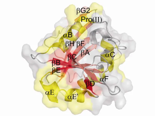
First, active-site residues to be mutated were identified on the basis of crystallographic and/or functional data available for hDHFR. Since we are interested in binding at the folate–binding site, target amino acids were all chosen within or close to that area, were they were more likely to be effective [110]. Target residues were identified before my arrival in the laboratory by PhD student Jordan Volpato and they cover all the folate-binding site (figure 1.8). Among these, five residues have been the main target of the investigation presented in this thesis: Ile7, Gly15, Trp24, Arg70 and Val115 (figure 1.8, in yellow). Each of the 5 targeted residues was randomized by saturation mutagenesis using NNS codon degeneracy (N: adenine/cytosine/guanine/thymine; S: cytosine/guanine). This means that at each position, all 20 amino acids were allowed.
1.5.1.2 - Screening for the properties of interest: activity and antifolate resistance
Previously, PhD student Jordan Volpato developed an efficient bacterial complementation approach to select MTX-resistant clones (described in chapter 2). Bacterial selection has the advantage of being very high-throughput, but unfortunately bacterial survival under selective conditions is not a direct read-out of catalytic activity and selected variants are not necessarily amenable to detailed characterization (where ‘amenable to’ refers to expression level, stability, solubility and/or activity) [111]. Since the subsequent steps of characterization are laborious and require the use of expensive compounds (NADPH and MTX), an improvement of the screening step was required. Therefore, a further step of screening using a plate reader-based activity assay in presence
35
of high concentrations of inhibitor was developed to identify only the best hits from bacterial selection, as it is described in chapter 3.
Moreover, in order to investigate discrimination in ligand binding, the scope of the project was extended by developing a screening strategy for further phenotypes of interest: activity (efficient binding of DHF upon mutation) and resistance/sensitivity to other antifolates (TMP and PMTX).
A protocol to quantify activity (kcat) and MTX resistance (IC50MTX) directly from cell lysates in microtiter plates was also developed.
1.5.2 – Structure-function relationship analysis
1.5.2.1 – Kinetic characterization of the mutants: determination of kinetic inhibition constants
Positive hits from second-tier screening were over-expressed and purified in order to determine more precisely the kinetic and inhibition parameters in vitro. The kinetic parameters KM and kcat describe productive binding of the mutants for the substrate DHF and the reaction rate of reduction of DHF to THF, respectively. The inhibition constant Ki describes productive binding of a competitive inhibitor to the mutants’ active-sites. The value obtained for these parameters were correlated to the different mutations for structure-activity relationship analysis.
We have also begun SAR with MTX by investigating the binding of its constituent fragments: DAP (2,4-diamino-6-(hydroxymethyl)pteridine), DAMPA (4-[N-(2,4-diamino-6-pteridinylmethyl)-N-methylamino]benzoic acid) and p-ABA-Glu (para-aminobenzoic acid-L-glutamate) to relevant MTX-resistant variants identified in our laboratory (figure 1.7; chapter 4).
1.5.2.2 – Structural characterization of the mutants by computer-based molecular modeling
As it is impossible to envisage resolving the structure of all the resistant variants identified, computer-based molecular modeling approaches were considered as tools for structural characterization.
First, energy minimization was used to compare the predicted structure of one novel identified MTX-resistant mutant to the crystal structure of the native enzyme complexed with MTX (chapter 2).
Then, docking was evaluated as a tool to mimic DHF and inhibitor binding in the active-site pocket of the native enzyme. Advantages and limits of this technique for SAR analysis will be discussed in chapter 5.
37
Table 1.1. Active-site interactions between hDHFR and folate or methotrexate
Folate (1DHF.PDB)[28] Methotrexate (1U72.PDB)[22] Dihydrofolate or
methotrexate component
hDHFR
residue Type of interaction
hDHFR
residue Type of interaction
I7
Backbone carbonyl: hypothesized H-bond with N8
DHF (not observed with FOL in 1DHF.PDB structure). Side-chain: hydrophobic
I7
V115
Backbone carbonyl: H-bond with 4-amino group.
Side-chain: hydrophobic T121 Side chain: H-bond with 4-amino group
L22
F31 F31 F34 Van der Waals/hydrophobic F34
Van der Waals/hydrophobic W24 W24
E30 E30
Pteridine ring
T136
H-bonding network involving structural waters, N3, O4 and
2-amino group T136
H-bonding network involving structural water 216, N1, N8 and 2-amino group
F31 F31 F34 F34 I60 I60 P61 P61 L67
Van der Waals/hydrophobic
L67
Van der Waals/hydrophobic
N10-methyl-p-ABA
N64 H-bond to p-ABA carbonyl oxygen N64 Side chain: H-bond to p-ABA carbonyl oxygen
R28
Carbonyl backbone: H-bonding network between
H2O410 and γ-COOH FOL
(rarely observed)
H2O198
H-bonding network between carbonyl backbone N64, backbone NH, K68, α-COOH
MTX
Q35 α− and γ−COOH FOL In proximity to both Q35 In proximity to both α− and γ−COOH MTX
L-glutamate
Table 1.2. Mutations providing MTX-resistance in hDHFR
a Ternary K
DDHF values. The KMDHF could not been determined cause of substrate inhibition. Since the rate of the chemical reaction is much slower than the rate of subsequent dissociation of THF, Nakano et al. used ternary KDDHF values as an approximation of KMDHF [45].
b Derived from plots of rate versus DHF concentration by least-square fit to the equation for substrate inhibition by Nakano et al.[45].
c Calculated from kcat/KM and KMDHF, using the approximation described in a and b[45].
d Ternary K
DDHF values [45]. e K
iMTX calculated from data in figure 5a of Thompson and Freisheim (1991) [46].
hDHFR variant KMDHF (µM) Ratio KM mut/ KM WT kcat (s-1) kcat/KM (μM-1s-1) KiMTX (nM) Ratio Ki mut/ Ki WT Native ≤0.075 1 10 92 0.034 1 Position 7 I7F [57] 20.5 270 5 0.25 24.6 7200 Position 15 G15W [59] 6.2 80 3.2 0.5 2.1 600 L22F [43] 3.9 50 23.8 6.1 0.5 150 L22R [43] 1.6 20 0.04 5 0.03 4.6 1300 L22W [43] 0.4 5 4.2 10 4.3 1300 Position 22 L22Y [43] 0.5 5 6.1 12 11 3200 F31A [65] 0.5 5 13.9 30 0.27 80 F31G [65] 0.4 5 11.3 26 0.35 100 F31S [65] 0.4 5 7.0 16 0.24 70 Position 31 F31R [57] 0.6 10 0.9 1.5 7.2 2100 F34A [45] 36 a 480 8.4 c 0.2 b 34d 10000 F34I [45] 24 a 320 13.7 c 0.6 b 13d 3800 F34S [45] 350 a 4600 6.0 c 0.02 b 210d 60000 F34T [45] 300a 4000 3.3 c 0.1 b 9.6d 2800 Position 34 F34V [45] 63a 800 30.7 c 0.5 b 10 3000 Position 70 R70K [46] 0.5 5 1.75 3.7 0.46e 13 L22F/F31G [66] 0.4 5 1.3 3.3 29 850 L22F/F31S [66] 0.4 5 1.6 3.6 26 7600 L22Y/F31G [66] 0.3 5 0.5 1.4 150 44000 Double mutants L22Y/F31S [66] 0.3 5 1.3 3.8 42 12000
39
Figure 1.1. Folate–dependent metabolic reactions. The abbreviation are: DHFR,
dihydrofolate reductase; TS, thymidylate synthase; GARFT, glycinamide ribonucleotide formyl transferase; AICARFT, aminoimidazole carboxamide formyl transferase; DHF, dihydrofolate; THF, tetrahydrofolate; 5-CH3-THF, 5-methyl tetrahydrofolate; 5,10-CH2
-THF, 5,10-methylene tetrahydrofolate; 5,10-CH--THF, 5,10-methenyl tetrahydrofolate; 5-CHO-THF and 10-5-CHO-THF, 5-formyl- and 10 formyl tetrahydrofolate, respectively. Image was adapted from Figure 1 in Zhao et al. 2003 [41].
Figure 1.1a: Synthesis of thymidylate (dTMP) from deoxyuridine monophosphate (dUMP). The enzymes dihydrofolate reductase (DHFR) and Serine hydroxymethyl
transferase are necessary for the recycling of the 5,5-CH2-THF. In dTMP, all the methylic
hydrogens (in red and blue) derive from 5,5-CH2-THF. Figure adapted from reference
39
Figure 1.1b: Role of the folate cofactor 10-formyl tetrahydrofolate (10-CHO-THF) in the synthesis of the inosine nucleus. A) Conversion of tetrahydrofolate (THF) to
10-formyl tetrahydrofolate (10-CHO-THF). B) Structure of inosine monophosphate (IMP), first intermediate to posses a complete purine nucleus. The carbons added by GARFT (glycinamide ribonucleotide formyl transferase) and AICARFT (aminoimidazole carboxamide formyl transferase), derived from formate and provided by 10-CHO-THF, are indicated in blue. Figure adapted from reference [2].
Figure 1.2. Reaction catalyzed by dihydrofolate reductase (DHFR). A) Reduction
catalyzed by vertebrate DHFRs. B) Schematic representation of the hydride transfer in the transition state: optimal carbon-carbon bond distance and -C⋅⋅⋅H⋅⋅⋅C- bond angle are indicated. Image adapted from Benkovic et al. 1988 [15].
41
Figure 1.3. Sequence and structural comparison of human, murine and E. coli dihydrofolate reductases (DHFRs). A) Sequence comparison. Sequence differences
from human DHFR are coloured in cyan for mouse DHFR and in red for E. coli DHFR.
B) Comparison of the backbone of human (1U72.PDB; green) and E. coli (1RX3.PDB;
magenta) and of human (1U72.PDB; green) and mouse (1U70.PDB; orange). Figure adapted from Cody et al. 2005 [22].
Figure 1.4. Secondary structure of hDHFR. Eight-stranded twisted β-sheet (βA, residues 4-10; βB, 47-53; βC, 71-76; βD, 88-90; βE, 108-116; βF, 130-139; βG, which is interrupted by a tight turn is composed by βG1, 157-159 and βG2, 168-172; βH, 175-185). Five α−helices (αB, 27-40; αC, 53-59; αΕ, 92−102 and αΕ’, 102-109; αF, 117-127), a polyproline-like helix (Pro(II), residues 21-26) and eight tight turns (residues 11-14, 18-21, 43-46, 61-64, 67-70, 83-86, 162-165, 172-175) connect the β-sheets among them.
43
Figure 1.5. Ligand binding at the active site of WT hDHFR. A) Bound folate (FOL; in
green) from 1DHF.PDB. B) Bound methotrexate (MTX; in magenta) from 1U72.PDB. The active site is shown. Active-site residues that interact with the ligands are shown in sticks representation and coloured in orange, yellow or blue to indicate interaction or proximity to the ligand’s pteridine, p-ABA or glutamate moiety, respectively. The 4-carbonyl and the 4-amino group of FOL (in A) and of MTX (in B) are circled to highlight the pteridine ring flip.