UNIVERSITÉ DE MONTRÉAL
CONCEPTION D’UN ADAPTATEUR MOLÉCULAIRE POUR L’IMMOBILISATION DE FACTEURS DE CROISSANCE SUR UN SUBSTRAT DE COLLAGÈNE
CYRIL ADDI
INSTITUT DE GÉNIE BIOMÉDICAL ÉCOLE POLYTECHNIQUE DE MONTRÉAL
MÉMOIRE PRÉSENTÉ EN VUE DE L’OBTENTION DU DIPLÔME DE MAÎTRISE ÈS SCIENCES APPLIQUÉES
(GÉNIE BIOMÉDICAL) AOÛT 2016
UNIVERSITÉ DE MONTRÉAL
ÉCOLE POLYTECHNIQUE DE MONTRÉAL
Ce mémoire intitulé :
CONCEPTION D’UN ADAPTATEUR MOLÉCULAIRE POUR L’IMMOBILISATION DE FACTEURS DE CROISSANCE SUR UN SUBSTRAT DE COLLAGÈNE
présenté par : ADDI Cyril
en vue de l’obtention du diplôme de : Maîtrise ès sciences appliquées a été dûment accepté par le jury d’examen constitué de :
M. JOLICOEUR Mario, Ph. D., président
M. DE CRESCENZO Gregory, Ph. D., membre et directeur de recherche M. GAILLET Bruno, Ph. D., membre
REMERCIEMENTS
Je tiens tout particulièrement à remercier mon directeur de recherche Gregory De Crescenzo, qui m’a soutenu et guidé tout au long de mon projet de maîtrise. Sa grande disponibilité et ses qualités humaines ont rendu cette expérience aussi enrichissante sur le plan intellectuel que sur le plan personnel, et je lui en suis particulièrement reconnaissant.
Je tiens également à remercier mes collègues de bureau, en particulier Frédéric qui m’a grandement aidé grâce à ses connaissances et son expérience dans le domaine, mais également Benoît, Eric, Florian, Adeline (qui fait partie du bureau, d’une certaine manière !), Nesrine, Kahina, Cécile, Yuan, mais également Samantha, Pauline et Charles.
Je remercie par ailleurs le programme MÉDITIS qui a permis de financer en partie mes travaux, ainsi que le Pr. Michael Buschmann qui m’a conseillé dans ce cadre.
J’ai également une pensée pour l’ensemble des personnels du génie chimique, qui m’ont aidé dans certaines démarches, en particulier Jean Huard.
Je remercie également Elisa pour son soutien quotidien, ainsi que ma famille et mes amis avec qui j’ai pu garder un lien fort, malgré ces deux années passés outre-Atlantique.
RÉSUMÉ
Les biomatériaux à base de collagène ont une place particulièrement importante dans le domaine de l’ingénierie tissulaire. Leur fonctionnalisation peut être facilement effectuée à l’aide de protéines chimères constituée d’une biomolécule, typiquement un facteur de croissance, fusionnée à un domaine de liaison au collagène. Cette fonctionnalisation peut conférer aux biomatériaux des propriétés mitogéniques et anti-apoptotiques, et influencer la colonisation de ce dernier par des cellules. De nombreuses études ont été menées en ce sens dans des domaines variés de la médecine régénératrice, comme la cicatrisation ou la régénération osseuse.
Nous avons choisi d’immobiliser des facteurs de croissance étiquetés avec une hélice alpha (Ecoil-GF) sur un substrat de gélatine en utilisant un adaptateur moléculaire constitué du domaine de liaison au collagène de la fibronectine, étiqueté avec l’hélice alpha partenaire (CBD-Kcoil). Les hélices alpha Ecoil et Kcoil interagissent par interaction superhélice d’une manière spécifique avec une forte affinité, ce qui permet la formation d’un complexe entre la gélatine, l’adaptateur moléculaire CBD-Kcoil et le facteur de croissance Ecoil-GF. Nous avons produit et purifié le facteur de croissance des fibroblastes basique étiqueté avec l’hélice Ecoil (Ecoil-bFGF), et nous avons comparé son immobilisation avec celle du facteur de croissance épidermique, en utilisant l’adaptateur moléculaire CBD-Kcoil.
ABSTRACT
Collagen-based biomaterials have attracted a lot of interest in the field of tissue engineering. Their functionalization can be easily performed using chimeric proteins composed of a biomolecule, typically a growth factor, that is fused to a collagen-binding domain. This functionalization may provide biomaterials with mitogenic and anti-apoptotic properties, and influence the cellular fate within the implant. Many studies have been conducted to this end, in various fields of regenerative medicine, such as wound healing and bone regeneration.
We chose to tether coil-tagged growth factors (Ecoil-GF) on a gelatin substrate using a molecular adaptor consisting of the collagen-binding domain of fibronectin, fused to the complementary coil (CBD-Kcoil). E and K coils interacted through coiled-coil interaction in a specific manner with high affinity, which enabled the formation of a ternary complex between the gelatin substrate, the molecular adapter CBD-Kcoil and the growth factor Ecoil-GF. We have produced and purified an Ecoil-tagged basic fibroblast growth factor (Ecoil-bFGF), and we have compared its tethering with that of the epidermal growth factor, using the molecular adaptor CBD-Kcoil.
TABLE DES MATIÈRES
REMERCIEMENTS ... III RÉSUMÉ ... IV ABSTRACT ... V TABLE DES MATIÈRES ... VI LISTE DES TABLEAUX ... IX LISTE DES FIGURES ... X LISTE DES SIGLES ET ABRÉVIATIONS ... XI
CHAPITRE 1 INTRODUCTION ... 1
CHAPITRE 2 DÉMARCHE GÉNÉRALE ... 4
CHAPITRE 3 REVUE DE LITTÉRATURE ... 5
3.1 Article 1 – The design and use of chimeric proteins containing a collagen binding domain for tissue engineering and regenerative medicine ... 5
3.2 Abstract ... 6
3.3 Introduction ... 6
3.4 Collagen-binding domains ... 8
Quantitative characterization of CBD-collagen interactions ... 9
Fibronectin ... 11
Von Willebrand Factor ... 12
Placental Growth Factor ... 13
C. histolyticum collagenases ... 14
Decorin ... 14
S. aureus adhesin ... 15
Engineered collagen-binding peptides ... 15
Collagen-mimetic peptide ... 16
3.5 Applications of collagen-binding fusion proteins ... 19
Chimeric collagen-binding proteins for wound healing ... 21
Chimeric collagen-binding proteins for bone regeneration ... 28
3.6 Conclusion ... 36
3.7 Acknowledgments ... 37
3.8 Reference ... 37
CHAPITRE 4 PRODUCTION ET PURIFICATION DE PROTÉINES CHIMÈRES – FONCTIONALISATION D’UN SUBSTRAT DE COLLAGÈNE... 55
4.1 Article 2 - The use of a chimeric collagen binding domain of fibronectin to recruit coil-tagged growth factors on gelatin-based biomaterials ... 55
4.2 Abstract ... 56
4.3 Introduction ... 56
4.4 Materials & Methods ... 58
Chemicals and reagents ... 58
Generation of the expression vectors ... 59
Expression of fusion proteins ... 59
Purification of Ecoil-bFGF protein ... 59
Purification of the CBD-Kcoil protein ... 60
Surface plasmon resonance (SPR) assays ... 60
ELISA ... 61
Proliferation assay ... 61
4.5 Results ... 62
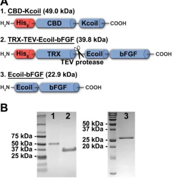
Expression and purification of fusion proteins ... 62
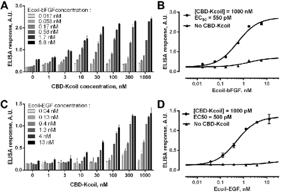
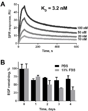
Ecoil-tagged growth factors can be recruited on gelatin through CBD-Kcoil interactions. ... 64
Stability of the gelatin/ CBD-Kcoil/Ecoil-GF complexes ... 65
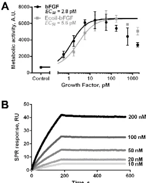
Bioactivity of Ecoil-bFGF ... 67
Tethered Ecoil-bFGF and Ecoil-EGF promote cell proliferation ... 69
4.6 Discussion ... 72
4.7 Conclusion ... 74
4.8 Acknowledgment ... 74
4.9 References ... 75
CHAPITRE 5 DISCUSSION GÉNÉRALE ... 80
CHAPITRE 6 CONCLUSION ET RECOMMANDATIONS ... 82
BIBLIOGRAPHIE ... 83
LISTE DES TABLEAUX
LISTE DES FIGURES
Figure 1.1. Structure du collagène de type I dans le corps humain ... 1 Figure 1.2. Stratégie de fonctionnalisation du collagène à l’aide d’une seule protéine chimère (A) ou de deux protéines interagissant par interaction superhélice (B). ... 3 Figure 3.1. Structure of several proteins which contains a collagen-binding domain (in red): human fibronectin (A), von Willebrand Factor (B), placental growth factor (C), C. histolyticum collagenases ColG and ColH (D), decorin (E) and S. aureus adhesin (F). ... 12 Figure 3.2. Biological effect of various chimeric proteins containing a collagen-binding domain (CBD). Collagen can be functionalized with growth factors or cytokines to stimulate cell proliferation or differentiation (A), cells can be engineered to provide collagen anchorage through a GPI-linked protein (B) and collagen can be decorated with cell-binding domains to promote cell adhesion (C). ... 20 Figure 4.1. Schematic illustration of an Ecoil-tagged GF tethered in an oriented manner on a gelatin-coated surface that had been functionalized with CBD-Kcoil. ... 58 Figure 4.2. Structure, production and purification of the coil-tagged chimeric proteins used in this study. ... 63 Figure 4.3. Tethering of Ecoil-bFGF (A) or Ecoil-EGF (C) on gelatin thanks to the CBD-Kcoil adaptor. ... 65 Figure 4.4. Characterization of the stability of the gelatin/CBD-Kcoil complex. ... 67 Figure 4.5. Characterization of the bioactivity of Ecoil-bFGF. ... 69 Figure 4.6. Bioactivity of Ecoil-bFGF and Ecoil-EGF tethered to collagen using a CBD-Kcoil adaptor. ... 71
LISTE DES SIGLES ET ABRÉVIATIONS
aFGF et FGF-1 acidic fibroblast growth factor bFGF et FGF-2 basic fibroblast growth factor BCA bicinchoninic acid assay
BMP bone morphogenetic protein
CBD collagen-binding domain
ColH and ColG collagenase H and G
DBM demineralized bone matrix
DMEM Dulbecco's modified eagle medium
EBM endothelial basal medium
EGF epidermal growth factor
ELISA enzyme-linked immunosorbent assay EPC endothelial progenitor cells
FBS fetal bovine serum
FDA Food and Drug Administration FGF fibroblast growth factor
FGFR fibroblast growth factor receptor
FN fibronectin
GPI glycosylphosphatidylinositol
HBS-EP HEPES buffered saline avec EDTA et Surfactant P20
HGF hepatocyte growth factor
HRP horseradish peroxidase
IGF insulin-like growth factor
IPTG isopropyl β-D-1-thiogalactopyranoside
GF growth factor
MBM mineralized bone matrix
MMP matrix metalloproteinase
MSCs mesenchymal stem cells
NGF nerve growth factor
OC osteocalcin
PBS phosphate buffered saline
PCL polycaprolactone
PDGF platelet-derived growth factor PlGF placental growth factor
PTH parathyroid hormone
SDF-1α stromal cell-derived factor 1α
SPR surface plasmon resonance
TEV tobacco etch virus
TGFβ transforming growth factor β
TRX thioredoxin
VEGF vascular epidermal growth factor VSMC vascular smooth muscle cells
CHAPITRE 1
INTRODUCTION
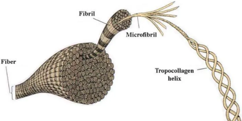
Le collagène est un matériau très populaire dans le domaine de l’ingénierie tissulaire, en particulier à cause de sa faible antigénicité et de son excellente biocompatibilité, ce qui en fait un matériau très sûr d’utilisation. Le collagène est une protéine fondamentale de la matrice extracellulaire, qu’on retrouve sous la forme d’une trentaine de paralogues, dont le collagène de type I, la protéine la plus abondante du corps humain (Figure 1.1). Il est exprimé dans la majorité des tissus comme la peau, les tendons, les ligaments, le cartilage, les os, les vaisseaux sanguins ou encore les muscles et confère une résistance mécanique aux tissus. Au vu de son importance, c’est une protéine extrêmement conservée au cours de l’évolution (97 % d’identité entre Homo sapiens et Bos taurus). Cela permet en particulier d’utiliser du collagène extrait de bœuf ou de porc pour fabriquer des biomatériaux qui présentent une bonne biocompatibilité chez l’homme. Un certain nombre d’entre eux sont déjà approuvés par les autorités de santé (la Food and Drug Administration et l’Agence Européenne des Médicaments).
Figure 1.1. Structure du collagène de type I dans le corps humain
Une approche prometteuse a été développée ces dernières années pour fonctionnaliser le collagène et lui donner, par exemple, des propriétés bio-inductives promouvant la migration, la prolifération, ou encore la différenciation cellulaire. Cette approche consiste à développer des protéines de fusion entre un domaine de liaison au collagène (issu de protéines qui se lient
naturellement au collagène, comme la fibronectine ou le facteur de von Willebrand) et un domaine bioactif (par exemple un facteur de croissance ou un domaine de liaison aux intégrines). Ces protéines chimères possèdent donc des propriétés de liaison au collagène en plus de leurs propriétés bio-inductives. De nombreuses études ont montré que ces protéines présentent un pouvoir de régénération beaucoup plus important que leur homologue d’origine (par exemple le facteur de croissance naturel), lorsqu’elles sont utilisées en adéquation avec un biomatériau à base de collagène. En particulier, leur biodisponibilité est augmentée car leur diffusion est limitée : ces protéines chimères peuvent en effet se lier au collagène endogène lorsqu’elles sont libérées du biomatériau. Elles ont prouvé leur utilité dans des domaines très variés de la médecine régénératrice (cicatrisation, régénération osseuse et neurale).
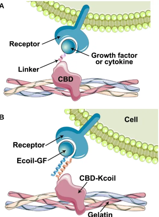
La production de ce type de protéines de fusion (Figure 1.2.A) peut être délicate, dans la mesure où l’ajout d’un domaine de liaison au collagène (CBD) à un facteur de croissance peut altérer le repliement de ce dernier, et affecter sa bioactivité.
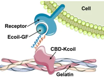
L’objectif de ce travail est de développer une stratégie de fonctionnalisation du collagène basée d’une part sur un adaptateur moléculaire constitué du CBD de la fibronectine, fusionné à une hélice alpha (Kcoil), et d’autre part sur des facteurs de croissance (GF) étiquetés avec l’hélice alpha complémentaire (Ecoil). L’interaction superhélice E/K étant une interaction de très forte affinité, un complexe peut se former entre le collagène (ou la gélatine), l’adaptateur moléculaire (CBD-Kcoil), et les facteurs de croissance (Ecoil-GF) (Figure 1.2.B). Cette approche peut théoriquement être appliquée à n’importe quel biomatériau obtenu à partir de collagène.
La première partie de ce mémoire présente une revue critique de la littérature sur les protéines chimères contenant un CBD. En particulier, les différents CBD reportés dans la littérature sont comparés et caractérisés, et les avantages des protéines chimères développées dans les domaines de la médecine régénératrice sont mis en évidence.
La seconde partie présente l’ensemble de la démarche de fonctionnalisation d’une surface de gélatine, en immobilisant des facteurs de croissance à l’aide d’un adaptateur moléculaire. Elle compile l’ensemble des résultats d’expériences, depuis la production et la purification des protéines de fusion, jusqu’à la caractérisation des interactions en jeu dans la fonctionnalisation de surface, et la réponse cellulaire engendrée.
A
B
Figure 1.2. Stratégie de fonctionnalisation du collagène à l’aide d’une seule protéine chimère (A) ou de deux protéines interagissant par interaction superhélice (B).
CHAPITRE 2
DÉMARCHE GÉNÉRALE
Le cœur de ce mémoire de maîtrise est composé de deux articles. Le premier, intitulé « The design and use of chimeric proteins with a collagen binding domain for tissue engineering and regenerative medicine » a été soumis à Advanced Drug Delivery Reviews en juillet 2016. Cet article compile l’ensemble des domaines de liaison au collagène reportés dans la littérature et compare leurs propriétés biophysiques. Les connaissances développées dans cette revue permettent de mieux situer le CBD employé au Chapitre 3 (issu de la fibronectine) par rapport aux autres CBD. En particulier, elles montrent que ce dernier a été employé depuis plusieurs années, seul ou sous forme de protéine de fusion, qui ont été testées in vivo avec succès. Cela démontre le grand potentiel de ce dernier, aussi bien au niveau de ses caractéristiques biophysiques (haute affinité pour le collagène) que de sa biocompatibilité (étant donné qu’il est dérivé de la fibronectine humaine). Le second article, intitulé « The use of a chimeric collagen binding domain of fibronectin to recruit coil-tagged growth factors on gelatin-based biomaterial », a été soumis à Acta Biomaterialia en juin 2016. Ces deux journaux scientifiques ont été sélectionnés pour leur niveau d’impact dans le domaine étudié ; Acta Biomaterialia étant un journal de référence concernant la fonctionnalisation de biomatériaux dans le domaine de la médecine régénératrice tandis que Advanced Drug Delivery
Reviews publie des revues de littérature sur les techniques émergentes dans le domaine des
stratégies thérapeutiques.
J’ai réalisé moi-même l’essentiel des travaux expérimentaux, avec l’aide et le soutien de Frédéric Murschel, Benoît Liberelle et Nesrine Riahi. J’ai aussi bénéficié de l’expérience de mon directeur de recherche Gregory De Crescenzo et de Frédéric Murschel concernant l’avancement général du projet de recherche et tout particulièrement pour la relecture des articles.
CHAPITRE 3
REVUE DE LITTÉRATURE
De nombreuses techniques ont été développées pour fonctionnaliser les biomatériaux avec des molécules bioactives afin d’influencer la migration, la prolifération ou encore la différenciation cellulaire. Parmi toutes ces approches, la création de protéines de fusion comprenant un CBD est particulièrement prometteuse pour fonctionnaliser les biomatériaux à base de collagène. De nombreuses protéines, comme des facteurs de croissances, ont ainsi été fusionnées à un CBD pour augmenter leur biodisponibilité, avec souvent un effet très bénéfique, y compris lors d’études in
vivo. Une revue de l’ensemble des travaux reportés dans ce domaine est présentée ici dans un
manuscrit récemment soumis à Advanced Drug Delivery Reviews.
3.1 Article 1 – The design and use of chimeric proteins containing a
collagen binding domain for tissue engineering and regenerative
medicine
Cyril Addi1, Frédéric Murschel1, Gregory De Crescenzo1,*
1 Department of Chemical Engineering, Biomedical Science and Technology Research Group, Bio-P2
Research Unit, École Polytechnique de Montréal, P.O. Box 6079, succ. Centre-Ville, Montréal (QC), Canada H3C 3A7.
3.2 Abstract
Collagen-based biomaterials are widely employed in the field of tissue engineering; they can be loaded with biomolecules such as growth factors to modulate the biological response of the host and thus improve their potential of regeneration. Recombinant chimeric growth factors fused to a collagen-binding domain have been reported to improve the bioavailability of these growth factors, especially when combined to an appropriate collagen-based biomaterial. This review first provides an extensive description of the various collagen-binding domains that have been characterized and fused to several proteins for application in the fields of tissue engineering and regenerative medicine. The second part of the review highlights the benefits of various collagen-binding domain /growth factor fusion proteins that have been designed for wound healing and bone regeneration.
Keywords— growth factor; collagen-binding domain; collagen; gelatin; biofunctionalization; scaffold; coating; delivery
3.3 Introduction
Collagen has attracted a lot of attention in the field of tissue engineering, given its excellent biocompatibility, biodegradability, weak antigenicity and safety[1]. Biomaterials made of collagen have been commonly used in various formulations, such as hydrogels, sponges and microparticles; they are often made of denatured collagen (i.e. gelatin)[2]. Importantly, some of them are already approved by the Food and Drug Administration[3]. Moreover, the mechanical properties of collagen scaffolds can be tuned by cross-linking, resulting in higher tensile strength and proteolytic resistance[4], [5] and allowing for their use in a wider scope of applications. Also, in order to improve their healing potential, these biomaterials have been loaded with various biomolecules to be released over time, be it small molecules such as steroids, antibiotics and chemotherapy agents, proteins such as growth factors and antibodies, or liposomes[4]. The potential benefits of their loading with biomolecules are numerous as it enhances effective local concentrations of drugs,
hence preventing side effects due to systemic distribution and it increases drug half-life in many cases[6].
Most of the collagen-derived biomaterials that have been developed for tissue engineering/regenerative medicine purposes simply trap the drug in their network and release it over time by diffusion through the pores of the scaffold[6]. In this delivery strategy, the difficulty to control drug release represents a major drawback: there is generally an initial burst release which is not followed by a sustained release over a long period[7], [8]. In addition to the rapid decrease of drug concentration in time, the initial burst itself may be an issue. For example, in the case of Bone Morphogenetic Protein-2 (BMP2) release, it was shown to promote inflammation and osteoclastic activity[7]. Conversely, reducing the size of the pores in order to extend the release hinders nutrient transport and thus severely affects cell viability[9].
Growth factors are frequently employed in therapeutic strategies such as bone and cartilage engineering[10], myocardial regeneration[11] and wound healing[12] because they can promote cell adhesion, migration, differentiation and proliferation. The optimal beneficial effect of these growth factors is determined by their spatiotemporal delivery, an essential trait that can affect cell fate[13]. Although no growth factor binding site has been reported in collagen yet[14], some growth factors, e.g. Transforming Growth Factor-β1 (TGF-β1), basic Fibroblast Growth Factor (bFGF) and BMP2, have a naturally strong affinity for collagen and stably interact with scaffolds made of collagen through ionic interactions[7], [15], [16]. However, these interactions mainly depend on the isoelectric point of the growth factor[7], [17] and the release profile is often characterized by an initial burst[7]. Therefore, it does not constitute the optimal manner to control the release of growth factors.
A variety of techniques have been developed for the covalent chemical conjugation of growth factors to collagen scaffolds so as to provide a highly localized and long-lasting signaling. The random covalent grafting of growth factors has proved worthwhile[18], [19], in particular through the NHS/EDC coupling chemistry that targets free amine groups on the growth factor[20] or by photo-irradiation[21]. However, the bioactivity of the immobilized proteins may be negatively impacted, since their attachment to the substrate may mask or alter their receptor binding moieties[22], [23]. The covalent binding of growth factors in an oriented manner with the help of
a cysteine tag has also been investigated[24]. However, the addition of a cysteine can hamper protein folding via the formation of unwanted disulfide bridges, hence resulting in a loss of bioactivity. Importantly, the covalent binding of growth factors may also prevent their endocytosis, which can be crucial for signaling[25].
The tethering of growth factors in a stable but non-covalent manner through specific affinity tags has also been extensively studied. The major strategies that have been tested include the interactions between biotin and streptavidin[26], coil peptides[27]–[29], DOPA-containing peptides[30] as well as the use of binding domains for heparin/heparan sulfate[31], laminin[32], fibrin[33] or fibronectin[34]. Among them, the use of several collagen-binding domain (CBD) fusion proteins in association with collagen-based biomaterials has been the subject of intense research. Indeed, type I collagen is the most abundant protein in the human body[35]; it is found in the extracellular matrix of bones, skin, tendons, cornea, artery walls as well as internal organs. Hence, a recombinant collagen-binding growth factor being tethered to a resorbable collagen biomaterial would also bear the potential to bind to the extracellular collagenous matrix after resorption of the implant, which would significantly improve its bioavailability over time.
The purpose of this review is (i) to examine the various CBDs that have been characterized and discuss how their origin and size affect their affinity and specificity for collagen, and (ii) to assess to which extent collagen biomaterials functionalized with CBD fusion proteins have emerged as powerful tools in the fields of wound healing and bone regeneration.
3.4 Collagen-binding domains
The term collagen-binding domain (CBD) encompasses very distinct polypeptide domains that are either engineered or derived from native collagen-binding proteins such as fibronectin, the von Willebrand Factor and several collagenases. This diversity of origin results in a great heterogeneity of sizes, as they range from a seven amino-acid long peptide to a whole protein domain of 42 kDa (Table I). Bearing in mind that these domains will be fused to growth factors and that controlled release is critical in tissue engineering and regenerative medicine, it is crucial to assess how their sequence and their length affect their interaction with collagen, especially in terms of stability.
Quantitative characterization of CBD-collagen interactions
For most of the CBDs studied in the literature, the strength of their interaction with collagen has been determined, either by reporting an apparent affinity, or thermodynamic association constant (KA, expressed in M-1), or its inverse, the thermodynamic dissociation constant (KD, expressed in
M). A plethora of dissociation constant values, spanning from the low nanomolar to the micromolar, have been determined for the various CBD-collagen interactions (see Table I). These differences in apparent affinities may not be surprising given the diverse origins of the CBDs and types of collagen. Such a range of affinities may be exploited to target specific types of collagen by selecting an appropriate CBD or to modulate the release of a given protein fused to a specific CBD by virtue of its affinity for collagen. However, in order to avoid any misinterpretation of the data presented in Table I, one may analyze them with caution.
Many techniques have indeed been employed by the research community to determine the affinities of the various CBDs for the different types of collagen. Those include Surface Plasmon Resonance (SPR)-based biosensor assays[36], Enzyme-Linked Immunosorbent Assays (ELISA)[14], radioactivity assays by using iodinated proteins[37] as well as fluorescence titration assays by measuring changes in anisotropy of fluorescein-labeled collagen chains[38]. All of these methods have their own merits and limitations to assess an apparent dissociation constant[39]. Interestingly, large differences can be pinpointed between thevalues derived from distinct techniques within the same report. For example, when characterizing the interactions between the von Willebrand A1 domain for type III collagen, Morales et al. determined an apparent KD of 8 nM by SPR and of 400
nM by ELISA[40]. Such a discrepancy can result from several biases inherent to the techniques at hand: in most SPR assays, the collagen is covalently attached to the biosensor surface, whereas, in ELISA, the substrate is non-specifically adsorbed at the bottom of the well. Both approaches may lead to distinct alterations of the three-dimensional conformation of the triple helix of collagen and, as a result, change the strength of its interaction with CBDs[40]. Other artefacts related to inappropriate assay conditions, as those documented in specialized reports[41], [42] may also be invoked to explain these discrepancies.
It is also not uncommon that distinct research groups report different apparent KD for the same
an apparent KD of 5.5 nM for the capture of the fusion protein TKKTLRT-PDGF (platelet-derived
growth factor) by a type I collagen membrane, whereas Lin et al. reported an apparent KD of 97
nM for the same protein incubated on type I collagen-coated wells[43], [44]. As much as the techniques, the procedure for data processing and KD determination may therefore be worth
questioning. In most of the articles here reviewed, the apparent KD values were indeed derived by
linearizing the experimental data, via a Scatchard plot for instance. Such an approach is prone to introduce errors and biases[45], especially when the plot deviates from linearity and when data are omitted[46], [47].
To overcome these limitations, we here propose a rationalized method for data processing. By curve-fitting the complete data set with the GraphPad Prism 6 Software (GraphPad Prism Software Inc., San Diego, CA), using a Hill-type equation that assumes a 1:1 interaction, the half-maximum effective concentration (EC50) of a CBD binding to collagen can be calculated:
𝑌 = 𝑌𝑚𝑎𝑥 1 +[𝐶𝐵𝐷]𝐸𝐶50
Where [CBD] and Y correspond to the incubated CBD concentration and the corresponding response (signal minus blank), respectively. Ymax, the theoretical maximal response, and EC50, the
half-maximum effective concentration, are outputs. Note that the Hill slope was fixed at 1.0 for the sake of comparison.
This curve-fitting method was applied to all the CBD-collagen interaction studies, when raw results were available, by extracting the data from the research article figures using the WebPlotDigitizer 3.9 software[48]. When applicable, the EC50 values derived from the fits, the coefficient of
determination (R²) as well as the number of experimental data points extracted from the figures (n) are given in Table I, in addition to the apparent dissociation constant value (KD) that was reported
by the authors. Although all R² values were close to 1, which is indicative of a good fit between the model and the data, some research groups have used a low number of CBD concentrations in their investigation. Given that the reliability of any curve-fitting is highly dependent on the number of experimental points, both the KD values they reported and the EC50 values we calculated need
to be treated with caution. Furthermore, we encountered several cases of biphasic interactions, for which we provided only the lowest EC50 (corresponding to the highest apparent affinity).
With this analytical approach, we found that the interaction of TKKTLRT-PDGF for type I collagen, with reported KD values of 5.5 nM and 97 nM by Sun et al. and Lin et al., respectively,
was best depicted by an EC50 of 1 100 nM in both cases[43], [44]. This calculated value is plausible,
given that the majority of the TKKTLRT fusion proteins have a dissociation constant in the low micromolar range (Table I).
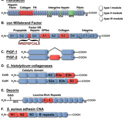
Fibronectin
Fibronectin (FN) is a 220-kDa protein of the ECM which exists as a dimer and binds to many biological partners such as collagen, fibrin, heparin and integrins (Fig. 1A). FN has a very high apparent affinity for the type I (KD = 13 nM)[49] and type II (KD = 58 nM) collagens[50], and it
binds even more strongly to their denatured form, gelatin[51], (KD = 2 nM)[50], which suggests
that the binding site(s) within collagen are, at least partially, masked in the native triple helix[49], [52].
The CBD of FN is a 42-kDa domain composed of six modules ( I6-II1-II2-I7-I8-I9, see Fig. 1A) that
are all required for full affinity[53], supporting a cooperative mode of interaction where all modules simultaneously participate[54], [55]. The FN type II modules are found in many other proteins, including the mannose receptor[56], the Factor XII[57] and the matrix metalloproteinase 2 (MMP2)[55], providing them with collagen-binding ability. Similar to the full-length FN, its CBD displays a higher affinity for gelatin than for properly folded collagens. The CBD of FN is however monomeric and has a moderate affinity for type I collagen (KD = 420-800 nM)[49], [58], hence
highlighting the importance of FN dimerization (and of the resulting avidity) for optimal binding to collagen. Numerous subdomains containing three or more modules have been studied, including a short peptide sequence extracted from the module I9, CQDSETRTFY, which binding to type I
Figure 3.1. Structure of several proteins which contains a collagen-binding domain (in red): human fibronectin (A), von Willebrand Factor (B), placental growth factor (C), C. histolyticum collagenases ColG and ColH (D), decorin (E) and S. aureus adhesin (F).
Von Willebrand Factor
The von Willebrand Factor (vWF) is a 275-kDa blood glycoprotein involved in hemostasis that is frequently encountered as a multimer (due to disulfide bonds), which improves its hemostatic potential[61]. The mature vWF has a very high affinity for type I (KD = 1.8 nM)[62], type III
(KD = 3.4 nM)[63] and type VI (KD = 8 nM)[64] collagens. It poorly binds to denatured collagen,
in stark contrast to FN[65], [66]. Several moieties of the vWF have been studied for their ability to bind to collagen (Fig. 1B), in particular the A1 (20.3 kDa) and A3 domains (19.3 kDa)[67]. Although conflicting results exist in the literature, the A1 domain of vWF does not seem to play a
significant role in the binding of vWF to type I/III collagens[68], [69] but is crucial for its interaction with the type IV/VI collagens[64], [70], [71]. On the contrary, the A3 domain is considered to be the main collagen-binding site of the vWF[72], [73] and have a moderate affinity for the type I (KD = 1 800 nM)[67] and type III collagens (KD = 1 600 nM)[40]. The A3 domain is
thought to interact with the RGQAGVMGF/RGEOGNIGF sequences of the α1/α2 chains of type I collagen and with the RGQOGVMGF sequence in type II and III collagens[69]. Shorter peptide sequences, derived from the bovine and human vWF have also been studied: WREPSFCALS and WREPGRCELN, respectively (Fig. 1B)[37]. These short peptides have often been mutated, that is, their cysteine residue has been replaced by a methionine (WREPSF[M]ALS or WREPGR[M]ELN)[74], [75] in order to avoid unwanted homodimerization or misfolding[76], [77]. The WREPSFCALS peptide is derived from a 21-kDa sequence (F570 - K682) of the bovine vWF (NP_001192237.1), described by Takagi et al.: this sequence is located at the end of the vWF propeptide[78] which is cleaved during posttranslational modifications of vWF[79]. As opposed to the A3 domain, the affinity of the WREPSFCALS peptide for type I collagen is very weak (EC50
= 29 000 nM, Table I)[37]. Nonetheless, it binds to all collagen types from I to V, in addition to gelatin[37]. Its 21-kDa parent fragment possesses the same characteristics but displays a higher affinity for type I collagen (EC50 = 780 nM, Table I)[37].
Placental Growth Factor
Another protein from which the potential of its short CBD has been recently highlighted is the Placental Growth Factor (PlGF, 16.7 kDa), a member of the VEGF family, that is essential for angiogenesis, in particular over the bone-marrow derived cells[80]. The sequence analysis of two splice variants of PIGF which interact differently with the ECM, PlGF-1 and PlGF-2, allowed the identification of a 2.8-kDa amino acid sequence that binds to collagen
(RRRPKGRGKRRREKQRPTDCHL, Fig. 1C)[14]. Indeed, while initially considered as a heparin binding domain[81], this sequence binds very strongly to type I collagen (KD = 126 nM) and even
more to Fibronectin, Vitronectin, Heparan Sulfate and other ECM proteins[14]. Although this sequence has a very high isoelectric point (pI = 12.0), electrostatic interactions only are not sufficient for collagen binding since a scrambled version of this peptide does not bind to type I collagen[14]. Conversely, Martino and colleagues assessed that the cysteine at the C-terminus of
the sequence can be mutated to a serine with little to no influence on the interaction with type I collagen[14]. No further experiment has been reported yet concerning the specificity of this CBD towards the other collagen types.
C. histolyticum collagenases
ColH (116 kDa) and ColG (126 kDa) are two collagenases from the pathogenic Clostridium
histolyticum (Fig. 1D)[82]. They feature a broad substrate specificity and target various types of collagens as well as gelatin[83], [84]. For instance the full-length ColH enzyme displays a strong
affinity for type I collagen (KD = 99.5 nM)[85]. Their CBD, corresponding to the S3 domain (13
kDa) for ColH and the S3a+S3b domains (26 kDa) for ColG, are often associated with the polycystic kidney disease-like domain that precedes them, that is S2a+S2b (20 kDa) for ColH and S2 (10 kDa) for ColG (Fig. 1D).
The fragment corresponding to the S2b+S3 domains of ColH displays a biphasic interaction with
collagen, presenting a strong affinity (KD = 339 nM) followed by a moderate affinity at higher
concentrations (KD = 2110 nM)[85], whereas the S3 domain alone poorly binds to type I collagen
(KD = 15 900 nM)[85].
The short fragment of ColG corresponding to the S3a+S3b domains (Fig. 1D) binds to all collagen types from I to IV, and even (POG)n (KD = 63 000 nM)[86], where (POG)n is a collagen-like
peptide, provided that the number of repeats, n, is large enough to allow the peptide to have a triple-helical conformation[87]. Likewise, it does not bind to gelatin, suggesting that this CBD recognizes the triple-helical structure of collagen[84], [86].
Decorin
Decorin is a small proteoglycan which interacts with collagen fibrils in all connective tissues[88, pp. 4–5]. Similarly to biglycan and fibromodulin, its core protein is mainly constituted of leucine-rich repeats (LRR). Although the glycosaminoglycan chains play a role in the binding of decorin to collagen, the core protein alone also binds to all types of collagen (I-VI) with strong affinity (KD
= 6 nM for type I collagen)[89]–[91]. Its CBD is located in the LRR 5-6[88], [89] of the core protein (NP_001911.1, Fig. 1E), it binds to type I collagen with moderate affinity (EC50 = 7 400
nM, Table I)[89]. This CBD contains a small sequence SYIRIADTNIT that is, when used as a single peptide, is able to specifically inhibit the interaction between decorin and type I collagen (Ki
= 4 000 nM)[89].
S. aureus adhesin
Staphylococcus aureus is a very common opportunistic pathogen which persistently colonizes
about 20% of the human population[92]. As a Gram-positive bacterium, S. aureus is covered with adhesive proteins, among which are adhesins[93]. CNA35 is a 35-kDa fragment of the S. aureus adhesin (CNA) that hooks the bacteria to collagen. This CBD is composed of two domains: N1 and N2, that can entwine the collagen triple helix (Fig. 1F)[94]. The interaction of CNA35 with the type I collagen shows a biphasic behavior characterized by a high affinity binding mode (KD = 500
nM) and a low affinity component of binding (our analysis indicated an EC50 > 300 000 nM)[95].
To a lower extent, this CBD also binds to collagen II, III and IV, but neither to collagen V nor to collagen VI[95].
V. mimicus metalloprotease
Vibrio mimicus is a pathogenic bacterium which is responsible for certain cases of gastroenteritis.
It expresses a metalloprotease named VMC, which CBD, LVLSRPGQFAQWAQT VKNLGEQYNAEFAVWLDT (3.8 kDa), contains two FAXWXXT repeats shown to be very important for its collagen-binding ability[96]. In particular, the second repeat is very conserved in several species of Vibrio[96]. This VMC metalloprotease targets type I, II and III collagens in addition to gelatin[97]. However, the 33 amino-acid-long CBD has a moderate affinity for type I collagen (KD = 4 000 nM)[96].
Engineered collagen-binding peptides
Another well-studied CBD that corresponds to the peptide sequence TKKTLRT has been engineered to be the antisense peptide of the collagenase-cleavage site within the α2 chain of type I collagen[98]. This peptide has almost the same hydrophilicity plot as SQNPVQP and SSNPIQP which are part of the matrix metalloproteinase 1 (MMP1) and the neutrophil collagenase, respectively[98]. TKKTLRT has a moderate affinity for type I collagen (our analysis indicated an
EC50 equal to 2300 nM, Table I)[98] and has the ability to bind to gelatin[99]. However, Fukata
and colleagues demonstrated that its maximal binding capacity is significantly weaker than that of the fragment corresponding to the S2b+S3 domains derived from the C. histolyticum ColH[100]. Although its isoelectric point is very high (pI = 11.2), this characteristic is not sufficient for its optimal interaction with collagen as De Souza and colleagues demonstrated that a scrambled version (LTTTKKR) did not bind to collagen[98].
Other collagen-binding peptides were identified by phage display[101] or by ribosome display, an in vitro method for selection and evolution of peptides (unpublished results)[102].
Collagen-mimetic peptide
The collagen-mimetic peptide, also known as collagen hybridizing peptide, is composed of 6-10 repeats of the sequence GPO (or GPP), where O is the hydroxyproline[103]. Since this peptide mimics collagen triple helix conformation, it has a high propensity to hybridize to collagen both in vitro and in vivo[103]. More particularly, it targets denatured collagen since it interacts with unfolded collagen strands to form a triple helical structure[104]. Collagen is frequently denatured in pathological conditions such as cancer, atherosclerosis, arthritis and fibrosis; the collagen-mimetic peptide can thus be employed to target the denatured collagen for therapeutic application in this context[101]. It has demonstrated high specificity and high affinity for type I-V collagen, displaying a dissociation constant close to 10 nM, depending of the number of the repeats[105]. However, since it spontaneously self-assemble into helical homotrimers, it needs to be heated or deprotected from a photo-cleavable group, in order to be single stranded[101].
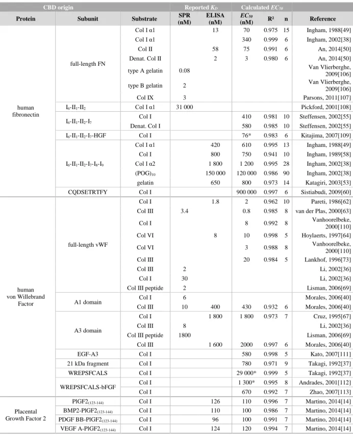
Table 3.1. Reported KD and calculated EC50 of various collagen-binding polypeptides.
See paragraph 3.4.1 for our methodological approach.
*: the plateau (saturation) has not been reached during the experiment.
CBD origin Reported KD Calculated EC50
Protein Subunit Substrate SPR
(nM) ELISA (nM) EC50 (nM) R² n Reference human fibronectin full-length FN Col I α1 13 70 0.975 15 Ingham, 1988[49] Col I α1 340 0.999 6 Ingham, 2002[38] Col II 58 75 0.991 6 An, 2014[50]
Denat. Col II 2 3 0.980 6 An, 2014[50]
type A gelatin 0.08 Van Vlierberghe,
2009[106]
type B gelatin 2 Van Vlierberghe,
2009[106]
Col IX 3 Parsons, 2011[107]
I6-II1-II2 Col I α1 31 000 Pickford, 2001[108]
I6-II1-II2-I7
Col I 410 0.981 10 Steffensen, 2002[55]
Denat. Col I 580 0.985 10 Steffensen, 2002[55]
I6-II1-II2-I7-HGF Col I 76* 0.983 6 Kitajima, 2007[109]
I6-II1-II2-I7-I8-I9 Col I α1 420 610 0.995 13 Ingham, 1988[49] Col I 800 750 0.941 10 Ingham, 1989[58] Col I α2 1 800 1 200 0.995 28 Ingham, 2002[38] (POG)10 150 000 120 000 0.986 90 Ingham, 2002[38] gelatin 650 800 0.973 14 Katagiri, 2003[53]
CQDSETRTFY Col I 900 000 0.997 6 Sistiabudi, 2009[60]
human von Willebrand
Factor
full-length vWF
Col I 1.8 2 0.962 10 Pareti, 1986[62]
Col III 3.4 0.8 0.985 8 van der Plas, 2000[63]
Col I 8 0.992 8 Vanhoorelbeke,
2000[110]
Col VI 8 10 0.998 5 Hoylaerts, 1997[64]
Col VI 3 0.988 8 Vanhoorelbeke,
2000[110]
Col III 20 0.984 5 Lankhof, 1996[73]
Col III 2 Li, 2002[36]
Col I 30 Li, 2002[36]
Col III peptide 2 Lisman, 2006[69]
A1 domain Col I 6 Morales, 2006[40]
Col III 10 400 430 0.932 6 Morales, 2006[40]
A3 domain
Col I 1 800 1 800 0.973 7 Cruz, 1995[67]
Col III 8 Li, 2002[36]
Col III peptide 1800 Lisman, 2006[69]
Col III 1 600 2000 0.997 6 Morales, 2006[40]
EGF-A3 Col I 580 0.998 5 Kato, 2007[111]
21 kDa fragment Col I 780 0.971 9 Takagi, 1992[37]
WREPSFCALS Col I 29 000* 0.999 5 Takagi, 1992[37]
WREPSFCALS-bFGF Col I 1 300* 0.995 8 Andrades, 2001[112]
Col I 670 0.992 7 Zhao, 2007[113] Placental Growth Factor 2 PlGF2(123-144) Col I 126 110 0.996 7 Martino, 2014[14] BMP2-PlGF2(123-144) Col I 110 100 0.986 7 Martino, 2014[14] PDGF BB-PlGF2(123-144) Col I 96 100 0.991 7 Martino, 2014[14]
CBD origin Reported KD Calculated EC50
Protein Subunit Substrate SPR
(nM) ELISA (nM) EC50 (nM) R² n Reference C. histolyticum Collagenase G S3b (POG)8 547 000 Matsushita, 2001[86] Col I 12 000* 0.915 10 Fukata, 2014[100]
S3aS3b (POG)8 63 000 Matsushita, 2001[86]
Col I 5 200* 0.976 10 Fukata, 2014[100]
C. histolyticum
Collagenase H
full-length ColH Col I 99.5 100 0.963 15 Matsushita, 1998[85]
S3 Col I 15 900 22 000* 0.990 17 Matsushita, 1998[85] Col I 12 000* 0.985 9 Fukata, 2014[100] S2bS3 Col I 339 2 000 0.980 17 Matsushita, 1998[85] Col I 2 100 0.934 9 Fukata, 2014[100] (POG)8 3 Brewster, 2008[114] FGF1-S2bS3 (POG)8 8 Brewster, 2008[114] S. aureus CNA N1-N2-N3 CNA55 Col I 2 100 1 800 0.979 7 Xu, 2004[115] Col I 1300 210 290 0.997 27 Rich, 1999[116] Col I 2200 Zong, 2005[94] N1-N2 CNA35 Col I 200 Zong, 2005[94] DBS4 3 Zong, 2005[94] (GPO)11 140 Zong, 2005[94] (GPP)11 7.5 Zong, 2005[94] Col I 500 260 0.992 11 Krahn, 2006[95] Col I 21 26 0.991 11 Xu, 2004[115] Col I 91 17 0.988 6 Kang, 2013[93] V. mimicus metalloprotease
Full-length VMC Col I 2100 5 Lee, 2005[96]
CBD Col I 4000 4 Lee, 2005[96]
Decorin full-length Decorin
Col I 6 4 0.977 8 Kalamajski, 2007[89] Col I 21 Nareyeck, 2004[91] Col VI 39 Nareyeck, 2004[91] LRR 5-6 Col I 7400 0.977 8 Kalamajski, 2007[89] Engineered peptide TKKTLRT Col I 2 300 0.980 7 De Souza, 1992[98]
TKKTLRT-EGF Col I 239 1 600 0.937 6 Yang 2009[117]
TKKTLRT-VEGF Col I 430 2 400 0.963 7 Zhang, 2009[118]
TKKTLRT-bFGF Col I 270 0.991 7 Zhao, 2007[113] TKKTLRT-BMP2 Col I 270 140 0.999 4 Chen, 2007[119] Col I 6 600 0.979 4 Zhao, 2009[120] TKKTLRT-NGF Col I 510 4 500 0.992 4 Sun, 2007[47] Col I 510 6 500 0.951 6 Sun, 2010[121] TKKTLRT-NT3 Col I 350 850 0.973 7 Fan, 2010[122] TKKTLRT-PDGF Col I 5.5 1 100 0.971 7 Sun, 2007[44] Col I 92 1 100 0.991 7 Lin, 2006[43] TKKTLRT-PTH Col I 292 1 200 0.996 7 Wu, 2013[123] TKKTLRT-BDNF Col I 470 1 100 0.995 8 Liang, 2010[124] Col I 420 1900 0.944 4 Han Q, 2009[46]
TKKTLRT-EphA4LBD Col I 430 1 500 0.990 7 Li, 2016[125]
TKKTLRT-PlexinB1LBD Col I 360 2 900 0.995 7 Li, 2016[125]
TKKTLRT-EGFR Ab Col I 460 2 800 0.955 8 Liang, 2015[126]
3.5 Applications of collagen-binding fusion proteins
Numerous recombinant collagen-binding proteins have been reported in the literature, most of which are growth factors or cell-binding domains. The majority of these fusion proteins have been produced in bacteria, as it can yield very high quantity of proteins, although these are not glycosylated. According to the UniProt database, all of the previously described CBDs - except those from fibronectin and decorin - are naturally non-glycosylated, and thus can be produced as recombinant protein in E. coli without any loss of collagen-binding activity[55], [67], [128]. In the specific case of FN, it is not clearly established if the glycosylation of the Asn511 residue (within the module I8) significantly impacts the affinity of this CBD for collagen[129]–[131].
This review will focus on the in vivo applications of these recombinant proteins, especially wound healing, bone regeneration and neuroregeneration.
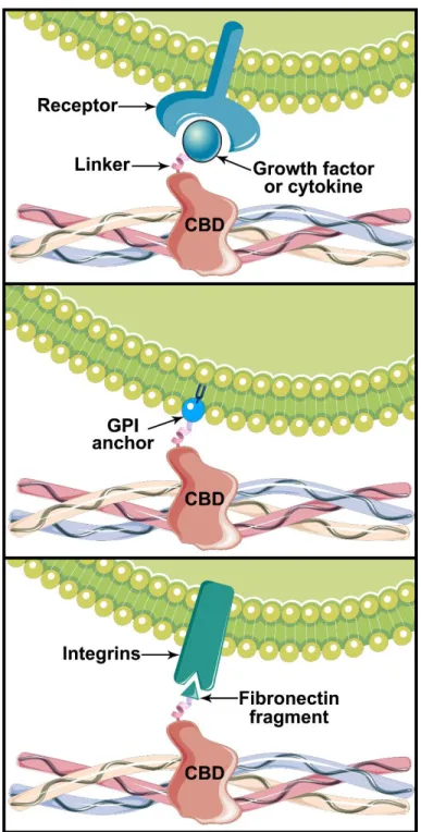
Figure 3.2. Biological effect of various chimeric proteins containing a collagen-binding domain (CBD). Collagen can be functionalized with growth factors or cytokines to stimulate cell proliferation or differentiation (A), cells can be engineered to provide collagen anchorage through a GPI-linked protein (B) and collagen can be decorated with cell-binding domains to promote cell adhesion (C).
Chimeric collagen-binding proteins for wound healing
Wound healing is a complex process initiated by an inflammatory phase (which involves the clotting cascade and the recruitment of neutrophils and macrophages), followed by a proliferative phase (characterized by a strong angiogenesis, fibroplasia, formation of granulation tissue and collagen deposition) and that ends with a remodeling phase[132]. Throughout this process, growth factors play crucial roles as chemoattractants (EGF, FGF-1/2, PDGF, TGF-β, SDF-1α)[133], [134], mitogens (EGF, FGF-1/2, HGF, PDGF)[133], [135] and promoters of angiogenesis (FGF-1/2, NGF-β, PDGF, TGF-β, VEGF)[136], [137]. All of these factors are released in a highly controlled manner for an optimal spatio-temporal distribution leading to the appropriate modulation of cell response[135]. Hence, providing exogenous growth factors may accelerate or improve the quality of the wound healing process, although the success of the approach greatly depends on the carrier used for their topical delivery[138]. One of the major drawbacks that limits the efficacy of bolus injections of growth factors for wound healing is their very short half-life in vivo[139], a problem that is sometimes addressed by daily administration of growth factors[137]. As an alternative, chimeric proteins corresponding to growth factors fused to CBD have been developed to overcome this limitation (Fig. 2A), so as to prevent the diffusion of the growth factors while maintaining their bioactivity.
EGF
It has been long known that the topical application of EGF may accelerate the rate of epidermal regeneration in the case of partial thickness skin wounds or chronic wounds, although repeated applications are required[12], [140]. In order to bolster EGF effect in vivo, several groups have engineered EGF fusion proteins displaying collagen-binding abilities.
One of the first CBD-containing fusion protein assessed in vivo corresponded to EGF linked to the WREPSFMALS peptide thanks to a 12 amino-acid-long linker[74]. This fusion protein, in contrast to the native soluble EGF, was shown to accumulate at the sites of inflammation of the colon in a nude mouse model of experimental colitis and to promote complete regeneration of the intestinal crypts after 3 days, when administered by enema[74]. The results indicated that one administration
of the fusion protein was sufficient to significantly improve healing when compared to native EGF and suggested that it bound to the collagen exposed at the site of injury site.
Ishikawa et al. developed a 46-kDa fusion protein with the CBD of Fibronectin (I6-II1-II2-I7-I8-I9)
and EGF, namely FNCBD-EGF[141]. This protein had a very stable and lasting interaction with collagen coated dishes but it did not bind to a large extent when applied alone in a full thickness wound, most probably because too little collagen was exposed at the wound site[141]. However, the functionalization of collagen sponges with FNCBD-EGF showed very good retention of the growth factor up to 4 days after implantation in diabetic wounds[141]. Oppositely, a collagen sponge loaded with native EGF showed almost no retention, most probably due to EGF diffusion around the wound[141]. Similar results have been obtained with a collagen hydrogel applied on a diabetic wound[142], highlighting that the fusion protein can be carried within various devices. In addition, the authors demonstrated that this fusion protein could directly bind to injured vessels when administered intravascularly in a rabbit model of injured carotid artery[142].
However, other designs of chimeric proteins containing a CBD and EGF led to mitigated results. The EGF-CBD developed by Nishi et al., which contained the CBD of ColH (S2b+S3 domains)[143] did not exhibit the mitogenic effect one would expect from its EGF moiety. Similarly, Kim et al. designed several chimeric proteins of EGF fused to the CBD of V. mimicus, either at the EGF N- or C- terminus[144]. They observed mitigated responses in term of EGF bioactivity as one of the CBD-EGF fusion protein did not exhibit any mitogenic effect[144]. The cause(s) of these loss of activity could be numerous: EGF misfolding due to the presence of a particular CBD (as already observed with other tagged EGF if not properly refolded[145] or steric hindrance of the CBD in absence of a long-enough linker[146].
In summary, successful CBD-EGF chimeras have been developed for enabling the capture of the epidermal growth factor in several collagen-based biomaterials that were loaded prior to implantation, as well as in vivo capture following topical delivery without the need for a carrier. The latter approach may however be more promising for vascular injuries than for skin wounds, most likely due to differences in endogenous collagen exposure.
FGF
Fibroblast growth factors (FGF) are potent mitogens and promote neovascularization. In particular, acidic (aFGF or FGF-1) and basic (bFGF or FGF-2) FGF are the most studied members of this family. Their fusion to CBDs has been extensively assessed due to the potential benefits these chimeras might bear in the field of wound healing.
Andrades et al. showed that the fusion protein WREPSFMALS-bFGF significantly reduced the healing time of wounds in both normal and diabetic rats when topically applied in a collagen hydrogel[112]. Zhao et al. compared the bioactivity of the same fusion protein with that of the chimeric protein TKKTLRT-bFGF, by implanting a functionalized collagen membrane in rats[113]. They observed that the collagen membrane functionalized with TKKTLRT-bFGF was more cellularized and vascularized after 7 days than the membrane functionalized with WREPSFMALS-bFGF[113]. Given that the latter protein has a lower affinity for collagen than the former (Table I), they suggested that the higher the affinity of the fusion protein to collagen, the higher the retention and the more significant the vascularization and cellularization[113].
A number of studies have then been conducted by this group to demonstrate that the TKKTLRT-bFGF fusion protein combined with collagen membranes could prove useful to counteract many tissue damage situations, be it for bladder regeneration[147], full-thickness abdominal wall defect repair[148], uterine horn reconstruction (all in rat)[149] and extrahepatic bile duct regeneration in pig[150]. In all cases, very encouraging results were obtained; for example, 90 days after the implantation of a collagen membrane functionalized with TKKTLRT-bFGF in a rat uterine horn damage model, the pregnancy rate increased from 33%, when untreated, to 60% when treated with bFGF and to 87% when treated with TKKTLRT-bFGF[149].
Altogether, since the bFGF moiety naturally binds to collagen and gelatin[151], the studies highlighted that CBD-bFGF fusions further enhanced the biological effects of basic fibroblast growth factor when incorporated in a collagen scaffold. Importantly, the choice of the CBD must be appropriately made in the design of a collagen-binding fusion protein, as the affinity for collagen mediates the retention and release rate of the growth factor, in turn impacting its effects in vivo.
The VEGF/PDGF family
Vascular endothelial growth factor (VEGF) and platelet-derived growth factor (PDGF) strongly promote angiogenic activity, in particular during the proliferative phase of wound healing[152], [153]. The translation of VEGF to clinical use in regenerative medicine is however compromised by its propensity to induce vascular permeability, which can lead to systemic hypotension, edema and even heart failure[154]. This limitation prevents its delivery in diffusible form in both peripheral and cardiovascular applications, due to its systemic biodistribution[14], [155]. A chimeric protein corresponding to VEGF fused to a CBD may thus eliminate this caveat by limiting VEGF diffusion.
Ishikawa and colleagues developed in that endeavor the FNCBD-VEGF121 fusion protein (where
FNCBD refers to full length CBD, i.e., I6-II1-II2-I7-I8-I9, Fig. 1A). After injecting the chimera into
the injured tibialis anterior muscle in mice, they observed that it was retained for more than 24 h at the injection site and promoted the proliferation of interstitial cells, as opposed to native VEGF121
or VEGF165[52]. Furthermore, in a mouse hindlimb ischemia model, the intramuscular injection of
FNCBD-VEGF121 did not promote the mobilization of endothelial progenitor cells in the blood
after 4 days, contrary to VEGF121 or VEGF165[52]. Altogether the results suggested that
FNCBD-VEGF121 was successfully retained in the muscle through its CBD without any systemic effect.
More recently, Martino et al. developed two chimeric proteins corresponding to VEGF121 and
PDGF BB fused to the CBD they identified in PlGF-2. The two proteins were tested in a diabetic mouse model of full-thickness wounds[14]. In comparison with the native growth factors, both fusion proteins significantly accelerated the closure of the wound, improved the granulation tissue formation and enhanced its neovascularization after 10-15 days[14]. Moreover, in comparison with native VEGF, the application of the VEGF-CBD drastically reduced the vascular permeability of the vessels when applied on the mouse ear skin, further suggesting that this fusion protein allowed for the separation of the angiogenic role of VEGF from its hyper-permeability activity[14]. Similarly, Zhang et al. fused the TKKTLRT peptide to the N-terminus of VEGF121 and
demonstrated that the functionalization of a collagen membrane with this chimeric protein significantly enhanced the neovascularization of the membrane after two weeks, when implanted subcutaneously[118]. More, the injection of this fusion protein in a rat infarcted myocardium
significantly reduced the scar size, enhanced capillary growth and improved the cardiac functions 4 weeks after injection, when compared to native VEGF121[118]. The fusion protein was also
almost undetectable in the serum 3 to 6 h post injection, although it was still present in the infarcted zone. In stark contrast, native VEGF121 had diffused into the circulation[118]. Gao et al. obtained
similar results with a collagen membrane functionalized with the same fusion protein in a rabbit infarcted myocardium model[156], providing strong evidence that myocardial infarction could be treated with a cardiac patch made of CBD-VEGF-functionalized collagen.
Encouraging results were also reported for the treatment of full thickness skin wound in diabetic rats, be it by repeated injections of the same fusion protein at the site of injury[157] or via the application of a functionalized collagen membrane[158]. Further attempts were made, with very promising results, to treat extensive urethral defect in dogs[159] and full-thickness injury uterus in rats[160]. For example, 60 days after injection in a rat scarred uterus, the pregnancy rate at the scar site increased from 6% in the control group to 19% in the group treated with VEGF and to 50% in the group treated with TKKTLRT-VEGF[160].
Similarly, Akimoto et al. constructed a fusion protein with the murine VEGF164 and the S3 domain
of C. histolyticum ColH, which was tested in a rat dorsal-skin flap model[161]. The fusion protein significantly decreased the necrosis rate and promoted neoangiogenesis after 7 days, when injected subcutaneously[161], suggesting that the bioavailability of the growth factor was prolonged. However, in this particular study, the injection of native VEGF164 did not show any effect on the
necrosis rate, in disagreement with previous studies[162], [163].
Lin et al. developed the fusion protein TKKTLRT-PDGF BB to decorate a collagen membrane[43]. 4 days after subcutaneous implantation, the biomaterial significantly promoted the cell ingrowth and neovascularization, when compared to the membrane loaded with pristine PDGF BB[43]. Moreover, when employed to treat rabbit dermal ischemic ulcer, the functionalized membrane significantly enhanced the re-epithelialization of the wound, the formation of granulation tissue and the neovascularization 14 days after implantation[44].
Altogether, CBD-VEGF fusions may have solved the major limitation of VEGF regarding its clinical translation, that is, the separation of its angiogenic properties from its negative impact on vascular permeability. More, the collagen-binding fusion proteins of VEGF and PDGF can be
administered to enhance the repair rate and extent in various types of wounds, by increasing the bioavailability of the growth factors, when compared to their native counterparts.
It is here worth mentioning that the high-molecular weight isoforms of VEGF possess a heparin-binding domain that can be used to improve their retention[164], be it via interaction with endogenous heparan sulfate or with a heparinized biomaterial[165]. However, the interaction of VEGF with endogenous heparan sulfate may be less stable than those of CBD-VEGF with collagen, since the administration of the native growth factor do not yield the same effects, as observed in most of the above-mentioned studies that used VEGF165 as control.
HGF
The hepatocyte growth factor (HGF, also named scatter factor) is an important cytokine in wound healing, given that it induces granulation tissue formation, promotes angiogenesis and accelerates re-epithelialization in vivo[166]. HGF synthesis and production are however complex insofar as they require the cleavage and processing of a single chain precursor by an enzyme, the HGF activator, leading to a mature heterodimer protein composed of the ɑ and β chains[167].
Kitajima et al. engineered a 120-kDa chimeric protein corresponding to a shortened CBD derived from Fibronectin (I6-II1-II2-I7) fused to HGF and expressed it in Sf9 cells, an insect cellular
platform that provides high levels of secreted proteins[109]. In vitro, the chimera stimulated the growth of human umbilical vein endothelial cells for more than 10 days, in contrast to the native growth factor that was rapidly internalized and degraded[109]. Seven days after the implantation of a collagen sponge in rats, the fusion protein significantly promoted neovascularization within the scaffold when compared to a sponge loaded with native HGF[109]. Moreover, the team reported that the density of blood vessels increased in a FNCBD-HGF dose-dependent manner[109], suggesting that the functionalization of the biomaterial could be fine-tuned for a specific application. Similarly to FNCBD-EGF, the fusion protein FNCBD-HGF did not accumulate in the wound to a larger extent than native HGF when applied topically in a dorsal full-thickness wound model[168], more likely due a limited exposure of endogenous collagen. This corroborates once again that the effectiveness of a collagen-binding fusion protein is bolstered when combined with the appropriate niche or carrier, such as a collagen hydrogel or a collagen membrane.
The FNCBD-HGF fusion protein was also tested in vivo by Ota et al. to treat a myocardial defect in pig, using a cardiac patch based on a collagen-containing urinary bladder matrix[169]. After 60 days, the functionalized patch significantly improved the recovery of the mechanical and electrophysiological functions of the myocardium when compared to a patch made of Dacron[169]. Of salient interest, Ohkawara et al. demonstrated that this protein accelerated the re-endothelialization of a rat carotid artery that had been injured by a balloon procedure. More precisely, re-endothelialization was observed when the protein was infused directly in the artery for 15 min, whereas infusion with native HGF did not exert such a healing effect[170]. However, this treatment significantly aggravated the hyperplasia of the neointima, most probably due to a higher local HGF concentration or a longer HGF half-life, which aggravated the stimulation of smooth muscle cells proliferation[170].
Altogether, although the production of recombinant HGF and HGF-derived chimeras is complex, the propensity of CBD-HGF fusions to enhance healing in vascular wounds has been demonstrated. Importantly, HGF potent mitogenic activity on smooth muscle cells requires their dosage to be tightly regulated, so as to mitigate hyperplasia.
Cell therapy
A significant improvement upon the localized administration of growth factors could be achieved by delivering specific cells at the site of injury and guiding them towards exposed endogenous collagen in vascular wounds.
This innovative concept was elegantly introduced by Tan and colleagues who engineered a fusion protein composed of the A3 domain of vWF and a glycosylphosphatidylinositol (GPI) anchor, a glycolipid naturally displayed at the cell membrane[171] (Fig. 2B). This glycosylated chimeric protein was produced in Chinese ovary hamster cells and used for the surface-functionalization of bone-marrow-derived endothelial progenitor cells (EPC). More specifically, EPCs were incubated with the vWF-GPI chimera which penetrated their lipid bilayer, thus providing them with a collagen-binding capacity. The CBD-displaying cells were then infused in the artery of a mouse suffering from a carotid injury. A significantly higher number of cells were incorporated at the site of injury, which led to an improved re-endothelialization of the artery when compared to the control
animals, in which the injected cells had their extracellular A3 domain cleaved from the GPI anchor prior to infusion[171].
A similar innovative approach has been developed by Shao and colleagues, who used a polypeptide containing both the collagen-binding sequence TKKTLRT and the cell-binding peptide EPLQLKM (the latter was identified by phage display and has a high specific affinity to bone marrow-derived mesenchymal stem cells, MSCs)[172]. Upon implantation in pig full-thickness wounds, collagen membranes functionalized with this polypeptide improved the healing rate, displaying numerous infiltrating cells and high blood vessel density, in stark contrast with the collagen membrane alone[172]. Although these results suggest that the polypeptide enhanced the capture of autologous MSCs at the wound site, further studies would be required to confirm the efficiency of this capture system.
Although cell-based therapy may still be controversial and in its early stages, CBD-cell fusions have proved to be effective in accelerating wound healing in animal models.
Chimeric collagen-binding proteins for bone regeneration
Bone regeneration is an intricate process regulated by numerous cytokines and growth factors. Similar to wound healing, these bioactive molecules play different roles, from chemoattraction (BMP, FGF, PDGF, VEGF, TGF-β)[173] and cell proliferation (FGF, IGF, PDGF, TGF-β)[173] to osteoinduction, i.e. differentiation of MSCs into mature bone cells (BMP, IL-11, TGF-β)[173]. Exogenous growth factors can thus be employed to functionalize scaffolds, such as demineralized bone matrix (DBM, made of spongy bone treated with HCl), in order to promote osteogenesis[33]. Some of these growth factors are already approved by the Food and Drug Administration (FDA) for bone regeneration purposes: for example, BMP2 can be administered to accelerate the healing of open tibial fractures[174]. However, the clinical translation of the other growth factors is seriously limited by the lack of control of their spatiotemporal release and the high doses that are required, which may lead to side-effects such as heterotopic bone formation or increased risks of cancer[33]. In this context, collagen-binding fusion proteins were shown to be very appealing to better control the localization of growth factors. In addition to being the main organic content of