Universite de Sherbrooke
A STUDY ON RADIOCHEMICAL ERRORS
IN POLYMER GEL DOSIMETERS
By
M AHBOD SEDAGHAT
Departement de medecine nucleaire et radiobiologie
Faculte de medecine et des sciences de la sante, Universite de Sherbrooke Sherbrooke (Quebec) J1H 5N4, Canada
Thesis subm itted to the Faculty o f Medicine and Health Sciences fo r the Degree o f Philosophiae Doctor (Ph.D.) in
Radiation Sciences and Biomedical Im aging
February 2012
Members o f the jury
Prof. Daniel Houde
Prof. Kim B. McAuley
Prof. Yves Dory
Prof. Martin Lepage
Dr. Rachel Bujold
President, Departement de medecine nucleaire et radiobiologie, Faculte de medecine et des sciences de la sante, Universite de Sherbrooke
Examiner, Department of Chemical Engineering, Faculty of Engineering and Applied Science, Queen’s University
Examiner, Departement de chimie, Faculte des sciences, Universite de Sherbrooke
Research Director, Departement de medecine nucleaire et de radiobiologie, Faculte de medecine et des sciences de la sante, Universite de Sherbrooke Research Director, Departement de medecine nucleaire et de radiobiologie, Faculte de medecine et des sciences de la sante, Universite de Sherbrooke
1+1
Library and Archives Canada Published Heritage Branch Bibliotheque et Archives Canada Direction du Patrimoine de I'edition 395 Wellington Street Ottawa ON K 1A0N 4 Canada 395, rue Wellington Ottawa ON K1A 0N4 CanadaYour file Votre reference ISBN: 978-0-494-94426-4 Our file Notre reference ISBN: 978-0-494-94426-4
NOTICE:
The author has granted a non
exclusive license allowing Library and Archives Canada to reproduce, publish, archive, preserve, conserve, communicate to the public by
telecomm unication or on the Internet, loan, distrbute and sell theses
worldwide, for commercial or non commercial purposes, in microform, paper, electronic and/or any other formats.
AVIS:
L'auteur a accorde une licence non exclusive permettant a la Bibliotheque et Archives Canada de reproduire, publier, archiver, sauvegarder, conserver, transmettre au public par telecomm unication ou par I'lnternet, preter, distribuer et vendre des theses partout dans le monde, a des fins com merciales ou autres, sur support microforme, papier, electronique et/ou autres formats.
The author retains copyright ownership and moral rights in this thesis. Neither the thesis nor substantial extracts from it may be printed or otherwise reproduced without the author's permission.
L'auteur conserve la propriete du droit d'auteur et des droits moraux qui protege cette these. Ni la these ni des extraits substantiels de celle-ci ne doivent etre imprimes ou autrement
reproduits sans son autorisation.
In compliance with the Canadian Privacy A ct some supporting forms may have been removed from this thesis.
W hile these forms may be included in the document page count, their removal does not represent any loss of content from the thesis.
Conform em ent a la loi canadienne sur la protection de la vie privee, quelques
form ulaires secondaires ont ete enleves de cette these.
Bien que ces form ulaires aient inclus dans la pagination, il n'y aura aucun contenu manquant.
Universite de Sherbrooke
UNE ETUDE SUR LES ERREURS
RADIOCHIMIQUE DANS LES DOSIMETRES
A GELS DE POLYMERE
Par
M AHBOD SEDAGHAT
Departem ent de medecine nucleaire et radiobiologie
These presentee a la Faculte de medecine et des sciences de la sante en vue de I 'obtendon du grade de philosophiae doctor
(Ph.D.) en sciences des radiations et imagerie biomedicale
Universite de Sherbrooke Sherbrooke (Quebec) J1H 5N4, Canada
Fevrier 2012
Membres du jury devaluation
President, Departement de medecine nucleaire et radiobiologie, Faculte de medecine et des sciences de la sante, Universite de Sherbrooke
Examinateur, Department o f Chemical Engineering, Faculty o f Engineering and Applied Science,
Q ueen’s University
Examinateur, Departement de chimie, Faculte des sciences, Universite de Sherbrooke
Directeur de recherche, Departement de medecine nucleaire et de radiobiologie, Faculte de medecine et des sciences de la sante, Universite de Sherbrooke Directeur de recherche, Departement de medecine Dr, Rachel Bujold nucleaire et de radiobiologie, Faculte de medecine et des sciences de la sante, Universite de Sherbrooke
© [Mahbod Sedaghat, 2012] Prof. Daniel Houde
Prof. Kim B. McAuley
Prof. Yves Dory
0
Table of Contents
List of illustrations (figures and tables)... VI Acronyms, abbreviation and symbols... VIII Thesis abstract... X
Resume de these en franfais...XII
Chapter 1. General introduction
1.1 Modem technologies in radiotherapy and related challenges...1
•1.1.1 Radiation delivery 1.1.2 Modem techniques and technologies 1.1.3 Modem technology; escalated risks 1.2 Gel dosimetry... 10
1.3 Polymer gel dosimeters... 15
1.4 Thesis outline and objectives... 29
References... 32
Chapter 2. Investigating the effect of oxygen scavengers on the
dose response, accuracy and reproducibility of polymer gel
dosimeters
Co-authorship... 412.1 A bstract... 42
2.2 Introduction... 44
2.3 Materials and methods...48
2.3.1 Oxygen diffusion in acrylamide-based gels 2.3.2 Oxygen diffusion in methacrylic acid-based gels 2.3.3 Varying [THPC] in water and gelatin gel 2.3.4 Magnetic resonance scanning 2.3.5. A practical example 2.4 Results... 55
2.4.1 Variations in the dose response of the gel dosimeters with varying [THPC]
2.4.3 Oxygen diffusion in PAG and PAGAT dosimeters 2.4.4 Oxygen diffusion in MAG and MAGAT dosimeters
2.4.5 Oxygen diffusion in MAGIC-type, PAGIC and normoxic PAGAT 2.4.6 Results of a practical measurement in clinic
2.5 Discussion... 68
2.5.1 Chemistry of normoxic polymer gel dosimeters 2.5.1.1 Effect of THPC 2.5.1.2 Effect of AscA 2.5.2 Dosimetric implications 2.6 Conclusions... 83 Acknowledgements... 84 References... 85
Chapter 3. Investigating the effect of exothermal polymerization
reaction and other physicochemical factors on the dose
response of polymer gel dosimeters
Co-authorship... 903.1 A bstract... 91
3.2 Introduction... 93
3.3 Materials and Methods...96
3.3.1 Preparation conditions 3.3.2 Phantoms and temperature probes 3.3.3 Gel fabrication 3.3.4 Irradiation, imaging and data analysis 3.3.5 Temperature variations during gelation 3.3.6 Virtual wedge experiment 3.4 Results...105
3.4.1 Acrylamide-based dosimeters 3.4.2 MAGAT dosimeter 3.4.3 NIP AM-based dosimeter 3.4.4 Cooling history 3.4.5 Virtual wedge 3.5 Discussion...117
3.5.1 Effect of temperature 3.5.2 Notable discrepancies between calibration vials and larger phantoms 3.5.3 NIPAM-based gel dosimeter 3.5.4 Siemens virtual wedge experiment 3.6 Conclusions... 134
Acknowledgement... 135
Chapter 4. Preliminary studies on the role and reactions of
Tetrakis(hydroxymethyl)phosphonium chloride in
polyacrylamide gel dosimeters
Co-authorship...141
4.1 Abstract...142
4.2 Introduction...144
4.3 Materials and Methods... 146
4.3.1 Varying the pH 4.3.2 FT-Raman spectroscopy studies 4.3.3 Scanning electron microscopy studies 4.4 Results... 150 4.4.1 pH modifications 4.4.2 FT-Raman spectroscopy 4.4.3 Electron microscopy 4.5 Discussion...156 4.6 Conclusion...163 Acknowledgement... 164 References...164
Chapter 5. Discussion
5.1 Summary of the project... 1675.2 Perspectives and future w o rk ... 175
References...176
Chapter 6. General conclusions...
178References... 181
A cknow ledgem ents... 182
Appendix
Permissions...183List of figures
Chapter 1 page
Figure 1.1 Schematic illustrating the key differences between conventional radiotherapy, conformal radiotherapy and intensity-modulated radiotherapy.
5
Figure 1.2 Schematic representation o f oxidation o f ascorbic acid and THPC 23
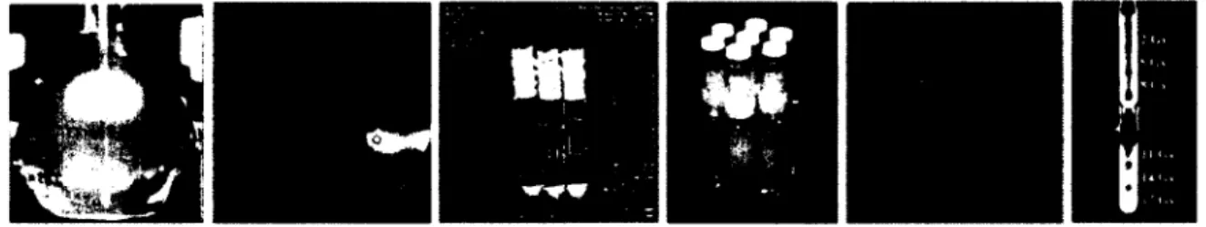
Figure 1.3 Photographs o f different calibration methods used in the polymer gel dosimetry literature
28
Chapter 2
Figure 2.1 Dose distribution in the Gammacell 220 sample chamber 51
Figure 2.2 Characteristics o f different polymer gel dosimeters 56
Figure 2.3 Progress o f polymerization inhibition front in (6%T, 50%C) acrylamide-based polym er gel dosimeter with varying THPC concentration
59
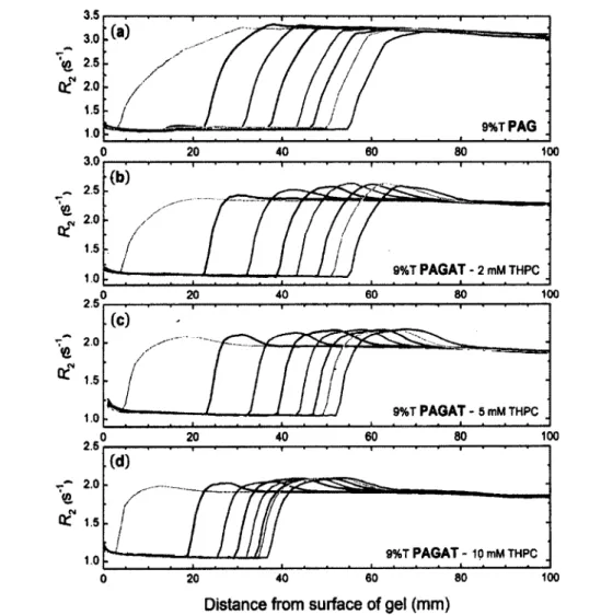
Figure 2.4 Progress o f polymerization inhibition front in (9%T, 50%C) acrylamide-based polym er gel dosimeter with varying THPC concentration
60
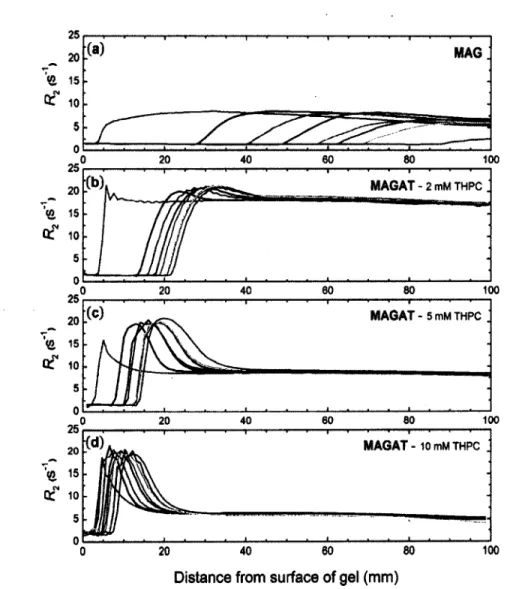
Figure 2.5 Progress o f polymerization inhibition front in methacrylic acid- based polymer gel dosimeter with varying THPC concentration
62
Figure 2.6 Progress o f polymerization inhibition front in MAGIC, PAGIC
and normoxic PAGAT dosimeters
64
Figure 2.7 Impact o f oxygen diffusion from plastic surfaces on the dose response o f PAGAT dosimeter as a function o f container size and time post-fabrication
66
Figure 2.8 Schematic demonstration o f the extent o f dose errors inflicted by a small change in the dose response o f a PAGAT dosimeter
81
Chapter 3
Figure 3.1 Photographs and schematic representation o f the phantoms
constructed for the experiments together with their preparation and set-up in the treatm ent room
98
Figure 3.2 Temperature increase as a function o f container size in (6%T) PAG dosimeter and its impact on the dosimeters dose response. The data obtained by calibration vials is also illustrated for comparison
106
Figure 3.3 Results showing temperature variations in (9%T) PAGAT
dosimeter, its impact on the dosimeters dose response and the data obtained by calibration vials for comparison
Figure 3.4 Results showing the extent o f temperature increase in methacrylic acid-based dosimeter and the corresponding dose response in different gel containers
110
Figure 3.5 Temperature increase in a modified NIPAM gel dosimeter and the
corresponding dose responses in different gel containers. The dose response obtained by calibration vials is also shown as a function o f time post fabrication
112
Figure 3.6 Temperature variations o f PAG in the four glass phantoms during
gelation
114
Figure 3.7 Temperature variations within two PAG dosimeters with different
volumes irradiated to identical doses with a Siemens virtual wedge. Profiles o f the dose response taken from the central axis o f the phantoms are also compared
116
Figure 3.8 Schematic illustrating the trends o f dose discrepancies observed in polym er gel dosimetry as related to the variations in their dose response
131
Chapter 4
Figure 4.1 Effect o f pH on transverse relaxation rates o f gelatin and dose responses o f PAG and PAGAT dosimeters
152
Figure 4.2 Comparison o f the consumption rate o f AA and BIS in PAG and
PAGAT dosimefers by FT-Raman spectroscopy
154
Figure 4.3 Scanning electron microscopy images o f gelatin, PAG and
PAGAT after 25 Gy
156
Figure 4.4 Scanning electron microscopy images o f gelatin-free poly
acrylamide solution polymerized by receiving 8 Gy and 25 Gy
157
List of Tables
Table 1.1 Summary o f polymerization reactions in polymer gel dosimeters 18
Table 2.1 Chemical components and concentration o f ingredients o f the gel
dosimeters studied in chapter 2
49
Table 3.1 Chemical components and concentration o f ingredients o f the gel
dosimeters studied in chapter 3
100
Table 3.2 Dose discrepancies related to polymer gel test-tube calibration reported in the literature
128
Table 4.1 The gels prepared for each study and the concentration o f the constituents
Symbols and Acronyms:
AA Acrylamide
ARAGIC A normoxic polymer gel composition consisting o f AA, BIS, gelatin, AscA,
HQ and copper sulfate
ADRT Adaptive radiotherapy
AscA Ascorbic acid
A gel dosimeter consisting of BIS and acrylamide in nitrogen environment
BANG and aqueous gelatin; it was later commercialized by MGS research Inc.; the
ingredients were subsequently modified under the same commercial name.
BEV Beam’s eye view
BIS N, N ’-methylene-bis-acrylamide
CRT Conformal radiotherapy
CT Computed tomography
ID, 2D, 3D, 4D one, two, three or four dimensional
3DCRT Three-dimensional conformal radiotherapy
4DRTP Four-dimensional radiotherapy
^max Depth of maximum dose in water
DO Dissolved oxygen
ES Echo spacing
FOV Field of view
FT-Raman Fourier transform Raman spectroscopy
Gy Gray; unit of absorbed dose per unit mass of irradiated material
HC1 Hydrochloric acid
HQ IGRT
Hydroquinone
Image guided radiotherapy IMRT
IMRT-CWG
Intensity modulated radiotherapy
IMRT Collaborative Working Group 2001; The US National Cancer Institute
Reaction rate constant
LCV Leuco-crystal violet
LCST Lower critical solution temperature
Linac Linear accelerator
LMG Leucomalachite green
MAA Methacrylic acid
MAG An anoxic gel dosimeter containing methacrylic acid and gelatin
MAGAT A methacrylic acid-based gel dosimeter containing THPC
MAGIC A methacrylic acid-based gel dosimeter containing AscA and copper sulfate
MLC Multi-leaf collimator
mM, pM Millimolar, micromolar
S^mbols^anc^ci™
N IPAM TV-isopropylacrylamide
NTCP Normal tissue complications probability
OAR Organ at risk
PAG An anoxic polymer gel dosimeter containing AA, BIS and gelatin
An anoxic polymer gel dosimeter consisting of poly(ethylene glycol) diacrylate, BIS and gelatin
PAGAT An acrylamide-based polymer gel dosimeter containing THPC
PAGAS An acrylamide-based polymer gel dosimeter containing AscA
PAGIC An acrylamide-based polymer gel dosimeter containing AscA and copper
sulfate
PDD Percentage depth dose in water
QA Quality assurance
R\ Spin-lattice nuclear magnetic relaxation rate (Ti )
Ri Spin-spin nuclear magnetic relaxation rate (T2 )
R m R.2 of an unirradiated gel sample
ROI Region(s) of interest
SEM Scanning electron microscope / microscopy
T, Longitudinal relaxation time
t2 Transverse relaxation time
TE Time o f echo
TCP Tumor control probability
THPC T etrakis(hydroxymethyl)phosphonium chloride
THPS Bis[tetrakis(hydroxymethyl)phosphonium] sulfate
TLD Thermoluminescent dosimeters
TPS Treatment planning system
TR Repetition time
VIPAR A polymer gel dosimeter consisting of A-vinylpyrrolidone, BIS and gelatin
[X\ Brackets indicate the concentration of compound X in mM
w/w % Weight percent; relative to the weight of the gel
%T Mass of total monomer relative to mass of mixture
Abstract:
A STUDY ON RADIOCHEMICAL ERRORS IN POLYMER GEL
DOSIMETERS
By
Mahbod Sedaghat
Departement de medecine nucleaire et radiobiologie
Faculte de medecine et des sciences de la sante, Universite de Sherbrooke Sherbrooke (Quebec) J1H 5N4, Canada
Thesis submitted to the Faculty o f Medicine and Health Sciences fo r the Degree o f Philosophiae Doctor (Ph.D.) in Radiation Sciences and Biomedical Imaging
The only true 3D dosimeters proposed to date are based on measuring a physical effect o f a series o f radiation-induced chemical reactions in a volume o f gel or plastic. While early investigations o f these 3D dosimeters raised high hopes, they have not yet found widespread use in radio-oncology departments for 3D quality assurance and verification o f modem radiotherapy treatments. Although laborious and extensive work is currently required to perform a satisfactory measurement with 3D dosimeters, the sole reason that hinders widespread clinical application o f these dosimeters is not their difficulty o f use; their dosimetric performance and reproducibility has yet to be improved. Among the proposed 3D dosimeters, those that have been mostly investigated and have found widest clinical applications are polymer gel dosimeters. A diverse range o f chemicals have been tested and proposed as potential compositions for polymer gel dosimetry. A significant work suggested use o f chemical oxygen scavengers in polymer gels as an alternative for mechanical oxygen removal. A growing number o f unexplained discrepancies in polymer gel dosimeters were reported in the literature between the results o f separate gel measurements or when attempts were made to calibrate their dose response.
This thesis was undertaken to investigate fundamental radiochemical sources o f error in polymer gel dosimeters. The basic assumption that chemical oxygen removal eliminates oxygen-related discrepancies was investigated and proved to be wrong. A
methodology was developed to isolate the effect o f oxygen on polym er gel dosimeters containing antioxidants and was applied on several gel com positions to enable comparison. Results revealed that the interplay between oxygen and antioxidant modifies the polymer yield and hence the dose response o f polym er gel dosimeters. Regardless o f the gel composition and antioxidant, this effect can induce severe dose errors in practical measurements whose origin may not be easily recognized, if the assumption o f minimal oxygen influence is considered valid. The results obtained can also be interpreted to postulate dominant reactions o f the antioxidants studied and their chemical pathways in polymer gel dosimeters.
Several other physicochemical factors were hypothesized to impact on the dose response o f polym er gel dosimeters. These factors included heat-induced polymer shrinkage and variations in the rates o f propagation/termination reactions during irradiation due to exothermal polymerization o f monomers. Detailed studies in different gel compositions refuted these hypotheses. In view o f the findings o f these investigations, discrepancies reported in the literature were analyzed and an attempt was made to categorize them and provide explanations on their origins.
Finally, the effect o f a specific oxygen scavenger, tetrakis(hydroxymethyl) phosphonium chloride (THPC), was studied on the polym er structure o f an acrylamide-based dosimeter. THPC is the antioxidant o f choice in most polym er gel dosimetry studies. FT-Raman spectroscopy and electron microscopy studies revealed that THPC takes part in reactions with the monomers during polymerization. Results were interpreted to postulate possible THPC-induced variations in the dose response o f polymer gel dosimeters.
It is concluded that although chemical oxygen removal from polymer gel dosimeters significantly facilitates their preparation, the accuracy and reproducibility o f these dosimeters may vigorously decline compared to their anoxic counterparts. Based on comprehensive studies performed, it is proposed that the major cause o f discrepancy in polym er gel dosimeters is yet oxygen.
Keywords: Polymer gel dosimetry, normoxic, calibration, discrepancy, free-radical reactions, dose error, polymerization, tetrakis(hydroxymethyl)phosphonium chlroride (THPC)
Resume:
UNE ETUDE SUR LES ERREURS RADIOCHIMIQUE DANS
LES DOSIMETRES A GELS DE POLYMERE
Par
M ahbod Sedaghat
Departement de medecine nucleaire et radiobiologie Universite de Sherbrooke
Sherbrooke (Quebec) J 1H 5N4, Canada
These presentee a la Faculte de medecine et des sciences de la sante en vue de I'obtention du grade de philosophiae doctor (Ph.D.) en sciences des radiations et
imagerie biomedicale
A ce jour, les seul dosimetres purement tridimensionnels (3D) sont bases sur la detection d’une propriete physique resultant d’une serie de reactions chimiques radio-induites dans un volume de gel ou de plastique. Les etudes initiales ont genere beaucoup d’enthousiasme mais ces dosimetres 3D ne sont pas parvenus a une utilisation repandue dans les departements de radio-oncologie pour l’assurance-qualite 3D et la verification de traitements complexes de radiotherapie modeme. Le temps et la complexity de la preparation de ces dosimetres 3D ne sont pas les raisons principals qui empechent leur deploiement clinique. Ce sont plutot leurs performances et la reproducibilite des mesures qui doivent etre ameliorees. Les plus etudies et les plus utilises parmi les differents types de dosimetres 3D sont les dosimetres a gels de polymere. De nombreuses compositions chimiques ont ete proposees et testees pour la dosimetrie a gels de polymere. L’utilisation d’un antioxydant s’est revelee une decouverte majeure dans le domaine puisque l’oxygene interfere avec le mecanisme de reponse des gels. Pourtant, un nombre croissant d’erreurs inexpliquees ont ete rapportees dans les ecrits et ont souleve un doute sur la reproductibilite et la justesse des doses determinees avec ces gels.
Cette these a ete entreprise pour etudier les sources radiochimiques d’erreurs dans les dosimetres a gels de polymere. 11 etait admis dans les ecrits que l’utilisation d ’un antioxydant permettait de contoumer tous les effets non-desires de l’oxygene. Cette these demontre que cela etait faux. Une methodologie a ete developpee pour isoler l’effet de l’oxygene dans un gel de polymere contenant ou non un antioxydant. Les resultats ont revele que la presence conjointe d’oxygene et d’antioxydant modifie la reponse du dosimetre. Independamment de
la composition du dosimetre et de l’antioxydant, cet effet peut induire des erreurs dosimetriques significatives dans des mesures simples et dont les origines etaient difficilement appreciates en ignorant l’effet de Foxygene et de l’antioxydant. Nos resultats nous permettent de postuler les reactions dominantes des antioxydants etudies et leur devenir chimique au sein d’un gel de polymere sous irradiation.
Puisque l’effet de l’oxygene et de l’antioxydant etait neglige, plusieurs autres facteurs physico-chimiques faisaient l’objet d’hypothese pour expliquer les erreurs observees. Ces facteurs incluent la compression des gels et des variations de propagation et de terminaison de la polymerisation reliees a la chaleur generee pendant la polymerisation. Des etudes detaillees de ces effets nous permettent de refuter ces hypotheses. Nos resultats nous permettent d’analyser, de categoriser et de proposer des explications sur l’origine des erreurs dosimetriques recensees dans les ecrits.
Finalement, l’effet d ’un antioxydant en particulier, le chlorure de tetrakis- (hydroxymethyle) phosphonium (THPC), a ete etudie en relation avec la structure du polymere forme dans un gel de polymere base sur l’acrylamide. Le THPC est l’antioxydant le plus utilise dans les differents dosimetres a gels de polymere. La spectroscopie FT-Raman et la microscopie electronique ont revele que le THPC reagit avec les monomeres pendant la polymerisation. Notre interpretation suggere un role important du THPC dans les variations de reponse a la dose dans les gels de polymere.
La conclusion generate de cette these est que l’elimination chimique de Foxygene par un antioxydant facilite grandement la preparation des dosimetres mais leur performance en est grandement alteree en comparaison avec des preparations dites anoxiques. L’oxygene demeure une source majeure d’erreur dans les dosimetres a gels de polymere.
Mots-cles: Dosimetrie a gel de polymere, normoxique, calibration, erreurs dosimetriques,
reactions radicalaires, radicaux libres de Feau, polymerization, chlrorure de tetrakis(hydroxymethyl)phosphonium (THPC)
Peer reviewed proceedings and oral presentations at
international conferences
i) Mahbod Sedaghat, Vincent Hubert-Tremblay, Luc Tremblay, Rachel
Bujold and Martin Lepage (2009) Volume-dependent internal
temperature increase within polymer gel dosimeters during irradiation J.
Phys.: Conf. Ser. 164 012009
ii) Mahbod Sedaghat, Rachel Bujold and Martin Lepage (2010) Impact o f
oxygen on the accuracy and precision o f polymer gel dosimeters J.
Phys.: Conf. Ser. 250 012017
Hi) M ahbod Sedaghat, Rachel Bujold and Martin Lepage (2010) Effect o f the
exothermal polymerization reaction on polymer gel dosimetric measurements J. Phys.: Conf. Ser. 250 012018
Chapter
I. General introduction
1.1 Techniques and technologies in modern radiotherapy and
related challenges
“Radiotherapy is, after surgery, the most successfully and most frequently used treatment modality fo r cancer. It is applied in more than 50% o f all cancer patients. At the time o f being diagnosed, about 60% o f all tumor patients are suffering from a malignant localized tumor which has not yet disseminated, i.e., no metastatic disease has yet occurred; thus, these patients can be considered to be potentially curable. Nevertheless, about one-third o f these patients (18% o f all cancer patients) cannot be cured, because therapy fails to stop tumor growth. This is the point where new technologies in radiation oncology, especially in 3D conformal radiotherapy, come into play: it is expected that they will enhance local tumor control ” (Schlegel 2006).
1.1.1. Radiation delivery:
Currently, treatment o f local cancers consists mainly in eliminating cancer cells with
variety o f methods, most notably surgery, chemotherapy and radiotherapy or
which single malignant cells can be individually targeted and eradicated. State o f the art
in all advanced therapies is therefore represented by those techniques that propose a more
accurate discrimination between a bulk o f cancerous cells (i.e., the tum or) and intact cells,
as eliminating cancerous cells is always accompanied with a degree o f damage to normal
and neighboring tissues. To achieve better discrimination between cancerous neoplasm
and normal tissue, radiation delivery technology for cancer therapy has vastly developed
through the past two decades. The core concern in all advanced technologies is accurate
tum or targeting. Tum or targeting should overcome two major issues in radiotherapy:
i) the first is that tumors acquire irregular shapes by invading contiguous structures
or lymph node regions (Neve 2006);
ii) the second is that many tumors may grow, shrink or move not only between the
time o f treatment planning and the start o f treatment but also during a single
treatment session or along the entire treatment process between the fractions o f
fractionated radiation therapy (Yartsev et al 2007, van de Bunt et al 2006, Webb
2006, Barker et al 2004, Bortfeld et al 2002).
The prescribed dose distribution has to be sculpted three-dimensionally to initially
conform to the tum or shape for the best treatm ent outcome; however, it is also necessary
to track the displacements o f the tum or and adapt the dose distribution to its changing
configuration during radiation delivery and/or along the treatment process. Ideally, this
will maximize tum or control probability (TCP) w hile simultaneously minimizing normal
tissue complications probability (NTCP) which is the optimum goal o f radiation dose
Techniques and technologies have been developed to address the first issue. The ongoing
integration o f these modem techniques with medical imaging devices specifically
developed for radiotherapy enables addressing the second issue. All these advances that
allow radiation oncologists to overcome both issues (/ and if) are relatively recent,
complex and still under constant development.
1.1.2. Modern techniques and technologies:
Tumors are characterized by their origin, volume, radio-resistance, location o f
occurrence in the body and invasiveness {i.e., Fielding et al 1992, Rami-Porta et al 2009,
Hall and Giaccia 2006, Peters et al 1982); delivering a sufficient dose to a tum or can
therefore be challenging depending on these factors, especially considering proxim ity o f
functional organs at risk (OAR) o f failure due to radiation. Any organ in the beam
pathway other than the tum or is potentially an OAR; depending on the location o f the
tumor, different organs can restrict the treatment delivery as they may possess certain
dose threshold tolerances beyond which their functionality would be severely damaged,
leading to health complications or death (M ilano et al 2007); such dose tolerances can be
found in the literature and have been established empirically. An important clinical
requirement is to deliver the prescription dose level such that it uniformly encompasses
the defined target volume. This is especially challenging near concave surfaces o f the
target within which an OAR may reside; such situations include, for example, a tumor
that is partially surrounding the spinal cord or brainstem (Webb 2003, Bortfeld 2006).
To spare adjacent healthy tissue to a tumor, the beam aperture was usually modified
each angle. Using X-ray computed tomography (CT) images, the concept o f beam 's-eye
view (BEV) was developed in radiotherapy to avoid geographic and dosimetric misses in
individual patients (Low et al 1990, Vijayakumar et a l 1992). Three-dimensional (3D)
conformal radiotherapy (3DCRT) is based on this concept such that a set o f fixed
radiation beams, shaped using the projection o f the target volume, are used to conform
the spatial distribution o f the prescribed dose to the 3D target volume {i.e., cancerous
neoplasm plus a margin for spatial uncertainties). The radiation beams normally have a
uniform intensity across the field or, where appropriate, have this intensity modified by
simple beam fluence modifying devices, such as wedges or compensating filters (Webb
2003, IMRT-CWG 2001). However, even the use o f many such conformal beams does
not guarantee that the final dose distribution will perfectly conform to the target volume.
The BEV o f the anatomy does not necessarily indicate the true 3D shape o f the structure
since all that is seen is projection o f the shape from each angle (M etcalfe et al 2007).
Creating a true conformal dose distribution for complex objects requires a 3D view o f the
object and its surroundings instead o f simply a series o f 2D projections. In 3DCRT even
when such a. volumetric description o f target and OAR structures is available, creating a
conformal 3D dose distribution is not straightforward, since this requires technology that
can produce nonuniform beam intensity profiles.
A radical change in the practice o f radiation therapy emerged when technical advances
enabled creating fields that have non-uniform radiation fluence and intensity (W ebb 2003,
IMRT-CWG 2001). With the development o f m ulti-leaf collimator (M LC) technology,
not only the boundaries o f a beam o f radiation could be dynamically shaped, but also
attenuators (leaves) in and out o f the field during irradiation using computer-calculated
patterns o f MLC / gantry movements (W ebb 2003, Bortfeld 2006). The MLC technology
laid the foundation o f intensity modulated radiotherapy (IMRT).
In principle IMRT allows not only to tailor the dose distribution to any irregular tumor
shape, but also to ‘paint’ the tum or with an optimized dose distribution taking into
m
CRT
\
homogeneous fluenceIMRT
inhomogeneous fluence I □ high intensity I low intensity (c) (d )Figure 1.1. Illustrating the key differences between (a) conventional radiotherapy, (b) conformal radiotherapy (CRT) without intensity-modulation and (c) CRT with intensity modulation (i.e., IMRT). For almost a century radiotherapy could only be delivered using rectangular-shaped fields with additional blocks and wedges (conventional radiotherapy). With the advent o f the multileaf collimator (MLC) more convenient geometric field shaping could be engineered (CRT). The most advanced form o f CRT is now IMRT whereby not only is the field geometrically shaped but the intensity is varied bixel-by-bixel within the shaped field. This is especially useful when the target volume has a concavity in its surface and/or closely juxtaposes organs-at-risk, e.g., as shown here in the head-and-neck, where tumors may be adjacent to spine, orbits, optic nerves and parotid glands. An IMRT fluence map is compared to a CRT field in (d) [from Webb 2003 with permission].
account the heterogeneities o f the tum or itself. For example, factors such as the
clonogenic cell density and oxygenation levels are non-uniformly distributed within a
tumor. One can hypothesize that the optimal dose distribution in the tumor is therefore
also non-uniform such that more dose is needed in regions with higher clonogenic cell
densities and in less oxygenated regions (Ling et al 2000, Bentzen 2005).
The evolution o f radiation therapy has been strongly correlated with the development
o f computer technology and the corresponding advances in diagnostic imaging
equipment. Imaging is involved in every key step o f the process, ranging from patient
staging, simulation, treatment planning, and radiation delivery, to patient follow-up. The
development o f 3DCRT and IMRT places more stringent requirements on the accuracy
o f beam targeting. In practice, large uncertainties exist in tumor volume delineation and
in target localization due to intra- and inter-organ motions. The utility o f modem
radiation technologies, such as 3DCRT and IMRT, cannot be fully exploited without
eliminating or significantly reducing these uncertainties (Xing et al 2006, Jaffray 2005).
The need to improve targeting in radiation treatment has begun an era in which medical
imaging devices are being integrated into the radiation delivery process such that organ
motion can be monitored before and/or during the treatment. This image-guided delivery
process enables verification o f the location o f the target before delivery or taking
compensating measures to revise or ‘adapt’ the delivery o f radiation to the target
displacements/deformations. Successors o f IMRT include techniques such as image
guided radiotherapy (IGRT), adaptive radiotherapy (ADRT) and four-dimensional
radiation therapy (4DRTP) all o f which are characterized by moving beams, small fields,
the tumor while minimizing the dose to healthy tissues without compromising the
prescribed target coverage (Li et al 2008, Dawson and Jaffray 2007, Xing et a l 2006,
Song et al 2005). With escalating doses however, there are greater concerns for precision
and accuracy in patient immobilization, and a greater need for better resolution in all
dimensions when imaging the patient and planning/delivering the treatment. As the safety
margins around the clinical target volumes are reduced in modem radiation therapy to
escalate the prescription doses, there is an increased concern about normal tissue
com plications that may occur as a result o f small delivery or dose calculation errors (Van
Dyk 1999). The increased complexity o f the modem technology o f radiation oncology
places greater pressure on quality assurance (QA) and quality control to ensure that
patients are treated safely (Low et al 2011, Dobler et al 2006, IMRT-CWG 2001).
1.1.3. Modern technology and escalated risks:
Challenges imposed by these new techniques on the dosimetry and QA are manifold.
M odem technologies rely on electronic data transfer networks, electronic communication
software, and software with sophisticated algorithms. The ability to test each individual
component would seem overwhelming or impossible. Some o f the major categories o f
accidents that have been reported in radiotherapy include: calculation errors, calibration
errors, inadequate review o f radiotherapy treatment plans, inadequate review o f
transcribed patient treatment charts, and error in anatomical area to be treated (M etcalfe
et al 2007). Severe dose errors or geometrical misses can still originate from an
individual mistake (human error), or software malfunction due to the complexity o f the
systems (TPS) use a variety o f dose calculation algorithms that approximate electron
transport and photon scattering in the interest o f calculation time (Ahnesjo and
Aspradakis 1999, Ahnesjo et a l 2006). Although for most usual cases these calculations
have shown adequate accuracy, care must be taken since approximations in some o f these
algorithms may result in non-accurate plans. This is true when the complexity o f the
treatment site increases (for example in head and neck treatments) and especially where
small IMRT segments are used (Knoos et al 2006, Muralidhar et al 2009). Another
example o f dose calculation errors has recently been reported by Jang et al (2009) who
reviewed IMRT treatm ent plans o f 23 patients with lung cancer including 15 cases with
severe pulmonary complications after radiotherapy. They found up to 25% dose error in
the low-dose regions o f their TPS-generated IMRT plans compared to a Monte Carlo
calculation, due to inadequate modeling o f MLC transmission and leaf scatter in their
commercial TPS.
Performing a fully 3D verification o f the intricate dose distributions as is the case in
most advanced radiotherapy treatments is very challenging. A recent study in the United
States (Ibbott et al 2008) indicated that a considerable number o f institutions who
participated in the survey (71 out o f 250 institutions, i.e., close to 30%) have failed to
deliver a dose distribution to a head-and-neck standard test phantom that falls within a
generous passing criteria (7% dose difference / 4 mm distance-to-agreement) compared
to their own TPS plan calculations.
Such shortcomings mainly originate from the fact that rapid advances in the
technology to deliver 3D radiation treatments have not been paralleled by corresponding
The tools presently used in the clinic for dose verification are either point dosimeters
or two-dimensional (2D) dosimeters (Low et al 2011). Point dosimeters include
ionization chambers and solid state dosimeters (e.g., different types o f diode detectors
and thermoluminescent dosimeters (TLDs)). 2D dosimeters include films (radiographic
and radiochromic) and arrays o f ion chambers or diodes. N um erous limitations (e.g.,
volume averaging due to the finite size o f the detector, energy dependence, dose rate
dependence, directional dependence, sensitivity limitations) restrict the use o f these
detectors to measure dose distributions in 3D, especially in IMRT cases where many
regions containing steep dose gradients exist, even within target volumes.
IMRT doses are also delivered dynamically; the incident fluence shape and intensity
are varying during the treatment such that specific points in the irradiated volumes
receive their final dose only over a total treatment time (Low et al 2011, IMRT-CWG
2001). Dynamic delivery is optimized by algorithms that use stochastic elements during
generation o f beam apertures such that apertures including single isolated leaves and
disconnected small segments are commonly created (Ceberg et al 2010). The delivery
generally includes a variable gantry speed, continuous leaf motion and dose rate
modulation. Therefore the resulting dose in each point is decoupled from the beam
geometry and may not be simply proportional to the dose rate. This makes scan-based
dose measurements impractical; i.e., dose measurements o f IMRT treatment plans are
limited to integrating dosimetric techniques (Low et al 2011). A comprehensive
verification o f a 3D dose distribution demands a laborious number o f measurements,
which present, at best, a partial sampling o f the distribution. A task group (TG 120) from
report highlighting the differences in dosimetry requirements between conventional
radiation therapy and IMRT (Low et a l 2011). The task group clearly states that radiation
dosimetry measurement techniques typically employed for conventional 3DCRT should
be reviewed and adapted for the unique challenges imposed by IMRT.
In the search for a fully 3D measurement tool, the first gel dosimeter emerged over a
quarter o f a century ago for medical applications (Gore et a l 1984). It was based on the
idea that a radiation-induced chemical alteration can be immobilized within a hydrogel
volume; if this chemical change is proportional to the absorbed dose, then appropriate 3D
imaging devices that are sensitive to the effect o f the chemical alteration can be used to
extract the distribution o f the chemical change which, ideally, should be identical to the
distribution o f absorbed dose in the volume o f the dosimeter. Several generations o f 3D
dosimeters have so far been developed and investigated, but the basic idea remained the
same; i.e., detecting an ideally stable radiation-induced chemical change that is ‘fixated’
within a holding 3D medium.
1.2 3D gel dosimetry
Gel dosimetry is in principle, a fully 3D dosimetry tool in the same sense as medical
imaging modalities such as magnetic resonance imaging (M RI) and X-ray computed
tomography (CT) can provide a 3D view o f patient anatomy. Radiotherapy gel dosimetry
is a form o f chemical dosimetry that relies on quantification o f a radiation-induced
chemical reaction. As this chemical reaction is locally preserved within a holding
chemical change can be mapped and quantified in three dimensions by analyzing the
images.
\
Essential constituents o f these dosimeters generally include water, a gelling agent (e.g.,
gelatin, polyvinyl alcohol or agarose) and a radiosensitive chemical that plays the role o f
a ‘radiation detector’. As gel dosimeters generally contain high water content (~ 80 to
90% o f their weight) they are effectively w ater equivalent for clinical photon and electron
energies. This makes them radiologically near soft-tissue-equivalent and eliminates the
need to correct for the absorbed dose in the dosimeter medium over a large range o f
clinical energies. The dosimeters are liquid during their preparation and can be poured
into shaped vessels and phantoms (including clinically interesting anthropomorphic
phantoms) prior to jellification (e.g., Gum et al 2002, Vergote et al 2004). The fact that
these dosimeters are simultaneously phantoms as well as dosimeters is another desirable
characteristic that makes them potentially very suitable for the verification o f complex
dose distributions as occurs in clinical settings, especially because there would be no
need for any corrections for the incident beam direction, beam perturbation or geometry
o f the phantom.
Proposed for modem clinical applications, currently three types o f gel dosimeters can
be distinguished; naming them chronologically by their first appearance in the literature,
these include:
i) Fricke gels (proposed first by Gore et al 1984);
ii) Polymer gels (first by Maryanski et al 1993); and
iii) Radio-chromic gels (several types were simultaneously proposed namely by
The three systems are based on a hydrogel matrix that preserves the spatial
distribution o f absorbed dose in the dosimeter. The chemical ‘detector’ that responds to
the absorbed dose in each type is different: i.e., the concentration o f ferric ions in Fricke
gels, the polym er yield in polymer gels and a change in the color o f the medium in radio-
chromic gels. Ionizing radiation triggers chemical reactions in each gel; the final
chemical yield is proportional to the energy absorbed in the dosimeter volume, but what
is measured at the end is a physical property related to the chemical change (Watanabe
2005); i.e., the 3D data extraction is not performed by measuring the chemical yield, but
by measuring a proportional physical consequence o f the chemical changes. Each class o f
gel dosimeters can produce an image contrast in one or several imaging modalities.
Depending on the type o f chemical reactions in each gel class, a certain imaging modality
can or cannot detect the effect o f the chemical changes in that specific gel class. For
example Fricke gels can primarily be scanned by MRI and optical CT (Schreiner 2004),
but not with X-ray CT or ultrasound. Polymer gels can produce a contrast in all imaging
modalities that have been investigated for gel dosimetry (Baldock et al 2010).
Radiochromic gels can be scanned only with optical scanners. Therefore gel dosimetry is
a system comprising o f a chemical aspect and an imaging aspect; the optimal accuracy o f
the measurements depends on simultaneous and inter-correlated optimization o f both
aspects. In principle this means that imaging param eters should be optimized for each
specific gel formulation that is developed. The choice o f the imaging modality depends
on the accessibility o f the scanner for a specific research group or radiotherapy center.
MRI was the first method used for gel dosimetry and is still the gold standard. However,
oncology centers. Consequently alternative imaging modalities such as X-ray CT,
ultrasound and optical CT were investigated (Hilts et al 2005, M ather and Baldock 2003,
Doran 2009). From these, optical CT is the second scanning method o f choice in gel
dosimetry due to its low cost and simplicity. However, the method is still under
development and is currently restricted to relatively small cylindrical gel phantoms to
reduce imaging artifacts.
Because chemical reactions are generally nonreversible, gel dosimeters are disposable
dosimeters and should be cast away securely after use according to safety instructions o f
the chemicals used.
Unfortunately, each class o f gel dosimeters suffers from a m ajor chemical drawback.
In early investigations o f Fricke gel dosimetry it became apparent that the dosimeter is
impaired by severe post-irradiation instability due to high rates o f ferric ion diffusion
(Olsson et al 1992, Schulz et al 1993, Harris et a l 1996, Baldock et a l 2001a). Therefore
spatial dose information was rapidly degraded particularly near high dose gradients and
in small fields. Some effort was given to the exploration o f alternative gelling agents
(Chu et al 2000) and systems with chelators {i.e., an organic chemical that forms two or
more bonds with a central ferrous or ferric ion) (Rae et al 1996), but these were found to
only partially suppress diffusion. This imposes practical time constraints from the start o f
irradiation to the end o f the dose measurement. It effectively prevents imaging o f dose
distributions produced by multiple field treatments or medium and low output rate
radiation sources. Currently these dosimeters require a relatively quick radiation delivery,
In polym er gel dosimeters while post-irradiation stability is not a detrimental issue as
in Fricke gels, the chemical drawback is sensitivity to atmospheric oxygen (Maryanski et
al 1993, Hepworth et al 1999). Oxygen inhibits the polymerization processes such that
polymer gels made under normal atmospheric conditions are not radiosensitive. As a
result, these gel dosimeters had to be manufactured in an oxygen-free environment, for
example in a glove box flushed with an inert gas such as nitrogen or argon (Baldock et al
1998a, De Deene et a l 1998a).
A solution for the problem o f oxygen inhibition in polymer gel dosimeters was
suggested in 2001 by Fong et al who proposed adding an antioxidant to the gel during
fabrication to chemically remove oxygen. The antioxidant binds dissolved oxygen
molecules in the gel such that radical polymerization is not hindered from proceeding by
oxygen radicals. This was considered a major development in the field o f gel dosimetry
as gel fabrication was significantly facilitated. One o f the main objectives o f this thesis
was to investigate further into the ambiguous dosimetric discrepancies observed in these
normoxic polym er gel dosimeters. It is unfortunate to report in this thesis that the major
chemical flaw for polym er gels still persists in a concealed manner in normoxic polymer
gel dosimeters.
Radiochromic gel dosimeters are relatively recent and their chemistry and dosimetric
performance has not yet been investigated or reported in detail. Several chemicals have
been suggested for radiochromic gel dosimetry. These include leuco-crystal violet (LCV),
leucomalachite green (LMG) and Turnbull’s blue dye (Babic et a l 2008, Sole and
Speva£ek 2008). These water-insoluble pigments are held artificially suspended within
genipin (a fruit extract) which is a natural gelatin cross-linker was introduced in 2008 that
forms a dark gelatin gel that bleaches upon irradiation (Jordan 2008). A recent study has
found severe dose rate dependence in LCV- and LMG-based dosimeters (Vandecasteele
et a l 2011), which significantly limits the use o f these dosimeters in most clinical
applications where dose rate is varied. Although post-irradiation stability o f radiochromic
gels is reported to be excellent (Babic et al 2008, Jordan 2008), and oxygen does not
need to be expelled from these gels as is the case for polym er gel dosimeters, it is not
unreasonable to suspect that other derogatory chemistry-related issues could be found in
them upon more research and usage o f radichromic gel dosimeters.
1.3 Polymer gel dosimeters
As mentioned earlier, a polymer gel dosimeter consists o f three major components: water
(typically between 80 to 90% by weight (w/w)), monomers (between 2-10 % w/w) and a
gelling agent (between 5-8% w/w). Monomers are small organic molecules, containing at
least one carbon-carbon double bond, that are capable o f reacting with identical or similar
molecules o f low molecular weight to form a large molecule (macromolecule) called a
polymer.
1.3.1. M onom ers:
Early polymer gel dosimeter formulations were based on acrylamide (AA) as a
monomer combined with an equal concentration o f a structurally similar co-monomer
called A /A ’-methylene-bis-acrylamide (BIS) (Maryanski et al 1993). BIS is the most
formulation was eventually given the acronym PAG (acronym base on poly-acrylamide
in gelatin) to distinguish in-house polymer gel formulations from a patented product
subsequently commercialized by MGS Research Inc. Since then several other acrylic
(Lepage et al 2001c, Fong et al 2001, Sandilos et al 2004, Saion et al 2005) or non
acrylic monomers (Pappas et al 1999, Senden et al 2006) were investigated and
subsequently suggested for gel dosimetry. Studies on alternative monomers other than
that originally proposed by Maryanski et a l (1993) were driven by the motivation to find
monomers with a higher sensitivity to radiation as in any experiment, a large response o f
the detector is desirable. Other monomers were studied for their lower toxicity (Senden et
al 2006), larger dose range (Kipouros et al 2001) or better dose response at low doses
(Sandilos et al 2004). Success was modest such that the original PAG formulation is still
the gold standard for new gel dosimeters to which their dosimetric performance is
generally compared (De Deene et al 2006a, Baldock et a l 2010). Several factors
contribute to this minimal success; for example solubility o f monomers in powder form is
limited in water (Koeva et al 2008). The use o f alternative water-soluble monomers in
liquid form is also restricted either by their low reactivity (Pappas et al 1999, Sandilos et
al 2004) or by the fact that gelatin may not set when a high concentration o f liquid
monomer is added to the solution. Other monomers that have been proposed lack
chemical stability as will be discussed further in this thesis. On the other hand, higher
chemical sensitivity is desirable as long as chemical changes can be measured with a low
uncertainty; therefore a polymer gel with a lower sensitivity whose response can be
higher uncertainty in measurements; this is where the power o f scanner in providing
accurate measurements faces its boundaries.
1.3.2. Basic reactions and oxygen sensitivity
The sensitivity o f polym er gel dosimeters to radiation and their large dynamic range is
based on one o f the most effective chain reactions known to chemistry, i.e., free radical
polymerization (W hittaker 2001). Free radicals that initiate polymerization in polymer
gel dosimeters originate from w ater radiolysis. This is the process in which water
molecules are dissociated in several highly-reactive radicals and ions upon irradiation. In
the case o f megavoltage radiotherapy the majority o f radiation dose is deposited by
collisions o f high energy electrons with w ater molecules that initiate a cascade o f
chemical reactions. In summary, the decomposition o f reactive intermediates can be
written as a simplified reaction o f which the dissociation rate (kD) is proportional to the
absorbed dose (Table 1.1). Radical species that may react with monomers are mostly the
hydroxyl radical (OH*), the hydroxonium ion (H3 0+) and particularly aqueous electrons
(W hittaker 2001, De Deene 2004a, Lepage and Jordan 2010, Baldock et al 2010). These
radicals initiate the polymerization o f monomers by binding to an electron o f the double
bond o f the monomer.
The growth o f polym er chains is a result o f chain propagation reactions by which the
monomer and polymer radicals react further with other monomers. This kinetic chain
length is typically 103-104 monomer units in most free radical polymerizations (W hittaker
2001, Lepage et al 2000). The general case in which a polym er radical with n monomer
heat ( 0 in the gel dosimeter as propagation reactions are exothermal (Salomons et al
2002).
Propagation continues until a termination occurs. Termination o f the polymerization
reaction occurs after two growing macroradicals encounter each other. The radicals may
undergo disproportionation or combination reactions, resulting in two dead polymer
chains or one, respectively. Primary radicals generated by w ater radiolysis can also react
with growing polymers and terminate their propagation. In addition to these termination
Table 1.1. A simplified summary of polymerization reactions in polymer gel dosimeters REACTION RATE Radiolysis H20 - + 2R* k[) Initiation R*+Mn -> M*n km , n=l or n> l Propagation M ’n+M m — RM Vm + Q kp(n,m ) mutual combination: R* + R* —►RR k,/ R*+ M*n —> Pn k,2(n) M n+ M m * P n+m kljjn .m j Termination disproportionation: (Oxygen-free environment) M*n+ M*m —* Pn + Pm ki-Kn.rn/ chain transfer: M*n+ SX PnX+S* k tr[M*][SX] S*+M — SM*
reactions, the growing polymer radical may also terminate by transfer o f the radical
group to other molecules
Baldock et al 2010). In g^
extent o f polymerization,
, for example gelatin (W hittaker 2001, Lepage et al 2001a,
dosimeters where increasing gelatin concentration reduces the
t was debated that the effective rate o f termination is increased
hold true for possible impurities in the gel solution that decrease the extent o f
polymerization.
The rate o f propagation and termination is dependent on the radical concentration; as a
result, polymerization becomes dose rate dependent. To what extent the dose response
depends on the dose rate is determined by the reactivity o f the various components in the
gel dosimeter (De Deene et a l 2006a). Chain termination reactions by combination and
by disproportionation involve reactions between two free radicals and as a result, the
rates o f these termination reactions are proportional to the square o f the radical
concentration. When radical concentrations are doubled, the rates o f propagation and
chain transfer reactions increase by a factor o f 2, whereas the rates o f bim olecular
termination reactions increase by a factor o f 4. Therefore polym er gel systems in which
the dominant termination reaction is chain transfer to gelatin show very little dose rate
dependence whereas in systems in which combination or disproportionation reactions are
dominant, polym er formation is much more dependent on the dose rate (Karlsson et al
2007, De Deene et al 2006a).
Peroxide radicals ([O-O] *2-) are created when oxygen is present in the gel (De Deene
et al 2002, Baldock et al 2010). These peroxide radicals will quickly react with other
radicals leading to termination. No polymerization occurs in gel dosimeters from which
oxygen has not been expelled. It was shown that trace amounts o f oxygen can have a
significant inhibitory effect on gel polymerization (Hepworth et al 1999).
Ideally in polymer gel dosimeters, the extent o f polymerization should only be a
function o f absorbed dose. In reality however, it is also a strong function o f the oxygen
only should polym er gel dosimeters be prepared in an oxygen-free environment in
specialized preparation facilities, but they should also be kept from oxygen diffusion
during storage and even after irradiation, as long as the polymerization reactions are not
terminated. In principle, this limits the choice o f gel container materials to very low
oxygen diffusing materials such as hard glass. M ost plastics adsorb oxygen and return it
back to the gel solution if used as a dosimeter container (Stevens 1992, Maryanski et al
1994). The extent o f oxygen contamination can never be exactly estimated and may not
be identical in different containers which will result in significant dosimetry errors.
1.3.3. Normoxic polymer gels and oxygen scavengers:
All these challenges were assumed to be overcome when a new gel dosimeter was
proposed that was radiosensitive under normal oxygen levels at room atmosphere (Fong
et al 2001). The gel also possessed significantly higher radiation sensitivity in MRI
compared to the traditional PAG formulation by using methacrylic acid as the monomer.
The brilliant idea in preparation o f this gel was that a small concentration o f ascorbic acid
(AscA), which is a well known chemical antioxidant, was added to the gel dosimeter
together with a small concentration o f copper sulfate to scavenge superoxide radicals
and/or to prevent oxygen from forming peroxide radicals by binding them into metallo-
organic complexes in a process initiated by copper sulfate.
The idea o f chemical oxygen removal from polymer gel dosimeters was further
investigated by De Deene et al (2002) and Maryanski (2002). Several antioxidants
including gallic acid, trolox, A-acetyl-cysteine, diethylhydroxylamine, glucose with
studied in polymer gel systems. N one o f these were found as effective as AscA; the
radiosensitivity o f gel systems prepared with these antioxidants was low such that none
o f them could be a proper substitute o f the recipe developed by Fong et al (2001).
However, the group at Ghent University found an oxygen-scavenging phosphorous
compound (De Deene et al 2002) that could produce a more radiosensitive polym er gel
than that originally reported by Fong et al (2001). M easurement o f oxygen levels in water
or polymer gel systems confirmed that oxygen concentration is reduced to a negligible
amount within a short time after addition o f chemical oxygen scavengers or antioxidants
(De Deene et a l 2002, Jirasek et al 2006). From studying oxygen inhibition effects in
PAG gel dosimeters, De Deene et al (2002) suggested that if a gel dosimeter shows any
dose response for doses below 5 Gy, this indicates that at least 98% o f oxygen in the gel
is chemically bound by the oxygen scavenger. Polymer gel formulations prepared with
oxygen scavengers showed no inhibition effects at low doses (Venning et a l 2005, Hurley
et al 2005); some formulation even had better dose sensitivity at low doses compared to
their hypoxic counterparts {e.g., Kozicki et a l 2007). This made the basis o f an
assumption that the dose response o f these dosimeters is not affected by oxygen and
potential systematic chemical uncertainties in their dose response are negligible
(Petrokokkinos et al 2009). Numerous authors subsequently investigated different
compositions and formulations o f this new generation o f polym er gels which became
known as ‘norm oxic’ gel dosimeters (Baldock et a l 2010).
So far, as mentioned earlier, only two oxygen scavengers have been found suitable for
fabricating normoxic polymer gel dosimeters; the first is ascorbic acid (AscA) and the
although oxidation o f these antioxidants has been investigated in aqueous solutions, their
mechanism o f action in polymer gel systems and especially their effects on the dosimetric
performance o f gel dosimeters is not fully understood. In an aqueous solution AscA
undergoes a two step reversible oxidation process from its predominant anion form (AH”)
to its dehydrated form (A) with the formation o f ascorbyl radical (A”’) as an intermediate
(Bendich et al 1986, Bielski 1982). The efficiency o f AscA as a strong radical scavenger
is due to the stability o f its radical. Because o f the delocalized nature o f the unpaired
electron in A”*, it tends to react preferentially with itself thus terminating the propagation
o f free radical reactions. Figure 1.2(a), adapted from Bendich et al (1986), is a schematic
representation o f oxidation o f AscA in aqueous solution.
Oxidation o f THPC in aqueous solution has been reviewed in the gel dosimetry
literature by Jirasek et al (2006) based on work by Vullo (1966). THPC is first reduced to
tetrakis(hydroxymethyl)phosphonium hydroxide (THPOH) as a result o f proton exchange
with water. THPOH then dissociates to tetrakis(hydroxymethyl)phosphine (THP). THP
scavenges oxygen by losing one hydroxymethyl group (CH2OH) and forming
tris(hydroxymethyl)phosphine oxide (TrHPO). Formaldehyde and hydrochloric acid are
byproducts o f the dissociation o f THPC in water solution. TrHPO will further react with
formaldehyde to produce more stable final products. Figure 1.2(b) schematically
illustrates oxidation o f THPC, based on the review by Jirasek et a l (2006).
The reactions o f these antioxidants in gel dosimeters are not necessarily restricted to
oxygen. They can potentially react with other constituents o f the gel either during gel
fabrication/storage, or during irradiation and even after irradiation, hence modifying the
performance and accuracy o f normoxic polymer gel dosimeters is one o f the main objectives o f this thesis.
(a)
2e , +2H+~ H+,
O
“ e .HO
OH
L-Ascorbic acid
Ascorbyl radical
( A - )
HO
e-^
3-C
Dehydro-L- ascorbic acid
(A)
<b>
H , ' " X'[or]
Hydrochloric acid
-^O NTrHPO
THPC
THPOH
■'r
Formaldehyde
Figure 1.2. Schematic representation of the oxidation of (a) ascorbic acid and (b) tetrakis- (hydroxymethyl) phosphonium chloride (THPC) in an aqueous medium. Refer to the text for abbreviations.


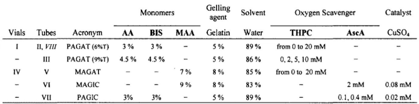

![Figure 2.2. Characteristics o f different polymer gel dosimeters. Dose-response curves of: (a) polyacrylamide gels (5% gelatin, 6%T); (b) polymethacrylic acid gels (8% gelatin, 7% M AA) with [THPC] from 0 to 20 mM](https://thumb-eu.123doks.com/thumbv2/123doknet/2590005.56969/71.924.226.694.71.930/figure-characteristics-different-dosimeters-response-polyacrylamide-polymethacrylic-gelatin.webp)