*Corresponding author: Jonathan Richir, Numerical Ecology of Aquatic Systems, University of Mons, Pentagone 3D08, 6, Avenue du Champ de Mars, 7000 Mons, Belgium, Tel: 3243662111; E-mail: jonathan.richir@alumni.ulg.ac.be
Received October 12, 2015; Accepted January 21, 2016; Published January 25, 2016
Citation: Richir J, Gobert S (2016) Trace Elements in Marine Environments: Occurrence, Threats and Monitoring with Special Focus on the Coastal Mediterranean. J Environ Anal Toxicol 6: 349. doi:10.4172/2161-0525.1000349 Copyright: © 2016 Richir J, et al. This is an open-access article distributed under the terms of the Creative Commons Attribution License, which permits unrestricted use, distribution, and reproduction in any medium, provided the original author and source are credited.
Trace Elements in Marine Environments: Occurrence, Threats and
Monitoring with Special Focus on the Coastal Mediterranean
Jonathan Richir
1,2*and Sylvie Gobert
21Numerical Ecology of Aquatic Systems, University of Mons, Pentagone 3D08, 6, Avenue du Champ de Mars, 7000 Mons, Belgium 2Laboratory of Oceanology, MARE Centre, University of Liege, B6C, 4000 Liege, Sart Tilman, Belgium
Keywords: Trace element; Toxicity; Aquatic environment;
Monitoring; Seagrass; Mussel; Mediterranean
Introduction
Such as terrestrial ecosystems, marine ecosystems are submitted
to increasing anthropogenic disturbances [1]. On the basis of expert
judgment, Halpern et al. [2,3] mapped the impact of 17 anthropogenic
drivers of ecological change (e.g., pollution, fishing, ocean acidification,
species invasion etc.) on marine ecosystems. Their analysis indicated
that no area remained unaffected by human activities, that a large
fraction of the oceans was strongly affected by those drivers (41%),
but that some large, less-impacted oceanic areas still remained
(3.7%, particularly near the poles). The ecologic, economic and social
importance of marine ecosystems being irrefutable [1,4], a
well-planned approach of managing the marine space is essential to achieve
sustainability [1]. Otherwise, entire ecosystems will stop functioning
under their actual form, as is the case for the highly productive hotspots
of biodiversity that are coral reefs [5], which is likely to lead to the
complete loss of goods and services derived from these ecosystems [6].
The Mediterranean is one of the richest regions of Europe in terms
of diversity of marine species with a high rate of endemism [7]. In the
Mediterranean, coastal ecosystems are dominated by macrophytes
(magnoliophytes and algae) [8], a globally net autotrophic system
displaying many ecological benefits (e.g., primary production, habitats,
source of food and oxygen, carbon well, stabilization of sediments etc.)
[9]. Despite their environmental, economic and social importance, a
growing number of reports document the occurring regression and/
or ecofunctional changes of these coastal ecosystems (e.g., Ref.
[10-12]). Pressures suffered by the Mediterranean make it a vulnerable
ecological unit, in particular because this sea is of too small dimensions
to ecologically self-counterbalance. Thus, the point of saturation of the
contaminants discharged in the Mediterranean will be more quickly
achieved than in the oceans [13]. And as regards the specific chemical
contamination by trace elements, the high levels currently measured in
the Mediterranean indicate non-stationary geochemical cycles which
result from an increase of external inputs [14]. In addition, the almost
total absence of tide does not allow the dilution of contaminants and
prevents the natural phenomena of depuration as encountered in
larger water bodies (i.e., in oceans). The Mediterranean also shows
a deficiency in the movement of its deep water masses and of its
surface currents which "turn in circles" in this almost closed basin.
The consequence of these specific features is that the answer of the
Mediterranean to environmental disturbances is more rapid than in the
larger oceans [15,16] which makes of this sea a privileged laboratory to
study environmental changes resulting from anthropogenic pressures
[17].
Trace elements
Definition
According to the International Union of Pure and Applied
Chemistry [18], trace elements are any element having an average
concentration of less than about 100 parts per million atoms (ppma)
or less than 100 µg g
-1. Such a precise definition does not exist in earth
sciences because the concentration of an element in a given phase
can be so low that it is considered a trace element, whereas the same
element can constitute a main part of another phase (e.g., Fe and
Al) [19]. Previously, scientists used the generic term "heavy metals"
when referring to trace elements. Today this appellation is discussed.
Effectively, some metals are not particularly "heavy" (e.g., Al, Ni). In
addition, some elements are not metals (e.g., As, Se). For these reasons,
the majority of researchers prefer today the name "metallic trace
elements" (if it is indeed metals) to the appellation "heavy metals", or
the formula "trace elements" when they are not metals (e.g., As, Se, B)
[20]. In the present review, we will consider trace elements (acronym
TEs used throughtout the paper) as any element, metallic or not, other
Abstract
Trace elements, as building blocks of matter, are naturally present in the environment. However, their extraction, production, use and release by men can lead to the increase of their environmental levels to concentrations that may be toxic for both men and the biota. The overall aim of this review is therefore to recall that trace elements remain contaminants of concern that still require scientific attention. Because marine coastal systems (and transitional environments in general) are particularly vulnerable to contamination processes, they deserve to be accurately monitored with quality indicator species. As an example, the 2 most widely quality indicator species used to assess the health status of the coastal Mediterranean are the seagrass Posidonia oceanica and the mussel Mytilus galloprovincialis. In this review, after a short introduction on human pressures on the World Ocean and the coastal Mediterranean in particular (1), we will redefine the term trace element from an environmental perspective and discuss their accumulation and toxicity for men and the biota (2). We will consider the benefits of using biological indicators instead of water and sediment measurements to assess the health status of the marine environment (3), and more particularly as regards the accurate and complementary indicators that are seagrasses (4) and mussels (5).
[26,37,39-41], the supply of quality water remains a major challenge
for humanity in the 21
stcentury [42]. The spectrum of adverse effects
caused by the consumption of contaminated water is huge and ranges
from relatively harmless to life threatening (Table 2) [38]. For example,
populations exposed to inorganic As via drinking water showed excess
risk of mortality from lung, bladder and kidney cancer and an increased
risk of skin cancer and other skin lesions such as hyperkeratosis and
pigmentation changes [43]. Japanese who consumed Cd polluted rice
and river water accumulated in their bodies a large amount of Cd that
lead to a serious osteoporosis-like bone disease, the “itai–itai byo”
or “ouch–ouch disease” [44]. And in some of the great Cairo cities,
Egypt, renal failure was related to Pb and Cd contamination of ground
drinking water [45].
Essential micronutrients may also show toxic effects when ingested
at too high levels, as reviewed by Goldhaber [46] and widely detailed in
Nordberg et al. [26]. The ingestion of very high doses of Cr causes liver
and kidney problems. Abdominal pain, cramps, nausea, diarrhea, and
vomiting have been caused by the consumption of beverages containing
high levels of Cu and liver damage has been seen in individuals with
diseases of Cu metabolism. The consumption of Mn-containing well
water has caused lethargy, tremor, and mental disturbances in Japan
and neurologic symptoms were reported in individuals exposed to
Mn-contaminated drinking water in Greece.
Sources of TEs in the sea
Continental runoff and atmospheric deposition are the primary
natural inputs of TEs in the marine environment [47]: crustal material
is either weathered on (dissolved) and eroded from (particulate) the
Earth’s surface or injected into the atmosphere by volcanic activity.
Forest fires and biogenic sources are of a lesser importance [48,49].
In addition to these natural sources, there exists a multitude of
anthropogenic emission sources, the major ones resulting from mining
and smelting activities [47]. Other important land-based anthropogenic
sources of TEs result from the growth of industrial, agricultural and
urban activities since the early-60s (e.g., Ref. [50-52]). According to the
United Nations [53], more than 80% of the pollution of the seas comes
from inland via the rivers or through runoff and discharges from the
coastal areas. As at least 60% of the world’s population live within 100
km of the coast, the contamination of coastal waters may pose serious
risks to human health as well as marine ecosystems [52].
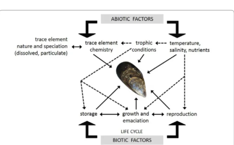
than the few major constitutive ones (i.e., C, H, N, S, O, P, Si, Cl, K, Na,
Ca and Mg) forming the bulk of living and mineral (except Fe and Al)
matter, whose concentrations are mostly below but sometimes above
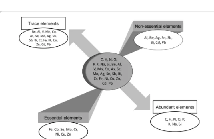
100 ppma according to the matrix analyzed (Figure 1) [21,22].
TEs can either be essential or non-essential (Figure 1). The essential
elements recognized by the World Health Organization (WHO) are
I, Zn, Se, Fe, Cu, Cr and Mo, the latter playing an important role in
biological systems [23,24]. Others TEs may/could also be essential,
such as Mn, Co, As, Ni or V. For these elements among others, the
essentiality is a characteristic which evolves according to our knowledge
and to the sensitivity of the authors who have a propensity more or
less strong to classify an element among the essential or not [25].
Non-essential TEs such as Hg, Pb or Cd play no physiological role, and are
often toxic even in very small quantity [26]. For these non-essential
TEs, only a threshold of toxicity exists, while essential TEs can be either
deficient in too small quantities, either toxic when they are absorbed in
high concentrations [25].
Production and uses
The world refinery and mine production of most TEs except a few
(e.g., As, Cd, Pb, Sn) have substantially increased these last decades
(e.g., Fe, Al, Mo) and particularly since the beginning of years 2000
(Table 1). World demand for minerals is affected by 3 general factors:
(i) uses for mineral commodities, (ii) the level of population that will
consume these mineral commodities and (iii) the standard of living
that will determine just how much each person consumes [27]. Today,
with the integration of India, the People’s Republic of China and other
populous developing and emerging countries (e.g., Brazil and Russia)
into the world economy, more than 50% of the world’s population
(instead of the previous 20%) account for the largest part of raw
materials consumption [28,29]. The increasing demand for mineral
raw materials further concerns numerous “emerging elements”. These
elements can be “truly emerging” because they have just gained entry
to the environment (new commercial uses and industrial releases) or
have nowadays become contaminants “of emerging concern” while
they were not in the past (new advances in analytical chemistry,
new knowledge on their toxicity, new environmental compartments
explored) [30,31].
The use of these emerging chemicals are multiple and diverse.
For example, V is regarded as one of the hardest of all metals. This
ubiquitous TE is employed in a wide range of alloys for numerous
commercial applications extending from train rails, tool steels,
catalysts, to aerospace [32]. Sb greatly increases the hardness and
the mechanical strength of Pb, and is found in batteries, antifriction
alloys, type-metal, small arms and tracer bullets, and cable sheathing. It
further has many uses as a flame retardant (in textiles, papers, plastics
and adhesives), as a paint pigment, ceramic opacifier, catalyst, mordant
and glass decolouriser, and as an oxidation catalyst [33,34]. Bi is largely
consumed in low-melting alloys and metallurgical additives, including
electronic and thermoelectric applications. The remainder is used for
catalysts, pearlescent pigments in cosmetics, pharmaceuticals, and
industrial chemicals [35].
Human toxicity
The major exposure media to TEs for humans are food, water
and air [36]. Safe drinking water supply is a high priority issue for
safeguarding the health and well-being of humans [37,38] and is an
important development issue [37]. Although standards legislate
the acceptable levels of contaminants in drinking water (Table 2)
Figure 1: Some essential and non-essential elements, abundant or in traces (modified after Ref. [25]).
Trace element Symbol Year 1990 2000 ↗(1990) 2010 ↗(1990) Aluminum Al 17,817 24,400 37% 40,800 129% Antimony Sb 83.2 122 47% 167 101% Arsenic As 47.6 36.9 -23% 52.8 11% Beryllium Be 0.286 0.226 -21% 0.203 -29% Bismuth Bi 3.33 3.75 13% 8.47 154% Cadmium Cd 20.2 20.2 0% 21.4 6% Chromium Cr 12,846 4,320 -66% 7,290 -43% Cobalt Co 37.1 33.3 -10% 89.5 141% Copper Cu 8,815 13,200 50% 16,000 82% Iron Fe 543,000 1,061,148 95% 2,590,000 377% Lead Pb 3,367 3,100 -8% 4,140 23% Manganese Mn 27.2 20.2 -26% 42.7 57% Molybdenum Mo 112 129 16% 242 117% Nickel Ni 1,029 1,250 21% 1,590 54% Selenium Se 1,789 1,460 -18% 2,120 19% Silver Ag 17.7 18.4 4% 23.1 31% Tin Sn 219 238 9% 265 21% Vanadium V 31.0 43.0 39% 57.6 86% Zinc Zn 7,325 8,730 19% 12,000 64%
Table 1: World production of 19 trace elements for the years 1990, 2000 and 2010, and percentage of increase since 1990 (data compiled from the Mineral Yearbooks published on the US Geological Survey website, www.usgs.gov).
Trace
element (ppm) from the WHO, Guideline values
the EPA and HC
Common sources of trace elements in drinking water Potential human health effects
Al WHO: 0.9 (<0.1 in
conventional treatment plants; <0.2 in other
treatment types)
Erosion of natural deposits; Al salts used as flocculents during
the treatment of drinking water. Little indication that orally ingested Al is acutely toxic to humans; no adverse health effect at levels found in drinking water; Al
exposure is a risk factor for the development or acceleration of onset of Alzheimer disease.
Sb WHO: 0.020; EPA,
HC: 0.006 refineries; fire retardants; ceramics; electronics; solders; Erosion of natural deposits; discharge from petroleum
contaminants from pipes and fittings.
Increase in blood cholesterol; decrease in blood sugar; microscopic changes in organs and tissues (thymus, kidney,
liver, spleen, thyroid).
As WHO, EPA: 0.01 Erosion of natural deposits (erosion and weathering of soils,
minerals, ores); runoff from orchards; runoff from glass and electronic production wastes.
Skin damage or problems with the circulatory system; increased risk of getting cancer (lung, bladder, liver, skin - classified as human carcinogen); neurological effects (numbness and tingling
of extremities).
Be WHO: 0.010; EPA:
0.004 discharge from electrical, aerospace and defense industries.Discharge from metal refineries and coalburning factories; Intestinal lesions; rarely found in drinking-water at concentrations of health concern.
Bi No guideline value Concentrations of Bi in drinking water have not been reported. Doses used in medicines are very much larger than the
estimated dietary exposure; dietary exposures to Bi are unlikely to be of toxicological concern.
Cd WHO: 0.003; EPA,
HC: 0.005 discharge from metal refineries; runoff from waste batteries Erosion of natural deposits; corrosion of galvanized pipes;
and paints; leaching from solders or black polyethylene pipes; industrial and municipal wastes.
Kidney damage; softening of bone; classified as human carcinogen.
Cr WHO, HC: 0.05; EPA:
0.010 Erosion of natural deposits; releases or spills from industrial uses (steel and pulp mills). Enlarged liver; irritation of the skin, respiratory and gastrointestinal tracts; kidney problems.
Co No guideline value Drinking water has a low-Co content, usually between 0.0001
and 0.0050 ppm. left ventricular failure, and enlarged hearts) observed in people Cardiovascular effects (cardiogenic shock, sinus tachycardia,
who consumed large amounts of beer over several years time containing Co sulfate as a foam stabilizer; gastrointestinal effects
(nausea, vomiting, and diarrhea), effects on the blood, liver injury, and allergic dermatitis have also been reported in humans
from oral exposure to Co.
Cu WHO: 2; EPA: TT
Action Level=1.3; HC<1.0
Erosion of natural deposits (erosion and weathering of rocks and minerals); corrosion of household plumbing systems;
contaminants from pipes and fittings; acidic mine water drainage; landfill leachates; sewage effluents; iron-related
industries.
Short term exposure: gastrointestinal distress; long-term exposure: liver or kidney damages. Cu is an essential element in
human metabolism; adverse health effects occur at levels much higher than the aesthetic objectives.
Fe HC: aesthetic
objectives≤0.3 Erosion of natural deposits (erosion and weathering of rocks and minerals); use of Fe coagulants; corrosion of steel and
cast iron pipes.
Not of health concern at levels causing acceptability problems in drinking-water.
Pb WHO, HC: 0.010; EPA: TT Action
Level=0.015
Erosion of natural deposits; corrosion of plumbing systems (pipes, solders, brass fittings and lead service lines);
contaminants from pipes and fittings.
Infants and children (under 6 years): delays in physical or mental development; neurobehavioural effects; children could show slight deficits in attention span and learning abilities. Adults: kidney problems; high blood pressure. Others: anaemia; central
nervous system effects; in pregnant women, can affect the unborn child; classified as probably carcinogenic to humans.
Mn WHO: 0.4;
HC: aesthetic objectives≤0.05
Erosion and weathering of rocks and minerals; naturally occurring in many surface water and groundwater sources, particularly in anaerobic or low oxidation conditions (the most
important source for drinking-water).
Not of health concern at levels causing acceptability problems in drinking-water.
Mo WHO: 0.02 Contaminations may occur in areas where Mo ore is mined. Occurs in drinking-water at concentrations well below those of
health concern.
Ni WHO: 0.07 Ni levels range from 0.002 to 0.010 ppm in fresh and tapwater;
water is generally a minor contributor to the total daily oral intake; the Ni contribution from water may be significant where
there is heavy pollution, in areas where Ni occurs naturally in groundwater, where there is use of certain types of kettles, of non-resistant material in wells or when water has come into
contact with Ni-plated taps; released from fittings; released from industrial Ni deposits.
Lack of evidence of a carcinogenic risk from oral exposure to Ni.
Se WHO: 0.04; EPA:
0.05; HC: 0.01 Naturally occurring (erosion and weathering of rocks and soils); discharge from petroleum and metal refineries;
discharge from mines.
Toxic effetcs: hair or fingernail losses at extremely high levels of exposure; numbness in fingers or toes; circulatory problems.
Ag WHO: available data
inadequate to permit derivation of health-based guideline value;
EPA: 0.1
Naturally occurring (erosion and weathering of rocks and soils); drinking water, not treated with Ag for disinfection purposes, usually contains extremely low concentrations of
Ag.
Water-soluble Ag compounds such as the nitrate have a local corrosive effect and may cause fatal poisoning if swallowed
accidentally.
Sn No guideline value Drinking-water is not a significant source of Sn; increasing use
of Sn in solder, which may be used in domestic plumbing, and proposed for use as a corrosion inhibitor.
Occurs in drinking-water at concentrations well below those of health concern; main adverse effect of excessive levels of Sn in canned beverages or other canned foods has been acute gastric
irritation; no evidence of adverse effects in humans associated with chronic exposure to Sn.
V No guideline value Typical values of V concentrations in drinking water are below
the detection limit. gastro-intestinal tract and mainly eliminated unabsorbed with the The main source of V intake is food; V little absorbed in the
faeces.
Zn HC: aesthetic
objectives≤0.05 leaching may occur from galvanized pipes, hot water tanks Naturally occurring; industrial and domestic emissions;
and brass fittings; Zn concentrations in water from active or inactive mines can be substantial.
Not of health concern at levels found in drinking-water; effects on human health by contamination on water supplies must be rare.
Table 2: Trace element guideline values (ppm) for drinking water, common sources of trace elements in drinking water and potential effects on human health. Data compiled from the World Health Organization (WHO) [37], the United States Environmental Protection Agency (EPA) [39], Health Canada (HC) [40], Nordberg et al. [26] and the Committee on Toxicity of Chemicals in Food, Consumer Products and the Environment (COT) [41]. Treatment Technique (TT) Action Level: US drinking water requiring a treatment process in order to reduce the level of a contaminant.
Removal processes from seawater
Unlike organic pollutants that can be degraded to less harmful
components by biological or chemical processes, TEs are considered
as non-degradable pollutants [19,54]. This persistent character of
TEs can alter, sometimes quite strongly, their natural biogeochemical
balance in contaminated environments [55]. Processes removing TEs
from seawater firstly include active biological uptake processes [56].
TEs are mainly transported into biological cells in ionic form through
ionic channels. In addition, specific transport mechanisms cross the
membrane barrier like binding with membrane carrier proteins or
transport through hydrophilic membrane channels. Lipid-soluble
(non-polar) metal forms including alkyl-TE compounds and neutral,
lipophilic, inorganically complexed TE species can cross biological
membrane by diffusion. TEs bound to very fine particles can also
be engulfed by endocytosis [57,58]. Following absorption, TEs are
transported to internal organs for utilization, storage, toxic effects, and
possibly release [57].
TEs can further be removed from seawater through passive
scavenging, i.e., the combined process of surface adsorption onto a
wide variety of relatively high affinity surface sites on both living and
dead particulate material followed by particle settling [56,59,60]. Much
of this particulate material (along with its associated TEs) is recycled
either in the water column or in superficial sediments. Labile bound
TEs can desorb from settling particles and resupply free TEs to the
dissolved pool [57]. Marine sediments can also act as a source of TEs
by releasing chemicals back to the overlying water column [54,61].
The primary flux processes between sediments and the water column
are resuspension and deposition, bioturbation, advection, upwelling/
downwelling, diagenesis reactions, and diffusion [61]. Because of
these remobilization processes, the effects of metal pollution on local
environments and organisms can be substantial and long lasting in
spite of years of restoration efforts [54].
TE toxicity on aquatic biota
TE metabolism and toxicity testing: The potential impact of a
contaminant on aquatic biota depends on the total concentration of
the contaminant, its speciation, interactions at receptors sites (e.g., at
a fish gill membrane or on an algal cell surface) and uptake into the
organism/cell, with either subsequent adverse effects or intracellular
detoxification [62]. The toxicity of each TE will therefore be related to
an organism-specific, metabolically available threshold concentration
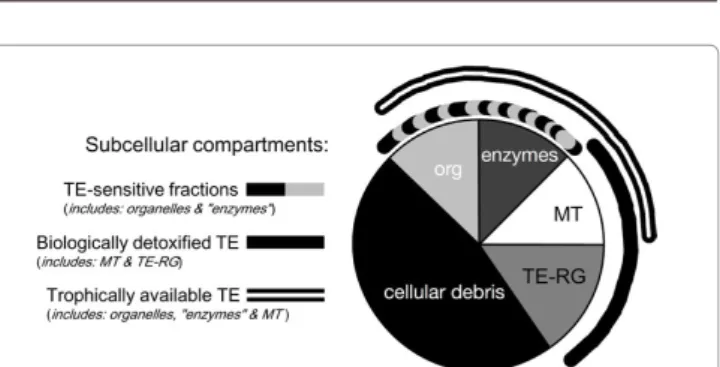
[63]. TEs accumulated into an organism occur in a bioreactive fraction
that is metabolically active and available and a fraction that has been
detoxified and is unavailable (Figure 2) [64]. TEs bound to inducible
TE-binding proteins such as metallothioneins or phytochelatins
or precipitated into insoluble concretions consisting of TE-rich
granules are biologically detoxified; on the contrary, TEs bound to
sensitive fractions such as organelles and heat-sensitive proteins can
be metabolically active. The higher the proportion of TE-sensitive
fractions, the greater the vulnerability to TE toxicity [65,66]. The
TE-sensitive fractions and TEs bound to metallothioneins are available
for trophic transfer to predators [66]. This compartmentalization of
TEs as defined by Wallace and Luoma [66] and Wallace et al. [65] is a
useful tool to interpret multiple ecotoxicological consequences of the
subcellular partitioning of TEs within organisms.
Mayer-Pinto et al. [67] critically reviewed studies on the effects
of TEs on aquatic assemblages and/or populations of invertebrates.
They pointed out that most studies in the field had been descriptive:
they generally demonstrated that the diversity of an assemblage
tended to decrease with an increase of environmental TE pollution
and that there were differences in the structure of assemblages facing
high pollution levels. Such descriptive studies are, however, unable
to demonstrate any causal relationship between the environmental
pollution and the changes observed [67]. Toxicity testing methods are
therefore required as a tool for predicting and assessing the impacts of
anthropogenic environmental stressors on organisms and ecosystems
[25]. Laboratory-based toxicity testing has the obvious advantage
to be unaffected by habitat or natural disturbances. This allows for
experimental evaluation of various parameters and conditions such
as temperature, toxicant threshold effect levels, mixture interactions,
life stages or exposure duration under strictly controlled conditions
[61]. Despite laboratory studies show lethal and sub-lethal effects of
TEs on organisms, extrapolating such findings to the field is however
little reliable [68]. Indeed, the exposure of aquatic organisms to
contaminants is mostly episodic due to changing water and sediment
quality. In addition, physicochemical characteristics of aquatic
ecosystems (e.g., temperature, pH, water hardness, dissolved organic
carbon) greatly influence contaminant bioavailability, toxicity and
bioaccumulation [69-71]. The naturally occurring variability of water
and sediment properties affecting TE chemical bioavailability cannot
be easily simulated in the laboratory [72]. Additional field experiments
are therefore necessary to validate laboratory results under relevant
environmental conditions.
Complementary results from laboratory and field toxicity tests
are useful for decision making, particularly if the responses of the
test organisms are severe and occur in multiple species [61]. Within
this perspective, detailed reviews on tested organisms from different
taxonomic groupings allow to identify species that may be suitable
candidates in a suite of toxicity test protocols [73] and to highlight
toxicity knowledge gaps that require to be addressed before using the
retained species in routine toxicity test procedures [74]. Such a work of
synthesis was performed for the Australian coastal waters [74,75]. Thus,
Van Dam et al. [74] reviewed the toxicity testing methods for water
column contaminants, summarizing data available for 16 taxonomic
groupings, among them vascular plants (seagrasses and mangroves)
and bivalve molluscs. Moreover, Adams and Stauber [75] reviewed
the whole sediment toxicity tests developed with numerous species
of 7 taxonomic groupings, of which bivalve molluscs. These authors
reported that bivalve molluscs were particularly relevant test organisms
for water and sediment toxicity tests and that seagrasses, as strong
accumulator of TEs, could act as integrated markers for environmental
TE exposure.
TE toxic effects on bivalve molluscs: Bivalve molluscs show many
physiological attributes (sensitive to contaminants, tolerant of a wide
range of abiotic factors, easy to grow and maintain in a laboratory
etc.) that make them appropriate bioassay organisms for toxicity
testing [76]. They are moreover good accumulators of organometallic
contaminants and TEs due to their behaviour and mode of feeding [77].
A number of standardized toxicity test protocols have been developed
for determining toxicity of single chemicals, complex effluents and
ambient samples of water or sediments to marine bivalves (e.g., Ref.
[61]; detailed guidance manuals available from the US Environmental
Protection Agency (EPA) and the American Society for Testing and
Materials (ASTM) [78]). Toxicity tests established on bivalve
embryo-larval developments are among the most sensitive in the EPA’s
national toxicity dataset, which is used to derive water quality criteria
[79]. Among bivalve molluscs, mussels from the genus Mytilus have
been largely used. TE toxicity on bivalve molluscs can be determined
at different structural levels, from genes to individuals, and response
parameters have included, in addition to larval-embryo developments,
changes in growth rates, clearance rates and survival rates,
DNA-damages, or changes in tissue morphology and in specific component
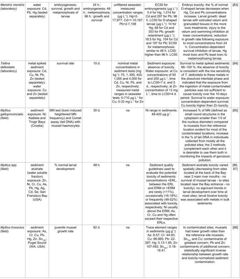
immunoreactivity (Table 3) [79-88].
Toxicity varies greatly between TEs. The median effect
concentrations (EC50) for Meretrix meretrix embryogenesis is 188
times higher for Cd than for Hg. Moreover, this difference in toxicity
directly relies upon the response parameter of interest. Thus, the EC50
for M. meretrix larval growth is only 6 times higher for Cd than for
Hg [83]. TE toxicity also fluctuates spatially and over time with water
properties, as demonstrated for Cu. The EC50 obtained by Cu spiking
of ambient water samples for mussel embryo development was lower
at sites located near the mouth of the San Diego Bay (California, USA)
compared to sites at the back of that Bay. This increase was consistent
with similar increasing trends in dissolved organic carbon (DOC)
and total suspended solids [79]. This protective effect of DOC on Cu
toxicity, experimentally demonstrated with contaminated filtered
seawater spiked with DOC, appeared related to higher fulvic acid and
lower humic acid contents [80].
TE toxic effects on seagrasses: Current knowledge on aqueous
toxicity of TEs in segrasses consists mainly of single TE exposures
Figure 2: A generalized ecotoxicological pie chart depicting subcellular compartments based on the biological significance of the various subcellular fractions in clams. Clams were homogenized, and differential centrifugation and tissue digestion techniques were used to obtain the following subcellular fractions (detailed procedure in Ref. [66]): trace element-rich granules (TE-RG), cellular debris, organelles (org), heat-sensitive proteins (“enzymes”) and heat-stable proteins (metallothioneins, MT). Subcellular fractions that are potentially vulnerable to TE exposure (i.e., organelles and “enzymes”; dashed arc) constitute the TE-sensitive fractions. Fractions that are involved with TE detoxification (i.e., metallothioneins and TE-rich granules; solid arc) constitute biologically detoxified TE. Fractions containing TEs that are readily available to predators (i.e., organelles, “enzymes” and metallothioneins; double arc) constitute trophically available TE (modified after Ref. [65]).in Sydney area (Australia). Photosynthetic efficiency and chlorophyll
pigment concentrations showed different sensitivities to Cu exposures
at the 3 sites, with seagrasses from the least developed estuary being the
most sensitive to Cu.
Marine vascular plants are still rarely used in ecotoxicological
testing, primarily because of difficulties in culturing/adapting and
testing with such large, slow growing organisms [74]. To overcome
these difficulties, and for greater environmental relevance, more recent
toxicity tests involved the in situ measurement of photosynthetic
endpoints (using PAM fluorometry) on wild plants in chamber
experiments (e.g., Ref. [93,94]). Field measurements of photosynthetic
efficiency can moreover be easily used as an efficient overall ecoindicator
of seagrass health (e.g., Ref. [97]). In addition, phytotoxic effect levels
for sediment-bound chemicals, spiked or in a whole sediment matrix,
are relatively unknown for seagrasses. But because concentrations of
several anthropogenic chemicals in rooted sediments exceed sediment
quality guidelines, there is a need to better understand the bioavailability
tested on 8 species (Table 4) [89-96]. Experimental designs have
varied considerably due, in part, to the lack of standardized toxicity
tests for marine vascular plants contrary to bivalve molluscs. Test
durations were between 6 hours and 51 days and response parameters
included photosynthetic activity, amino acid concentrations, tissue
growth, pigment contents or leaf cell mortality. Cu, Cd, Pb, and Zn
were the most commonly tested compounds with Cu toxicity being
particularly high, leading to seagrass leaf necrosis and decay (e.g., Ref.
[93,96]). Interspecific differences in sensitivity to the same TE have
been reported. Prange and Dennison [91] incubated 5 seagrass species
(Halophila ovalis, Halophila spinulosa, Halodule uninervis, Zostera
capricorni and Cymodocea serrulata) with Fe or Cu (1 ppm); seagrass
responses to pollutants were assessed by changes in PSII photochemical
efficiency and free amino acid content. Fe additions only affected
Halophila spp. while Cu additions affected the other seagrass species
as well. TE sensitivities can even differ between populations of a same
species. Macinnis-Ng and Ralph [94] in situ monitored the effects of
Cu and Zn on 3 geographically isolated populations of Z. capricorni
Species Studied trace
elements and sites
Measured
parameters Test duration concentrationsExperimented concentrations and Field or effect
measured effects
Supplementary comments Ref.
Mytilus galloprovincialis (laboratory)
water exposure: Cu; San Diego Bay (CA, USA)
embryo-larval
development 48 h. unfiltered seawater; nominal tested
concentration range
(µg L-1): 0-50; filtered
seawater used for reference toxicant
tests
absence of ambient toxicity to bivalve embryos; reference
toxicant test EC50 value=6.43 ± 1.36 µg
L-1; EC50 of Cu spiked
ambient water samples: 1.7 to 3.4 times lower
at sites located near the mouth of the Bay compared to sites at the back of the Bay.
Normally developed bivalve larvae possess a hinged D-shaped shell
(prodissoconch); differences bewteen unfiltered Cu spiked water
samples indicate a gradient in complexation capacity increasing
from the mouth to the back of the Bay (consistent with similar increasing trends in DOC and TSS).
[79]
Mytilus trossolus
(laboratory) exposure: water
Cu, Zn, Ni, Cd (tested separetely)
embryo-larval
development 48 h. filtered seawater; measured
concentration ranges (µg L-1): Cu=1.1-71.0; Zn=5-576; Ni=<DL-760; Cd=<DL-1,200; effect of DOC addition on metal toxicity tested
for Cu
EC50 (in μg L-1): 9.6
for Cu, 99 for Zn, 150 for Ni, and 502 for Cd; experimental addition of DOC reduced Cu
toxicity.
Normally developed bivalve larvae possess a hinged D-shaped shell (prodissoconch); protective
effects of DOC on Cu toxicity are influenced by their distinct physicochemical properties: protection appears to be related to higher fulvic acid and lower humic
acid contents. [80] Mytilus galloprovincialis (laboratory) water
exposure: Cr gill morphology and immunoreactivity to
components involved in gill motility; total glutathione content;
activities of GSH-related enzymes, of catalase, and of key glycolytic enzymes; mRNA expression of selected genes 96 h. artificial seawater; nominal tested concentrations (µg L-1): 0.1, 1, 10 Morphological, biochemical and molecular changes in mussel gills when exposed to concentrations ranging from 0.1 to 10 µg L-1. Progressive changes in gill morphology and in immunoreactivity to components
involved in neurotransmission; increased activities of GSH-related
enzymes and total glutathione content suggesting Cr detoxication/ reduction at the site of metal entry; increased activity of glycolytic enzymes, indicating modulation
of carbohydrate metabolism; significant changes in transcription
of different genes (sex- and concentration-related differences).
[81]
Mytilus edulis
(laboratory) exposure: Niwater clearance rate; haemolymph
genotoxicity and cytotoxicity 120 h. filtered seawater; nominal tested concentrations (µg L-1): 4.6 (control), 18, 56, 180 56 µg L-1: clearance rate decreases; 180 µg L-1: NRR decreases
and % tail DNA increases in mussel
haemocytes.
Clearance rate is Ni concentration-dependent (decreased by 30 % at the highest concentration); NRR
assays, designed to assess the viability of the cells based on the penetration of a weakly cationic dye across lysosomal membranes,
indicate a cytotoxic response; Ni has a genotoxic effect on the integrity of the DNA in haemocytes.
Meretrix meretrix
(laboratory) exposure: Cd, water
Pb, Hg (tested separately)
embryogenesis; survival, growth and
metamorphosis of larvae 24 h.: embryogenesis; 48 h.: metamorphosis; 96 h.: growth and survival unfiltered seawater; measured concentration ranges (µg L-1): Hg=2-17,977; Cd=1-10,167; Pb=2-7,158 EC50 for embryogenesis (µg L-1): 5.4 for Hg, 1,014 for Cd and 297 for Pb; 96 h. LC50 for D-shaped larvae (µg L-1): 14 for Hg, 68 for Cd and 353 for Pb; growth retardment (µg L-1): 18.5 for Hg, 104 for Cd and 197 for Pb; EC50
for metamorphosis: similar to 48 h. LC50; higher than 96 h. LC50.
Embryo toxicity: the % of normal D-shaped larvae decreases when
Hg, Cd and Pb concentrations increase. Larval growth: dead larvae with extruded velum and
granulated tissues in the more toxic treatments; injury to the velum and swimming inhibition at
lower concentrations; reduction in growth rate following exposure
to most concentrations from 24 h. Concentration-dependent survival inhibition of larvae. Hg most toxic and Pb least toxic to
metamorphosing larvae. [83] Tellina deltoidalis (laboratory) metal spiked sediment exposure: Cd, Cu, Ni, Pb, Zn (tested separetely); water exposure: Cu and Zn (tested separetely)
survival rate 10 d. nominal metal
concentrations in sediment tests (mg
kg-1): 75, 1,,300, 420,
1,000 and 4,000 for Cd, Cu, Ni, Pb, and Zn, respectively; measured metal ranges in seawater tests: 0-710 µg L-1 for Cu; 0-33 mg L-1 for Zn Sediment exposure: absence of toxicity. Water exposure: at Cu concentrations of 50 and 200 µg L-1, time to LC50=7 d. and 5 d., respectively; at Zn concentration of 13 mg L-1, time to LC50=4 d.
Survival to metal spiked sediments: 88–100 %; the absence of toxicity is consistent with the low sensitivity
of T. deltoidalis to these metals in the dissolved intertidal phase and indicated that the exposure from the ingestion of metal-contaminated
particles was not sufficient to cause toxicity over the 10-days period. Survival to water exposure:
concentration-dependent survival; Cu toxicity higher than Zn toxicity.
[84] Mytilus galloprovincialis (field) sediment exposure: Ni; Kaštela and Trogir Bays (Croatia)
MN test (toxin induced heightened MN frequency) and Comet
assay (tail DNA) with mussel haemocytes
30 d. na Ni range in sediments:
48-420 µg g-1. Increased % of MN (defined as small round structures in the
cytoplasm smaller than 1/3 of the nucleus diameter) compared
to mussels from the reference location evident for most of the contaminated locations; increase in the % of tail DNA in individuals collected from mostly all the polluted sites; the 2 methods complement each other and it is desirable to use them both in monitoring the impacts of genotoxic
pollution.
[85]
Mytilus spp.
(field) sediment elutriate
(water soluble fraction) exposure: Zn, Ni, Cr, Cu, As, Pb, Hg, Ag, Cd, Se; San Fransisco Bay
(USA)
% normal larval
development 48 h. na guidelines used to Sediment quality
evaluate the potential toxicity of sediments: concentrations <ERL, between the ERL and ERM or >ERM
are rarely (<11%), occasionally (16-18%) or frequently (48-52%) associated with toxicity, respectively; Ni usually above the ERM; As, Cr, Cu and Hg often exceed their respective
ERLs.
Sediment elutriate toxicity varied spatially (decreasing from sites
located at the back of the Bay near 2 main river mouths - no survival of mussel larvae - to sites located near the Bay entrance - no toxicity); no signicant trends in larval development over time at most sites; larval bivalve toxicity was associated with metals in bulk
sediments.
[86, 87]
Mytilus trossulus
(field) exposure: As, sediment
Cr, Cu, Pb,
Hg, Zn, SnTBT;
Puget Sound (WA, USA)
juvenile mussel
growth rate 82 d. na Trace element ranges in sediments (µg g-1):
As: 8-57; Cr: 44-93; Cu: 66-965; Pb: 22-297; Hg: 0.13-1.95; Zn:
107-592; SnTBT:
0.18-18.41.
In contaminated sites, mussels had lower growth rates than
the reference site mussels;
SnTBT and Cu: contaminants of
greatest concern; Pb and Zn: contaminants of additional concern;
statistically significant inverse relationship between growth rate and toxicity-normalized sediment
contamination.
[88]
Table 3: Biological responses in bivalves exposed to dissolved and sediment trace elements under laboratory or field conditions. h.: hour; d.: day; na: not applicable; EC50: the median effect concentration; LC50: the median lethal concentration; DOC: dissolved organic carbon; TSS: total suspended solids; DL: detection limit; Glutathione (GSH)-related enzymes; NRR: neutral red retention; MN: micronucleus; ERL: effects range-low; ERM: effects range-median.
and phytotoxicity of sediment-bound contaminants [72].
The Monitoring of the Marine Environment
Biomonitoring
Until the early 70s, the monitoring of terrestrial and marine
environments mainly relied on the detection and quantification of
pollutants in physical environments - air, water, soils and sediments
[25,98]. During the years 80s, almost all environmental monitoring
networks stopped monitoring the water itself to assess the quality of
aquatic ecosystems. Reasons for this abandon were diverse, among
them: the measured concentrations of chemicals are generally low (in
the order of a few ng L
water-1for most TEs) and often close to the limits of
detection of the analytical techniques; the risk of secondary accidental
contamination makes the measurements sensitive; the concentrations
of dissolved substances may vary considerably over time (e.g., with
tidal cycles, water run-off, seasons etc.) and episodic pollution events
can be missed; the measurement of dissolved pollutants do not
provide an assessment of the portion which is available for uptake and
accumulation by aquatic organisms [25,99]. The analysis of sediments
overcomes some of these disadvantages. Contaminants accumulated
in sediments, particularly in organically rich sediments, are more easy
to monitor and much less susceptible to accidental contaminations.
Sediments also offer a degree of time integration. However, sediment
accumulation of contaminants is much affected by sediment
characteristics (particle size, mineralogy, organic carbon content) and
measured concentrations again do not provide an assessment of their
bioavailable fractions [25,99].
When compared with the conventional chemical analysis of water
and sediments, the monitoring relying upon the biota exhibits obvious
predominance. Biomonitoring reveals the biological changes of
organisms affected by chemicals and the integrated effects of multiple
pollutants on these latest; has high sensitivity because of the rapid
responses induced in organisms exposed to pollutants; realizes the
monitoring of pollutants at low levels because of their chronic toxicities
under long-term exposure; allows widely sampling even at remote
areas; avoids the limits of the conventionnal chemical analysis such as
continuous sampling, needs of expensive instruments etc. [100].
Bioindicators
Since the ultimate purpose of pollution monitoring is the
protection of ecosystems and human beings, the main interest of the use
of quantitative sentinel organisms with regard to water or sediments is
their capacity to give information on the bioavailability of pollutants
[101]. Currently, the term "bioindicator" is a deeply ambiguous term
which has different meanings in different contexts [102]. To prevent
problems due to different interpretations of this term, we use the
definition of Blandin [103]: “a biological indicator (or bioindicator)
is an organism or a set of organisms that allows, by reference to
biochemical, cytological, physiological, ecological or ethological
variables, in a practical and safe way, to characterise the status of an
ecosystem or an eco-complex and to highlight as early as possible their
changes, natural or caused”. Bioindicators therefore allow to accurately
assess the effects of anthropogenic activities on ecosystems.
To be considered as a good bioindicator of environmental
contamination, the selected species must meet a number of criteria,
as listed by Cossa [101] or Rainbow [104]: the sentinel organism
should be a net strong accumulator of contaminants and should not
regulate the total concentration of a contaminant in its body tissues;
it should be sedentary and reasonably abundant; it should have a
sufficiently long life to permit sampling of more than one-year class;
it should be large enough to provide sufficient tissue for analyses
and should bioaccumulate sufficiently to allow direct measurement
without preconcentration; it should be resistant to handling stresses;
a correlation should exist between the level of contaminants in the
organism and in the surrounding environment; it should be tolerant of
exposure to environmental variations in physicochemical parameters
and the effects on the organism of these variations should be known. No
single species however combines all these qualities, and a compromise
must be found [101].
For marine pollution monitoring, the bioindicator species used
belong to numerous taxonomic groupings of which micro- and
macro-algae, seagrasses, ascidians, sponges, bivalve and gastropod
molluscs, polychetes, crustaceans, fishes, seabirds, marine reptiles and
mammals [105,106], each bioindicator showing some special merits
when compared to the others [100]. In the two next sections, we will
present two bioindicator species widely used in the monitoring of the
health status of the coastal Mediterranean, from punctual surveys to
international monitoring programs: the Neptune grass Posidonia
oceanica and the Mediterranean mussel Mytilus galloprovincialis.
These two bioindicators respond appreciably and quantitatively to the
coastal pollution by TEs and complement one another: the two species
accumulate contaminants dissolved in the water column; P. oceanica,
deeply rooted in sediments, also reflects the contamination of this
compartment; mussels, as a filter feeder, accumulate contaminants from
their particulate phase. Together, they give an estimate of the overall
pollution (water, sediments, suspended matter) of Mediterranean
coastal environments [107].
Posidonia oceanica
Biology
P. oceanica is a marine magnoliophyte endemic to the
Mediterranean [108,109]. It grows on sandy and rocky bottoms and
forms patchy and continuous meadows regarded as one of the climax
communities of the Mediterranean [110]. It colonizes large areas of the
infralittoral floor from the surface to maximal depths of 45 m [110,111].
P. oceanica beds cover a surface estimated between 25,000 and 50,000
km
2, i.e., between 1 and 2% of the Mediterranean [112], and is only
missing in zones under the influence of large estuaries (Po, Rhone,
Nile - diminution of salinity and increase of turbidity) [113]. The
light and the transparency of the water are determining factors for its
growth [114]. P. oceanica has the same morphology as the other marine
magnoliophytes: below-ground parts consist of roots for anchoring and
rhizomes for mechanical support; above-ground parts consist of shoots
bearing several leaves [115]. Rhizomes grow horizontally (competition
for space: plagiotropic rhizome) or vertically (competition for access
to the light: orthotropic rhizome). The progressive silting and the two
types of rhizome growth result in a typical terraced formation called
“matte” consisting of the intertwining of various strata of rhizomes,
roots, and sediments [113].
P. oceanica plays various ecological and functional roles (reviewed
in Ref. [12,16,116]). First of all, P. oceanica meadows are considered
to be among the most productive ecosystems of our planet. This
ecosystem is made up by the juxtaposition of two types of primary
production: the net primary production of P. oceanica which is on
average of 420 g
DMm
-2year
-1and can reach 1,300 g
DM
m
-2year
-1; the
net primary production of the epiphytes which is between 100 and 500
g
DMm
-2year
-1. On a global scale, only seagrass ecosytems display this
specific feature. P. oceanica exhibits structural roles: P. oceanica leaf
Species Studied trace elements and
sites
Measured
parameters durationTest concentrationsExperimented Effect concentrations and measured effects Supplementary comments Ref.
Zostera marina (laboratory) Cu, Cr, Cd, Hg, Zn, Pb (tested separetely) growth rate 0.5, 2, 5, 8, 12 and 19 d. 0.1, 0.5, 5 and
50 µM 12 d. at 5 µM and after 8 d. at 50 Growth rate inhibition - Cd: after
µM; Cu: after 5 d. at 5 µM and after 2 d. at 50 µM; Hg: rapid effect at all concentrations; Zn:
after 2 d. at 50 µM.
Growth rate inhibited by Cd, Cu, Hg, Zn; no effect of Cr and Pb exposures; toxicity of metals decreases in the order: Hg ≥ Cu > Cd ≥ Zn > Cr and Pb; cellular substances are leached to
the water and plants turned black in the Cu 50 µM experiment and in the Hg 5 µM and 50 µM experiments.
[89]
Halophila stipulacea (laboratory)
Al leaf cell viability 12 d. from 10-4 to 10-9
mol L-1 Cellular damages from 10
-4 to 10-8
mol L-1. categories (except in the mid-rib Protoplast necrosis in all cell
cells); plasmatic resistance decreases in the order mid-rib, mesophyl,
epidermal, teeth cells.
[90] Halophila ovalis, Halophila spinulosa, Halodule uninervis, Zostera capricorni and Cymodocea serrulata (laboratory) Fe, Cu (tested
separetely) changes in PSII photochemical
efficiency (Fv/Fm) and free amino acid
content
12 d. of exposure;
5 d. of recovery
1 mg L-1 + EDTA Fe and Cu: 1 mg L-1; decline in
PSII photochemical efficiency and in amino acid contents; effects
are species-specific.
Fe addition experiments: declines in PSII photochemical efficiency in H. ovalis and H. spinulosa correspond
with the replacement of fresh seawater (12 days), suggesting that these species became acclimatized to the new environmental conditions; Z. capricorni exhibited a decline in total free amino acid contents (could
be a precursor signal of Fe induced stress). Cu addition experiments: Fv/ Fm ratio response was highly variable
between the 5 seagrass species (death of H. spinulosa); decline in
amino acid concentrations in Z. capricorni and H. uninervis.
[91]
Halophila ovalis
(laboratory) Cu, Cd, Pb, Zn (tested
separately) chlorophyll a fluorescence; pigments (chlorophyll a, b, and carotenoids) 4 d. 1, 5 and 10 mg L-1 Cd - 1 to 10 mg L-1: limited stress. Cu - 5 and 10 mg L-1: lethal effect. Pb - 1 to 10 mg L-1:
limited effect on fluorescence; chl. a, b decrease. Zn - 1 to 10
mg L-1: changes to the chl. a
fluorescence responses; various effects on pigment contents.
Variety of effects on the photosynthetic processes, with Cu and Zn having greater effects than Pb and Cd; quantum yield is the most sensitive measure of the photosynthetic processes; pigment contents generally confirm the chlorophyll a fluorescence responses.
[92]
Zostera
capricorni (field) Cd, Cu, Pb, Zn (tested separetely); 1 reasonably pristine site at Pittwater (NSW, Australia) photosynthetic efficiency (ΔF/Fm'); pigments (chlorophyll a, b, and carotenoids) 10 h. of exposure; 3 d. of recovery
0.1 and 1 mg L-1 Cu, Zn - 0.1 and 1 mg L-1:
photosynthetic efficiency decreases during the exposure
period; Cu - 0.1 and 1 mg L-1:
after 96 h., carotenoid pigments decline, the chlorophyll a/b ratio is depressed and the chlorophyll/
carotenoid ratio is elevated.
Samples exposed to Zn recover to pre-exposure levels but those exposed to Cu do not; browning of leaves and some leaf loss occurred due to exposure to Cu; Cd and Pb do
not have impact on the chlorophyll a fluorescence and the pigment data
support these findings.
[93] 3 isolated populations of Zostera capricorni (field) Cu, Zn (tested separetely); 1 semi-pristine and 2 impacted sites in Sydney area (Australia) photosynthetic efficiency (ΔF/Fm'); pigments (chlorophyll a, b, and carotenoids) 10 h. of exposure; 3 d. of recovery
0.1 and 1 mg L-1 Cu in the semi-pristine site - 0.1
mg L-1: fluorescence decreases; 1
mg L-1: chlorophyll concentration
decreases. Cu in the 2 impacted
sites - 1 mg L-1: fluorescence
decreases.
Lack of response due to Zn exposure; different sensitivities to Cu: greater
impact of Cu on the more naïve population (higher decrease of fluorescence during exposure and
lower recovery).
Posidonia oceanica (laboratory) Cd DNA methylation and chromatin reconfiguration; expression of PoMT2k and PoCMT1; nuclear chromatin ultrastructure 6 h., 2 d.,
4 d. 10 and 50 µM PoMT2k expression - Cd 50 µM: increase in PoMT2k expression
after 6 h. in apical tips and leaves; Cd 10 µM: increase in PoMT2k expression after 2 d. in leaves. Changes in DNA methylation and in PoCMT1 expression: Cd 10 µM after 6 h. Chromatin reconfiguration: Cd 50
µM after 2 d.
Cd treatment induces a DNA hypermethylation (time- and dose-dependent) as well as an
up-regulation of CMT, indicating that de novo methylation occurs; a high
dose of Cd leads to a progressive heterochromatinization of interphase
nuclei and apoptotic figures are observed after long-term treatment;
Cd perturbs the DNA methylation status through the involvement of a specific methyltransferase; such changes are linked to nuclear
chromatin reconfiguration likely to establish a new balance of expressed/repressed chromatin; the data show an epigenetic basis to the mechanism underlying Cd toxicity in
plants.
[95]
Halophila ovalis
(laboratory) Pb, Cu (tested separetely) fluctuating asymmetry growth rate; leaf
and dimension
51 d. Pb: 10 and 50 mg
L-1; Cu: 0.5, 2 and
4 mg L-1
Growth rate decrease - Cu: 0.5
mg L-1; Pb: 10 mg L-1. Reduced
leaf dimension - Cu: 0.5 mg L-1;
Pb: 50 mg L-1. Increased leaf
asymmetry - Cu: 2 mg L-1.
Reduced growth rate of the seagrass observed both in Pb and Cu treatments; leaf size of the plant reduces as the metal concentrations increase and when the plants are
exposed to the metals for longer duration; increased leaf asymmetry
more apparent at the 2 ppm Cu treatment; no increase in fluctuating
asymmetry in Pb treatments; the mortality of leaves is especially high
in Cu treatments.
[96]
Table 4: Biological responses in seagrasses exposed to dissolved trace elements under laboratory or field conditions. EDTA: ethylenediaminetetraacetic acid; PSII: photosystem II; PoMT2k: Posidonia oceanica Metallothionein (MT) 2k, an important metal tolerance gene; PoCMT1: one member of the Posidonia oceanica chromomethylase (CMT) family, a DNA methyltransferase. Fv: variable fluorescence; Fm: maximum fluorescence; ΔF: fluorescence yield; Fm’: light-adapted maximal fluorescence.


![Table 5: Annual balances of trace element (TE) amounts in Posidonia oceanica (tons y -1 ) for the whole Mediterranean [147]](https://thumb-eu.123doks.com/thumbv2/123doknet/5843135.141667/13.892.63.442.155.476/table-annual-balances-element-amounts-posidonia-oceanica-mediterranean.webp)