INTERACTION BETWEEN ESTROGEN AND INTERFERON- SIGNALING PATHWAYS IN THE REGULATION OF MAJOR HISTOCOMPATIBILITY
COMPLEX CLASS II EXPRESSION IN BREAST CANCER CELLS
By
Jorge Alfonso Leon Machado
Thesis presented to the Département de Biologie as condition for the achievement of a Master’s degree in science (M. Sc.)
FACULTÉ DES SCIENCES UNIVERSITÉ DE SHERBROOKE
January 20 2017
Members of the jury
Professor Viktor Steimle Directeur de recherche
Université de Sherbrooke, Département de Biologie
Professor Nicolas Gévry Président rapporteur
Université de Sherbrooke, Département de Biologie
Professor Richard Blouin Membre interne
i SUMMARY
Activation of the antigen presentation mechanisms by cancer cells is one of the main pathways used by the immune system for tumor detection and suppression. Induction of the expression of molecules of the Major Histocompatibility complex class II (MHC-II) by the interferon- (IFN) is important for the efficient presentation of tumor antigens. Nevertheless, it has been observed that expression of these molecules is supressed in tissular contexts where the concentration of estradiol (E2) is high.
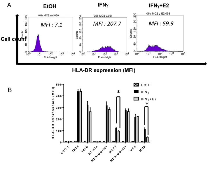
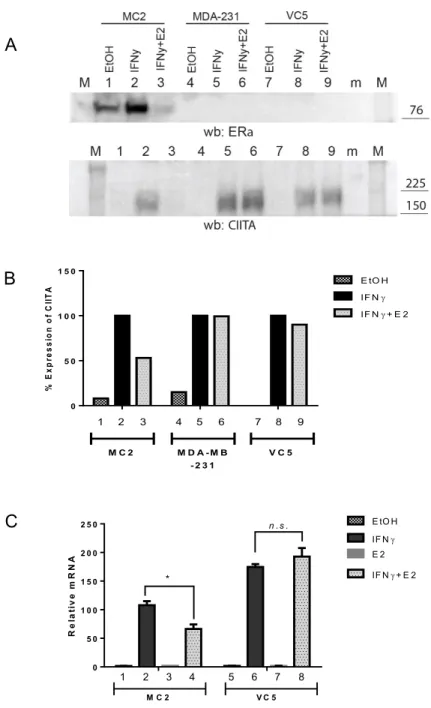
In this work we attempted to explain if the down-regulation exerted by estradiol on the expression of the MHC-II molecules in breast cancer cells was mediated by a silencing effect of the estrogen receptor- (ER) through a possible estrogen receptor binding site (ERBS) in the locus of promoter IV (pIV) of the master regulator of MHC-II expression, the class II transactivator (CIITA). The breast cancer cell line MDA-MB-231 (ER-/ERβ-) and its stable transfectants MC2 (ER+) and VC5 (empty vector) were used as model cell lines. Expression of the MCH-II molecules is controlled by CIITA, and stimulation with IFN activates the transcription of the pIV of CIITA. Stimulation of these cell lines with IFN induced expression of the MCH-II molecules and addition of E2 repressed such expression only in the MC2 cell line, as observed by flow cytometry analysis. Six other breast cancer cell lines were tested, with only the MCF7 cell line showing a significant inhibition. Then we analyzed if the inhibition of the MHC-II expression was due to a down-regulation of CIITA. Protein analysis performed by western blot and mRNA quantification by RT-qPCR both revealed down-regulation of CIITA in the cells exposed to IFN+E2 compared to those treated only with IFN. However, reporter gene analysis did not demonstrate any influence of our candidate ERBS in the inhibition of the activation of CIITA-pIV. ChIP-seq analysis of the VC5 and MC2 cell lines for ER also failed to demonstrate any binding of the receptor anywhere in the vicinity of the CIITA locus. However gene ontology and disease ontology analysis
ii of the sequencing data showed a higher activation of tumorigenic cellular pathways in the cells treated with IFN + E2 than in the cells treated only with E2. These results suggest that activation of the inflammatory pathways by IFN could exert a detrimental effect on the cancer development.
Key words: breast cancer, antigenic presentation, CIITA, MHC-II, estrogen, interferon gamma, estrogen receptor binding site.
iii INTERACTION ENTRE LES VOIES D’ACTIVATION DE L’ESTROGÈNE ET DE L’INTERFÉRON - DANS LA RÉGULATION DE L’EXPRESSION DU COMPLEXE
MAJEUR D’HISTOCOMPATIBILITÉ DE CLASSE II DANS DES CELLULES DE CANCER DU SEIN
par
Jorge Alfonso Leon Machado
Thèse présentée au Département de Biologie en vue de l’obtention du grade de maîtrise en sciences (M. Sc.)
FACULTÉ DES SCIENCES UNIVERSITÉ DE SHERBROOKE
iv SOMMAIRE
L’activation des mécanismes de présentation antigénique par les cellules cancéreuses est l’une des voies principales employées par le système immunitaire pour la détection et la suppression des tumeurs. L’induction de l’expression de molécules du complexe majeur d’histocompatibilité de classe II (CMH-II) par l’interféron- (IFN) est importante pour la présentation efficace des antigènes tumoraux. Cependant, il a été observé que l’expression de ces molécules est supprimée dans certains tissus dans lesquels la concentration d’estradiol (E2) est élevée.
Dans ce travail, nous avons tenté de déterminer si l’inhibition exercée par l’estrogène (E2) sur l'expression des molécules du CMH-II dans des cellules de cancer du sein est médiée par un effet de silençage du récepteur de l’estrogène- (ER) à travers d’un possible site de liaison de récepteur d'estrogène (ERBS) dans le locus du promoteur IV du régulateur clé de l’expression du CMH-II, CIITA. La lignée cancéreuse mammaire cellulaire de cancer de sein MDA-MB-231 (ER-/ERβ-) et ses transfectants stables MC2 (ER+) et VC5 (vecteur vide) ont été utilisés comme des lignées cellulaires modèles. L'expression des molécules du CMH-II est contrôlée par CIITA, et la stimulation avec l’IFN active la transcription du pIV de CIITA. La stimulation de ces lignées cellulaires avec l’IFN induit l'expression des molécules du CMH-II et l'addition d’E2 réprime de cette expression seulement dans la lignée cellulaire MC2, telle qu'elle est observée par analyse de cytométrie de flux. Six autres lignées de cancer de sein ont été testées et seulement la lignée cellulaire MCF7 montrait une inhibition significative. Ensuite, nous avons analysé si l'inhibition de l'expression du CMH-II était due à une régulation de CIITA. L'analyse des protéines effectuée par Western blot et la quantification de l'ARNm par RT-qPCR quantitative ont révélé une inhibition de CIITA dans les cellules exposées à l’IFN + E2 par rapport à celles traitées seulement avec l’IFN. Cependant, des analyses avec un gène rapporteur n'ont pas démontré une
v influence quelconque de notre site de liaison de récepteur d'estrogène candidat dans l'inhibition de l'activation de CIITA-pIV. Des analyses de ChIP-seq dans les lignées cellulaires MC2 et VC5 pour l’ER n’ont également pas démontré la présence d’une liaison du récepteur dans le voisinage du locus de CIITA. Cependant, des analyses sur l'ontologie des gènes et des maladies sur les données de séquençage ont montré une activation accrue des voies cellulaires cancéreuses dans les cellules traitées avec IFN + E2 comparé avec les cellules traitées uniquement avec E2. Ces résultats suggèrent que l'activation des voies inflammatoires par l’IFN pourrait exercer un effet plus négatif qu’anticipé sur le développement du cancer.
Mots clés: cancer du sein, présentation antigénique, CIITA, CMH-II, estrogène, interféron gamma, site de liaison du récepteur de l’estrogène.
vi ACKNOWLEDGEMENTS
First and foremost I would like to thank my director Prof. Viktor Steimle, a person that over the last four years has become more than a boss as I consider him my Mentor and the example of the scientist I would like to become. The door to Prof. Viktor office was always open whenever I ran into a trouble spot or had a question about my research or writing. Also I would like to thank him for his patience and support during the rough spots.
I would also like to thank all the students and post-doctoral personnel from Prof. Gévry laboratory who took the time to teach me the different techniques that were needed for the execution of this project, mainly Stephanie Bianco, Maika Jangal, Martin Morin and Mylène Brunelle. Without their help and patience this project would have been all but impossible to accomplish.
I would also like to acknowledge Prof. Pierre-Étienne Jacques for the time taken to guide me through the use of the Galaxy-GenAP application and for his suggestions for the analysis of the ChIP-seq results and Prof. Marc Bélisle for the help given with the statistical analysis of the flow cytometry results.
I also thank my counselors Prof. Nicolas Gévry and Prof. Richard Blouin for their feed-back and advice all along the project.
The contributions from the technical staff were also of great help for the continued and timely execution of the experiments, for this I thank Ms. Manon Dufresne for the help with the cellular culture and Daniel Garneau for the programming of the robo-pipetter that allowed me to easily perform the hundreds of qPCRs needed during this project.
-Are you ready to rock Jorge? -Shields-up. Weapons online!
vii INDEX
SUMMARY ... i
FRENCH TITLE ... iii
FRENCH SUMMARY ... iv
ACKNOWLEDGEMENTS... vi
INDEX ... vii
ABBREVIATIONS ... x
FIGURE INDEX ... xiv
Chapter I. INTRODUCTION ... 1
1.1 Historical context ... 1
1.2 Breast cancer pathology and development ... 3
1.3 Molecular characterization of breast cancer ... 7
1.4 Tumorigenesis ... 10
1.5 Tumor heterogeneity ... 12
1.6 Estrogens and estrogen receptors ... 15
1.6.1 Normal function ... 15
1.6.2 Non genomic activities of ERs ... 18
1.6.3 Activation of ERs non-E2-dependent ... 19
1.7 Breast cancer resistance to anti-estrogen therapy ... 20
1.8 Immune response to cancer ... 22
1.8.1 Antigenic activation of the immune response ... 23
1.8.2 T cell mediated tumor suppression ... 25
1.9 Antigenic presentation and regulation ... 29
1.10 CIITA regulation ... 36
viii
1.12 Objectives and hypothesis ... 38
Chapter II MATERIALS AND METHODS ... 43
2.1 Cell culture ... 43 2.2 Transfections ... 44 2.3 Stimulations ... 44 2.4 Western blotting ... 44 2.5 Flow cytometry ... 45 2.6 ChIP assays ... 46
2.7 Library preparation for Illumina sequencing ... 46
2.8 ChIP-seq analysis ... 46
2.9 Quantitative PCR (ChIP) ... 47
2.10 Quantitative RT-PCR (mRNA) ... 47
2.11 Reporter gene assays ... 48
2.12 Depletion experiments by shRNA ... 49
Chapter III RESULTS ... 50
3.1 Major histocompatibility complex class II expression is differentially modulated by E2 in different BCC lines ... 50
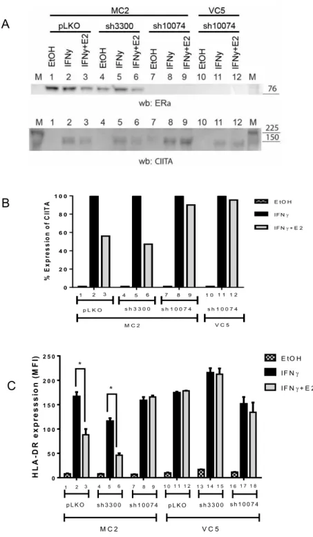
3.2 E2 induced down-regulation of the HLA-DR expression is dependent on ER and mediated by the down-regulation of CIITA 52 3.3 Down-regulation of CIITA gene expression may be induced by activity of the ER as a silencer ... 55
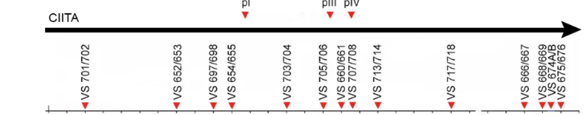
3.4 Analysis of distal regulatory CIITA elements by ChIP-PCR ... 60
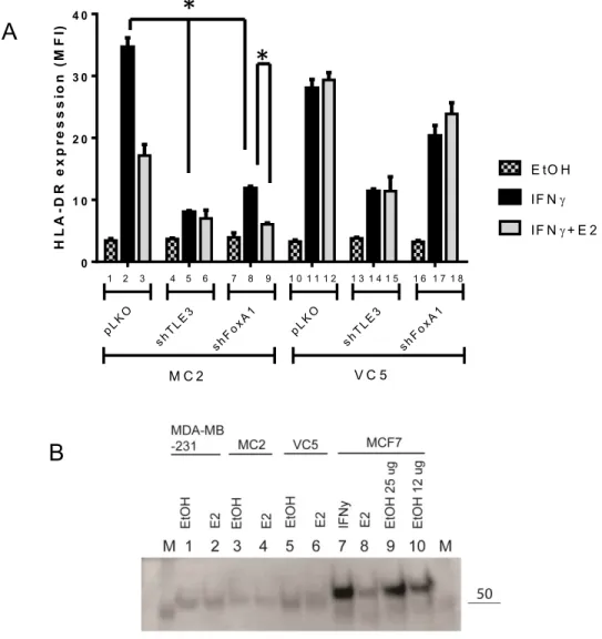
3.5 Inhibition of the ER partners TLE3 and FoxA1 does not prevent Down-regulation of the HLA-DR expression ... 61
3.6 Genome wide analysis of ER binding by ChIP-seq ... 66
3.7 Genomic analysis of ER ChIP-seq results reveals changes in the binding motifs of the ER with the combination of IFN+E2 .... 70
ix 3.8 Genomic analysis of ER ChIP-seq results shows that IFN
inclusion increases tumorigenic phenotype ... 70
Chapter IV DISCUSSION ... 76
Chapter V PERSPECTIVES ... 86
ANNEXE I ChIP-qPCR primer sequences ... 88
ANNEXE II shRNA sequences ... 90
ANNEXE III RT-qPCR primer sequences ... 90
x ABBREVIATIONS
AR Androgen Receptor
APC Antigen Presenting Cell
BC Breast Cancer
BCC Breast Cancer Cell BCR B Cell Receptor
BLS Bare Lymphocyte Syndrome
BRCA1/2 BReast CAncer gene 1 and 2 cAMP cyclic Adenosine MonoPhosphate CBP CREB Binding Protein
cDNA complementary DNA
ChIP Chromatin ImmunoPrecipitation
ChIP-seq Chromatin ImmunoPrecipitation followed by sequencing CIITA Class II TransActivator
CREB cAMP Responsive Element Binding
COSMIC Catalogue Of Somatic Mutations In Cancer CTA Cancer Testis Antigens
CTL Cytotoxic T Lymphocytes
DBD DNA Binding Domain
DC Dendritic Cells
DCIS Ductal Carcinoma In-Situ DNA DeoxyriboNucleic Acid
E1 estrone
E2 estradiol
xi E-box Enhancer box
EGF Epidermal Growth Factor
ER Estrogen Receptor
ERBS Estrogen Receptor Binding Site
ERE Estrogen Response Element
ER Estrogen Receptor alpha ERβ Estrogen Receptor beta ERR Estrogen Related Receptors FIGF c-Fos Induced Growth Factor
GAS (interferon) Gamma Activated Site GEP Gene Expression Profile
GM-CSF Granulocyte-Macrophage Colony-Stimulating Factor
GTP Guanosine TriPhosphate
HAT Histone Acetyl Transferase
HER-2 Human Epidermal growth factor Receptor 2 HLA-DR Human Leukocyte Antigen DR
HLA-DM Human Leukocyte Antigen DM HSP90 Heat Shock Protein 90
IBC Inflammatory Breast Carcinoma IDC Invasive Ductal Carcinoma
IFN InterFeroN gamma
IFNGR InterFeroN gamma Receptor
IGF-I Insulin-like Growth Factor I
IHC ImmunoHistoChemistry
IL-2 InterLeukin 2 IL-6 InterLeukin 6 IL-11 InterLeukin 11
xii
ILC Invasive Lobular Carcinoma
IRF-E Interferon Regulatory Factor-Element
JAK: JAnus Kinase
KD Knock-Down
LBD Ligand Binding Domain
LCIS Lobular Carcinoma In-Situ
Ii Invariant chain
MAPK Mitogen-Activated Protein Kinase MEME Multiple Em for Motif Elicitation
MHC-I Major Histocompatibility Complex class I MHC-II Major Histocompatibility Complex class II
mRNA messenger RNA
NFkB Nuclear Factor kappa-light-chain-enhancer of activated B cells NFY Nuclear transcription Factor Y
NK Natural Killer cell
NR Nuclear Receptor
qPCR quantitative Polymerase Chain Reaction pDC plasmacytoid Dendritic Cell
PDK1 Phosphoinositide-Dependent protein kinase 1
PKA Protein Kinase A
PKB Protein Kinase B
PKC Protein Kinase C
PR Progesterone Receptor
RNA RiboNucleic Acid
RT-qPCR Reverse Transcription quantitative-PCR
SDS-PAGE Sodium Dodecyl Sulfate – PolyAcrylamide Gel Electrophoresis
xiii STAT1 Signal Transducer and Activator of Transcription 1
TAA Tumor Associated Antigens
TCGA The Cancer Genome Atlas
TCR T Cell Receptor
TEC Thymic Epithelial Cells TFIIB Transcription Factor II B TFIID Transcription Factor II D TGF Transforming Growth Factor
Th T helper cell
TNFα Tumor Necrosis Factor alpha TSA Tumor-specific Antigens TSS Transcription Start Site
TSLP Thymic Stromal LymphoPoietin USF1 UpStream transcription Factor 1
WB Western Blot
xiv FIGURE INDEX
Figure 1. Morphology of the human breast ... 4
Figure 2. Breast cancer classification by intrinsic molecular subtype ... 9
Figure 3. Targeted elimination of cancer subpopulations ... 14
Figure 4. Schematic representation of ER activation and signaling ... 21
Figure 5. Antigenic processing and presentation by MHC-I and MHC-II molecules ... 30
Figure 6. MHC-II enhanceosome structure ... 35
Figure 7. CIITA promoter activation ... 37
Figure 8. ChIP-seq in MCF7 cell line ... 42
Figure 9. HLA-DR expression in different BCC lines ... 51
Figure 10. E2-induced inhibition of CIITA expression ... 53
Figure 11. CIITA expression in ER-KD cells ... 56
Figure 12. Luciferase assay for CIITA intronic candidate ERBS ... 58
Figure 13. Primer pair positions for the regions of interest in the CIITA locus 62 Figure 14. ChIP-qPCR enrichment for target sequences ... 63
Figure 15. HLA-DR expression in TLE3/FoxA1-KD cells ... 65
Figure 16 Call peak modulation for different treatments ... 68
Figure 17. ChIP-seq for ER at the HLA-DR and CIITA loci. ... 69
Figure 18. ER binding motifs identified by MEME ... 71
Figure 19. Genome wide average distances of the ERBS respective to the nearest TSS ... 73
Figure 20. Biological function and pathological conditions associated to the ER binding pattern as determined by GREAT ... 74
1 CHAPTER I
INTRODUCTION
1.1 Historical context
The history of humanity is often defined as the succession of striking events that mark the collective consciousness. Unfortunately, often these events have been great wars; from the Mongol invasions of the XIII century to the world wars of the XX century. These acts were marked by the great, and relative sudden, loss of lives. And yet, when we speak about history we often neglect to consider that illness has been the major cause of death through history, and thus, one of the great factors in the moulding of human civilization. Among all the illness a human can suffer from, infectious diseases are the ones that have had the deepest impact through history. The Black plague pandemic of the XIV century in Europe reduced the population almost by a quarter and the consequent socio-cultural changes dictated the future to come. However, among all these conditions there is one that is inherent to us and has plagued humanity since the very beginning, the cancer. The first recorded depiction of this illness dates back to ancient Egypt, circa 1300 B.C., and clearly describes the characteristics of a breast cancer (BC). Unfortunately for the majority of human history the cause of this condition remained a mystery, and the implementation of archaic treatments was often useless. However with the arrival of the scientific method and the subsequent development of modern medicine during the XIX, XX and XXI centuries, new ways of treating this condition were found. With innovations in surgery methods, such as the general anaesthesia in 1846 by William Morton, and the mastering of mastectomy procedure in the 1880s by Halsted, medicine started an arms race against cancer. Later in the XX century, new tools in the form of chemical compounds were found to preferentially attack the tumor cells. During the post WWII period an old weapon of war, nitrogen mustard, or mustard gas as it was known, was the first drug to be approved in North
2 America for the treatment of cancer (Gilman, 1963) The compound acts by alkylating the cancer cell’s DNA, thus damaging it and inducing cell death (Goodman et al., 1946). This discovery marked the birth of chemotherapy as a form of treatment for cancer. With the help of these and many other developments humanity was finally able to control to some extent the development of cancer, transforming what was previously a death sentence into a fighting hope for those inflicted by the condition. However as any other arms races we have witnessed in human history, they are never unilateral. Very quickly it was realized that the heterogeneity of cancers was extensive and that they behave quiet differently from patient to patient, even in the presence of the same type of cancer. It was then realized that the complexity of the cancer pathology was far greater than we could have ever imagined. Quickly enough it was observed that therapeutic treatments were inducing physiological changes in the tumor that were rendering the treatments less effective. Parallel to these observations, it was realized that tumours were composed of a highly heterogeneous population of cells with different metabolic patterns and cytological characteristics. Elimination of one specific sub-population by a particular treatment was allowing the further development of a different sub-population resistant to the treatment and with different characteristics. This scenario greatly resembles a Darwinian competition within the cancer population where the fittest cells are the ones able to resist the treatments and thus, the ones to survive (Tabassum and Polyak, 2015).
With the progressive reduction of tobacco consumption among the western population breast cancer has taken the place of lung cancer as the leading cause of death among women (Canadian Cancer Society, 2015). According to a report presented by the Public Health Agency of Canada on cancer statistics in 2015, around 42% of Canadian women will develop a form of cancer during their lives, and of these, about 26% will be cases of breast cancer. In total, it is estimated that about 5000 people died from this condition in 2015 in Canada. It is also important to mention that the relative survival
3 ratio for breast cancer has improved only about 4% from 1992 to 2008 (Canadian Cancer Society, 2015).
Even after 70 years since the beginning of chemotherapy and after thousands of scientific studies, statistics like these remind us that a definite cure for breast cancer, and cancer in general, is still to come and that the continuing investigation of the cancer pathology is as important as it was a century ago.
1.2 Breast cancer pathology and development
Breast cancer pathology is defined as an abnormal cellular growth originating within the breast. Given the highly diverse structural composition of any human organ, breast cancer can arise from any cellular type present in it, from stromal cells (myofibroblastoma) to neurons (neuroendocrine carcinoma), however the vast majority of breast cancers arise from the epithelial cells of the mammary glands, either from the milk ducts or the cuboidal cells (milk secreting cells) (International Agency for Research on Cancer, 2008).
An overview of the structure of the breast is shown in Fig. 1. The classification of breast cancer is a complex procedure that takes into account several characteristics of the tumor such as morphology, origin, extension, and histological and genetic markers. Because of this complexity the classification has undergone several changes over the years. Nevertheless the most common form of classification is based on immunohistocytological observations with the more precise classifications, such as the molecular and genetic markers, being used to clarify the portrait of each specific case. After years of compiled cases, it is possible nowadays to associate each BC profile with a usual outcome (Dai et al., 2015).
4 According to the histopathology classification of the tumor, breast cancer cases are classified in four main classes (International Agency for Research on Cancer, 2008):
Carcinomas in situ contain two categories depending on the origin of the tumor:
Ductal carcinoma in situ (DCIS): is the most common type of non-invasive breast cancer. The denomination in-situ means that tumor is still limited to the interior of the lining of the milk ducts, and that it has not yet spread through the walls of the ducts. Breaching of the ducts allows invasion of the surrounding tissues and more importantly, infiltration into the lymph nodes. Infiltration of the lymphatic system is one of the more usual ways for spreading of the cancer cells to other parts of the body (metastasis).
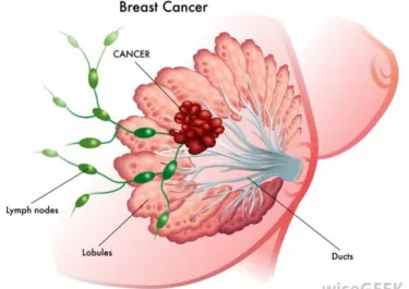
Figure 1. Morphology of the human breast. 1) Lobules are an assembly of alveolar structures containing specialised cuboidal epithelial cells that secrete milk. 2) The lactiferous duct are connections that form a branched system for the collection of the milk, the ducts join together and drain the milk into an opening in the nipple. 3) Lymph nodes are part of the lymphatic
5 system and participate to the defense of the breast by circulating the lymphatic fluid containing immune cells. (Picture from www.wisegeek.org)
Detection is usually difficult, as it does not present any of the usual symptoms, such as inflammation or noticeable masses. Most of the time this kind of tumor is hormone receptor positive. Even if it is not life threatening the presence of this kind of tumor is associated with reoccurrence and development of invasive breast cancer.
Lobular carcinoma in situ (LCIS): Similar to the DCIS, this type of cancer occurs when the abnormal cells develop in the milk-making glands (lobules) and remain in-situ. The danger of the LCIS is usually very low and is not life threatening, but it is an indicator that a woman is at increased risk for developing invasive breast cancer in either breast in the future. Given the in-situ characteristic of this tumor, its removal is usually easy and does not require chemotherapy.
Infiltrating breast carcinomas
Invasive ductal carcinoma (IDC): is the most common type of invasive breast cancer accounting for around 80% of invasive breast cancers. Opposite to the in-situ type, this form of cancer is characterised by infiltrating cells (invasive) that have broken through the wall of the duct and invaded the surrounding breast tissue. The probability of invasion to the lymph nodes is high in this form of cancer.
IDC can be broken down into different subtypes, based on the appearance and behaviour of the cancer cells. These include invasive mucinous carcinoma, tubular
6 carcinoma, medullary carcinoma and invasive micropapillary carcinoma. In some cases, when the observed morphology of the cancer cells does not present any feature characteristic of a particular type of cancer, the IDC is classified as invasive not otherwise specified or non-specific type.
Invasive lobular carcinoma (ILC): Makes up about 10% of invasive breast cancer cases. ILC originates from the milk-making glands (lobules) of the breast and the subsequent invasion is similar to the one in IDC. Particular to this type of cancer is a type of growth in which the cells grow in single lines that lead to a hardening of the breast.
Inflammatory breast carcinoma (IBC): This uncommon type of invasive breast cancer accounts for about 1-3% of all breast cancers. It tends to be more common in younger women, and women of African ancestry. In its early stages, IBC is often mistaken for infection; this is due to a particular inflammatory reaction induced by the tumor cells present in lymph vessels. IBC begins in the milk ducts of the breast and spreads to the lymph vessels. This type of cancer has a higher chance of spreading and it can be more aggressive and harder to treat than invasive ductal or lobular cancer. Treatment usually involves a combination of approaches including chemotherapy, surgery, radiation, and hormone and HER-2 targeted therapy, if appropriate.
Pager’s disease: This type of condition is characterised by a cellular growing in and around the nipple. It is an unusual type of breast cancer, accounting for less than 5% of all cases and is more common in women over the age of 50. However
7 this condition occurs sometimes in combination with another form of breast cancer such as DCIS or IDC.
1.3 Molecular characterization of breast cancer
The classification presented above gives a general description about the localisation of the tumor, its cytological origin and more importantly, about its containment status. However, this tells little about their specific metabolic or genetic features. With the development of simpler and cheaper molecular approaches over the last couple of decades it has become usual to observe the gene expression profile (GEP) of the cells. The GEP allows for further characterisation of the tumor with respect to certain mutations, such as mutations in the breast cancer 1 and 2 genes (BRACA1/2), which will be discussed later. Along with the GEP some specific proteins serve as molecular markers that help with the characterisation of the tumor.
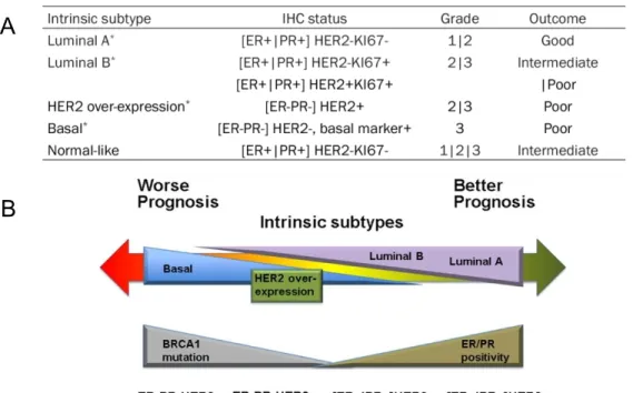
Even though several researchers and organizations have presented different arguments over the years about what genes describe better the cancer phenotype, it was the work of Sorlie and colleagues in the early 2000s (Perou et al., 2000; Sorlie et al., 2001) that established the standard for the molecular subtyping of breast tumors. These works identified five different subtypes, each one associated with a particular set of immunohistochemistry (IHC) markers (Fig. 2A), and grades describing the aggressivity of the cells based on their differentiation status, with well differentiated cells being less aggressive. Sorlies’s classification is based on the status for three hormone receptors identified as IHC markers, these cover: the progesterone receptor (PR), the human epidermal growth factor receptor 2 (HER-2) and the estrogen receptor (ER), with the status of these receptors being crucial in the prognosis of the patients (Fig. 2B) (Dai et al., 2015). KI67 is a nuclear protein associated with cellular proliferation
8 that is normally expressed at low levels in normal breast tissue (Fig. 2A). However the use of KI67 as a molecular marker for breast cancer prognosis has been greatly debated and though it was used by Sorlie and colleagues in their works (Sorlie et al., 2001), the absence of a standard agreement for its quantification has led to its disuse in pathology tests (Inwald et al., 2013). The importance of these factors resides in their ability to induce growth-promoting signals; the progesterone is a steroid sex hormone that participates in the control of the menstrual cycle, pregnancy and embryogenesis and HER-2 belongs to a family of receptor tyrosine kinases that can interact with different signaling molecules, but in most cases induces cellular proliferation (Roy et al, 2009). Estradiol (E2) plays a central role in the breast cancer pathology as it possesses the ability, via the ER, to induce expression of genes encoding growth factors, their receptors and different signaling molecules that can activate cell proliferation and survival stimuli (Shou et al., 2004). Such is the role played by ER that it is called the defining and driving transcriptional factor of breast cancer, since its target genes can direct the cellular growth and the endocrine response. Crosstalk between these three receptors can lead to unusual activation of pathways that exacerbates the growth and division of the cancer cells. For example, activation of the mitogen activated protein kinase (MAPK) by HER-2 induces phosphorylation of ER, thus simulating the binding of E2 and inducing transcription of ER controlled genes (Ali and Coombes, 2002).
A very important fourth marker, this one a genetic mutation, is also used in combination with the previous three to establish the prognosis of the patients; mutations in the Breast cancer 1 and 2 genes (BRCA1/2). Germ line mutations of BRCA1 are present in approximately 90% of familial breast and ovarian cancers and in approximately 50% of familial breast cancers alone. BRCA1 is a nuclear protein of 220 kDa and 1,863 amino acids, it is highly expressed in rapidly proliferating mammary epithelial cells during pregnancy and is down-regulated during lactation (Marquis et al., 1995). Several pleiotropic biological functions have been associated with BRCA1, most of them related to transcriptional regulation, chromatin remodeling, DNA damage repair, cell cycle
9 regulation, and checkpoint control (Scully and Livingston, 2000). The tumor suppressor role presented by these genes is given by their participation in the DNA repair mechanisms and, therefore, allowing them to play a role in ensuring the stability of the cell’s genetic integrity. Dysfunctional mutations in either one of these genes results in DNA damage that may not be repaired properly and as a result, the cells are more likely to develop additional genetic alterations that can lead to cancer.
Figure 2. Breast cancer classification by intrinsic molecular subtype. A) Breast cancer subtype dependent on the immunohistochemistry status of the receptors: estrogen (ER), progesterone (PR) and the human epidermal growth factor 2 (HER-2) showing the usual grades and outcomes associated with each subtype. (Perou et al., 2000) B) Usual prognosis in function of the molecular subtypes and the status of BRCA1 (Dai et al., 2015).
A
10 Specific inherited mutations in BRCA1/2 increase the risk of female breast and ovarian cancers and they have been associated with increased risks of several additional types of cancer. The effects of mutations in BRCA1/2 are seen even when a person’s second copy of the gene is normal (Campeau et al., 2008). Combination of the status of these four markers is usually the main argument for decision making regarding the treatments to be implemented in the patients, and have been associated with different prognosis (Fig 2B) (Spitale et al., 2009).
The high level of heterogeneity evidenced by Sorlie’s classification reveals a classical characteristic that underlays the very nature of the BC pathology, a high mutational rate that is especially problematic for the treatment of the illness (Osz et al., 2012).
1.4 Tumorigenesis.
The precise molecular mechanisms behind the triggering of tumorigenesis have been intensively studied during the last decades with the discovery of proto-oncogenes by J.M. Bishop and H.E. Varmus during the 80s, establishing a conceptual landmark in the study of cancer (Bishop, 1982; Varmus, 1985). This discovery revealed that cancer is generated by our own genes if mutations in their sequence alter their function. Nevertheless not one modification can start development of cancer by itself, this is greatly due to the redundancy and safety mechanisms of the cells, where failure of one specific pathway is usually compensated by activation of a parallel path or overexpression of a similar one (Gerlinger et al., 2014). Because of this, cancer has been characterised as an aging condition as it takes time for a healthy cell to accumulate the necessary mutations to undermine the safety features and become a tumor. How many of these genetic mutations are necessary to trigger the transformation is still debated, however it has been agreed that transformation from
11 normal acting cells to cancer proliferation is driven by a relative limited number of genes known as cancer drivers (Kim, 2015).
More precisely, these cancer drivers are defined as genes that are mutated in more than one type of cancer. In a statistical sense this means that every cancer harbors at least one of these mutations, empirically, a tumor tends to contain between two to eight cancer drivers (Vogelstein et al., 2013). Two types of drivers are known today, oncogenes and tumor suppressors. Oncogenic modifications are often missense mutations that change the amino acid residues and alter the protein function. These modifications are more disturbing for the cellular homeostasis when the result is a gain of function. A classic example is the mutation of the KRAS gene, where the mutation of glycine 12 (G12D or G12V) results in an constitutively active k-ras protein that remains in a GTP-bound state (Okudela et al., 2010). In a general manner, gain of function is a hallmark of oncogenic mutations. On the other hand, characteristic tumor suppressor mutations usually result in the disabling of the protein, either as consequence of a missense or nonsense modifications. The most common examples are the mutations of the previously discussed BRCA1 gene. Mutations in this gene are found all over the coding region with no specific mutation being yet identified as more relevant than others among the 389 mutations documented (Vogelstein et al., 2013). In a general manner nonsense mutations in the coding region are typical in a tumor suppressor gene.
A closer look at the function of two of cancer driver genes, will serve to illustrate the reason why this type of genes is primordial in the triggering of the cancer phenotype. The prevalence of mutations in TP53 and PIK3CA in breast cancer is about 33%, this characteristic was observed by a statistical analysis of the mutations presented in the cancer genome database Catalogue of somatic mutations in Cancer (COSMIC) and The Cancer Genome Atlas (TCGA) (Kandoth et al., 2013). The TP53 gene is mutated in 42% of all cancers and its product, p53, plays a central role in the DNA damage response by inducing cell cycle inhibitors that arrest the cellular division, thus giving the
12 cell time to repair the genomic damage. In the absence of function of p53, these damages are carried through to the next generation of cells, thereby increasing the risk of cellular dysfunction and the proliferation of more damages. The second highly common cancer driver is PIK3CA with mutations in this gene being present in about 18% of all cancers. The product of PIK3CA is the p110 catalytic subunit of the 3-kinase (PI3k). This unit phosphorylates phosphatidylinositol-(4,5)-biphosphate to phosphatidylinositol-(4,5)-triphosphate (PIP3) (Shaw and Cantley, 2006). Mutations in PI3k often result in a gain of function that increases the lipid kinase activity. As described before, gain of function is an oncogenic hallmark and in this case the overproduction of PIP3 by PI3k activates multiple kinases such as the phosphatidylinositol dependant kinase (PDK1) and the protein kinase B (PKB), both of which have been shown to inhibit pro-apoptotic molecules thus conferring abnormal survival capabilities to the cell (Engelman, 2009).
In general, all four molecular subtypes of breast cancer described in Figure 2A (excluding the basal subtype) harbor mutations in Tp53 or PIK3CA, however their prevalence is different and they are remarkably mutually exclusive, but the reason why these mutations are never present together is not yet understood.
1.5 Tumor heterogeneity
One very important factor to consider in the discussion of cancer development is the high level of heterogeneity present within the cell population of the tumor. As important as the activity of the cancer drivers is for cancer development, it is not the only factor to consider. Tumors are rarely composed of monoclonal populations and different genetic expressions from different subpopulations result in different cellular pathways being activated. The effect of cross-talk among these different pathways is a factor impossible to neglect when considering the development and treatment of cancer.
13 While cancer was studied for many decades as a clonal disease (Nowell, 1976), early studies in murine mammary tumours revealed that certain subpopulations from different sections of the same tumour differ in growth rate, immunogenicity, drug response and ability to metastasize, thereby demonstrating functional and phenotypic heterogeneity (Heppner, 1984). However, even when we are far from understanding the complexity of the interactions that act among these subpopulations, there is growing evidence that cancer cells behave as communities and present several characteristics of ecological behaviour. Due to this, increasing attention is now being directed towards the cooperative behaviour of sub-clones that can influence disease progression, and cancer is now studied as an evolving ecosystem (Kanaseki et al., 2003; Merlo et al., 2006). As expected, these interactions add a greater layer of complexity to therapeutic treatments where, often, tumors presenting a high level of heterogeneity lead to a poor prognosis (Tabassum and Polyak, 2015).
Nevertheless not all of these behaviours act in favour of the survival of the tumor. As in any ecological community there are several kinds of interactions that have been observed in tumors. These interactions can be classified as negative or positive depending on the proliferative capabilities that they give to the tumor (Tabassum and Polyak, 2015). Negative interactions are mainly competitive ones that arise from the limitations in nutrients and oxygen, this type of competition usually results in the elimination of one subpopulation and the expansion of one more fit (Pepper et al., 2009). On the other hand, positive interactions can present different characteristics and can be classified as commensalistic, where one population profits from the other without damaging it or synergistic, where two subpopulations cooperate to produce new characteristics that are not necessarily indispensable for the community, or mutualistic when the subpopulations cooperate to benefit all the parties (Pienta et al., 2008). These positive interactions are of great interest for the treatment of cancer as they can heavily affect the outcome of the patient (Michelson, 1987). For example, targeted therapies that eliminate a tumor subpopulation that exerted a negative interaction over other
14 subpopulations will favour a later tumor regrowth of the remaining cells, not only because the treatment fails to eliminate the totality of the tumor but also because the remaining subpopulations are no longer under the pressure of a negative interaction (Pepper et al., 2009) (Fig. 3A.). In a similar way, the elimination of a subpopulation that exerts a positive interaction over the tumor community greatly affects their development as these subpopulations often help tumor expansion via the secretion of growth or angiogenic factors such as the c-fos induced growth factor (FIGF) and interleukin 11 (IL-11), respectively (Marusyk et al., 2014). Elimination of a positive interacting subpopulation will result in a rapid decrease of the size of the tumor and a prolonged regression but inevitably will also end up allowing for tumor regrowth because of the same failure to eliminate all the cancer cells.
Figure 3. Targeted elimination of cancer subpopulations. A) Elimination of a selfish subpopulation exerting a negative interaction over the other subpopulations. In absence of the negative interaction a faster tumor regrowth should be expected. B) Elimination of positive interacting subpopulations that help tumor growth can delay tumor regrowth (Tabassum and Polyak, 2015).
15 The problem presented by the existence of different subpopulations within the tumor constitutes an important challenge for the current treatments, as many of them are based on the precise targeting of cellular characteristics that are only common to some subpopulations. In the breast cancer pathology for example, the treatment with tamoxifen, an anti-estrogenic compound, of ER-positive cases will face two problems: first, the existence of ER-negative subclones among the tumor, these subpopulations will present resistance from the beginning of the treatment and will proliferate better as the competing cells are eliminated. Once the ER-positive cells are eliminated, the ‘second generation’ tumor will stop responding to the treatment thus aggravating the condition. Another possible outcome is the induction of tamoxifen resistance via mutation of the ER-positive cells, induced by the selective pressure of the treatment (Barkhem et al., 2004). Such a scenario would result in the co-existence of two different subpopulations resistant to tamoxifen, a situation presenting a worst prognosis.
As presented before, the status of ER in breast cancer cells is one of the main factors in BC decision treatment and prognosis prediction, however its importance goes beyond the cancer pathology as E2 is a central factor in the regulation of several developmental mechanisms and tissular functions in mammalian organisms.
1.6 Estrogens and estrogen receptors
1.6.1 Normal function
One of the best models for E2-driven transformations is the breast, an organ that by its nature has evolved to be highly sensitive to hormonal variations. During puberty, tissue development is directed by estrogen along with other sexual hormones, the same is
16 true during pregnancy, when the same hormones initiate further development and activation of functions. Because of these characteristics, estrogen is known as the main sexual hormone and besides its function in the breast, it drives the development of the female reproductive organs and secondary sex characteristics (Barkhem et al., 2004).
The estrogen family of steroidal hormones is composed of three molecules, estrone (E1), estradiol (E2) and estriol (E3). Estradiol is the most common form and its ubiquitous expression throughout the body is most significant during the fertile years. Its activity is replaced by E1 after the onset of menopause and by E3 during pregnancy. Also, E2 has been identified as the hormone possessing the highest affinity for its receptors, making it the most relevant form of the hormone for not pregnant and pre-menopausal women. (Nilsson et al., 2001) For the rest of this work, every mention of estrogen should be understood as referring to the estradiol (E2) form.
E2 is one of the main sexual hormones and as such acts as a messenger; its activity is mainly mediated via a family of dedicated hormone receptors, the estrogen receptors (ERs). Around the 1970s, Jensen and Jacobsen (1967) observed that the biological effects of estrogen had to be mediated by a receptor protein, based on the specific binding of E2 in the uterus. Continued experimentation for the next decade concluded in two groups reporting the cloning of the estrogen receptor (ER) in 1986 (Green et al., 1986). Until 1995, it was assumed that there was only one ER and that it was responsible for mediating all of the physiological and pharmacological effects of natural and synthetic estrogens and anti-estrogens. However, in 1995, a second ER, estrogen receptor beta (ERβ), was cloned from a rat prostate cDNA library (Kuiper et al., 1996). The first ER is now called ERα (Chu and Fuller, 1997).
17 Estrogen is produced primarily in the ovaries in females and testes in males, and is central to the regulation of growth, development, and physiology of the reproductive system in humans (Swedenborg et al., 2009). The biological function of estrogen is mediated by ERα and ERβ. The signaling and activation or inhibition of estrogen target genes depends on a tightly controlled balance between the expression of ERα and ERβ in the target organs. ERs belong to the superfamily of steroid hormone nuclear receptors (NRs) (Choi and Jeung, 2003; Osz et al., 2012). This family also includes the estrogen-related receptors (ERR), progesterone receptors (PR), androgen receptors (AR), glucocorticoid and mineralocorticoid receptors. Steroid receptors (SRs), such as the ER, are ligand-dependent transcription factors, and they often control cell cycle activities (Weigel and Moore, 2007). ERs in particular have been shown to regulate certain cell proliferation pathways (Lee et al., 2012).
The complex structure of ERs, containing several functional sites, is evidence of the multiple activities they can execute. Similar to other NRs, ERs are comprised of five domains with distinct functions (Fig. 4A) (Skafar and Zhao, 2008). First, the N-terminus of the A/B domains contains an activation function 1 (AF1) module, which contributes to the transcriptional activity of ERs and is an essential domain for interaction with co-regulators. This domain is prone to post-transcriptional modifications that stimulate AF1 activity (Kong et al., 2003). A central region named C domain encodes for a DNA-binding domain (DBD) that is essential for the DNA-binding of ERs to DNA (Geserick et al., 2005). The D domain is a hinge region that includes sequences for nuclear localization signaling and facilitates post-translational modification of ERs. Usually these modifications contribute to the activation of ER signaling in the cells. Finally, at the C-terminal region we find the E/F domain, which contains a ligand-binding domain (LBD) that serves also as an interaction site with co-regulators, in addition to the ligand-dependent activation function 2 (AF2) module. Both modules have the ability to regulate the activity of ERs (Edwards, 2000). Certain differences between the F domain of ER
18 and ERβ may be responsible for the selectively transcriptional control of ERs in specific target genes (Skafar and Zhao, 2008).
Binding of different ligands to the LBD (either steroid hormones or synthetic compounds) induces a specific conformation of the receptor thus promoting precise co-regulatory interactions and associations of the function domains AF1 and AF2 (Bramlett et al., 2001; Nilsson and Gustafsson, 2011). In the classical pathway, binding of estrogen to the receptor induces its phosphorylation and the formation of homodimers and heterodimers, that are then translocated into the nucleus (Fig. 4B) (McDevitt et al., 2008). ERs modulate the transcription of target genes by binding to estrogen response elements (EREs) in the DNA sequence (Park et al., 2011).This binding promotes DNA bending and looping, allowing for the interaction with co-regulators and the general transcriptional machinery. Some of these regulators are activators, co-repressors, co-integrators, and histone modifiers such as acetyltransferases and deacetylases (Edwards, 2000). In the absence of estrogen, ERs remain in an inactivated state thanks to the association with the heat shock protein 90 (Hsp90). This protein has been associated with protein stabilization, binding affinity of receptors to ligands, and signaling cascades (Sanchez, 2012).
1.6.2 Non genomic activities of ERs
Even if the activity of ERs is most often mediated via the binding to the EREs and the consequent chromatin modifications, it is also possible for them to modulate the cellular activity without directly binding to DNA. Characteristic of the non-genomic activities is the fast response times of these pathways, with cellular mechanism responding in seconds to minutes of E2 stimulation compared to hours for the canonical DNA-dependent pathway (Marino et al., 2006). The mechanisms by which the non-genomic
19 activities are exerted are highly heterogeneous. For instance, inhibition of the expression of the cytokine IL-6 by E2 is mediated by an interaction between ER and the c-rel subunit of the NFkB complex, where the ER prevents NFkB from binding to the IL-6 promoter (Galien and Garcia, 1997). Similar is the activation of ER by E2 that induces subsequent interaction and activation of the IGF-1 receptor resulting in activation of the MAPK signaling. The majority of the non-genomic activities are mediated by membrane bound ERs (Fig 4B). In these cases other membrane bound receptors such as g-protein coupled receptor 30 (GPR30) or the metabotropic glutamate receptors (mGLUR) interact with the ER via cytoplasmic kinases to transduce signals to targets in the cytosol or in the nucleus (Levin, 2009).
1.6.3 Non-E2-dependent activation of ERs
In the same manner as the ERs can regulate certain cellular activities independently of their DNA binding properties, some activities can be induced in absence of E2 (Bunone et al., 1996). Frequently, this alternative activation of the ERs is exerted by signaling pathways that phosphorylate it, thus imitating the modifications induced by the binding of E2. Some of the factors that can induce such modifications include regulators of the general cellular phosphorylation state, such as protein kinase A (PKA) (Aronica and Katzenellenbogen, 1993) or protein kinase C (PKC) (Ignar-Trowbridge et al., 1996) and certain extracellular signals such as peptide growth factors, cytokines, or neurotransmitters (Chalbos et al., 1994). Among these factors, some are known to completely mimic the effects of E2, for example the epidermal growth factor (EGF) in the mouse reproductive tract (Curtis et al., 1996). Other growth factors which activate ER signaling include insulin (Newton et al., 1994), insulin-like growth factor I (IGF-I) (Aronica and Katzenellenbogen, 1993), and transforming growth factor (TGF)-β (Ignar-Trowbridge et al., 1996). As mentioned before, a very important example of this is the activation of ER by HER-2 via phosphorylation, thus leading to ER-activation and
20 induction of E2-controlled genes in absence of the hormone (Nilsson et al., 2001). This phenomenon is often observed in breast cancer patients that have developed resistance to anti-estrogen therapy.
1.7 Breast cancer resistance to anti-estrogen therapy
The genetic deregulations described above are also very important in the context of tumor survival, as the resistance developed to several types of treatments is dependent on the activation of non-targeted cancer driver mechanisms that allow for cellular proliferation even with active inhibition of the ‘original’ driver.
Given the importance of E2 for breast cancer development, the targeted treatment of ER has been one of the most usual therapies for this condition. As ER-positive breast cancer represents around 70% of all cases of breast cancer, the ER inhibitor Tamoxifen has been widely used (Jordan, 2006). Tamoxifen acts as an agonist of ER by competing with E2 to bind the receptor. Once bound, Tamoxifen does not inhibit but greatly modifies the activity of ER (Wang et al., 2004). However, about 30% of all cases treated with Tamoxifen show resistance and end up relapsing at a rate of 1-5% per year (Goss et al., 2008).
Different molecular approaches have been used to identify the mechanisms behind this resistance with several different pathways showing some kind of activity responsible for the phenomena. Overexpression of the MYC oncogene, activation of the insulin/Igf receptor signaling and mutation of the PI3k are some of the observed cancer drivers that are activated in the resistance phenotype (Engelman and Janne, 2008)
21 Figure 4. Schematic representation of ER structural domains, activation and signaling. A) Functional domains of the ER. The A/B domain at the N-terminus contains the function domain AF-1 (transcription activation function 1) that interacts with other TF. The C/D domain has a DNA binding function that contains a two-zinc finger structure. The E/F domain at the C-terminus possess the ligand binding pocket and the second function domain AF-2, that interacts with co-activators. B) Activity of the ER can be executed by three different pathways. 1) The classical pathway is mediated by binding of nuclear ER to an ERE, recruitment of co-regulators and subsequent induction of target genes. 2) The secondary pathway is mediated by cytoplasmic or membrane bound ER. Interaction with other membrane bound receptors, such as the mGLUR and cytoplasmic kinases can control transcription factor and regulate gene
A
22 transcription. 3) The third pathway is mediated by non-ER receptors such as the GPR30/GPER. Activation of these receptors induce signaling through cytoplasmic kinases and finally regulation of gene transcription (Nilsson and Gustafsson, 2011).
However these back-up cancer drivers are not only activated in breast cancer, but have been also observed in many other cancer types under treatment, further showing the importance of these mechanisms to the proliferative phenotype. Among the cancer drivers in the breast the most usual cause responsible for anti-estrogen resistance is a mutation in the estrogen receptor alpha (ESR1) gene. Certain mutations generate amino acid substitutions that mimic the active conformation of ER in the absence of ligand, thus rendering the cell non-dependent of E2 (Robinson et al., 2013; Toy et al., 2013). Interestingly enough, these mutations are usually not present in the primary tumor and are evidence for Darwinian survival under the selective pressure of anti-estrogen therapy (Kim, 2015).
1.8 Immune response to cancer
All tumors have a common characteristic regarding their relationship towards the immune response, the presence of a complex assembly of immune cells and the consequent cellular interactions established between the two cellular classes (Coussens et al., 2013). Tumor cells become immunogenic early during the tumorigenic process and are able to be eliminated by the immune system. Tumor suppression is mainly the result of cytotoxic activity by activated T cells directed to tumor cells expressing tumor-specific antigens. Tumor cells possess a high mutational rate that generates changes in the proteins, these changes can be identified by the T cells via the antigenic presentation pathway (Dunn et al., 2004). Tumor escape is most often the result of interference of the chronic inflammation mechanism that disrupts the cytotoxic
23 T cell activity. Such scenario is usually observed in tumors evolving from chronically inflamed tissues, where tissue repair pathways are overexpressed (Coussens et al., 2013). An example of the importance of the T cell immune response for the control of cancer was evidenced when mice lacking a functional immune system were shown to be severely more susceptible to induced cancers. In humans, the effect of interleukin-2 (IL-interleukin-2), secreted by T-cells, showed an increased ability of the lymphoid compartment to suppress tumors (Schreiber et al., 2011).
1.8.1 Antigenic activation of the immune response
Observations from these and other experiments revealed that the T cell compartment is central to the immune response thanks to its ability to recognize peptide epitopes that are displayed by major histocompatibility complex molecules (MHCs) on the surface of the tumor cells. In theory, three classes of antigens may mediate such cancer rejection. These classes are broad classifications of potential antigens found within tumors. The first class is known as tumor specific antigens (TSAs), these are antigens that are not encoded by the normal genome and may represent either oncogenic viral proteins or abnormal proteins that arise as a consequence of somatic mutations; these are known as neo-antigens (Hanahan and Weinberg, 2000). For the large majority of human tumors without a viral cause, such neo-epitopes are only produced by tumor-specific DNA modifications that result in the formation of new protein sequences. In the case of virus-associated tumors, such as cervical cancer and a subset of head and neck cancers, epitopes derived from viral open reading frames also contribute to the pool of neo-antigens (Schumacher and Schreiber, 2015). During cancer initiation and progression, tumor cells acquire protein-altering mutations that are either responsible for transformation, as the cancer driver mutations mentioned before, or are a by-product of the genomic instability that accompanies cellular transformation, these later ones are known as passenger mutations (Hanahan and Weinberg, 2011). The
24 alterations that actually result in expression generate mutant proteins with a loss of self-identity and can be perceived as foreign proteins by the immune system (Heemskerk et al., 2013). Another factor that induces the development of neo-antigens is as a by-product of anticancer treatments, like chemotherapy or radiation therapy, or by targeting epigenetic control mechanisms or drugs intervening with DNA repair pathways.
The second class of antigens is known as tumor associated antigens (TAAs). These TAAs include proteins encoded in the normal genome and may be either normal differentiation antigens or other normal proteins that are aberrantly expressed. Overexpressed normal proteins that possess growth/survival promoting functions, such as Wilms tumor 1 (WT1) (Ohminami et al., 2000) or the breast cancer inducing HER-2 (Kawashima et al., 1999), are examples of TAAs that directly participate in the oncogenic process. Some TAAs are composed of proteins carrying posttranslational modifications such as phosphorylation (Doyle et al., 2006). Because TAAs are normal proteins, their antigenicity depends on abnormal expression levels or abnormal context to circumvent naturally occurring mechanisms of immunological tolerance (Pardoll, 2003). Along these lines, TAAs usually have lower T cell receptor (TCR) affinity compared with TSAs or foreign antigens (Stone et al., 2015). Because of this, TSAs are less likely to be susceptible to mechanisms of immunological tolerance and therefore may represent more visible targets for immune-mediated tumor control (Heemskerk et al., 2013).
The final category are the cancer-germline/cancer-testis antigens (CTAs) (Simpson et al., 2005).This category represents the antigens that are normally expressed in testis, fetal ovaries, and trophoblasts, but can also be expressed in cancer cells (Simpson et al., 2005).Because they are encoded in the normal genome but display highly restricted tissue expression, CTAs have received considerable attention as attractive targets for
25 immunotherapy (Scanlan et al., 2002). Given the fact that the reservoir of T cells recognizing the non-mutated self-antigens is controlled by the maturation process in the thymus, the neo-antigens constitute an important fraction of the cancer antigens that can elicit the immune response (Gilboa, 1999).
1.8.2 T cell mediated tumor suppression
Shortly after development, tumors become organized and acquire tissue-like characteristics with local and systemic connections with immune cell populations of myeloid and lymphoid origin, each one interacting with the tumor in different patterns that may drive further tumor development to different outcomes (Palucka and Coussens, 2016).
It is important to understand that the interaction between the tumor and the immune system is a dynamic process, guided by the pressure exerted by the immune response, answered by the tumor resistance, and modulated by a damage control mechanism that protects the surrounding tissues (Coussens et al., 2013). When the immune response to cancer is high and the immune-suppression processes are low, the tumor development is kept under control. However, a strong anti-tumor response will induce certain physiological mechanisms designed to inhibit the effector T cells in order to prevent tissue damage and maintain tissue homeostasis (Coussens et al., 2013). In the absence of a precise set of stimuli, the immune response has evolved to be transitory. It is designed to maintain the integrity of the organism and to establish coexistence with the environment by protecting it from external threats. Given that the threats are usually transitory, the suppression often prevails. Multiple pathways of suppression are at play in tumor microenvironments, including cells such as Th2-polarized macrophages, immature and suppressive monocytes, regulatory B cells, and regulatory T cells, as well as certain molecules that are key in the activation of checkpoints that control T cell
26 differentiation (for example, CTLA-4 and IDO) and effector function (such as PD-1). It has been shown that pharmacological blockade of these inhibitory pathways can tip the balance toward anti-cancer effector T cells (Coussens et al., 2013). The latter ones can be primed or boosted by antigen-presenting cells (DCs) and/or by co-stimulatory signals (for example CD137 ligands). Tumor antigens in general can be presented to T cells in exogenous vaccines, as well as endogenously via DCs that captured dying neoplastic cells. When T cells specific to defined antigens kill neoplastic cells, such a process can enable generation of responses to other antigens, so called epitope spreading. A critical factor in the balance between immunogenicity and suppression is inflammation; this is due to the fact that the type of inflammation can direct the continued immune response to the tumor. A Th1 oriented response is more effective to destroy the tumor, while a Th2/Th17 oriented may result in tumor promotion. (Palucka and Coussens, 2016).
To better understand how the type of immune response can drive the direction of the tumor response, it is important to have a closer look at the interactions and functions of the different compartments of the immune system with the tumor.
The myeloid cells present different functions related to the maintenance of homeostasis of the organism; these functions involve the capture and degradation of antigenic molecules by macrophages or for presentation by dendritic cells (DCs); tissue repair induction by the macrophages, and some effector functions by the mast cells, monocytes and granulocytes, such as release of different cytokines (Tae Chul Moon 2014). However, in some cases tumor cells can alter the steady-state activity of the myeloid immune cells present in the vicinity of the tumor, known as tumor micro-environment (TME) (DeNardo et al., 2011). The cells present in the TME include tissue-resident and blood derived cells and their activity can be regulated by the tumor cells by secreting factors such as interleukin-6 (IL-6) or granulocyte-macrophage
colony-27 stimulating factor (GM-CSF). These secretions have the ability to increase recruitment and proliferation of immature myeloid cells in proportions that are atypical under physiological conditions (Gabrilovich et al., 2012).The myeloid cells possess an important functional plasticity in response to environmental signals, and can drive the immune response to different outcomes, for example antigen degradation or antigen presentation, when macrophages acquire DC capabilities (Banchereau et al., 2000). Alternatively, they can drive the immune response towards tissue repair rather than inflammation when macrophages are polarized toward type 2 states, and protective or non-protective T-cell immunity when programmed by cancer-derived factors (Balkwill et al., 2005).
Among the myeloid compartment, the dendritic cells play a central role in the immune response, as they are able to trigger both the humoral and cell mediated branches of the immune system by presenting the antigens to B and T lymphocytes (Lanzavecchia and Sallusto, 2001). In the cancer pathology the tumor antigens are presented to T cells either at the tumor sites or at the lymph nodes. The dendritic cells can display these tumor antigens via the classical pathways by major histocompatibility molecules of class I (MHC-I) or class II (MHC-II). Lipid antigens can also be presented via non-classical pathways by CD1 molecules that allow selection of rare antigen-specific T cells (CD4+ or CD8) and NK-T cells (Lanzavecchia and Sallusto, 2001). If the antigenic presentation by dendritic cells induces a better activation of naïve CD8+ T cells, these will differentiate into T cytotoxic lymphocytes (CTL) and direct the immune response towards a Th1 polarized immune response. In contrast, preferential activation of naïve CD4+ T cells will produce helper cells with different cytokine expression patterns and FoxP3+ regulatory cells. These cells have a dampening effect on the cytotoxic activity to prevent autoimmune responses, however this type of response is also less anti-carcinogenic (Zhu and Paul, 2008). In a similar way, non-maturation of the dendritic cells by exposure to interleukin-10 for example, will induce the antigenic presentation for T cell suppression (Pulendran, 2015).
28 The advantage of a Th1-polarized CD4+ response is its increased cytotoxic effect. This type of response is characterised by secretion of interleukin-2 (IL-2), tumor necrosis factor alpha (TNF) and interferon gamma (IFN). These cytokines in combination with CD8+ T cells promote macrophage cytotoxic activity (Stout and Bottomly, 1989) and can induce upregulation of antigen processing and expression of MHC molecules in dendritic cells (Lanzavecchia and Sallusto, 2001). On the other hand the Th2-polarised response is characterised by secretion of interleukins-4,-5,-6,-10 and -13. This pattern of secretion can induce T-cell anergy and loss of cytotoxicity, increase the humoral immunity and regulate the tumor-promoting activities of macrophages (DeNardo et al., 2011).
These different outcomes of the immune response have led to a change in the classical view, according to which the immune cells simply facilitate tumor rejection, and has been replaced by a more complex view, where the lymphocytes can present both tumor-promoting and tumor-inhibiting properties (Coussens et al., 2013). The best example of this phenomenon is the link between the type of inflammation and the associated type of Th immune response, and how each of these inflammation profiles shows opposing effects on tumors. Th2-associated chronic inflammation promotes neoplastic cell survival, angiogenesis, tissue remodeling, and metastasis, while Th1-associated acute inflammation, triggers neoplastic cell destruction. As the immune response pressures the tumor cells, several mechanisms are implemented by the cancer cells to escape elimination. First, the cells become antigen-edited as the more immunogenic cells are killed off by the immune system. Then the cells that escape elimination become less immunogenic. This progression is exerted by two mechanisms. The main factor is the down-regulation of MHC molecules while a great proportion of them also activate intrinsic gene expression programs that are directed to suppress T cells and stimulate myeloid cells. These two characteristics being typical of a Th2 oriented response. Cytokines related to this kind of response include transforming growth factor β (TGFβ); IL-4, -13, -8, and -10; thymic stromal