Abies balsamea oleoresin
Serge Lavoie‡1, Charles Gauthier‡1,2, Jean Legault1, Sylvain Mercier1,
Vakhtang Mshvildadze1 and André Pichette*1
Full Research Paper
Open AccessAddress:
1Université du Québec à Chicoutimi, Chaire de Recherche sur les Agents Anticancéreux d'Origine Végétale, Laboratoire d'Analyse et de Séparation des Essences Végétales (LASEVE), Département des Sciences Fondamentales, 555 boul. de l'Université, Chicoutimi (Québec) G7H 2B1, Canada and 2Université de Poitiers, Institut de Chimie IC2MP, UMR-CNRS 7285, 4 rue Michel Brunet, 86022 Poitiers, France
Email:
André Pichette* - andre_pichette@uqac.ca * Corresponding author ‡ Equal contributors
Keywords:
Abies balsamea; cycloartane; lanostane; oleoresin; triterpenoids
Beilstein J. Org. Chem. 2013, 9, 1333–1339.
doi:10.3762/bjoc.9.150
Received: 15 April 2013 Accepted: 12 June 2013 Published: 04 July 2013
Associate Editor: A. Kirschning
© 2013 Lavoie et al; licensee Beilstein-Institut. License and terms: see end of document.
Abstract
Phytochemical analysis of A. balsamea oleoresin led to the isolation of three new 3,4-seco-lanostane triterpenoids 1–3, one new cycloartane triterpenoid 4 along with fourteen known terpenoids. Structure determinations were based on extensive 1D/2D NMR, IR and MS spectroscopic analyses, and comparison with literature data. The isolated compounds were evaluated in vitro for their cytotoxicity against human cell lines (A549, DLD-1, WS1) and their antibacterial activity against E. coli and S. aureus. Abiesonic acid (6) exhibited weak cytotoxic activity against A549 (IC50 = 22 µM) while compounds 1 and 4 were weakly active against
S. aureus (MIC = 25 µM).
Introduction
The genus Abies (Pinaceae) comprises 46 species of evergreen conifers [1]. Most of them are found in temperate and boreal regions of the northern hemisphere. The first phytochemical investigation of Abies species was undertaken 75 years ago by Takahashi [2]. Since then, more than 277 secondary metabo-lites have been isolated, and mainly identified as terpenoids, flavonoids and lignans [3]. Balsam fir Abies balsamea (L.) Mill., a popular Christmas tree in Canada, has been used
tradi-tionally by North American aboriginal people as an antiseptic, tuberculosis remedy, and venereal aid [4]. In recent years, we have become interested in studying the bioactive constituents of
A. balsamea. Our work allowed the identification of
antibacte-rial sesquiterpenoids, active against S. aureus, from balsam fir essential oil [5]. We also isolated two cytotoxic tetraterpenoids from the cortical oleoresin of the tree bark, featuring an unprecedented C40 scaffold [6]. Herein, we describe the further
Figure 1: Structures of isolated compounds 1–18.
phytochemical study of A. balsamea oleoresin, which led to the isolation and structure elucidation of three 3,4-seco-lanostane-type triterpenoids 1–3, one cycloartane-3,4-seco-lanostane-type triterpenoid 4 and fourteen known terpenoids. The antibacterial (E. coli and
S. aureus) and cytotoxic (A549, DLD-1 and WS1) activities of
the isolated compounds are also reported.
Results and Discussion
The oleoresin of A. balsamea (1st lot) was fractionated by silica gel column chromatography with hexanes/EtOAc (100:0 → 93:7) and MeOH as eluent. Both hexanes/EtOAc 93:7 and
MeOH fractions were combined and concentrated under reduced pressure. Purification of this extract using a combina-tion of silica gel or polyamide column chromatography and reversed phase C18 HPLC resulted in the isolation of three new (1–3) and six known terpenoids (Figure 1). In another experi-ment, oleoresin (2nd lot) was triturated with hexanes. The precipitate was subjected to successive silica gel column chro-matography followed by reversed phase C18 HPLC to give one new (4) as well as three known terpenoids. Similarly, purifica-tion of the filtrate afforded five known terpenoids. Based on their spectroscopic data (IR, MS and NMR) and comparison
with literature values, the structures of the known compounds were elucidated as awashishinic acid (5) [7], abiesonic acid (6) [6], firmanoic acid (7) [8], (22Z)-3,4-seco-9βH-lanosta-4(28),7,22,24-tetraen-23,26-olid-3-oic acid (8) [9],
(25R)-3,4-seco-9βH-lanosta-4(28),7-diene-3,26-dioic acid (9) [10],
abiesolidic acid (10) [10,11], (23R,25R)-3,4-seco-17,14-friedo-9βH-lanosta-4(28),6,8(14)-trien-26,23-olid-3-oic acid (11) [10], (24E)-3,4-seco-9βH-lanosta-4(28),7,24-triene-3,26-dioic acid (12) [12], abiesanordine C (13) [13], methyl 13-oxo-podocarp-8(14)-en-15-oate (14) [14], 15-hydroxydehydroabietic acid (15) [15], methyl 15-hydroxydehydroabietate (16) [16], (12E)-15-nor-12-labden-14-al (17) [17] and 8-hydroxy-14,15-dinor-11-labden-13-one (18) [13,18] (Figure 1). 1H and
13C NMR spectroscopic data of known compounds (5–18) are given in Supporting Information File 1.
Compound 1 was isolated as a white amorphous powder. Its molecular formula was established as C31H44O5 from the [M + H]+ peak at m/z 497.3261 (calcd 497.3262) in the positive HRESIMS, indicating ten degrees of unsaturation. The IR spec-trum displayed strong absorption bands at 1692 and 1736 cm−1
indicative of carboxylic acid functionalities. The 13C NMR and DEPT spectroscopic data (Table 1) exhibited 31 carbons including one carbonyl carbon at δC 202.4, and two carboxylic carbons at δC 172.4 and 174.8. The 1H NMR data (Table 2) exhibited six olefinic signals at δH 4.73 (s), 4.77 (s), 4.78 (s), 4.86 (s), 5.48 (dd, J = 6.2, 3.1 Hz) and 7.11 (br s), one methoxy methyl at δH 3.67 (s), four tertiary methyl at δH 0.90 (s), 0.92 (s), 1.75 (s) and 2.18 (s) and one secondary methyl at δH
0.85 (d, J = 6.4 Hz). Detail analysis of the above NMR informa-tion, together with 1H–1H COSY, HSQC and HMBC analyses indicated that 1 shares the same structure with abiesonic acid (6), previously isolated from A. balsamea [6], but with an addi-tional methoxy group. An HMBC cross-peak between this methyl signal and the carbon at δC 174.8 (C-3) allowed the assignment of compound 1 as (−)-rel-abiesonic acid 3-methyl ester.
Compound 2, obtained as a white amorphous powder, possessed a molecular formula of C30H42O4 with ten degrees of unsatura-tion based on the [M + H]+ peak at m/z 483.3087 (calcd 483.3105) in the positive HRESIMS. The IR absorption bands showed the presence of carboxylic acid (1702 cm−1) and olefin (1635 cm−1) functionalities. The 13C NMR spectroscopic data of 2 (Table 1) displayed 30 carbon signals, which by the assis-tance of a DEPT experiment, were identified as six methyl, seven sp3 methylene and three sp3 methine groups, three sp3
quaternary carbon atoms, one sp2 methylene and three sp2
methine groups, and seven sp2 quaternary carbon atoms. A
1H–1H COSY experiment provided correlations from H2-1 to H2-2, H-6 to H-5 and H-7, H2-11 to H-9 and H2-12, H2-15 to
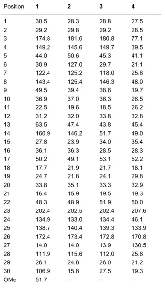
Table 1: 13C NMR spectroscopic data (100 MHz, CDCl3) of com-pounds 1–4. Position 1 2 3 4 1 30.5 28.3 28.8 27.5 2 29.2 29.8 29.2 28.5 3 174.8 181.6 180.8 77.1 4 149.2 145.6 149.7 39.5 5 44.0 50.6 45.3 41.1 6 30.9 127.0 29.7 21.1 7 122.4 125.2 118.0 25.6 8 143.4 125.4 146.3 48.0 9 49.5 39.4 38.6 19.7 10 36.9 37.0 36.3 26.5 11 22.5 19.6 18.5 26.2 12 31.2 32.0 33.8 32.8 13 63.5 47.4 43.8 45.4 14 160.9 146.2 51.7 49.0 15 27.8 23.9 34.0 35.4 16 36.1 36.3 28.5 28.3 17 50.2 49.1 53.1 52.2 18 17.7 21.9 21.7 18.1 19 24.7 21.8 24.1 29.8 20 33.8 35.1 33.3 32.9 21 16.4 15.9 19.5 19.3 22 48.3 48.9 51.9 50.0 23 202.4 202.5 202.4 207.6 24 134.9 133.0 134.4 46.1 25 138.7 140.4 139.3 133.9 26 172.4 173.4 172.8 170.8 27 14.0 14.0 13.9 130.5 28 111.9 115.6 112.0 25.8 29 26.1 24.8 26.0 21.2 30 106.9 15.8 27.5 19.3 OMe 51.7 – – –
H2-16 and H-20 to H3-21 and H2-22 (Figure 2). Analysis of HMBC spectra indicated correlations from H3-19 to C-1, C-5, C-9 and C-10; from H3-29 to C-4, C-5 and C-28; from H-7 to C-8; from H3-18 to C-12, C-13, C-14 and C-17; from H3-30 to C-13, C-16, C-17 and C-20; from H3-21 to C-17, C-20 and C-22; from H2-22 and H-24 to C-23; and from H3-27 to C-24, C-25 and C-26. The relative configuration of 2 was determined by analysis of a NOESY experiment, which provided correla-tions (Figure 2) of H-5 to H2-2; H-28Z to H-9; H-22a (δH 2.85) to H3-18 and H3-21; H3-18 to H-22b (δH 2.16) and H-24; H-24 to H-20 and H-22b. These correlations indicated the α-orienta-tion of H-5 and H3-30 and the β-orientation of H-9, H3-18 and H3-19. All these facts suggested that compound 2 was strongly similar to cis-sibiric acid [19]. Since the chemical shift of H-24 in cis-sibiric acid (δH 6.15) was upfield of the signal for 1 (δH
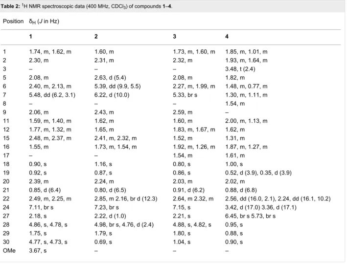
Table 2: 1H NMR spectroscopic data (400 MHz, CDCl3) of compounds 1–4. Position δH (J in Hz) 1 2 3 4 1 1.74, m, 1.62, m 1.60, m 1.73, m, 1.60, m 1.85, m, 1.01, m 2 2.30, m 2.31, m 2.32, m 1.93, m, 1.64, m 3 – – – 3.48, t (2.4) 5 2.08, m 2.63, d (5.4) 2.08, m 1.82, m 6 2.40, m, 2.13, m 5.39, dd (9.9, 5.5) 2.27, m, 1.99, m 1.48, m, 0.77, m 7 5.48, dd (6.2, 3.1) 6.22, d (10.0) 5.33, br s 1.30, m, 1.11, m 8 – – – 1.54, m 9 2.06, m 2.43, m 2.59, m – 11 1.59, m, 1.40, m 1.62, m 1.60, m 2.00, m, 1.13, m 12 1.77, m, 1.32, m 1.65, m 1.83, m, 1.67, m 1.62, m 15 2.48, m, 2.37, m 2.41, m, 2.32, m 1.52, m 1.31, m 16 1.55, m 1.73, m, 1.54, m 1.92, m, 1.26, m 1.87, m, 1.27, m 17 – – 1.54, m 1.61, m 18 0.90, s 1.16, s 0.80, s 1.00, s 19 0.92, s 0.87, s 0.86, s 0.52, d (3.9), 0.35, d (3.9) 20 2.39, m 2.24, m 2.03, m 2.02, m 21 0.85, d (6.4) 0.80, d (6.5) 0.91, d (6.2) 0.88, d (6.8) 22 2.49, m, 2.25, m 2.85, m 2.16, br d (12.3) 2.64, m 2.32, m 2.56, dd (16.0, 2.1), 2.24, dd (16.1, 10.2) 24 7.11, br s 7.23, br s 7.15, s 3.42, d (17.0) 3.36, d (17.1) 27 2.18, s 2.22, d (1.0) 2.21, s 6.45, br s 5.73, br s 28 4.86, s, 4.78, s 4.98, br s, 4.76, d (2.4) 4.88, s, 4.82, s 0.95, s 29 1.75, s 1.79, s 1.80, s 0.88, s 30 4.77, s, 4.73, s 0.69, s 1.04, s 0.90, s OMe 3.67, s – – –
that the trans-stereoisomer was isolated instead of the cis-one (See Table 2 and Supporting Information File 1). This was further confirmed by NOESY correlation of H-24 to H-20 and H3-30, but not to H3-27. Consequently, the structure of 2 was determined as (−)-rel-(24E)-23-oxo-3,4-seco-9βH-lanosta-4(28),6,8(14),24-tetraen-3,26-dioic acid.
Compound 3, a white amorphous powder, possessed a molec-ular formula of C30H44O5 based on the [M + H]+ peak at m/z 485.3250 (calcd 485.3262) in the positive HRESIMS, suggesting nine degrees of unsaturation. The IR spectrum implied the existence of carboxylic acid (1703 cm−1) and olefin (1633 cm−1) functionalities. The 13C NMR spectroscopic data of 3 resembled those of (24E)-3,4-seco-9βH-lanosta-4(28),7,24-triene-3,26-dioic acid (12) [12] except for change at δC 33.3 (C-20), 19.5 (C-21), 51.9 (C-22), 202.4 (C-23), 134.4 (C-24), 139.3 (C-25), 172.8 (C-26) and 13.9 (C-27) (See Table 1 and Supporting Information File 1). The HMBC correlations from H-24 to C-23 indicated the presence of a ketone group at C-23 (Figure 2). This conclusion was confirmed from the downfield δC of C-22 (+16.4) in comparison with 12. The relative con-figuration was established with the NOESY spectrum
(Figure 2). Briefly, the configuration at C-5, C-9, C-10, C-13 and C-17 was determined by cross-peaks from H-28Z to H-9; H-5 to H3-19 and H3-29; H3-18 to H-9 and H-20; H3-30 to H-17; and H3-21 to H2-12. NOESY correlation between H-24 and H3-27 was not observed, suggesting that the geometry of the C-24,25 double bond was E. This was confirmed by δH
comparison of H-24 with that of 1, 2, 6 and 7 (See Table 2 and Supporting Information File 1). On the basis of these spectro-scopic evidences, the structure of 3 was assigned as (−)-rel- (24E)-23-oxo-3,4-seco-9βH-lanosta-4(28),7,24-triene-3,26-dioic acid.
The HRESIMS of 4, isolated as a white amorphous powder, showed a pseudomolecular [M + H]+ ion peak at m/z 471.3463, corresponding to the formula C30H46O4 (calcd. 471.3469), indi-cating eight degrees of insaturation. The IR absorption bands at 3416, 1708 and 1633 cm−1 suggested the presence of hydroxyl, carbonyl and olefin functionalities. The 13C NMR and DEPT-135 spectra of 4 showed signals for 30 carbons designated as five methyl; twelve methylene, including one alkene at δC
130.5; five methine, including one secondary alcohol at δC 77.1; and eight quaternary carbons, including those at δC 170.8 and
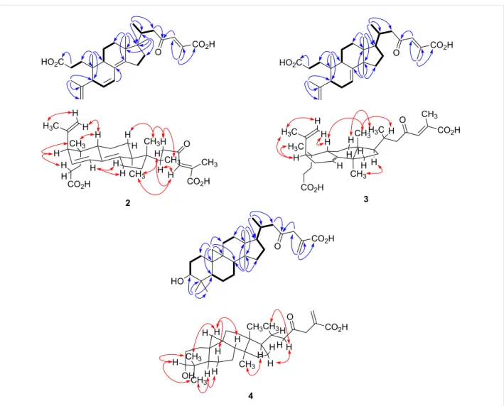
Figure 2: Selected COSY (▬), HMBC (blue arrows) and NOESY (red arrows) correlations for compounds 2–4.
207.6 representing carboxylic and ketone carbonyls, respective-ly (Table 1). The 1H NMR spectrum showed two doublets at δH
0.35 (J = 3.9 Hz) and 0.52 (J = 3.9 Hz) characteristic of a cyclo-propane ring (Table 2), suggesting that 4 is a member of the cycloartanes, which is an important triterpenic family in the genus Abies [3]. In the 1H–1H COSY spectrum, correlations between H2-2 to H2-1 and H-3; H2-6 to H-5 and H2-7; H2-7 to H-8; H2-16 to H2-15 and H-17; and H-20 to H3-21 and H2-22 were observed (Figure 2). HMBC correlations from H2-19 to C-1, C-5, C-8, C-9, C-10 and C-11 connected together three different fragments in the vicinity of the cyclopropyl group. Other correlations between H3-18 to C-12, C-13, C-14 and C-17; H3-21 to C-17, C-20 and C-22; H2-27 to C-24, C-25 and C-26; H3-28 and H3-29 to C-3, C-4, C-5, C-28 and C-29; H3-30 to C-8, C-13, C-14 and C-15; and H2-22 and H2-24 to C-23 were observed and completely assigned the carbon skeleton of the molecule (Figure 2). The relative configuration was deter-mined with the help of a 2D NOESY experiment showing correlations from H-19β to H-6β, H-8 and H3-29; H-5 to H3-28
and H-6α; H3-30 to H-11α and H-17; and H-22b to H-20 and H2-16 (Figure 2). The α-orientation of the hydroxy group at C-3 was deduced from the small coupling constant of H-3 (J = 2.4 Hz), and from the NOESY correlations with both H3-28 and H3-29. Accordingly, the structure of compound 4 was defined as (+)-rel-3α-hydroxy-23-oxocycloart-25(27)-en-26-oic acid.
The absolute stereochemistry of the new compounds (1–4) has not been determined experimentally. However, the previously described compounds 7, 9, 10 and 11 have been shown to pos-sess the usual configuration for triterpenes [8,10,11]. The struc-tures of many other triterpenes isolated from the genus Abies were also reported with this absolute configuration according to their X-ray crystallographic data [20-22].
The structure of compound 8 was reported by Xia et al [9]. In their paper, the configuration at Δ22 was determined as E but it was not supported by any spectroscopic data. Since 1H and
13C NMR data obtained for 8 were identical to those reported by Xia within 0.01 and 0.1 ppm respectively (see Supporting Information File 1), we supposed that both molecules were the same. However, the geometry at Δ22 should be assigned to Z because of the clear NOESY correlation between H-22 and H-24. Interestingly, lanostane with E geometry at Δ22 has never been isolated so far. Moreover, triterpenes with this kind of side chain bearing an E configuration for Δ22 have only been reported by Guo et al [23,24]. During their work on Schisandra spp., they isolated many nortriterpenes having both Δ22
configurations. A statistical analysis of the 1H chemical shift for H-22 and H-24 was conducted: for E-configured Δ22, δH are 5.9 ± 0.2 and 7.8 ± 0.1 while for Z-configured Δ22, δH
are 5.3 ± 0.1 and 7.2 ± 0.2, respectively. Since δH measured for compound 8 was 4.98 and 6.97, it should be assigned as (22Z)-3,4-seco-9βH-lanosta-4(28),7,22,24-tetraen-23,26-olid-3-oic acid.
The isolates were evaluated in vitro for their cytotoxic activi-ties against two human cancer cell lines, namely lung carci-noma (A549) and colon adenocarcicarci-noma (DLD-1), as well as against one healthy cell line (WS1) using the resazurin reduc-tion test [25]. Etoposide was used as a positive control (IC50 ≤ 1.0 µM). None of the compounds were found to be active (IC50 > 25 µM) with the exception of abiesonic acid (6), which showed a weak cytotoxic activity against A549 (IC50 = 22 µM). The antibacterial activity of isolated com-pounds was also evaluated in vitro against E. coli and S. aureus using the microdilution assay [26] with gentamycin as a posi-tive control (MIC < 0.1 µg/mL). No activity was observed for all the tested compounds (MIC ≥ 50 µM) except for triter-penoids 1 and 4, which were weakly active against S. aureus (MIC = 25 µM).
Supporting Information
Supporting Information File 1
Experimental procedures, product characterization and 1H and 13C spectra for compounds 1–18.
[http://www.beilstein-journals.org/bjoc/content/ supplementary/1860-5397-9-150-S1.pdf]
Acknowledgements
We acknowledge "Chaire de Recherche sur les Agents Anticancéreux d'Origine Végétale" and NSERC for funding, and Catherine Dussault for biological assessment. The photo-graph from our photo-graphical abstract is from Robert H. Mohlen-brock @ USDA-NRCS PLANTS Database / USDA NRCS. 1995. Northeast wetland flora: Field office guide to plant species. Northeast National Technical Center, Chester.
References
1. Mabberley, D. J. Mabberley's plant-book: a portable dictionary of
plants, their classification and uses, 3rd ed.; Cambridge University
Press: Cambridge, 2008; pp 1 ff.
2. Takahashi, T. J. Pharm. Soc. Jpn. 1938, 58, 888–901.
3. Yang, X.-W.; Li, S.-M.; Shen, Y.-H.; Zhang, W.-D. Chem. Biodiversity 2008, 5, 56–81. doi:10.1002/cbdv.200890015
4. Herrick, J. W.; Snow, D. R. Iroquois medical botany; Syracuse University Press: Syracuse, NY, 1995; p 278.
5. Pichette, A.; Larouche, P.-L.; Lebrun, M.; Legault, J. Phytother. Res. 2006, 20, 371–373. doi:10.1002/ptr.1863
6. Lavoie, S.; Legault, J.; Gauthier, C.; Mshvildadze, V.; Mercier, S.; Pichette, A. Org. Lett. 2012, 14, 1504–1507. doi:10.1021/ol300237f 7. Shang, N.; Guerrero-Analco, J. A.; Musallam, L.; Saleem, A.;
Muhammad, A.; Walshe-Roussel, B.; Cuerrier, A.; Arnason, J. T.; Haddad, P. S. J. Ethnopharmacol. 2012, 141, 1051–1057. doi:10.1016/j.jep.2012.04.002
8. Roshchin, V. I.; Raldugin, V. A.; Baranova, R. A.; Pentegova, V. A.
Chem. Nat. Compd. 1986, 22, 613–614. doi:10.1007/BF00599284
9. Xia, J.-H.; Zhang, S.-D.; Li, Y.-L.; Wu, L.; Zhu, Z.-J.; Yang, X.-W.; Zeng, H.-W.; Li, H.-L.; Wang, N.; Steinmetz, A.; Zhang, W.-D.
Phytochemistry 2012, 74, 178–184.
doi:10.1016/j.phytochem.2011.11.011
10. Wada, S.-i.; Iida, A.; Tanaka, R. J. Nat. Prod. 2002, 65, 1657–1659. doi:10.1021/np020282b
11. Raldugin, V. A.; Gatilov, Y. V.; Rybalova, T. V.; Rashkes, Y. V.
Chem. Nat. Compd. 1986, 22, 645–651. doi:10.1007/BF00598342
12. Kim, H. J.; Choi, E. H.; Lee, I.-S. Phytochemistry 2004, 65, 2545–2549. doi:10.1016/j.phytochem.2004.07.007
13. Yang, X.-W.; Li, S.-M.; Feng, L.; Shen, Y.-H.; Tian, J.-M.; Liu, X.-H.; Zeng, H.-W.; Zhang, C.; Zhang, W.-D. Tetrahedron 2008, 64, 4354–4362. doi:10.1016/j.tet.2008.02.069
14. Abad, A.; Arnó, M.; Peiró, M.; Zaragoza, R. J. Tetrahedron 1991, 47, 3829–3844. doi:10.1016/S0040-4020(01)80907-8
15. Zhao, Y.-X.; Zhou, L.; Guo, L.; Luo, X.-D.; Zhou, J.
J. Asian Nat. Prod. Res. 2005, 7, 259–264.
doi:10.1080/10286020410001690163
16. Alvarez-Manzaneda, E. J.; Chahboun, R.; Guardia, J. J.; Lachkar, M.; Dahdouh, A.; Lara, A.; Messouri, I. Tetrahedron Lett. 2006, 47, 2577–2580. doi:10.1016/j.tetlet.2006.02.037
17. Wahlberg, I.; Vogt, C.; Wallin, I.; Nishida, T.; Enzell, C. R.
Acta Chem. Scand., Ser. B 1982, 36, 573–576.
doi:10.3891/acta.chem.scand.36b-0573
18. Wahlberg, I.; Karlsson, K.; Curvall, M.; Nishida, T.; Enzell, C. R.
Acta Chem. Scand., Ser. B 1978, 32, 203–215.
doi:10.3891/acta.chem.scand.32b-0203
19. Shevtsov, S. A.; Raldugin, V. A. Chem. Nat. Compd. 1989, 25, 182–187. doi:10.1007/BF00598407
20. Li, Y.-L.; Gao, Y.-X.; Yang, X.-W.; Jin, H.-Z.; Ye, J.; Simmons, L.; Wang, N.; Steinmetz, A.; Zhang, W.-D. Phytochemistry 2012, 81, 159–164. doi:10.1016/j.phytochem.2012.05.032
21. Yang, X.-W.; Li, S.-M.; Wu, L.; Li, Y.-L.; Feng, L.; Shen, Y.-H.; Tian, J.-M.; Tang, J.; Wang, N.; Liu, Y.; Zhang, W.-D.
Org. Biomol. Chem. 2010, 8, 2609–2616. doi:10.1039/c001885f
22. Li, Y.-L.; Yang, X.-W.; Li, S.-M.; Shen, Y.-H.; Zeng, H.-W.; Liu, X.-H.; Tang, J.; Zhang, W.-D. J. Nat. Prod. 2009, 72, 1065–1068. doi:10.1021/np800790h
23. Wang, J.-R.; Kurtán, T.; Mándi, A.; Guo, Y.-W. Eur. J. Org. Chem. 2012, 5471–5482. doi:10.1002/ejoc.201200557
24. He, F.; Li, X.-Y.; Yang, G.-Y.; Li, X.-N.; Luo, X.; Zou, J.; Li, Y.; Xiao, W.-L.; Sun, H.-D. Tetrahedron 2012, 68, 440–446. doi:10.1016/j.tet.2011.11.026
25. O'Brien, J.; Wilson, I.; Orton, T.; Pognan, F. Eur. J. Biochem. 2000,
267, 5421–5426. doi:10.1046/j.1432-1327.2000.01606.x
26. Banfi, E.; Scialino, G.; Monti-Bragadin, C. J. Antimicrob. Chemother. 2003, 52, 796–800. doi:10.1093/jac/dkg439
License and Terms
This is an Open Access article under the terms of the Creative Commons Attribution License
(http://creativecommons.org/licenses/by/2.0), which permits unrestricted use, distribution, and reproduction in any medium, provided the original work is properly cited. The license is subject to the Beilstein Journal of Organic
Chemistry terms and conditions:
(http://www.beilstein-journals.org/bjoc)
The definitive version of this article is the electronic one which can be found at: