Université de Montréal
Régulation de l'hepcidine et le rôle de la lipocaline 2 dans
l'homéostasie du fer / Novel insights into the regulation of
hepcidin and the role of lipocalin 2 in iron homeostasis
par Hua Huang
Programme de Sciences Biomédicales Faculté de Médecine
Thèse présentée à la Faculté de Médecine en vue de l’obtention du grade de Doctorat
en Sciences Biomédicales
2014
Université de Montréal
Faculté des études supérieures et postdoctorales
Cette thèse intitulée :
Régulation de l'hepcidine et le rôle de la lipocaline 2 dans l'homéostasie du fer / Novel insights into the regulation of hepcidin and the role of lipocalin 2 in iron homeostasis
Présentée par : Hua Huang
a été évaluée par un jury composé des personnes suivantes :
Dr Richard Bertrand, président-rapporteur Dr Manuela Santos, directeur de recherche
Dr Hongyu Luo, membre du jury Dr Kostas Pantopoulos, examinateur externe Dr Sylvie Lesage, représentant du doyen de la FES
Résumé
Le fer, un métal de transition, est requis pour la survie de presque tout les organismes vivant à cause de son habilité à accepter ou donner un électron et donc à catalyser plusieurs réactions biochimique fondamentales. Cependant, la même propriété permet aussi au fer ionique d’accélérer la formation de radicaux libres et donc le fer peut potentiellement avoir des effets néfastes. Conséquemment, l’homéostasie du fer doit être étroitement régulé, tant au niveau cellulaire que systémique. Notre étude met l’emphase sur deux molécules importante pour régulation du métabolisme du fer : la lipocaline 2 (Lcn2) et l’hepcidine.
Lcn2, une protéine de phase aiguë, est impliquée dans le transport du fer par les sidérophores. Lcn2 est un candidat potentiel comme transporteur du fer qui pourrait être responsable de l’accumulation excessive du fer non lié à la transferrine dans le foie des patients atteints d’hémochromatose héréditaire (HH). Nous avons généré des souris double-déficiente HfeLcn2 pour évaluer l’importance de Lcn2 dans la pathogenèse de surcharge en fer hépatique dans les souris knock-out Hfe (Hfe -/-). Notre étude révèle que la délétion de Lcn2 dans les souris Hfe -/-n’influence pas leur accumulation de fer hépatique ou leur réponse à une surcharge en fer. Le phénotype des souries HfeLcn2-/- demeure indiscernable de celui des souris Hfe-/-. Nos données impliquent que Lcn2 n’est pas essentiel pour la livraison du fer aux hépatocytes dans l’HH.
L’hepcidine, un régulateur clé du métabolisme du fer, est un petit peptide antimicrobien produit par le foie et qui régule l’absorption intestinale du fer et son recyclage par les macrophages. L’expression de l’hepcidine est induite par la surcharge en fer et l’inflammation, tandis que, à l'inverse, elle est inhibée par l'anémie et l'hypoxie. Dans certaine situations pathologique, l’hepcidine est régulée dans des directions opposées par plus d’un régulateur.
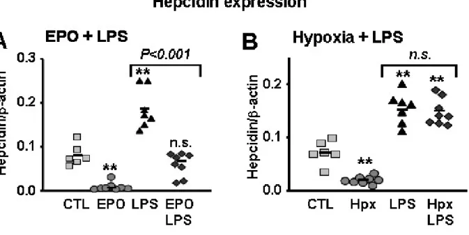
avons examiné la régulation de l’hepcidine en présence de stimuli opposés, ainsi que la contribution des médiateurs et des voix de signalisation en aval de l’expression de l’hepcidine. Nous avons démontré que l'érythropoïèse, lorsque stimulé par l’érythropoïétine, mais pas par l’hypoxie, diminue l’expression de l’hepcidine d’une façon dépendante de la dose, même en présence de lipopolysaccharides ou de surcharge de fer alimentaire, qui peuvent agir de manière additive. De plus, l’entraînement érythropoïétique inhibe tant la voix inflammatoire que celle de détection du fer, du moins en partie, par la suppression du signal IL-6/STAT3 et BMP/SMAD4 in vivo. Au total, nos données suggèrent que le niveau d’expression de l’hepcidine en présence de signaux opposés est déterminé par la force du stimulus individuel plutôt que par une hiérarchie absolue. Ces découvertes sont pertinentes pour le traitement de l’anémie des maladies chronique et les désordres de surcharge en fer.
Abstract
Iron, a transition metal, is required for survival by almost all living organisms due to its ability to accept or donate electrons and thus to catalyze many fundamental biochemical reactions. However, the same properties also allow ionic iron to accelerate the formation of free radicals and as such iron has the potential for deleterious effects. Consequently, iron homeostasis must be tightly regulated at both cellular and systemic levels. Our studies focused on two important molecules in the regulation of iron metabolism, namely, lipocalin 2 (Lcn2) and hepcidin.
Lcn2, an acute phase protein, is involved in iron trafficking via siderophores. Lcn2 has emerged as a candidate iron-transporter that may be responsible for excessive non-transferrin-bound iron (NTBI) accumulation in the liver of hereditary hemochromatosis (HH) patients. We generated HfeLcn2 double-deficient mice to evaluate the importance of Lcn2 in the pathogenesis of hepatic iron loading in Hfe knockoutmice. Our studies revealed that deletion of Lcn2 in Hfe-knockout mice does not influence hepatic iron accumulation in Hfe -/-mice, or their response to iron loading, as the phenotype of HfeLcn2-/- mice remained indistinguishable from that of Hfe-/- mice. Our data imply that Lcn2 is not essential for iron delivery to hepatocytes in HH.
Hepcidin, a key regulator of iron metabolism, is a small antimicrobial peptide produced by the liver that regulates intestinal iron absorption and iron recycling by macrophages. Hepcidin expression is induced by iron-loading and inflammation while, conversely, being inhibited by anemia and hypoxia. Under certain pathologic situations, hepcidin is regulated in opposite directions by more than one regulator. We further investigated how different factors influence
mediators and downstream signaling pathways of hepcidin expression. We show that erythropoiesis drive, when stimulated by erythropoietin but not by hypoxia, down-regulates hepcidin in a dose-dependent manner, even in the presence of lipopolysaccharide or dietary iron-loading, which may act additively. Moreover, erythropoietic drive inhibited both the inflammatory and iron-sensing pathways, at least in part, via the suppression of IL-6/STAT3 and BMP/SMAD4 signaling in vivo. Altogether, our data suggest that hepcidin expression levels in the presence of opposing signaling are determined by the strength of the individual stimuli rather than by an absolute hierarchy. These findings are pertinent for the treatment of the anemia of chronic disease and iron-loading disorders.
Table of contents
RÉSUMÉ ... III
ABSTRACT ... V
LIST OF TABLES ... 4
LIST OF FIGURES ... 5
LIST OF ACRONYMS AND ABBREVIATIONS ... 6
ACKNOWLEDGEMENTS ... 10
CHAPTER 1 INTRODUCTION ... 11
1. The importance of iron for biological systems ... 12
1.1 Iron-containing proteins ... 12 1.1.1 Hemoproteins ... 13 1.1.2 Iron-sulfur proteins ... 13 1.1.3 Iron-binding proteins ... 13 1.2 Iron toxicity ... 15 2. Iron homeostasis ... 18
2.1 Cellular iron homeostasis ... 18
2.1.1 Cellular iron uptake ... 19
2.1.2 Cellular iron storage ... 22
2.1.3 Iron usage in the mitochondria ... 23
2.1.4 Cellular iron export ... 24
2.1.5 Regulation of cellular iron homeostasis ... 25
2.2 Systemic iron homeostasis ... 28
3. Hepcidin ... 30
3.1 The discovery of hepcidin ... 30
3.2 Regulation of hepcidin synthesis ... 34
3.4 Molecular signaling pathways regulating hepcidin expression ... 39
3.4.1 BMP/SMAD signaling pathway ... 39
3.4.2 IL-6/STAT3 signaling pathway ... 42
3.4.3 Crosstalk between signaling pathways ... 43
4. Interactions between immune system and iron metabolism ... 45
Lactoferrin... 45
Nramp1 ... 46
Lipocalin 2 ... 47
5. Disorders of iron metabolism ... 49
5.1 Hereditary hemochromatosis ... 51
5.2 β-thalassemia ... 52
5.3 Anemia of chronic diseases ... 55
6. Rational and objectives of study ... 58
CHAPTER 2 IS THE IRON-DONOR LIPOCALIN 2 IMPLICATED IN THE PATHOPHYSIOLOGY OF HEREDITARY HEMOCHROMATOSIS?... 59
Abstract ... 60
Introduction ... 61
Materials and Methods ... 63
Results ... 65
Discussion ... 67
Acknowledgments ... 69
Figures and Legends ... 70
CHAPTER 3 CONTRIBUTION OF STAT3 AND SMAD4 PATHWAYS TO THE REGULATION OF HEPCIDIN BY OPPOSING STIMULI ... 72
Abstract ... 73
Introduction ... 76
Results ... 82
Discussion ... 87
Acknowledgements ... 91
Figures and Legends ... 92
CHAPTER 4 GENERAL DISCUSSION ... 100
Lipocalin 2 and other NTBI transporters ... 101
Hepcidin signaling pathway ... 103
Therapeutic applications of hepcidin ... 109
Iron metabolism and immune system interaction ... 113
Concluding remarks ... 116
List of tables
List of figures
Figure 1 The importance of iron for biological systems ... 14
Figure 2 The basis for iron toxicity. ... 17
Figure 3 Depicition of iron import (top) and export (bottom) pathways in a generic mammalian cell. ... 27
Figure 4 Hepcidin-25 structure ... 33
List of acronyms and abbreviations
ACD, anemia of chronic disease β2m, β2-microglobulin .
BMP, bone morphogenetic protein BMT, Bone marrow transplantation
GPx, catalase, glutathione peroxidase C/EBP, CCAAT/enhancer-binding protein alpha GVHD, chronic graft-versus-host disease CDA, congenital dyserythropoietic anemia
CI, carbonyl-iron
DFX, deferasirox DFP, deferiprone DFO, deferoxamine holoTf, diferric-Tf 2,5-DHBA , 2,5-dihydroxybenzoic acid DMT1, divalent metal transporter 1 DcytB , duodenal cytochrome B EPO, erythropoietin
Erk, extracellular signal-regulated kinase ESAs , erythropoiesis stimulating agents
Fe-S, iron-sulfur
FtH, ferritin heavy chains FtL, ferritin light chains
FtMt, mitochondrial ferritin FPN, ferroportin
GDF15, growth differentiation factor 15 GPx, glutathione peroxidase HCT, hematocrit Hb , hemoglobin HJV, hemojuvelin Hepc, hepcidin HH, hereditary hemochromatosis HIF, hypoxia-inducible factor
H2O2, hydrogen peroxide
●HO, hydroxyl radical holo, iron loaded ISC, iron-sulfur cluster
IL-6, interleukin-6 IFN-γ, interferon-gamma IREs, iron-responsive elements
IRF-8, interferon regulatory factor 8
IRIDA, iron-refractory iron deficiency anemia IRPs, iron regulatory proteins IDA, iron deficiency anemia
JAKs, Janus kinases
JH, juvenile hemochromatosis Lcn2, lipocalin 2 LPS, lipopolysaccharide
LEAP-1, liver-expressed antimicrobial peptide
MCV, mean corpuscular volume m-HJV, membrane-bound hemojuvelin Nramp1, natural resistance-associated macrophage protein-1
Nramp2, natural resistance-associated macrophage protein-2
NGAL, neutrophil gelatinase associated lipocalin
NTBI , non-transferrin bound iron NMR, nuclear magnetic resonance spectroscopy OsM, oncostatin M
O2●-, superoxide anion
PBS, phosphate-buffered saline PHD, prolyl hydroxylase domain
PNS, peripheral nervous system ROS, reactive oxygen species
rhEPO , recombinant human EPO
RBC, red blood cells RGMC, repulsive guidance molecule C
RNAi, RNA interference
Scara5, scavenger receptor class A, member 5 s-HJV, soluble hemojuvelin
shRNA, short hairpin RNA
SMAD4, SMA- and mothers against decapentaplegic-related protein siRNA, small interfering RNA
SOD, superoxide dismutase
STAT-3, signal transducer and activator of transcription 3
TIM-2, T cell immunoglobulin and mucin domain containing 2 TNF-tumor necrosis factors-
Tf, transferrin TfR, transferrin receptor
TMPRSS6,Transmembrane protease, serine 6 TWSG1, twisted gastrulation protein
USF2, upstream stimulatory factor 2 UTRs, untranslated regions
VHL, von-Hippel–Lindau ZIP14, Zrt/Irt-like protein 14
Acknowledgements
I would like to express my great gratitude to my supervisor Dr . Manuela Santos for her guidance, encouragement, and support during the course of my studies. Her enthusiasm and the effort she invested in the supervision of my work have made this experience a true success.
I would like to thank to Alexander Reuben, my colleague, for the excellent work on the English correction.
I would like to thank you my fellow graduate students, postdoctoral fellows and technicians who contributed to the friendly and collaborative atmosphere in the Dr Santos laboratory. In particular, I wish to thank Dongmei Wang, Annie Calvé, Gabriela Fragoso, Antonio Layoun for the technical support, Edward Bagu for the helpful discussion, and Annie Calvé for translating the abstract of my work into French.
Special thanks for my husband, Tianbo, for his encouragement, understanding, and support throughout these stressful years.
I would like to acknowledge the following organizations for financial support during the course of my study: Canadian Institutes of Health Research (grant no. MOP44045) and the Natural Sciences and Engineering Research Council of Canada (grant no. 298518-06).
CHAPTER 1 INTRODUCTION
Iron, a transition metal, is required for survival by almost all living organisms due to its ability to switch between two different redox states and as such catalyze many fundamental biochemical reactions. However, ionic iron can accelerate the formation of free radicals and as such has a potential for deleterious effects. Therefore, iron levels need to be tightly regulated such as to meet body iron needs, while at the same time avoiding toxicity. The importance of iron in biological systems is best illustrated by the ever-increasing number of diseases associated with deregulated iron metabolism. Thus, understanding the mechanisms for the regulation of iron homeostasis is essential for human health.
1. The importance of iron for biological systems
Iron is the fourth most abundant element on the surface of our planet [1]. Despite this relative abundance in the rocks constituting the earth's crust, iron is present in rather small quantities in living matter. However, iron remains an essential part in many metabolic processes essential for life.
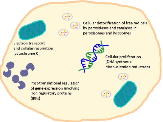
The metabolic importance of this element comes from its chemical properties, as iron can form up to six coordination bonds by accepting an electron pair on each of its six atomic orbitals. Electronegative elements such as oxygen, nitrogen and sulfur thus have the ability to bind iron. This fundamental property allows iron to associate with many biologically-relevant proteins. Furthermore, as a transition metal, iron can exist in two forms, the reduced (Fe2+, ferrous iron) and oxidized forms (Fe3+, ferric iron). This ability to alternatively accept and give electrons makes iron the cofactor of choice for many redox enzymatic reactions (Figure 1). Therefore, the widespread use of iron can be accounted for by its ability to shuttle electrons, flexibility for binding ligands in diverse orientations, and its high bioavailability during the early stages of evolution under the reducing conditions of a sulphur-rich atmosphere.
1.1 Iron-containing proteins
Many proteins that have crucial roles in cellular physiology require iron to function. Interestingly, most iron-binding proteins are highly conserved across prokaryotes and eukaryotes, and the central position of iron in cellular metabolism is maintained in almost all forms of life. Depending on how the metal is bound to the protein, iron-containing proteins can be classified into three classes: hemoproteins, iron-sulfur proteins and iron-binding proteins.
1.1.1 Hemoproteins
Hemoproteins have a tetrapyrrole core at the center of which is encased an iron atom. When the iron atom is in the reduced form, the prosthetic group is called heme, whereas it is called hemin when the iron atom is in the ferric form [2]. The most representative hemoproteins include hemoglobin (Hb), myoglobin [3], cytochromes [4], catalases and peroxidases. Hemoglobin and myoglobin are responsible for stabilizing, transporting and storing oxygen. Oxygen transport through hemoglobin is one of the most important biological functions of iron. Cytochromes constitute major components of the mitochondrial electron transport chains (cytochromes a, b, c) that participate in electron transport and energy metabolism [5], while catalases and peroxidases are hemoproteins that play a fundamental role in the elimination of hazardous reactive oxygen species (ROS) and as such have antioxidant functions.
1.1.2 Iron-sulfur proteins
This type of protein contains what is called an iron-sulfur center (or cluster) consisting of combinations between iron and sulfur atoms of variable stoichiometry. Iron atoms bridge with inorganic sulfides and are bound to proteins through cysteine residues to form the iron-sulfur (Fe-S) proteins [6]. [2Fe-2S], [4Fe-4S] and [3Fe-4S] are the most common cluster variants in eukaryotes [7]. Iron-sulfur proteins include ferredoxins, NADH dehydrogenases, hydrogenases, cytochrome c reductase and nitrogenases. They play a critical role in a wide range of cellular activities as components of the respiratory electron transport complexes, and of tricarboxylic acid cycle enzymes, aconitase and succinate dehydrogenase [7, 8]. In addition, Fe-S clusters are components of DNA repair enzymes fundamental for the recognition of DNA damage and repair.
Examples of iron-binding proteins include ribonucleotide reductase necessary during the S phase of DNA synthesis, and lipoxygenases that catalyze the oxidation of fatty acids. Other iron-binding proteins are fundamental for the transport and storage of iron, namely transferrin and ferritin.
Figure 1 The importance of iron for biological systems
Iron is vital for almost all living organisms due to participation in a wide variety of cellular processes such as cellular respiration, proliferation, detoxification and differentiation [9-13].
1.2 Iron toxicity
Although iron is indispensable for life, it may also become toxic. The toxicity of iron is mainly associated with its potential participation in the excessive production of ROS, a byproduct of oxygen metabolism that describes a variety of molecules and free radicals (chemical species with one unpaired electron).
Under normal physiological conditions, superoxide anion (O2●-) and hydrogen peroxide (H2O2)
are continuously produced in tissue cells as byproducts of aerobic metabolism [14, 15]. More than 500 liters of oxygen is utilized daily by tissue cells of a normal human subject, and 1-5% of the oxygen consumed by the respiratory chain is incompletely reduced to O2●- and H2O2 [14,
16]. If not efficiently removed, hydroxyl radical (●HO) may be generated from H2O2 via the
Fenton reaction in the presence of Fe2+ and cause oxidative damage to cellular components, including nucleic acids [17], proteins [18] and lipids [19] .
To cope with the oxidative stress elicited by aerobic metabolism, mammalian cells have developed an ubiquitous antioxidant defense system, which consists of superoxide dismutases (SODs), catalase, glutathione peroxidases (GPxs) and glutathione reductase together with a number of low molecular weight antioxidants such as ascorbate, α-tocopherol and glutathione [20, 21] (Figure 2). However, this antioxidant defense system may be overwhelmed by various pathological or environmental factors so that a fraction of ROS may escape destruction and form the far more reactive hydroxyl radical (●HO) [20, 21]. An increase in ROS-elicited oxidative damage to DNA and other biomolecules may impair normal functions of tissue cells and lead to human aging and disease [17, 20].
Because of its destructive potential, iron is suspected to play a role in many pathological conditions, including carcinogenesis, atherosclerosis, and a number of neurodegenerative disorders, such as Parkinson's or Alzheimer's disease [22-27]. To minimize these potential toxic effects, highly sophisticated mechanisms and specialized molecules for the acquisition, transport, and storage of iron in soluble, non-toxic forms have evolved to meet cellular iron requirements and to systemically-regulate iron homeostasis.
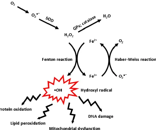
Figure 2 The basis for iron toxicity.
The Haber-Weiss reaction refers to the net reaction (•O2- + H2O2 → •OH + OH- +O2).The
reaction generates hydroxyl radical (•OH) from hydrogen peroxide (H2O2) and is catalyzed by
iron. The first step involves reduction of ferric iron to ferrous (Fe3+ + •O2− → Fe2+ + O2). The
second step is the Fenton reaction (Fe2+ + H2O2 → Fe3+ + OH− + •OH). The highly reactive
hydroxyl radical (•OH) can lead to oxidative stress-induced protein oxidation, lipid peroxidation, mitochondrial dysfunction, and DNA damage. SOD - superoxide dismutase; GPx – glutathione peroxidase.
2. Iron homeostasis
The chemical properties of iron place two limitations on the biological behavior of this element. First, although iron is abundant, the metal is most commonly found in nature as the insoluble ferric hydroxide. Therefore, organisms have evolved complex mechanisms to obtain iron from their environment. Second, iron is potentially toxic by participating in the generation of toxic oxygen radicals. Consequently, while iron is accumulated in amounts sufficient for metabolism, organisms must ensure that their intracellular concentration of "free" iron does not reach toxic levels.
To avoid iron excess and its potential toxic consequences while at the same time providing enough of iron for growth and development, iron homeostasis must be tightly controlled at both the cellular and systemic levels. Iron needs to be transported and safely stored, processes that are mediated by a number of specialized proteins involved in maintaining iron homeostasis such as to ensure the acquisition of iron and its distribution to different organs and tissues, as well as intracellular organelles.
2.1 Cellular iron homeostasis
At the cellular level, iron homeostasis is achieved through the coordinated regulation of iron uptake, storage, export and management of intracellular iron distribution. Importantly, cellular iron homeostasis seems to be differentially regulated in iron-acceptor cells (primarily erythroid precursors) and in iron-donor cells (macrophages, hepatocytes and intestinal epithelial cells in the duodenum). Figure 3 summarizes the general mechanisms that intervene in the uptake, intracellular distribution and transport of iron to cell populations.
2.1.1 Cellular iron uptake
Under normal physiological conditions, virtually all iron in circulation is bound to transferrin (Tf), an abundant plasma glycoprotein that can bind one or two ferric ions with high affinity [28]. Therefore, nearly all-cellular acquisition of iron from blood occurs via transferrin receptor 1 and 2 (TfR1- and TfR2-) mediated uptake.
TfR-mediated iron uptake
In circulation, iron-free Tf, or apoTf, captures extracellular ferric iron and forms the diferric-Tf (holoTf) complex that has high affinity for TfR1 expressed at the cell surface. The Tf/TfR1 complex is then internalized via endocytosis into endosomes. Acidification of the early endosomes through the entry of protons (pH=5.5) triggers conformational changes in both Tf and TfR1, facilitating the release of ferric iron from the Tf/TfR1 complex. Subsequently, the STEAP3 ferrireductase (six-transmembrane epithelial antigen of prostate 3) reduces ferric iron to ferrous iron (Fe2+) [29] allowing its transportation across the endosomal membrane into the cytoplasm by the divalent metal transporter 1 (DMT1; also known as Natural Resistance Associated Macrophage Protein 2, or NRAMP2) [30]. The apoTf/TfR1 complex is then recycled back to the cell surface and apoTf is released into the bloodstream to recapture ferric iron, thereby completing the transferrin cycle (Figure 3).
The TfR1 is ubiquitously expressed at lower levels in normal cells and at higher levels on proliferating cells and cells that require high amounts of iron, such as intestinal epithelial cells [31], placental trophoblasts and erythroid precursors [32]. A homologue of TfR1, termed TfR2, has additionally been identified [33]. Unlike TfR1, TfR2 is exclusively expressed in liver hepatocytes, duodenal crypt cells and early erythroid precursors [34] [35]. Similar to TfR1,
uptake during embryonic development, particularly from day 13 to postnatal day 1, a period during which TfR1 expression is suppressed while TfR2 is activated in hepatocytes [35].
Non-transferrin bound iron (NTBI) uptake
Besides Tf iron, which represents the normal form of circulating iron, a second mechanism of iron uptake occurs through a transferrin-independent process. This NTBI transport process is considered to have a minor role in iron uptake under normal physiological conditions but becomes the primary uptake mechanism when serum iron is severely elevated and surpasses the iron-biding capacity of Tf, as seen in primary and secondary iron overload disorders [37]. NTBI has been directly connected with the production of harmful ROS and ensuing tissue damage, as most organs, including the liver, heart, pancreas and brain have a high capacity to rapidly uptake NTBI. The mechanism(s) responsible for NTBI uptake remain to be fully understood, however several molecules, including DMT1, ZIP14 (also known as Slc39a14), and L-type voltage-dependent calcium channels (L-type VDCC) have all been implicated in NTBI uptake.
DMT1. Divalent metals such as Mn and Zn are able to inhibit NTBI uptake by mouse
hepatocytes indicating that NTBI may share the same transporter with other divalent metals. DMT1 has been proposed to be a putative transporter of NTBI in hepatic cells under iron overloading situations, because DMT1 expression is upregulated in the liver of iron-overloaded mice [38]. However, DMT1-deficient mice are able to develop hepatic iron overload, suggesting that another alternative pathway could play a major role in Fe3+ uptake and indicating that there is an alternative iron-transporter for hepatic NTBI uptake [39, 40].
ZIP14. The Zrt/Irt-like protein 14 is a zinc transporter that is also involved in NTBI uptake by
hepatocytes [41]. When ZIP14 is overexpressed in the AML12 mouse hepatocyte cell line, NTBI uptake increases, while the opposite occurs when the expression of the endogenous
ZIP14 is suppressed [41].
LVDCC. The L-type voltage-dependent calcium channels, where “L" stands for long-lasting
activation, represent a group of calcium channels that allow the influx of Ca2+ essential for normal excitability and excitation-contraction coupling in cardiomyocytes. Several findings have been shown to support the role of LVDCC in cardiac NTBI uptake [42]. For example, LVDCC agonists can augment myocyte iron uptake by perfused rat hearts, while conversely, LVDCC blockers have an inhibitory effect [43]. Moreover, treatment of iron-loaded mice with LVDCC blockers, such as amlodipine and verapamil, were shown to inhibit LVDCC current in cardiomyocytes, reduce myocardial iron accumulation and improve survival [44]. Conversely, overexpression of LVDCC in transgenic mice was shown to lead to increased myocardial iron accumulation and oxidative stress, resulting in impaired cardiac function in comparison with wild-type mice [44]. More recently, it has been demonstrated that, unlike wild-type mice, treatments of DMT1-deficient mice with the LVDCC blocker nifedipine had no effect in reducing iron accumulation in the liver, suggesting that this effect of nifedipine-mediated modulation of iron transport occurs trough DMT1 [45]. However, McKenzie et al reported that photodegraded nifedipine is an iron-specific ionophore, and the ionophore effect was independent of DMT1[46]. Despite these discrepancies regarding the mechanism of action, these findings suggest that nifedipine could possibly be beneficial in iron overload cardiomyopathy.
Alternative iron uptake systems
Some specific cell types have additional means to take up iron, usually involving receptor-mediated endocytosis of other forms of protein-bound iron. For example, survival of kidney cells in culture has been shown to be regulated by lipocalin 2-dependent endocytosis of
cell immunoglobulin and mucin domain containing 2) ferritin receptors [48, 49].Another source of iron for some specialized cells is represented by heme. For example, SLC48A1 has been identified as a heme import molecule [50]. Macrophages can uptake heme indirectly, through phagocytosis of senescent and dying red blood cells (RBC). Finally, in plasma, hemoglobin and free heme arising from intravascular hemolysis are cleared by specific scavenger systems: hemoglobin forms a complex with haptoglobin that is delivered to macrophages via CD163-mediated endocytosis [51, 52], while free heme binds to hemopexin and the complex is endocytosed via the CD91 receptor present on the surface of macrophages, hepatocytes, and other cell types [53].
2.1.2 Cellular iron storage
Once uptaken into the cell, iron that is not immediately utilized or exported is stored within ferritin, the major iron-storage protein in the body. Ferritin is a conserved protein that assembles into a large shell-like structure. Its structure possesses a cavity that provides space to accommodate up to 4500 iron atoms. Thus, ferritin can sequester excess intracellular iron in a redox inactive form to achieve the purpose of storage and detoxification. Ferritin is composed of a combination of 24 subunits of heavy (FtH) and light (FtL) chains with the expression ratio of FtH/FtL chains varies depending on the tissue. For example, FtH is highly expressed in the heart, while FtL expression is increased in the liver. The FtH/FtL ratio also changes in response to inflammation and infection. Functionally, FtH has a potent ferroxidase activity that catalyzes the oxidation of ferrous iron, essential for iron internalization and packing into the mineral core. In turn, the FtL subunit is involved in iron nucleation and protein stability. Iron stored in ferritin is thought to be bioavailable and is mobilized for cellular utilization mainly during lysosomal and proteasomal turnover [54]. Serum ferritin is mostly composed of the FtL isoform and as such, contains negligible iron levels but is useful as a marker for body iron storage levels if no inflammatory or infectious conditions are present
[55]. In addition to this major form of ferritin, mitochondria also contain a nuclear-encoded H-type ferritin homopolymer, designated mitochondrial ferritin (FtMt) [56]. FtMt is mostly detected in mitochondrial-rich tissues, including heart, pancreas and kidney. Under normal conditions, FtMt is not involved in mitochondrial iron utilization. However, the expression of FtMt in patients suffering from sideroblastic anaemia rises significantly in iron-loaded ring erythroblasts (sideroblasts), indicating that FtMt may play an important role in the detoxification of iron within this organelle [57].
2.1.3 Iron usage in the mitochondria
Mitochondria perform an important role in the control of cellular iron metabolism. They constitute the major subcellular site of iron utilization, as the sole site for heme biosynthesis[58], as well as the major site for Fe–S cluster protein assembly [59]. The exact mechanism(s) by which mitochondria acquire iron is not fully understood. The inner mitochondrial membrane contains iron transporters that traffic iron from the cytosol into the mitochondrial matrix. Mitoferrin has been recently identified as a mitochondrial iron importer important for iron uptake in both erythroid and non-erythroid cells [60, 61], as exemplified by the fact that mutating mouse mitoferrin leads to impaired heme synthesis due to defective mitochondrial iron uptake [60]. In addition to mitoferrin, an endogenous mammalian siderophore, namely 2,5-dihydroxybenzoic acid (2,5-DHBA), has been proposed to be involved in mitochondrial iron uptake. In this case, iron is imported into the mitochondria in the form of an iron-siderophore complex [62]. Several studies suggest that in erythroid cells, iron-loaded endosomes directly deliver iron to mitochondria through a transient and rapid contact, in a process that has been termed “kiss-and-run” [63].
the cytosol and further inserted into hemoproteins, such as hemoglobin or in cytochromes. In response to iron deficiency, mammalian cells enhance mitochondrial iron uptake to maintain vital mitochondrial functions that are Fe-dependent [64], and suppress multiple Fe-dependent pathways, such as Fe-S cluster scaffold proteins, Nfu1 and Isa1, to limit the utilization of cellular iron [65]. A matrix protein called frataxin [66] has been identified as a possible regulator of mitochondrial iron export [67] and storage [68]. In humans, inappropriate expression of frataxin due an intronic GAA triplet repeat expansion causes the neurodegenerative disorder Friedreich ataxia [69], an autosomal recessive disease in which accumulation of iron within the mitochondria has been reported both in humans [70] and animal models [71, 72].
2.1.4 Cellular iron export
Ferroportin (FPN, also known as Ireg1 and MTP1) is the only known mammalian iron exporter identified so far [73-75]. While FPN is ubiquitously expressed, higher levels are found in cells that are crucial for cellular iron homeostasis, such as duodenal enterocytes, macrophages, hepatocytes and, interestingly, also in cells of the central nervous system. Intestinal enterocytes are responsible for iron transfer into the body, while macrophages are the major sites for iron recycling. FPN-mediated iron export depends on the activity of the copper-dependent ferroxidase hephaestin in enterocytes, while all other cells require ceruloplasmin, another copper-dependent ferroxidase present in the plasma, to successfully export iron. Cherukuri et al proposed ceruloplasmin could share responsibility with hephaestin for iron absorption under stress in the intestine [76]. These ferrioxidases convert Fe2+ to Fe3+, the iron form that can then bind plasma transferrin. Mice with hephaestin deficiency develop anemia due to defective iron-export from enterocytes into the body to complete the process of intestinal iron absorption [77]. In humans, mutations in the ceruloplasmin gene result in iron accumulation in macrophages, hepatocytes, and cells of the central nervous system, causing
iron-restricted anemia and neurodegeneration, as seen in aceruloplasminemia, an autosomal recessive disorder [78, 79].
FPN-mediated iron-export is highly regulated and this regulation is essential for iron acquisition, utilization and storage. At the transcriptional level, FPN mRNA levels are augmented in response to increased erythropoiesis [80], iron, heme and other transition metals [81-84], and conversely, mRNA levels are inhibited during inflammation [85]. At the translational level, expression of FPN is controlled by the iron regulatory proteins (IRPs) 1 and 2. FPN can additionally be regulated at the post-translational level by a small antimicrobial peptide called hepcidin [86]. Last but not least, in addition to FPN-mediated iron export, cells may also export iron in the form of heme and ferritin, as previously mentioned.
2.1.5 Regulation of cellular iron homeostasis
To achieve cellular iron homeostasis, iron uptake, storage, utilization, and export must be coordinately regulated. The post-transcriptional regulation of these processes by IRPs through their interaction with iron-responsive elements (IREs), the so-called IRE/IRP system, is very well characterized. IRPs belong to the iron-sulfur cluster (ISC) isomerase family and include two IRP homologue proteins, IRP1 and IRP2. IRPs bind to evolutionarily-conserved
cis-regulatory hairpin structures called IREs, which are located at the 5′ or 3′ mRNA
untranslated regions (UTRs) [52]. Cellular iron levels regulate the interaction between IREs and IRPs through different mechanisms. More specifically, IRP1 is regulated in a reversible manner through Fe/S assembly/disassembly, while IRP2 is regulated in an irreversible manner, at the level of protein stability. In the presence of high intracellular iron levels, IRP1 binds to a [4Fe-4S] cluster, namely cytoplasmic aconitase, leaving the IREs free. When cellular iron levels are low, IRP1 binds to IREs [87]. The conversion between holo-IRP1 and apo-IRP1
2-oxoglutarate-dependent oxygenase(s) may be involved in IRP2 degradation in response to iron[88]. Non-iron signals are also involved in IRE/IRP binding regulation. For example, nitric oxide (NO) favors apo-IRP1 formation and inhibits IRP2 degradation while hypoxia conditions induce holo-IRP1 formation and stabilize IRP2.
How intracellular iron regulates iron uptake and storage via the IRE/IRP system is best exemplified by the regulation of TfR1 and ferritin [52]. For TfR1, IREs are located at the 3′ UTR mRNAs, while ferritin IRE is located at the 5′ UTR of the mRNAs encoding FtH and FtL chains. In iron-deficient cells, IRE/IRP interaction with the 3′ UTR stabilizes TfR1 mRNA [89]. However, 5′ UTR binding of IRPs to their IREs inhibits the early steps of the translation process [90]. As a result, TfR1 expression levels are increased and de novo ferritin synthesis is suppressed, leading to increased cellular iron uptake via TfR1 and decreased iron storage to counteract iron deficiency. In iron-abundant cells, the binding of IREs to IRPs is inhibited, resulting in TfR1 mRNA degradation and ferritin mRNA translation. This will limit iron uptake via TfR1 preventing iron excess while augmenting ferritin synthesis to safely store iron within the cell.
Besides their presence in TfR1 and ferritin, IREs have been identified also at the 5′ UTR of mRNAs encoding ALAS2 (iron utilization), an enzyme that catalyzes the first step of heme synthesis in the mitochondria of erythroblasts. Both IRPs when bound to ALAS2 IRE can inhibit ALAS2 mRNA translation [91]. IREs are also present at the 5′ UTR of mRNAs encoding ferroportin (export) [73], mitochondrial aconitase (a citrate cycle enzyme) [92] and hypoxia-inducible factor- (HIF-) 2α [93], and at the 3′ UTR of mRNAs encoding DMT1 (uptake) [94]. In contrast to TfR1, the mRNAs encoding TfR2 and mitochondrial ferritin do not possess IREs, and as such are not regulated through the IRE/IRP system.
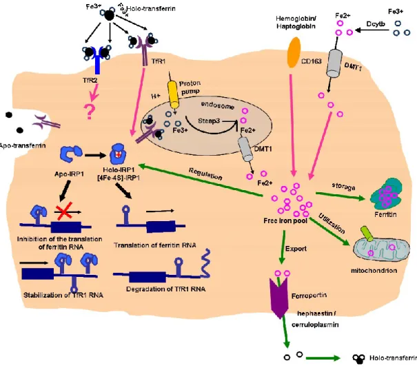
Figure 3 Depicition of iron import (top) and export (bottom) pathways in a generic mammalian cell.
Iron uptake, storage and export mechanisms and some regulatory elements are shown. TfR1 - transferrin receptor-1; TfR2 - transferrin receptor-2; DMT1 – divalent metal transporter 1; Dcytb - duodenal cytochrome b-like ferrireductase; CD163 - hemoglobin scavenger receptor; IRP-1 - iron regulatory protein-1. Steap-3 - six-transmembrane epithelial antigen of prostate 3.
2.2 Systemic iron homeostasis
Because mammals have no regulated mechanism for the elimination of excess iron, the control of iron balance requires a fine adjustment in iron absorption by intestinal enterocytes and the recycling of iron by macrophages. Regulatory effectors that modulate intestinal iron absorption probably also regulate the release of iron from tissue macrophages and hepatocytes.
The levels of intestinal iron absorption are regulated by both systemic and local enterocyte iron status. Changes in enterocyte iron concentrations serve as a fine-tuning mechanism of intestinal iron absorption through the “mucosal block” that occurs several hours after consumption of a large dose of iron [96]. The decrease in intestinal iron absorption occurring following an oral dose of iron is associated with increased enterocyte iron levels, as assessed by IRP activity and ferritin immunoblotting. Reduced iron absorption is also accompanied by a rapid decrease in expression of the mRNAs encoding the brush border iron transport molecules Dcytb and DMT1 containing the IRE splice variant[97]. Thus, in the brush border, but not in the basolateral border, iron transport components are regulated locally by enterocyte iron levels, which support the hypothesis that systemic stimuli exert their primary effect on basolateral transport molecules.
More recently, Bruno Galy et al[98] found that IRPs inhibit the expression of both FPN and DMT1 in adult mice. However, intestine-specific IRP-deficient mice fail to absorb adequate amounts of iron in spite of the high expression levels of the iron transporters DMT1 and FPN. IRP ablation also results in iron retention in enterocytes. For over a half century, people have believed enterocytic ferritin can limit dietary iron intake [99, 100]. Thus, the mucosal block in IRP-deficient mice likely results from iron withholding by the large excess of mucosal ferritin. Bruno Galy et al further showed ferritin actually only traps a minor fraction of the iron transiting through enterocytes. Their results suggest a dual function of IRPs in the regulation
of iron absorption: IRPs limit apical and basolateral iron transport and also ensure sufficient passage of iron through the enterocyte by counteracting the diversion of the metal into ferritin.
Besides the IRP/IRE interaction, some iron sensing proteins are additionally involved in the regulation of iron absorption. HIF2α is shown to be involved in directly regulating the transcription of the gene encoding DMT1, the major intestinal iron transporter [101] and FPN, the only known iron exporter [98, 102]. Intestinal specific deletion of HIF2α results in the decrease of hepatic and serum iron levels [101]. HIF2α levels are dependent on prolyl hydroxylase domain (PHD) enzymes that require ferrous iron for their enzymatic activity [103]. Therefore, PHD enzymes serve as iron sensing proteins that regulate iron absorption.
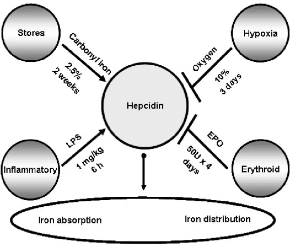
Intestinal iron absorption is modulated by 4 major regulators: body iron stores, erythropoiesis, hypoxia and inflammation [104]. The regulation of these factors results in the variation of hepcidin levels, a small antimicrobial peptide secreted by hepatocytes [105, 106] which functions as an inhibitor of iron absorption [107]. The discovery of hepcidin and its central role in iron metabolism have clarified the pathological mechanisms of most common iron disorders. Mice deficient in hepcidin develop iron overload [108], while mice overexpressing it present profound iron deficiency [109]. A change in hepcidin levels subsequently targets the expression of the iron-exporter FPN [107] in duodenal absorptive cells and macrophages, and consequently the amount of iron entering the body and being recycled by macrophages.
3. Hepcidin
The discovery of the iron regulatory hormone hepcidin has provided a consistent model of iron homeostasis that allows for a better understanding of the importance of sensors at the cellular level for iron needs, as well as of the pathophysiology of hereditary iron metabolism disorders such as hemochromatosis and anemia of inflammation (or anemia of chronic disorders) [110].
3.1 The discovery of hepcidin
Hepcidin, also named LEAP-1, was initially identified as an antimicrobial peptide in human urine and serum by two independent groups [105, 111]. The link between hepcidin and iron metabolism was first discovered by Sophie Vaulont’s group [108]. The group determined that USF2 (upstream stimulatory factor 2) knockout mice unexpectedly experienced severe iron overload with iron deposition in the liver and pancreas. The iron dysregulation observed in USF knockout mice was found to be related to the accidental knockout of a neighbor gene, identified as hepcidin, in the construct used for generating the USF2 knockout mice [108]. Further evidence of hepcidin involvement in iron metabolism was then demonstrated with the generation of transgenic mice overexpressing hepcidin under the control of a liver-specific promoter, leading to the development of severe iron deficiency anemia and premature death of transgenic mice [109].
Further studies showed that the hepcidin gene is evolutionarily conserved across vertebrate species from fish to humans [112]. The human genome contains only one copy of the hepcidin gene [112], whereas there are duplicate copies in the mouse (Hepc1 and Hepc2) genome [106,
108] and two or more copies in certain fishes [113, 114]. In mice, both Hepc1 and Hepc2 are homologous to human hepcidin, but only hepc1 plays a role in iron metabolism [115]. Hepc2 is not an inactive pseudogene since it is expressed in high levels and subjected to certain regulation. It shares some structural similarities with fish hepcidin-like peptides, which suggests Hepc2 serves a similar function as these fish hepcidin-like peptides, that is, antimicrobial activity [115].
The human hepcidin gene (HAMP) maps to chromosome 19q13.1. It comprises two introns and three exons encoding a precursor protein of 84 aa, called preprohepcidin. Preprohepcidin undergoes enzymatic cleavage during its export from the cytoplasm resulting in a 64 aa prohepcidin that is released into the endoplasmic reticulum [116]. Next, a 39 aa pro-region peptide is post-translationally removed by a furin-like proprotein convertase which results in the production of mature bioactive hepcidin-25 (25-aa form), identified both in blood and urine [105, 111]. In urine, two N-terminally truncated isoforms of 22-aa and 20-aa have also been detected. These two isoforms do not seem to have any identified iron-regulatory activity, suggesting that they are degradation products of the hepcidin-25 form [105].
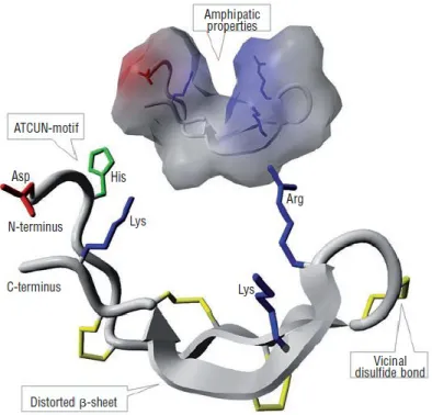
Further studies using nuclear magnetic resonance spectroscopy (NMR) showed that hepcidin-25 it’s a simple hairpin-shaped, cysteine-rich peptide with distorted -sheets linked by four disulfide bridges. Three disulfide bridges stabilize the two anti-parallel strands while the fourth bridge is located in the vicinity of the hairpin loop which is more stressed and could have greater chemical reactivity [117] (Figure 4). Structure function analysis indicated that the iron regulatory activity requires the full-length peptide (hepcidin-25). The 20-aa, N-terminally truncated hepcidin has no iron-regulating bioactivity, indicating that the five N-terminal amino acids are crucial for this activity [118, 119].
Hepcidin structure closely resembles the cysteine-rich antimicrobial peptide defensins. It has a positively charged hydrophilic residue located on the concave side, and a hydrophobic residue distributed on the convex side, a typical structure of antimicrobial peptides known to disrupt bacterial membranes [120]. Several studies have shown that hepcidin indeed possesses antimicrobial properties [105, 116, 121, 122]. However, very high levels of hepcidin are required in order to achieve antimicrobial activity, about 100-fold higher than necessary for its iron regulatory activity[105].
Several studies suggest that the hepcidin gene is expressed in multiple tissues. The highest expression is predominantly in the liver of mammals and most fishes [105, 111-113, 123-126], and weaker expression can also be detected in the heart, lungs, spinal cord, stomach, intestine, pancreas, adipocytes, skeletal muscle, and testis [127-133]. Myeloid cells, including monocytes, macrophages and neutrophils, also express hepcidin [134-138]. Hepcidin expression is regulated differently in macrophages and in hepatocytes. Direct iron-loading does not induce hepcidin production in mouse macrophages [134], much as BMP6 treatment fails to, but LPS and IL-6 stimulation leads to hepcidin induction in these cells [139]. In addition, hepcidin can also be induced in macrophages by gram-positive and gram-negative bacteria wall constituents, through toll-like receptor (TLR) 2 and TLR4, which favors iron retention in macrophages and promotes host defense [135, 140]. Moreover, TLR-mediated hepcidin induction requires signaling through the MyD88-dependent pathway [140].
Figure 4 Hepcidin-25 structure
Front: overview of the structure of hepcidin-25. Distorted β-sheets are shown as grey arrows,
and the peptide backbone is colored gray. The disulfide bonds are colored yellow, highlighting the position of an unusual vicinal bond between adjacent cysteines at the hairpin turn. Positive residues of arginine (Arg) and lysine (Lys) are pictured in blue, the negative residue of aspartic acid (Asp) in red, and the histidine-containing Cu2+- Ni2+ (ATCUN)-binding motif in the N-terminal region is colored green. Background: hepcidin-25 molecule displayed with solvent-accessible surface that illustrates the amphipathic structure of the molecule. The molecule is colored gray, except for the side-chains of positive (blue) and negative (red) residues. Figure obtained from [141] (permission obtained from Journal Haematologica).
3.2 Regulation of hepcidin synthesis
Considering the close relationship between iron metabolism and hepcidin, the same factors that influence dietary iron absorption, namely iron stores, erythropoietic activity, hemoglobin, oxygen content and inflammation, are also capable of regulating the expression of hepcidin in hepatocytes. Recent advances in understanding how hepcidin is regulated at the molecular level have expanded our knowledge about the pathways regulating iron metabolism.
3.2.1 Iron-sensing pathway of hepcidin regulation
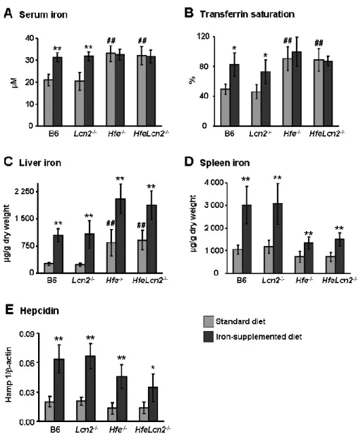
The regulation of hepcidin by body iron levels acts through a negative feedback mechanism. In both mice and men, hepcidin expression is induced by iron loading [106, 142] leading to inhibition of intestinal iron absorption and iron release from macrophages. Conversely, hepcidin expression is suppressed in iron deficiency [143], promoting intestinal iron absorption and iron release from macrophages into the circulation. Hepcidin is mainly expressed in the liver, and it is therefore most likely that iron-sensing takes place in this organ. However, direct iron treatment of both primary hepatocytes and hepatocytes cell lines leads to reduced hepcidin expression rather than activation [142], indicating that iron-sensing by hepatocytes in vivo involves the collaborative interaction between cell types, such as Kupffer cells. Studies in patients with an iron-overload disease, hereditary hemochromatosis (HH) and juvenile hemochromatosis (JH), revealed several proteins that are involved in hepcidin regulation by iron. HH and JH are a group of genetic disorders caused by mutations in the HFE, TfR2, hepcidin, and hemojuvelin (HJV) genes [144-149]. Despite iron accumulation in several organs in patients with HH and JH, hepcidin expression is inappropriately defective, clearly indicating that all molecules involved (HFE, TfR2, hepcidin and HJV) are crucial for hepcidin activation by iron.
3.2.2 Inflammatory pathway of hepcidin regulation
Hepcidin synthesis is also significantly increased during inflammation due to infection, autoimmune disease, and cancer. Excess hepcidin production in these settings inhibits dietary iron absorption and sequesters iron inside macrophages, resulting in low levels of circulating iron in the plasma, or hypoferremia. The finding that hepcidin-deficient mice do not develop hypoferremia upon inflammatory stimuli further demonstrates that hepcidin is a crucial player in the hypoferremic response accompanying inflammation [150]. Hypoferremia is often understood as a host defense response aimed at limiting iron availability to microorganisms. However, sustained hypoferremia over significant time may lead to the development of the anemia of chronic disease, or functional anemia [151].
Hepcidin induction by inflammatory stimuli is predominantly mediated by interleukin-6 (IL-6) [150, 152]. IL-6 administration in mice and humans rapidly (within hours) induces hepcidin excretion, and is concomitantly accompanied by a significant decrease in serum iron and transferrin saturation [150]. How does hypoferremia develop so rapidly? The plasma transferrin compartment contains approximately 2–4 mg of iron, which accounts for 1% of total body iron content. This transit compartment flows about 20 mg of iron each day, thus, the entire content of iron in plasma turns over every 3–4 hours[9]. Only 1 to 2 mg of iron are obtained from intestinal absorption, and most iron is recycled from senescent erythrocytes. If hepcidin could completely block iron recycling, in one hour plasma iron would drop by at least 25%, consistent with the degree of hypoferremia observed in humans infused with IL-6 [150]. In vitro studies further demonstrate that primary hepatocytes treated with IL-6 are also capable of increasing hepcidin synthesis, an induction that can be efficiently blocked by pre-treatment with anti-IL-6 antibodies [150].
hepatocytes [152] and Hep3B cells [150]. However, later studies showed that IL-6-deficient mice still respond to LPS and produce hepcidin [153]. Therefore, other cytokines must also contribute to the regulation of hepcidin in hepatocytes. In murine hepatocytes, IL-1 and IL-1 are able to stimulate hepcidin transcription, while IFN- shows an inhibitory effect [154]. Similarly, in other cell types, hepcidin is also regulated by cytokines. In human monocytes, IL-6 and IFN- are capable of inducing hepcidin, while TNF- can inhibit hepcidin induction by IL-6 [155]. A recent study found that hepcidin expression is induced by both IFN- and IL-6 in human airway epithelial cell lines [156]. The study also demonstrates that hepcidin induction by IFN- is independent of IL-6 and that it signals via the JAK/STAT1 cascade to activate hepcidin.
3.2.3 Regulation of hepcidin in response to erythropoietic demand
Erythropoiesis is the process by which RBCs are produced, by consuming most of the iron absorbed from the diet or recycled from hemoglobin by macrophages. Increased erythropoietic activity suppresses hepcidin expression promoting dietary iron absorption and mobilizing iron release from storage sites, resulting in more iron being available for erythropoiesis. Both hypoxia and anemia stimulate erythropoiesis through the production of erythropoietin (EPO). Accordingly, EPO administration in humans and mice suppresses hepcidin expression [157, 158]. Recent studies have shed some light on whether EPO acts directly or indirectly to suppress hepcidin expression. In mice that received full-body irradiation to block erythropoiesis, EPO-mediated suppression of hepcidin is prevented [159]. Similarly, when mice that were rendered anemic by bleeding or chemical hemolysis are pretreated with erythropoiesis inhibitors, such as carboplatin or doxorubicin, EPO-mediated hepcidin suppression is also abolished [159, 160]. These studies indicate that EPO acts indirectly, possibly through soluble mediators, to mediate hepcidin suppression and it requires erythropoiesis activity. More recent studies suggest that growth differentiation factor 15
(GDF15), and twisted gastrulation protein (TWSG1) both of which are released by erythroid precursors during accelerated erythropoiesis, are the most likely candidates mediating hepcidin suppression in response to erythropoietic demand [161, 162]. GDF15, a member of the transforming growth factor-β superfamily, was found at high levels in the serum of patients suffering from -thalassemia and congenital dyserythropoietic anemia (CDA) [161, 163, 164]. Furthermore, human hepatocytes treated with serum from -thalassemia patients showed a decreased hepcidin expression, which could be reversed by depletion of GDF15 [161]. Thus, GDF15 acts as an erythroid factor by down-regulating hepcidin expression in these patients. However, immunoprecipitation of GDF15 in the sera of -thalassemia patients did not completely abolish hepcidin suppression in exposed hepatocytes, suggesting that GDF15 is likely not the sole mediator of hepcidin suppression. In fact, further studies identified yet another mediator, TWSG1, found to be elevated in a murine model of -thalassemia major [162]. TWSG1 is a BMP-binding protein that can suppress hepcidin expression in human hepatocytes through a BMP-dependent mechanism. Further studies showed that this regulation involved the inhibition of BMP-dependent activation of the SMAD-mediated signal transduction pathway [162].
Both in vivo and in vitro studies have additionally showed that hepcidin expression is also suppressed by hypoxia [143, 158]. Hypoxia per se induces EPO production in the kidney and liver, and as such would contribute to increase erythropoiesis and consequently suppress hepcidin production. Hypoxia-inducible transcription factors (HIFs) are the central mediators of hypoxia-induced erythropoiesis. HIFs consist of an O2 and iron-sensitive α subunit (HIF-1α and HIF-2α,) and a constitutively expressed β subunit, HIF-β. Under normoxic conditions, HIF is subjected to degradation by the von-Hippel–Lindau (VHL) ubiquitin ligase, a tumor suppressor protein via an iron-dependent mechanism[165]. Under hypoxic conditions, HIFs
iron-deficient through an iron-depleted diet. Furthermore, mice with liver-specific deletion the VHL gene express extremely low levels of hepcidin [167]. Thus, the VHL/HIF axis is essential for oxygen sensing and hepcidin regulation. The study of Mastrogiannaki M et al. indicated that hepcidin suppression by hepatic HIF-2 required enhanced erythropoietic activity [168]. Similarly, Liu Q et al found that Hamp1 suppression requires Epo-induced erythropoiesis [169]. The human hepcidin promoter contains several consensus-binding sites for HIF suggesting that hypoxia may also directly regulate hepcidin via HIF at the transcriptional level [167]. In addition, ROS are also involved in the hypoxic regulation of hepcidin. Last but not least, HepG2 cells incubated in hypoxic conditions markedly increased ROS levels and decreased hepcidin expression, and anti-oxidant treatments can reverse this inhibition. Further studies examined the role played by several transcriptional factors and found that C/EBPα and STAT3 dissociate from hepcidin under hypoxic conditions, an effect that also can be reversed by anti-oxidant treatment. Thus, ROS may suppress hepcidin expression via preventing C/EBPα and STAT3 binding to the hepcidin promoter during hypoxia [170] .
3.3 The hepcidin/ferroportin axis
Substantial progress has been made recently in elucidating the mechanism of action of hepcidin. Hepcidin acts by inhibiting the expression of the FPN protein both in enterocytes and macrophages [171, 172].
The concentration of FPN on the cell surface is controlled by both the rate of FPN synthesis and degradation among other factors. In vitro studies demonstrated that hepcidin mediates FPN internalization and degradation [86]. Hepcidin-mediated FPN internalization requires the activity of two of FPN lysines that may be targets of ubiquitination [173]. Internalized FPN is subjected to mono-ubiquitination and is targeted for degradation [86, 172]. The hepcidin-FPN
interaction forms an iron homeostasis loop: iron induces hepcidin production, which then regulates FPN concentration at the cell surface. When iron levels are high, hepcidin is produced and targets FPN for degradation on the basolateral membrane of absorptive enterocytes, thereby blocking the transfer of iron from enterocytes into the plasma. Within two days, short-lived absorptive enterocytes are shed into the intestinal lumen and the iron sequestered in these cells is consequently removed from the body [174]. Similarly, in macrophages, hepcidin-mediated FPN degradation results in iron sequestration within these cells.
Besides post-translational regulation, FPN expression is additionally regulated at the transcriptional level by iron and heme [75, 134, 175-177]. Iron, and some other transition metals including zinc, copper, manganese, cobalt, and cadmium, can directly induce FPN transcription via unknown mechanism(s) [178, 179]. It has been observed that animal products such as meat, fish and poultry enhance non-heme iron absorption in humans. A proposed mechanism involves the induction of FPN expression by heme at the transcriptional level via the transcription factor Bach1 [81]. Finally, and as already mentioned, FPN levels are also regulated post-transcriptionally via the IRE/IRP system [180, 181].
3.4 Molecular signaling pathways regulating hepcidin expression
Intensive research during the past 10 years has been conducted to identify the signaling pathways involved in hepcidin transcriptional regulation. At the present, two major signaling pathways have been identified: the BMP/SMAD iron-sensing pathway and the IL-6/STAT3 inflammatory pathway, depicted in Figure 7.
superfamily. The SMAD proteins are homologs of both the drosophila protein "mothers against decapentaplegic" (MAD) and the Caenorhabditis elegans protein SMA, with its name deriving from a combination of the two. In vivo and in vitro studies demonstrated that several BMPs, such as BMP2, 4, 6, and 9 can activate HAMP transcription [182, 183]. Further studies in liver-specific Smad4-knockout mice revealed that lack of Smad4 in hepatocytes results in a 100-fold lower hepcidin transcriptional expression than wild-type mice accompanied by massive hepatic iron overload [184]. Furthermore, knockdown of Smad4 in mouse primary hepatocytes also ablated the hepcidin induction by BMP4 [184]. These results establish that the BMP/SMAD4 pathway is important for hepcidin regulation through the iron-sensing pathway. The BMP/SMAD4 iron-sensing pathway can be described as follows: iron induces BMP6 expression, which then binds to cell-surface BMP receptor (BMPR) complexes that include type I and II (BMPRI and BMPRII) serine/threonine kinase receptors. Upon ligand binding, BMPRII phosphorylates BMPRI, leading to further phosphorylation of SMAD1/5/8 in the cytosol. Phosphorylated SMDS1/5/8 then form a complex with the common mediator SMAD4, translocate into the nucleus and bind to BMP-responsive elements (BMPRE) located in the hepcidin promoter and activate hepcidin transcription.
Several elements participating in the iron-sensing pathway mediated by SMAD4 signaling have been identified, including BMP6, HJV, TfR2, HFE and TMPRSS6.
BMP6: BMP6 has emerged to be the essential molecule sensing iron for the BMP/SMAD4 pathway. In vitro studies showed that BMP2, 4, 6, and 9 can all induce hepcidin production [183, 185], however only BMP6 transcription is actually regulated by dietary iron in vivo [186]. Accordingly, mice with targeted disruption of BMP6 have reduced phosphorylated Smad1/5/8 levels, develop massive iron overload and show undetectable levels of hepcidin mRNA in the liver [187, 188]. Finally, administration of exogenous BMP6 directly into mice has been shown to induce hepcidin [187]. Collectively, these studies demonstrate that BMP6 is essential for hepcidin induction and is a key player in iron sensing.
HJV: HJV is a GPI-linked cell-surface protein also known as repulsive guidance molecule C (RGMC). HJV interacts with BMP6, BMP4, and BMP2 and functions as a BMP co-receptor [189]. Thus, HJV regulates hepcidin expression by enhancing BMP/SMAD4 signaling [190]. Mutations in the HJV gene cause juvenile hemochromatosis (JH) in humans and mice, characterized by severe iron overload in the liver, pancreas and heart [149, 191]. There are two forms of HJV with opposing effects: a membrane-bound form (m-HJV) that can induce hepcidin expression and a soluble form (s-HJV) that suppresses hepcidin expression. s-HJV is the product of cleavage at the C-terminus of m-HJV by a proprotein convertase, furin [192]. Neogenin, a receptor membrane protein, is then required for the release of the s-HJV [193]. Neogenin binds to HJV and this interaction is crucial for BMP-dependent signaling of hepcidin expression [194]. In addition, s-HJV can also bind to BMPs, thereby preventing the interaction between m-HJV and BMPs [190] and functioning as a competitive antagonist of m-HJV. Importantly, s-HJV is increased during iron deficiency and hypoxia, possibly due to furin activation in these conditions [195]. Conversely, increased iron concentrations reduced s-HJV expression in cells overexpressing HJV[195].
TfR2: TfR2 is a homolog of TfR1 mainly expressed in hepatocytes [33]. Similarly to TfR1, TfR2 also binds to iron-loaded transferrin, but with much weaker affinity [33, 34]. Homozygous mutations in the TfR2 gene cause type 3 HH in humans. Accordingly, mouse models with mutated or knockdown of TfR2 present a phenotype similar to human HH, associated with reduced hepcidin expression [146, 196]. Unlike TfR1, TfR2 transcription is up regulated by increased serum iron possibly due to the stabilization of the TfR2 protein upon binding to diferric transferrin [197]. TfR2-mediated hepcidin regulation occurs through its interaction with HFE [198].
binding to TfR1 and consequently inhibits cellular iron uptake [201, 202]. In addition, HFE is also able to bind to TfR2. The presence of iron-loaded transferrin displaces HFE from TFR1, and allows HFE to rather bind to TfR2 and form the HFE/TfR2 complex. The HFE/TfR2 complex subsequently induces SMAD1/5/8 phosphorylation expression via the Erk (extracellular signal-regulated kinase) activation pathway, and hence induces hepcidin expression [203]. Thus, it has been proposed that BMP6, BMPR, HFE, TfR2 and HJV form a super complex at the hepatocyte surface that senses body iron levels [204] and activates hepcidin expression through a SMAD4-dependent pathway.
TMPRSS6: TMPRSS6 (transmembrane protease, serine 6), also called matriptase-2, is a transmembrane serine protease mainly expressed in the liver [205] that is required for sensing iron deficiency. Once activated during iron deficiency, it cleaves m-HJV into small fragments [206], resulting in the suppression of BMP signaling and hence hepcidin expression. Alternatively, TMPRSS6 is involved in an yet to be identified transmembrane signaling pathway activated by iron deficiency, which would then bind to inhibitory elements located in the HAMP promoter region [207]. The mechanisms through which TMPRSS6 senses iron deficiency are not fully elucidated, however mutations in this gene lead to iron-refractory iron deficiency anemia (IRIDA) in humans and mice [207-209].
3.4.2 IL-6/STAT3 signaling pathway
As mentioned previously, IL-6 is a crucial cytokine that induces hepcidin during inflammation. Further studies have demonstrated that this induction is mediated by STAT3 [210-212]. One study showed that IL-6 directly induces hepcidin and activates STAT3 in mouse hepatocytes, as assessed by measuring phospho-STAT3 expression. Furthermore, in mice with disrupted gp130, the signal transducing unit in the IL-6/IL-6 receptor (gp80), both hepcidin induction and STAT3 phosphorylation are blunted. This blunted response suggests that STAT3 is the