The cross-talk between dendritic and regulatory T cells:
good or evil?
Pascale Hubert,1Nathalie Jacobs, Jean-Hubert Caberg, Jacques Boniver, and Philippe Delvenne Department of Pathology, CRCE-CBIG, B35, University of Liege, CHU of Liege, Liege, Belgium
Abstract: Immune responses against pathogens re-quire fine regulation to avoid excessive inflammation, which could be harmful to the host. Moreover, the immune system must be tolerant to nonpathogenic antigens to prevent allergy, autoimmunity, and transplant rejection. There is accumulating evi-dence that interactions between dendritic cells (DC) and regulatory T (Treg) cells play a crucial role in the balance between immune response and tolerance. Communications between these cells are complex, bidirectional, and mediated by soluble or cell surface molecules. The maturation status of DC, which may be influenced by different micro-environmental factors, is considered as an impor-tant checkpoint for the induction of peripheral tolerance through modifications of the activation status of T cells. Moreover, several lines of exper-imental evidence suggest that different subsets or the functional status of DC are also involved in the promotion of Treg cell differentiation. A better knowledge of the regulatory mechanisms of the immune response induced or inhibited by DC via their interactions with Treg cells could be relevant for the development of new, immunotherapeutic approaches. J. Leukoc. Biol. 82: 781–794; 2007.
Key Words: tolerogenic䡠regulation䡠immunity
INTRODUCTION
The adaptive immune system is a highly effective system specialized to protect the host from dangerous pathogens. Therefore, the immune system distinguishes not only self from foreign structures but also harmful foreign pathogens from innocuous antigens to prevent self-destructive immune re-sponses. Moreover, under infectious conditions, negative reg-ulation also prevents excessive inflammatory responses to in-vading microorganisms, thus reducing host tissue damage and providing sufficient time to develop a long-lasting memory. Unfortunately, these negative regulatory processes are also heavily exploited by invading pathogens for their own survival. Among the important players in immune suppression are the regulatory T (Treg) cells and specialized subsets of dendritic cells (DC). Indeed, the functional properties of DC are tradi-tionally associated with the efficient induction of primary T cell responses. However, increasing evidence exists that DC are also involved in the induction and maintenance of
antigen-specific peripheral T cell tolerance by acting on Treg cells. This article aims to report recent developments in the role of different DC subsets in tolerance, their interactions with Treg cells, and the positive and negative aspects of these bidirec-tional cell communications.
TOLEROGENIC DC
In addition to their central role in initiating adaptive immu-nity to infections and other nonself antigens, DC have been found recently to be involved in the induction of regulatory tolerance [1].
TOLEROGENIC DC SUBSETS
Until recently, the immature developmental stages of DC dif-ferentiation were believed to induce T cell anergy or Treg cells, whereas DC, which are transformed into mature DC by virtue of activation stimuli, were thought to represent immunogenic DC capable of inducing strong, primary T cell responses (Fig. 1). This bimodal concept of immature versus mature DC has been challenged recently by studies demonstrating that bone marrow-derived, antigen-bearing mature DC can expand Treg cells with suppressive properties in vitro and in vivo [2–5]. Steady-state, immature myeloid DC
The first evidence that immature DC are able to induce toler-ance derives from studies showing that peripheral CD4⫹ T cells acquire regulatory properties after repeated in vitro stim-ulation with immature DC [6]. These T cells were then able to block the proliferation of conventional effector T cells. This inhibition was mediated by cell– cell contact and was indepen-dent of soluble mediators. These observations were confirmed in in vivo models with OVA or hen egg lysozyme (HEL) covalently linked to anti-DEC-205 antibodies and injected into mice [7–9]. These antigen-antibody conjugates target the DC-specific antigen receptor DEC-205, which mediates uptake and presentation to OVA- and HEL-specific T cells without further activating the DC in situ. The analysis of the immune
re-1Correspondence: Department of Pathology B35, CHU of Liege, University
of Liege, 4000 Liege, Belgium. E-mail: [email protected]
Received November 23, 2006; revised May 30, 2007; accepted May 30, 2007.
sponses induced after targeting DC in the steady-state revealed that tolerance was obtained. Possible mechanisms included the disappearance of antigen-specific T cells [7, 8] and the induc-tion of Treg cells [9]. These mechanisms are not mutually exclusive, as induction of Treg cells was observed approxi-mately 8 days after loading the DC in vivo, and T cell deletion was recorded 3 weeks after the original challenge of the DC. Although the exact mechanism of action is not yet clear, the activation of these two pathways is corroborated further by additional results. For example, the in vivo occurrence of CD8⫹T cells with regulatory properties has been demonstrated after injection of immature DC [10]. In contrast, direct “kill-ing” of T cells by induction of apoptosis after contact with DC has been reported by Suss and Shortman [11]. They showed that a certain subtype of DC expresses the Fas ligand (FasL), enabling these DC to kill Fas-bearing, activated T cells. More-over, a subset of DEC-205⫹/CD8⫹ DC identified in mouse spleens was shown to be able to curb proliferation of CD8⫹and CD4⫹T cells in vitro [12–14].
Immature DC may function as “the police” of the immune system, which actively maintains tolerance to self-antigens derived from the processing and presentation of the apoptotic cell. In general, when DC encounter apoptotic cells, a nonin-flammatory and tolerogenic phenotype typical of immature DC is observed [15]. For example, Stuart et al. [16] showed that DC pretreated with apoptotic cells exhibited an impaired capacity to stimulate T lymphocytes. In addition, Kim et al. [17]
dem-onstrated that phosphatidylserine from apoptotic cells induces regulation of DC activation. Downstream events following ap-optotic cell contact or phosphatidylserine contact with DC resulted in the inhibition of IL-12p35 transcription and thus, IL-12p70 synthesis [17]. In addition to maintaining an imma-ture phenotype, exposure to apoptotic cells induces the expres-sion of IL-6 and IL-12p40, further enhancing the tolerogenic function of the DC. Consequently, differentiation of naı¨ve T lymphocytes into type 1 effectors is suppressed [18].
Modulated, immature myeloid DC
Immature myeloid DC treated with agents, which inhibit their maturation, acquire tolerogenic activity. Such agents include vitamin D3 [19], immunosuppressive drugs (such as cyclospor-ine) [20], and IL-10 [21], which inhibit differentiation, matu-ration, activation, and survival of DC in vivo and in vitro, leading to impaired activation of alloreactive T cells. The secretion of IL-12 and proinflammatory cytokines and the expression of costimulatory molecules are reduced significantly in these modulated DC, resulting in an impaired capacity of DC to induce allogeneic or autologous T cell activation [22].
The inhibitory effect of IL-10 on the APC function of DC is attributed to the down-regulation of MHC-class II and costimu-latory molecules and to the inhibition of inflammatory cytokine synthesis inducing an antigen-specific anergy in alloreactive and melanoma antigen-specific CD4⫹ and CD8⫹T cells, and the anergic CD8⫹cells lose their capacity to lyse target tumor
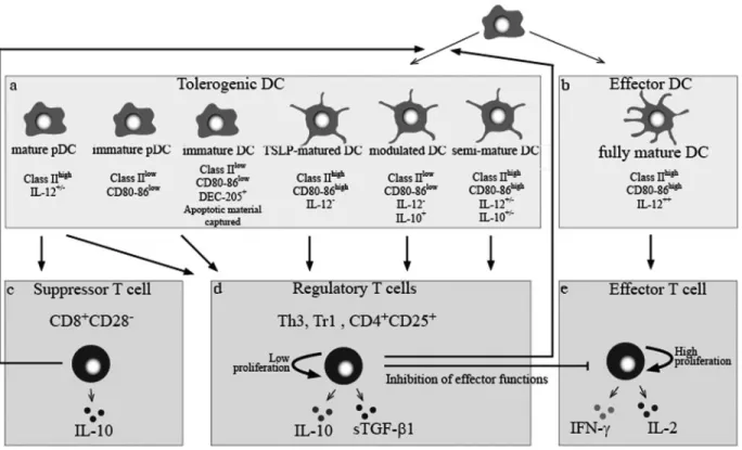
Fig. 1. Schematic representation of the interplay between tolerogenic DC subsets and Treg cells. (a) Tolerogenic DC are composed of different subsets of cells
with specific phenotypic profiles. (b) Fully mature DC express high levels of CD80/CD86 and HLA-DR, produce significant levels of IL-12, and stimulate T cell immunity and vigorous T cell-proliferative responses (e), whereas tolerogenic DC induce suppressor T cells (c) and Treg cells (d) with an inherent, low ability to proliferate in response to mitogens or antigens. Treg cells inhibit not only effector T cells but also act on DC to temper immunity efficiently. pDC, Plasmacytoid DC; TSLP, thymic stromal lymphopoietin; Tr1, Treg 1 cell; sTGF-1, soluble TGF-1.
cells [21, 23, 24]. Treatment of immature DC with TGF- and IL-10 has been shown to have a similar effect, resulting in the generation of CD4⫹and CD8⫹Treg cells [25]. It is important that DC with properties similar to the IL-10-modulated DC generated in vitro have also been detected in vivo in IL-10-producing tumors [26].
Treg cell-treated, immature myeloid DC
Human CD8⫹CD28⫺ T cells are a distinct Treg cell popula-tion, which suppresses antigen-specific CD4⫹T cell responses by inhibiting their capacity to produce IL-2 and to up-regulate CD40L expression [27]. Chang et al. [28] found that human CD8⫹CD28⫺ T cells can modulate the function of immature, monocyte-derived DC. These modulated DC do not express CD80 and CD86 and are able to anergize alloreactive memory CD4⫹T cells [28]. This suppressive effect by CD8⫹CD28⫺T cell-treated DC is MHC-restricted and antigen-specific. The induction of CD4⫹T cell anergy is not caused by a suppressor effect mediated by soluble factors but requires direct interac-tions between effector CD4⫹ T cells and pretreated DC. The mechanism for generating tolerogenic DC has been analyzed extensively in this system. Exposure of immature DC to CD8⫹CD28⫺ T cells in an antigen-specific manner results in interference with CD40 –CD40L-mediated signaling, which normally induces the functional maturation of DC and a high level of expression of CD80 and CD86. Moreover, the tolero-genic influence of CD8⫹CD28⫺T cells is associated with the induction of the inhibitory molecules Ig-like transcript 3 (ILT3) and ILT4 at the DC surface [28]. It is interesting that CD8⫹CD28⫺ T cells, which can up-regulate these molecules in donor APC, are present in the circulation of human heart transplant recipients, especially in rejection-free patients [28]. As with CD8⫹CD28⫺ Treg, CD4⫹CD25⫹ Treg cells have been shown to act directly on DC in a cytokine-independent manner, inducing the up-regulation of the inhibitory receptors ILT3 and ILT4. These inhibitory receptors are crucial to the tolerogenic phenotype acquired by DC, as the suppressive effect of Treg cells on T cell proliferation can be abrogated by antibodies to ILT3 and ILT4 [29].
Semimature myeloid DC
Lutz and Schuler [30] have described semimature DC, which originate from the exposure of immature DC to tissue-derived TNF-␣ in the absence of a pathogenic motif. These cells acquire part of the characteristics of fully mature DC, includ-ing the expression of costimulatory molecules and the ability to migrate to the draining lymph nodes. However, they produce low levels of proinflammatory cytokines such as IL-1, IL-6, TNF-␣, and IL-12p70. Semimature DC lack the capacity to produce polarizing signals and as a consequence, predomi-nantly drive the development of adaptive Treg cells. It is important that on subsequent pathogenic challenge, such semi-mature DC can still develop into fully immunogenic, IL-12-producing mature DC, and they drive effector Th1 cell re-sponses [30]. For example, in vitro-generated, peptide-loaded DC, which have been matured by TNF-␣ and subsequently injected into mice, have been shown to act in a tolerogenic manner by preventing experimental autoimmune
encephalomy-elitis through the induction of IL-10-producing CD4⫹ Treg cells [31, 32]. However, the mechanisms used to induce toler-ance remained unclear [33].
Immature pDC
Immature pDC, freshly enriched from human peripheral blood, can induce an anergic state in antigen-specific CD4⫹ T cell lines [34]. This effect is associated with a loss of IL-2 produc-tion and is completely or partially reversible in the presence of a high concentration of exogenous IL-2 in the secondary cul-tures [34]. T cell anergy induction by immature pDC requires cognate contact via TCR-MHC engagement [34]. This process is cytokine-independent, but the precise interactions between pDC and CD4⫹ T cells, which induce T cell anergy, remain unclear. The mechanism for T cell anergy induction could be explained by TCR-MHC engagement without costimulatory signals through CD80 and CD86, as freshly isolated pDC do not express or express only minimal levels of CD80 and CD86 [35]. However, as CD40-activated mature pDC are also tolero-genic, it is unlikely that their capacity to elicit the generation of Treg cells is related only to inefficient T cell stimulation. CD40L-activated pDC
The induction of tolerance versus immunity may not simply be determined by the maturity of the DC. For example, naı¨ve CD8⫹ T cells primed with allogeneic, CD40L-activated pDC differentiate into CD8⫹T cells, which display poor secondary-proliferative and cytolytic responses. In addition, they produce, respectively, high and low levels of IL-10 and IFN-␥ upon restimulation [36]. These IL-10-producing CD8⫹ Treg cells can be generated directly from naı¨ve CD8⫹T cells by a single round of stimulation with mature pDC, and they share many similarities with CD4⫹ Treg cells. These CD8⫹ Treg cells, allostimulated with mature pDC, suppress primary T cell ac-tivation through IL-10 secretion [36 –38].
Mature DC
Current literature suggests that T cells recognizing antigen on mature DC differentiate into effector T cells, whereas tolerance is induced when antigen is presented by immature DC.
Liu [39] reported the only study of DC requirement for the differentiation of Treg cells from the thymus. Within the me-dulla of the human thymus, a subset of mature DC positively selects Treg cells, protecting the medium to high-affinity, self-reactive T cells from negative deletion and inducing their differentiation into Treg cells in the thymus. How DC can have a negative role in the deletion of high-affinity, self-reactive thymocytes and simultaneously, a positive role in the selection of high-affinity, self-reactive Treg cells seems to be a paradox [39]. A recent study by Watanabe et al. [40] showed that the epithelial cells within Hassall’s corpuscles are developmen-tally programmed to express TSLP, which activates a subpopu-lation of DC in the thymic medulla to express CD80 and CD86. TSLP does not induce activated myeloid DC to produce proin-flammatory cytokines such as IL-12, IL-6, TNF-␣, and IL-1. Although the immature DC within the cortico-medullary junc-tion may be critical for negative selecjunc-tion, TSLP-DC in the central part of the medulla may be critical for the positive
selection of high-affinity, autoreactive T cells and for their differentiation into CD4⫹CD25⫹Treg cells [40].
In the periphery, mature DC also expand functional Treg cells. These latter cells may be important during infection, when DC are presenting microbial antigens, as there is likely a concomitant presentation of self and environmental antigens. When mature DC are inducing immune responses in the lymph node, with concomitant IL-2 production, bystander CD25⫹CD4⫹T cells would be expected to expand. In addition, when maturing DC are themselves presenting cognate self-antigens, thymic-derived CD25⫹CD4⫹ T cells would be ex-pected to expand vigorously and to serve to suppress autore-active responses by other DC [2, 41, 42]. Moreover, Verhasselt et al. [43] observed that a significant fraction of CD4⫹T cells cultured with mature, autologous DC acquired regulatory prop-erties. Indeed, when added to an allogeneic MLR, these CD4⫹ T cells suppressed the response of alloreactive T lymphocytes to the priming DC [43].
FEATURES OF TOLEROGENIC DC
Tolerogenic DC subsets share common features, such as low MHC expression and little or no expression of CD80 and CD86, except for the CD40-activated mature pDC. They also produce reduced levels of IL-12 and low amounts of costimu-latory CD28L. The induction of Treg cells requires a direct cell– cell contact between T cells and DC, and suppression of T cells is MHC-restricted and antigen-specific. There are sev-eral candidate molecules expressed by DC, which may induce key signals to generate Treg cells, such as IL-10, programmed death-ligand 1 (PD-L1), ILT3, ILT4, and indoleamine 2,3-dioxygenase (IDO) [33, 44, 45].
The inhibitory receptors ILT3 and ILT4 belong to a family of molecules, which are structurally and functionally related to killer cell-inhibitory receptors, and they display a long, cyto-plasmic tail containing immunoreceptor tyrosine-based inhib-itory motifs. ILT3 and ILT4 mediate inhibition of cell activa-tion by recruiting the tyrosine phosphatase Src homology-2-containing tyrosine phosphatase 1 and interfere with CD40 – CD40L-mediated signaling. Although the ligand for ILT3 is currently unknown, ILT4 bond HLA-A, -B, -C, and -G [28]. Recently, it has been demonstrated that the up-regulation of ILT3 and ILT4 induced on DC upon interaction with suppres-sor CD8⫹CD28⫺ T cells renders DC tolerogenic [46]. It is interesting that similar effects are exerted by exogenous IL-10 and IFN-␣ [47]. Blocking of the inhibitory receptors with specific antibodies against ILT3 and ILT4 restores the stimu-lating activity of the DC, preventing them from inducing T cell anergy [48].
Another potentially important aspect of tolerogenic DC is IDO, which catalyzes the depletion of the essential amino acid tryptophan. This depletion enhances the production of immu-noregulatory kynurenine metabolites, which inhibit T cell pro-liferation, and induces T cell suppression by activation of GCN2 kinase in T cells [49]. IDO expression is detected constitutively in human regulatory pDC and can be induced by classical DC maturation stimuli, namely IFN-␥ and LPS or PGE2, which contribute to their immunoregulatory capacity
[50]. Inversely, murine CD4⫹CD25⫹Treg cells can help DC to express functionally active IDO [51].
Other molecules, which are certainly promising for future study, are novel, negative, costimulatory B7 family members (reviewed in ref. [52]), including PD-L1 (also known as B7-H1), B7-H3 (also known as B7RP-2), B7-H4 (also known as B7S1 or B7x), and the ligand of the B and T lymphocyte attenuator. Although glucocorticoid-induced TNFR (GITR) ex-pression has been associated with activated, naturally occur-ring Treg cells [53], more recent literature indicates that the distribution of GITR expression is much wider and that GITR ligation suppresses rather than induces tolerance [54]. Consid-ering the wealth of negative regulatory molecules, it might be anticipated that further subclassification of regulatory DC sub-sets will be necessary in the near future.
TREG CELL POPULATIONS
Treg cells consist of different subsets of T lymphocytes char-acterized by their ability to suppress proliferation of conven-tional effector T cells by various mechanisms (see Communi-cation between DC and Treg cells and regulatory functions below).
The list of candidate markers for Treg cells increases con-tinuously and includes CD45RB, CD103, or CD122 and the transcriptional repressor forkhead box P3 (Foxp3) [55, 56], which is currently considered as the most promising marker for Treg cells. In addition, Treg cells are characterized by the constitutive expression of GITR family-related protein, L-se-lectin (CD62L), and CTLA-4 (or CD152) [57, 58]. Thus, no specific marker (except the transcription factor Foxp3) has been identified to date. In particular, no defined cell surface molecules can be targeted with antibodies specifically to de-plete or to purify these cells. This problem is an ongoing challenge for the study and manipulation of these suppressive cells. Concerning the Foxp3 itself, one question remains: whether its expression is an absolute marker of Treg cells in humans or mice. In the mouse, there is an excellent correlation between the expression of Foxp3 and CD25, but a minor population of Foxp3⫹ cells is CD25–. In humans, almost all CD4⫹CD25hicells are Foxp3⫹, whereas a variable percentage of CD25intcells expresses lower amounts of Foxp3. Moreover, whereas the TGF--induced murine Foxp3⫹ T cells manifest suppressor activity in vitro and in vivo, Foxp3 expression in human T cells does not correlate uniformly with suppressor activity. In addition, populations of CD4⫹CD25⫹ Treg cells producing IL-10 do not seem to express Foxp3 [59].
Over the past few years, several phenotypically distinct Treg cell populations have been described [59 – 61] (Table 1). The classic Treg cells are thymus-derived CD4⫹CD25⫹Foxp3⫹ T cells, and many investigators have termed these cells “natural” Treg in contrast to Treg cells, which develop in peripheral lymphoid tissues and are frequently Foxp3– and have been termed “adaptive” or “induced” Treg cells. Several studies have raised the possibility that CD4⫹Foxp3⫹cells might also be generated in peripheral lymphoid tissues from naı¨ve CD4⫹ Foxp3–progenitors [59].
Among all the subsets described to date, the most studied are the naturally occurring CD4⫹CD25⫹Treg cells, the anti-gen-induced CD4⫹CD25⫹ Treg cells, and the CD8⫹CD28–T cells (or T suppressive cells).
The naturally occurring CD4⫹CD25⫹Treg cells represent a small subset (5– 6%) of the overall CD4⫹T cell population and are generated in the thymus. They mediate immune suppres-sion by inhibiting the activation and proliferation of Th and cytotoxic T cells through a cell– cell contact and antigen-nonspecific mechanism [67]. However, in some models, Foxp3⫹ natural Treg cells have been shown to be antigen-specific [68], suggesting that the distinction between the dif-ferent subsets of Treg cells is not at all clear and that the notion of antigen induction is not adequate to distinguish the different Treg cells.
The antigen-induced CD4⫹CD25⫹ Treg cells are detected in peripheral tissues after MHC/peptide stimulation of conven-tional CD4⫹CD25–precursors [69]. These Treg cells include Tr1 cells, secreting IFN-␥, IL-10, and to a lesser extent, TGF-, and Th3 T cells, secreting high levels of TGF- and IL-10 [70]. The suppressive activity of the antigen-induced Treg cells is cell contact-independent and is mediated by the release of TGF- and IL-10 [71]. Th3 and Tr1 cells display suppressive properties on Th1 and Th2 cells, but only Th3 cells provide help for IgA synthesis [72]. Tr1 cells regulate the function of naı¨ve and memory T cells in vitro and in vivo and can suppress responses to tumors, alloantigens, and pathogens [72]. These two subsets of Treg cells also differ in the expres-sion of distinct integrins, mainly ␣41 and␣47[73], which
have been shown to be homing receptors for the migration of T lymphocytes to inflamed tissues and to mucosal sites, respec-tively [74]. These data suggest that ␣41⫹CD25⫹ Treg cells
migrate in vivo to inflamed tissues, where they inhibit effector
T cell responses, whereas␣47⫹CD25⫹Treg cells could
pre-vent chronic mucosal inflammations by counteracting autore-active T cells.
It is generally understood that being CD4⫹T cells, naturally occurring Treg cells as well as antigen-induced Treg cells need to be activated by MHC class II-bound epitopes on DC. It is widely believed that although antigen-induced Treg cells need TCR ligands and costimulation, naturally occurring Treg cells need only a TCR-driven signal for functional activation. None-theless, as only a limited class of cells expresses MHC class II molecules, naturally occurring Treg cells need DC for their activation. Thus, just as DC are indispensable in activating, naı¨ve effector T cells, they are also needed for the activation of Treg cells [75].
A subpopulation of Treg cells expresses CD8 and can me-diate immune suppression in an antigen-dependent manner [76]. CD8⫹Treg cells suppress antigen-activated CD4⫹T cells by a TCR/MHC class Ib molecule restriction [77]. CD8⫹ CD28–Treg cells also suppress APC, which present the same peptide/MHC complexes to which the CD8⫹ Treg cells were primed previously [78]. In contrast to naturally occurring CD4⫹CD25⫹ Treg cells, an important feature of CD8⫹Treg cells is that they are generated or induced only after antigen priming [79].
COMMUNICATION BETWEEN DC AND TREG CELLS AND REGULATORY FUNCTIONS
There is accumulating evidence that tolerogenic DC can stim-ulate Treg cell expansion (Fig. 2). For example, tumors con-vert DC into TGF--expressing DC, which are capable of promoting Treg cell proliferation [80]. In addition to expansion
TABLE 1. Subsets of T-cells
CD4⫹T cells CD8⫹T cells CD4⫹CD25⫹ (CD45RA⫹) Tr1 (CD45RO⫹) Th3 (CD45RO⫹) CD8⫹CD25⫹ CD8⫹CD28– CD8⫹CD28– (CD45RO⫹) CD8⫹CD122⫹ Markers CD25 ⫹⫹ ⫹ ⫹ ⫹ – ND – GITR ⫹⫹ – ND ⫹ ⫹ ND ND CTLA-4 ⫹⫹⫹ ⫹ ⫹⫹ ⫹ ⫹ ND ND CD62L ⫹ ⫹ ⫹ ND ND ND ⫹ Foxp3 ⫹⫹ ⫹(?) (?) ⫹⫹ ⫹ ND – IL-10 production ⫹ ⫹⫹⫹ ⫹ ND ⫹ ⫹⫹ ⫹⫹ TGF- production ⫹ ⫹ ⫹⫹⫹ ⫹⫹⫹ ⫹ ND ⫹⫹ Main regulatory mechanisms Cell–cell contact in vitro Multiple mechanisms of action in vivo IL-10 TGF- Cell–cell contact TGF- and CTLA-4 Cell–cell contact Targeting ILT3 and ILT4 on DC
IL-10 IL-10 and additional unknown mechanisms
Target cells T cells/APC T cells/APC T cells T cells DC/APC T cells/APC T cells Suggested
origin
Thymus Periphery Periphery Thymus Periphery Periphery Periphery
References [40, 62] [63] [64] [65] [28] [38] [66]
of Treg cells, DC can mediate the conversion of naı¨ve CD4⫹CD25–T cells. Kretschmer [81] reported the conversion of truly naı¨ve CD4⫹ T cells into suppressor cells expressing Foxp3 by the targeting of peptide-agonist ligands to DC. The conversion was achieved by minute antigen doses with subop-timal DC activation. The addition of TGF- or the absence of IL-2 production, which reduces proliferation, enhanced the conversion rate [81].
Once activated in an antigen-specific manner by DC, Treg cells might also suppress effector T cells, which are specific for other antigens. This happens if these antigens are expressed by the same APC or target cells, as well as by cells, which are in close vicinity, a phenomenon known as bystander suppression [82]. Suppressive mechanisms of Treg cells have been ad-dressed using many in vitro and in vivo mouse models. Mul-tiple suppressive mechanisms including cell– cell contact and soluble factors have been proposed [83– 85].
Activated human Treg cells express granzyme A and kill T cells and APC through perforin [86]. However, another study using granzyme B-deficient and perforin-deficient mice re-ported that Treg cells mediate suppression through a granzyme
B-dependent but perforin-independent mechanism [60, 87]. Soluble factors and cell– cell contacts are involved in the suppressive function of Treg cells. For example, competition between Treg and conventional T cells for IL-2 has been suggested as a suppressive mechanism [57, 88]. Besides their role in the effector functions of T cells and in DC viability, Treg cells also have a profound impact on DC function. In this respect, human CD4⫹CD25⫹ Treg cells may render DC inef-ficient as APC [89]. Treg cells are also able to enable DC to express functional IL-10 [90]. CD4⫹CD25⫹ Treg cells have been shown to inhibit the expression of CD80 and CD86 costimulatory molecules on DC in a CTLA-4-dependent way [91, 92]. Treg cells impede DC function by down-regulating the activation of NF-B. The suppression mechanism requires TGF- and IL-10 and is associated with the induction of the Smad signaling pathway and activation of the STAT3 transcrip-tion factor [93].
Moreover, Sato et al. [94] showed that Treg cells suppress CD25⫺CD4⫹ T cell responses, at least in part by inhibiting IL-12 production from DC, and they themselves can undergo proliferation with the mediation of CD25⫺CD4⫹ T cells in vitro. These results offer a novel, negative-feedback system involving a tripartite interaction among CD25⫹CD4⫹ and CD25⫺CD4⫹ T cells and DC, which may contribute to the termination of immune responses.
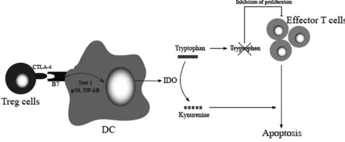
Another mechanism is the induction of IDO-expressing DC. IDO is an enzyme that degrades the essential amino acid tryptophan, which is required for T cell activation [95]. IDO⫹ human DC reduce tryptophan, and as this amino acid is an essential, proliferative stimulus for effector T cells, its defi-ciency provokes T cell apoptosis and thus, promotes tolerance in the tumor microenvironment (Fig. 3) [95]. Mouse CTLA-4⫹ Treg cells mediate their suppressive activity by inducing the expression of IDO in DC through CTLA-4 [96].
B7-H4 (also known as B7x, B7S1), which is a recently discovered member of the B7 family of T cell costimulatory molecules, regulates T cell responses negatively. It is interest-ing that Treg cells, but not normal T cells, induce B7-H4 expression by DC and render them immunosuppressive [97].
CD4⫹CD25⫹ Treg cells can convey suppressor activity to conventional CD4⫹T cells, a phenomenon consequently called infectious tolerance [98, 99]. Human CD4⫹T cells, which have been cocultured with CD4⫹CD25⫹ Treg cells after isolation, are able to suppress the proliferation of freshly prepared, conventional CD4⫹T cells. This “infectious” suppressor func-tion, transferred from CD4⫹CD25⫹Treg cells, has been shown to be cell contact-dependent and partially mediated by TGF- [99]. However, there are also indications that CD4⫹CD25⫹ Treg cells can mediate their suppressor activity via the release of IL-10 [100 –102].
Suciu-Foca et al. [29] described a cascade of suppression where allospecific CD8⫹CD28⫺Treg cells first tolerize the DC. These tolerized DC anergize alloreactive CD4⫹T cells, which recognize MHC class II alloantigens on their membrane-induc-ing Treg cells. In turn, these Treg cells tolerize fresh DC. Finally, DC tolerized by the CD4⫹CD25⫹ Treg cells may inhibit the alloreactivity of other CD4⫹T cells, thus continuing the T suppressor cell cascade. These tolerized DC with
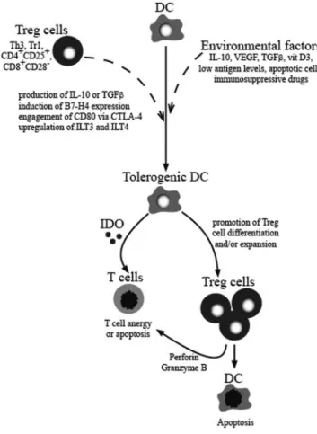
up-Fig. 2. Schematic representation of the interplay between DC and Treg cells.
Environmental factors and Treg cells may alter the function of DC. The mechanisms responsible for the induction of tolerogenic DC by Treg cells are multifactorial and include the production of IL-10 or TGF-, the induction of B7-H4 expression, the engagement of CD80, or the up-regulation of ILT3 and ILT4. These tolerogenic DC may subsequently promote Treg cell differentia-tion and/or expansion and induce T cell anergy or apoptosis by the producdifferentia-tion of IDO by tolerogenic DC. Activated Treg cells may also kill target cells directly (T cells or DC) through perforin or granzyme B-dependent pathways. VEGF, Vascular endothelial growth factor.
regulated expression of the inhibitory receptors ILT3 and ILT4 spread the unresponsiveness to antigen-specific T cells.
In conclusion, Treg cells control the expansion of effector T cells, which respond to self and nonself antigens through a variety of mechanisms, including cognate or noncognate inter-actions with DC or activated effector T cells, direct regulation by the killing of undesirable T cell specificities, or secondary activation of new Treg cell subsets. In addition to T cells, Treg cells can therefore temper immunity by targeting DC, and multiple Treg cell-mediated, suppressive mechanisms are likely to be operative in vivo.
BENEFICIAL AND ADVERSE EFFECTS OF THE CROSS-TALK BETWEEN DC AND TREG CELLS
There is convincing evidence that Treg cells play a protective role against autoimmune diseases, allograft rejections, and allergic conditions, where they suppress potentially patho-genic, immune responses mediated by effector Th1 cells, Th2 cells, or CTLs [103, 104]. As these effector T cell responses also play an important role in the protection against pathogens, it might seem counterintuitive that Treg cells could have a protective role in infection. However, in many infectious dis-eases, important collateral damage to host tissues potentially induced by immune responses against pathogens is prevented by immunoregulatory mechanisms, including the induction of Treg cells [105]. Moreover, recent studies suggest that Treg cells can recognize microbial antigens and that this recognition is associated with their function [68]. Although Treg cells are beneficial to the host by preventing immunopathology and enabling the development of immune memory, they can also be beneficial to the pathogen, enabling it to establish a chronic infection. Many pathogens have evolved strategies that facili-tate their persistence, largely through their ability to evade or subvert the host immune response. One strategy is to induce a
state of immunosuppression through direct interference with host immune effector mechanisms or through the production of immunosuppressive cytokines. For example, distinct families of pathogen-derived molecules, including filamentous hemag-glutinin and adenylate cyclase toxin from Bordetella pertussis, cholera toxin, hepatitis C virus nonstructural protein 4, and Schistosoma mansoni-specific phosphatidylserine, interact with pattern recognition receptors, including CD11b–CD18, gangli-oside GM1, or TLR2 on the surface of DC. These interactions stimulate IL-10 production and inhibit IL-12 production by macrophages and DC and activate DC to become semimature and to promote the induction of Treg cells (for review, see ref. [105]).
Treg cells are also implicated in the tolerance against com-mensal microorganisms and innocuous agents in the gastroin-testinal system or in the respiratory tractus [106, 107]. Indeed, mucosal surfaces are obligatory sites for tolerance induction against numerous exogenous antigens. Therefore, the mucosal surface seems to provide a microenvironment, where IL-10 and TGF- are highly expressed, DC effector functions affected, and Treg cells induced [108].
It is interesting that Treg cells are thought to have an enormous potential in suppressing pathological immune re-sponses in transplantation. Treg cells have been described in tolerated tissues, such as accepted skin grafts. T cells from such tolerated tissues can repopulate the peripheral immune system of immunodeficient recipients and prevent graft rejec-tion by naı¨ve T cells [109].
An emerging model of Treg cell action is that organ-specific Treg cells acquire suppressive activity through activation by DC expressing organ-derived antigens. Thus, the efficacy of Treg cell-based therapy should be increased by using organ-specific Treg cells rather than polyclonal Treg cells. This necessitates the ability to identify relevant antigens and to expand rare, antigen-specific Treg cells from diverse poly-clonal populations. Yamazaki et al. [2] showed in a mouse model that DC are able to expand antigen-specific Treg cells,
Fig. 3. Treg cells inhibit effector T cells via the induction of IDO. The expression of IDO by DC is mediated partially by the interaction of CTLA-4 expressed
on Treg cells and B7 expressed on DC. IDO catalyzes the degradation of tryptophan, which is an essential, proliferative stimulus for effector T cells and leads to the apoptosis of these cells in a tryptophan-deprived environment.
particularly polyclonal Treg cells directed to alloantigens. When triggered by specific antigen, these Treg cells act back on immature DC to block the up-regulation of CD80 and CD86 costimulatory molecules.
Serial studies of the phenotype displayed by T cells from heart allograft recipients have demonstrated a significant in-crease of the CD8⫹CD28⫺CD27⫹ perforin-negative T cell population in rejection-free patients [110]. The Treg cells from these recipients inhibited the up-regulation of CD80 and CD86 on CD40-ligated DC from the donor. This inhibitory effect is MHC class I-allorestricted, demonstrating the antigen speci-ficity of these in vivo-generated Treg cells [111]. Treg cells from these patients also induced the up-regulation of ILT3 and ILT4 on donor DC. Serial determination of the suppressor activity displayed by CD4⫹CD25⫹T cells of patients yielded similar results. However, CD4⫹CD25⫹Treg cells become de-tectable at later times following transplantation. The persis-tence of allospecific Treg cells in rejection-free patients late after transplantation indicates that these T cells, which inhibit the direct, allorecognition pathway, are stimulated continu-ously by donor APC. However, as donor DC migrate out of the graft during the early post-transplantation period, it is likely that they are stimulated by endothelial cells of the transplant, which are known to act as semiprofessional DC [112].
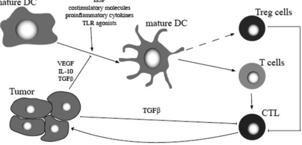
In contrast to their protective role in autoimmune diseases, Treg cells are likely to interfere negatively with immunity against a wide variety of human tumors. Numerous studies in recent years have described an increased frequency of CD4⫹CD25⫹T cells, and some or all of the characteristics of Treg cells were in the blood and the tumor microenvironment of patients with cancers originating in the head and neck [113], lung [114, 115], liver [116], gastrointestinal tract [117, 118], breast [119, 120], or skin [121]. Moreover, Treg cells with specificity for antigens expressed by human tumors have been identified recently [85, 122], and the extent of Treg cell accu-mulation in tumor tissues has been shown to predict poor survival [123]. Tumor environmental factors such as IL-10, TGF-, VEGF, and PGs suppress DC differentiation and func-tion, resulting in immature and/or partially differentiated DC [124]. These tumor-converted DC are able to promote Treg cell expansion or proliferation [80]. In addition to this induction of tolerogenic DC, Treg cells themselves might be recruited by tumors and secondarily, can alter the DC function, activating the suppressor cascade [125].
Whereas Treg cells can impede anticancer surveillance activities, it is likely that suppression of active inflammation by these cells can prevent or control inflammation-associated cancer [126, 127].
Autoimmune diseases develop as a consequence of break-down in central and/or peripheral tolerance. Treg cells may function in vivo by interrupting the effector development pro-gram through limiting the autoreactive T cell access to DC. In this context, failure of Treg cells to maintain tolerance may be the underlying cause of autoimmune diseases [128]. In psori-asis, it was found that a CD4⫹T lymphocyte subpopulation in peripheral blood or in psoriatic plaques, phenotypically resem-bling Treg cells (CD25high, CTLA-4⫹, Foxp3high), was deficient in its suppressor activity. This was associated with accelerated proliferation of CD4⫹ responder T cells in psoriasis [129].
Thus, it is speculated that this dysfunction in the skin and in the blood of psoriatic patients may cause hyperproliferation of psoriatic, pathogenic T cells in vivo.
THERAPEUTIC STRATEGIES
The manipulation of the DC–T cell interactions using different pharmacological or biological agents could be exploited to improve antitumor strategies or to control a variety of chronic inflammatory conditions, such as autoimmune diseases, post-transplantation graft-versus-host disease (GVHD), and allo-graft rejection.
Treg cells can be expanded by ex vivo cultures with alloge-neic splenocytes and IL-2, and the administration of these cells at the time of grafting delays or prevents GVHD [130]. The addition of TGF- in the ex vivo cultures has been shown to enhance the functionality of Treg cells strongly [131]. The observation that Treg cells expressing high levels of CD62L inhibit GVHD lethality efficiently suggests that they can act in secondary lymphoid tissues [132] in addition to the GVHD target tissues such as skin, liver, lung, and the gastrointestinal tract. In this regard, expression of the chemokine receptor CCR5 by Treg cells was shown to play a critical role in the migration to target tissues and to improve survival significantly after donor lymphocyte infusion [133]. However, a major lim-itation for the current therapeutic use of Treg cells is the difficulty to obtain sufficient amounts of these relatively rare cells from a single donor.
DC also serve as potential targets for suppression of alloim-mune reactivity and promotion of tolerance induction. Indeed, the DC-mediated immune regulation (i.e., expansion/induction of Treg cells, such as CD4⫹CD25⫹T cells or Tr1 cells, which produce IL-10) is an important mechanism by which DC exert their tolerogenicity. In transplantation, one of the potential difficulties associated with the administration of immature, regulatory DC is that in the context of the danger signals related to surgical trauma and ischemia-reperfusion injury, the administered DC may mature and accelerate graft rejection or at least be unable to diminish immune alloresponses. This problem could be overcome if the regulatory DC were admin-istered before the transplantation. As demonstrated in most experimental animal models, this approach could help the DC to exert their tolerogenic effects by the time of transplantation. A potential solution to decrease the risk of DC maturation induced by the inflammatory environment is to manipulate the DC in vitro to produce maturation-resistant, immature DC or “alternatively activated” DC with stable, tolerogenic proper-ties. Presently, the enhancement of DC tolerogenicity was achieved by the use of pharmacological inhibition of DC mat-uration or by the use of genetically engineered DC expressing immunosuppressive molecules [134]. A novel strategy involv-ing the use of DC for the regulation of T cell responses consists of the generation of cytokine-modified regulatory DC. Several growth factors, including IL-10, TGF-, G-CSF, hepatocyte growth factor (HGF), and vasoactive intestinal peptide (VIP), may modulate DC maturation and favor the differentiation of tolerogenic DC [135]. In particular, DC differentiated in the presence of exogenously added TGF-, IL-10, GM-CSF, and
IL-4 possess the immunophenotypic and functional features of immature DC and induce CD4⫹CD25⫹Treg cells, which sup-press the response of antigen-primed CD4⫹T cells to alloge-neic, normal mature DC [136]. In addition, the release of proinflammatory cytokines, i.e., IL-1, IL-6, TNF-␣, and most markedly, IL-12, is abolished after IL-10 treatment [137]. The therapeutic use of IL-10 is promising, as the injection of IL-10-modified DC has been shown to be able to prevent autoimmunity in a murine model of multiple sclerosis [138] or to prolong graft survival [139]. Although most of these results were obtained with DC exposed to IL-10 in vitro, there is also recent evidence that IL-10-driven DC modulation may play a role in the generation of Treg cells in vivo [140, 141]. In addition to IL-10, TNF-␣ may play a role in the induction of tolerogenic DC [32]. Gonzalez-Rey et al. [142] have shown that the injection of VIP-induced regulatory DC improves the clin-ical severity of inflammatory bowel diseases. The therapeutic effect was associated with the generation of IL10⫹ Treg cells with suppressive capacity on autoreactive T cells.
The role of immunosuppressive drugs in the induction of regulatory DC was reviewed extensively elsewhere [134]. Among them are the vitamin D3 metabolite 1␣,25-(OH)2D3,
N-acetyl-l-cysteine, and common immunosuppressive drugs, such as corticosteroids, cyclosporin A, rapamycin, and aspirin [22, 143–145]. All of them interfere with DC differentiation and maturation and consecutively, induce Treg cells. Cortico-steroids do not affect DC viability but down-regulate the ex-pression of costimulatory molecules on DC, prevent DC mat-uration, and impair their immunostimulatory activities by in-hibiting the NF-B pathway. Glucocorticoids also inhibit
LPS-or CD40L-induced DC maturation and DC production of IL-12 and TNF-␣. DC exposed to dexamethasone fail to prime Th1 cells efficiently, and the repeated stimulation of T cells with these DC generates IL-10-producing Treg cells.
The modulation of costimulatory molecule expression on DC is another way to induce the expansion of Treg cells, and the blocking of molecules of the B7 family, including B7-1, B7-2, or CTLA-4, has been used successfully for inducing immuno-logic tolerance in vitro [146].
Recent data suggest that Treg cells expanded with DC are able to suppress autoimmune diseases in vivo. For example, islet autoantigen-specific murine Treg cells can be expanded ex vivo by culturing CD25⫹ CD4⫹ T cells from diabetics diagnosed recently with DC, loaded with islet antigens derived from islet tissues or from peptide mixtures of identified autoan-tigens [42]. Moreover, the injected Treg cells induce new Treg cells in vivo directly, by expansion or differentiation through cytokines or costimulatory molecules, or indirectly, by licens-ing DC to induce the differentiation of new Treg cells (Fig. 4) [147]. The advantage of directing therapy to specific antigens is the avoidance of generalized immunosuppression. However, the potential disadvantage is the possibility that generating tolerance to one antigen may not block a disease mediated by T cells with many specificities.
Strategies aiming to suppress tumor tolerance by inducing the maturation of DC in vivo are also under active investiga-tion. In this context, tumor-specific CTL, which do not lead to an adequate anti-tumor response as a result of the presence of Treg cells, may be activated to eradicate established tumors [148, 149]. Simple vaccines have been described already,
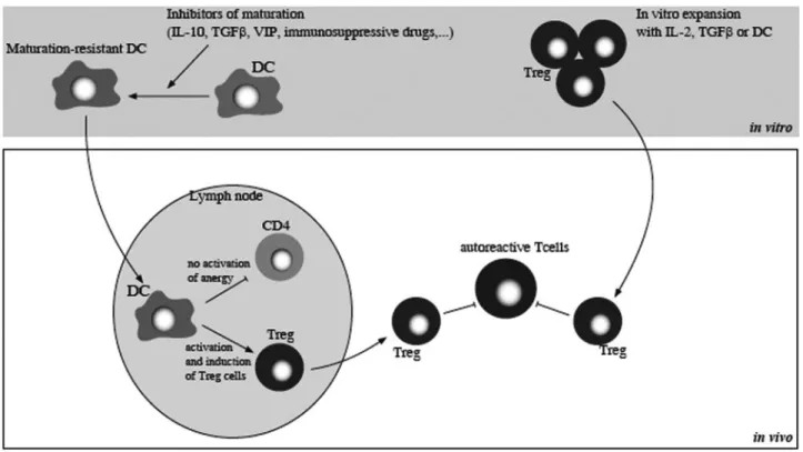
Fig. 4. Therapeutic strategies in the context of autoimmune diseases and allograft rejection. DC are manipulated in vitro to produce maturation-resistant DC with
stable tolerogenic properties by the use of pharmacological inhibitors of DC maturation (IL-10, TGF-, G-CSF, GM-CSF, HGF, VIP, immunosuppressive drugs) and serve to the expansion/induction of Treg cells. Conversely, Treg cells can also be expanded by ex vivo cultures with IL-2, TGF-, or DC and have inhibitory effects on autoreactive T cells.
which take advantage of CpG DNA motifs (a TLR9 agonist) coinjected with or conjugated to a protein antigen [150, 151]. Vaccination with heat shock protein (HSP)-peptide complexes can similarly stimulate the maturation of DC in vivo and induce immunologic and clinical responses in melanoma patients [152]. Ex vivo-derived DC may become mature in situ by preconditioning the injection site with TLR agonists [153]. DC can also be activated at the site of maturation through the coexpression of local costimulatory molecules, proinflamma-tory cytokines, and TLR agonists (Fig. 5) [154]. Recently, Nair et al. [155] showed that vaccination of mice against Foxp3, using Foxp3 mRNA-transfected DC, is capable of stimulating specific CTL, which cause the preferential depletion of Foxp3-expressing Treg cells in tumors but not in the periphery.
CONCLUSIONS
Currently, there is growing evidence that several factors, such as the cell surface phenotype, maturation status, and immuno-logical context, may determine whether a DC is tolerogenic or immunogenic. Because of the ability of several DC populations to induce antigen-specific Treg cells, manipulation of tolero-genic DC is a potential strategy to suppress harmful T cell responses in patients with autoimmune and allergic diseases as well as those who experience transplant rejection.
However, tolerance is also considered as a potential obstacle to tumor immunotherapy, as the suppressive milieu present within established tumors may inhibit effective immune re-sponses. Promising protocols should emerge in the future to shift the balance back to a proinflammatory environment, to promote DC activation, and to enhance anti-tumor immunity.
ACKNOWLEDGMENTS
The authors thank the Belgian Fund for Medical Scientific Research, the Centre Anti-Cancereux pre`s l’Universite´ de Lie`ge, Televie, Bonjean-Oleffe, L. Lacroix, and Le´on Fredericq funds for their support.
REFERENCES
1. De Jong, E. C., Smits, H. H., Kapsenberg, M. L. (2005) Dendritic cell-mediated T cell polarization. Springer Semin. Immunopathol. 26, 289 –307.
2. Yamazaki, S., Patel, M., Harper, A., Bonito, A., Fukuyama, H., Pack, M., Tarbell, K. V., Talmor, M., Ravetch, J. V., Inaba, K., Steinman, R. M. (2006) Effective expansion of alloantigen-specific Foxp3⫹ CD25⫹ CD4⫹ regulatory T cells by dendritic cells during the mixed leukocyte reaction. Proc. Natl. Acad. Sci. USA 103, 2758 –2763.
3. Tarbell, K. V., Yamazaki, S., Steinman, R. M. (2006) The interactions of dendritic cells with antigen-specific, regulatory T cells that suppress autoimmunity. Semin. Immunol. 18, 93–102.
4. Kubach, J., Becker, C., Schmitt, E., Steinbrink, K., Huter, E., Tuetten-berg, A., Jonuleit, H. (2005) Dendritic cells: sentinels of immunity and tolerance. Int. J. Hematol. 81, 197–203.
5. Tan, J. K., O’Neill, H. C. (2005) Maturation requirements for dendritic cells in T cell stimulation leading to tolerance versus immunity. J. Leu-koc. Biol. 78, 319 –324.
6. Jonuleit, H., Schmitt, E., Schuler, G., Knop, J., Enk, A. H. (2000) Induction of interleukin 10-producing, nonproliferating CD4(⫹) T cells with regulatory properties by repetitive stimulation with allogeneic im-mature human dendritic cells. J. Exp. Med. 192, 1213–1222. 7. Mahnke, K., Qian, Y., Knop, J., Enk, A. H. (2003) Induction of CD4⫹/
CD25⫹ regulatory T cells by targeting of antigens to immature dendritic cells. Blood 101, 4862– 4869.
8. Bonifaz, L., Bonnyay, D., Mahnke, K., Rivera, M., Nussenzweig, M. C., Steinman, R. M. (2002) Efficient targeting of protein antigen to the dendritic cell receptor DEC-205 in the steady state leads to antigen presentation on major histocompatibility complex class I products and peripheral CD8⫹ T cell tolerance. J. Exp. Med. 196, 1627–1638. 9. Hawiger, D., Inaba, K., Dorsett, Y., Guo, M., Mahnke, K., Rivera, M.,
Ravetch, J. V., Steinman, R. M., Nussenzweig, M. C. (2001) Dendritic cells induce peripheral T cell unresponsiveness under steady state conditions in vivo. J. Exp. Med. 194, 769 –779.
10. Dhodapkar, M. V., Steinman, R. M. (2002) Antigen-bearing immature dendritic cells induce peptide-specific CD8(⫹) regulatory T cells in vivo in humans. Blood 100, 174 –177.
11. Suss, G., Shortman, K. (1996) A subclass of dendritic cells kills CD4 T cells via Fas/Fas-ligand-induced apoptosis. J. Exp. Med. 183, 1789 – 1796.
12. Belz, G. T., Behrens, G. M., Smith, C. M., Miller, J. F., Jones, C., Lejon, K., Fathman, C. G., Mueller, S. N., Shortman, K., Carbone, F. R., Heath, W. R. (2002) The CD8␣(⫹) dendritic cell is responsible for inducing peripheral self-tolerance to tissue-associated antigens. J. Exp. Med.
196,1099 –1104.
13. Kronin, V., Wu, L., Gong, S., Nussenzweig, M. C., Shortman, K. (2000) DEC-205 as a marker of dendritic cells with regulatory effects on CD8 T cell responses. Int. Immunol. 12, 731–735.
14. Kronin, V., Fitzmaurice, C. J., Caminschi, I., Shortman, K., Jackson, D. C., Brown, L. E. (2001) Differential effect of CD8(⫹) and CD8(–) dendritic cells in the stimulation of secondary CD4(⫹) T cells. Int. Immunol. 13, 465– 473.
Fig. 5. Strategies aiming to suppress
tu-mor tolerance. The in vivo maturation of DC is a critical event to induce immunologic and clinical responses against tumors. Ex vivo-derived DC may become mature in situ by preconditioning the injection site with HSP, costimulatory molecules, proinflam-matory cytokines, or TLR agonists. Tumor-specific CTL, which do not lead to an ade-quate anti-tumor response as a result of the presence of Treg cells and tumor factors, may then be activated to eradicate cancer cells.
15. Steinman, R. M., Turley, S., Mellman, I., Inaba, K. (2000) The induction of tolerance by dendritic cells that have captured apoptotic cells. J. Exp. Med. 191, 411– 416.
16. Stuart, L. M., Lucas, M., Simpson, C., Lamb, J., Savill, J., Lacy-Hulbert, A. (2002) Inhibitory effects of apoptotic cell ingestion upon endotoxin-driven myeloid dendritic cell maturation. J. Immunol. 168, 1627–1635. 17. Kim, S., Elkon, K. B., Ma, X. (2004) Transcriptional suppression of interleukin-12 gene expression following phagocytosis of apoptotic cells. Immunity 21, 643– 653.
18. Wallet, M. A., Sen, P., Tisch, R. (2005) Immunoregulation of dendritic cells. Clin. Med. Res. 3, 166 –175.
19. Penna, G., Adorini, L. (2000) 1 ␣,25-dihydroxyvitamin D3 inhibits differentiation, maturation, activation, and survival of dendritic cells leading to impaired alloreactive T cell activation. J. Immunol. 164, 2405–2411.
20. Lagaraine, C., Lebranchu, Y. (2003) Effects of immunosuppressive drugs on dendritic cells and tolerance induction. Transplantation 75, 37S– 42S.
21. Steinbrink, K., Wolfl, M., Jonuleit, H., Knop, J., Enk, A. H. (1997) Induction of tolerance by IL-10-treated dendritic cells. J. Immunol.
159,4772– 4780.
22. Schlichting, C. L., Schareck, W. D., Nickel, T., Weis, M. (2005) Den-dritic cells as pharmacological targets for the generation of regulatory immunosuppressive effectors. New implications for allo-transplantation. Curr. Med. Chem. 12, 1921–1930.
23. McBride, J. M., Jung, T., de Vries, J. E., Aversa, G. (2002) IL-10 alters DC function via modulation of cell surface molecules resulting in im-paired T-cell responses. Cell. Immunol. 215, 162–172.
24. Steinbrink, K., Graulich, E., Kubsch, S., Knop, J., Enk, A. H. (2002) CD4(⫹) and CD8(⫹) anergic T cells induced by interleukin-10-treated human dendritic cells display antigen-specific suppressor activity. Blood
99,2468 –2476.
25. Sato, K., Yamashita, N., Baba, M., Matsuyama, T. (2003) Modified myeloid dendritic cells act as regulatory dendritic cells to induce anergic and regulatory T cells. Blood 101, 3581–3589.
26. Enk, A. H., Jonuleit, H., Saloga, J., Knop, J. (1997) Dendritic cells as mediators of tumor-induced tolerance in metastatic melanoma. Int. J. Cancer 73, 309 –316.
27. Cortesini, R., LeMaoult, J., Ciubotariu, R., Cortesini, N. S. (2001) CD8⫹CD28- T suppressor cells and the induction of antigen-specific, antigen-presenting cell-mediated suppression of Th reactivity. Immunol. Rev. 182, 201–206.
28. Chang, C. C., Ciubotariu, R., Manavalan, J. S., Yuan, J., Colovai, A. I., Piazza, F., Lederman, S., Colonna, M., Cortesini, R., Dalla-Favera, R., Suciu-Foca, N. (2002) Tolerization of dendritic cells by T(S) cells: the crucial role of inhibitory receptors ILT3 and ILT4. Nat. Immunol. 3, 237–243.
29. Suciu-Foca, N., Manavalan, J. S., Cortesini, R. (2003) Generation and function of antigen-specific suppressor and regulatory T cells. Transpl. Immunol. 11, 235–244.
30. Lutz, M. B., Schuler, G. (2002) Immature, semi-mature and fully mature dendritic cells: which signals induce tolerance or immunity? Trends Immunol. 23, 445– 449.
31. Voigtlander, C., Rossner, S., Cierpka, E., Theiner, G., Wiethe, C., Menges, M., Schuler, G., Lutz, M. B. (2006) Dendritic cells matured with TNF can be further activated in vitro and after subcutaneous injection in vivo which converts their tolerogenicity into immunogenicity. J. Immu-nother. 29, 407– 415.
32. Menges, M., Rossner, S., Voigtlander, C., Schindler, H., Kukutsch, N. A., Bogdan, C., Erb, K., Schuler, G., Lutz, M. B. (2002) Repetitive injections of dendritic cells matured with tumor necrosis factor␣ induce antigen-specific protection of mice from autoimmunity. J. Exp. Med. 195, 15–21. 33. Mahnke, K., Enk, A. H. (2005) Dendritic cells: key cells for the induc-tion of regulatory T cells? Curr. Top. Microbiol. Immunol. 293, 133–150. 34. Kuwana, M. (2002) Induction of anergic and regulatory T cells by plasmacytoid dendritic cells and other dendritic cell subsets. Hum. Immunol. 63, 1156 –1163.
35. Ito, T., Inaba, M., Inaba, K., Toki, J., Sogo, S., Iguchi, T., Adachi, Y., Yamaguchi, K., Amakawa, R., Valladeau, J., Saeland, S., Fukuhara, S., Ikehara, S. (1999) A CD1a⫹/CD11c⫹ subset of human blood dendritic cells is a direct precursor of Langerhans cells. J. Immunol. 163, 1409 –1419.
36. Gilliet, M., Liu, Y. J. (2002) Generation of human CD8 T regulatory cells by CD40 ligand-activated plasmacytoid dendritic cells. J. Exp. Med.
195,695–704.
37. Ochando, J. C., Homma, C., Yang, Y., Hidalgo, A., Garin, A., Tacke, F., Angeli, V., Li, Y., Boros, P., Ding, Y., Jessberger, R., Trinchieri, G., Lira, S. A., Randolph, G. J., Bromberg, J. S. (2006) Alloantigen-presenting
plasmacytoid dendritic cells mediate tolerance to vascularized grafts. Nat. Immunol. 7, 652– 662.
38. Wei, S., Kryczek, I., Zou, L., Daniel, B., Cheng, P., Mottram, P., Curiel, T., Lange, A., Zou, W. (2005) Plasmacytoid dendritic cells induce CD8⫹ regulatory T cells in human ovarian carcinoma. Cancer Res. 65, 5020 – 5026.
39. Liu, Y. J. (2006) A unified theory of central tolerance in the thymus. Trends Immunol. 27, 215–221.
40. Watanabe, N., Wang, Y. H., Lee, H. K., Ito, T., Wang, Y. H., Cao, W., Liu, Y. J. (2005) Hassall’s corpuscles instruct dendritic cells to induce CD4⫹CD25⫹ regulatory T cells in human thymus. Nature 436, 1181– 1185.
41. Yamazaki, S., Iyoda, T., Tarbell, K., Olson, K., Velinzon, K., Inaba, K., Steinman, R. M. (2003) Direct expansion of functional CD25⫹ CD4⫹ regulatory T cells by antigen-processing dendritic cells. J. Exp. Med.
198,235–247.
42. Tarbell, K. V., Yamazaki, S., Olson, K., Toy, P., Steinman, R. M. (2004) CD25⫹ CD4⫹ T cells, expanded with dendritic cells presenting a single autoantigenic peptide, suppress autoimmune diabetes. J. Exp. Med.
199,1467–1477.
43. Verhasselt, V., Vosters, O., Beuneu, C., Nicaise, C., Stordeur, P., Gold-man, M. (2004) Induction of FOXP3-expressing regulatory CD4pos T cells by human mature autologous dendritic cells. Eur. J. Immunol. 34, 762–772.
44. Rutella, S., Lemoli, R. M. (2004) Regulatory T cells and tolerogenic dendritic cells: from basic biology to clinical applications. Immunol. Lett.
94,11–26.
45. Schreiner, B., Mitsdoerffer, M., Kieseier, B. C., Chen, L., Hartung, H. P., Weller, M., Wiendl, H. (2004) Interferon- enhances monocyte and dendritic cell expression of B7–H1 (PD-L1), a strong inhibitor of autol-ogous T-cell activation: relevance for the immune modulatory effect in multiple sclerosis. J. Neuroimmunol. 155, 172–182.
46. Suciu-Foca, N., Manavalan, J. S., Scotto, L., Kim-Schulze, S., Galluzzo, S., Naiyer, A. J., Fan, J., Vlad, G., Cortesini, R. (2005) Molecular characterization of allospecific T suppressor and tolerogenic dendritic cells: review. Int. Immunopharmacol. 5, 7–11.
47. Vlad, G., Piazza, F., Colovai, A., Cortesini, R., Della Pietra, F., Suciu-Foca, N., Manavalan, J. S. (2003) Interleukin-10 induces the upregula-tion of the inhibitory receptor ILT4 in monocytes from HIV positive individuals. Hum. Immunol. 64, 483– 489.
48. Manavalan, J. S., Rossi, P. C., Vlad, G., Piazza, F., Yarilina, A., Cor-tesini, R., Mancini, D., Suciu-Foca, N. (2003) High expression of ILT3 and ILT4 is a general feature of tolerogenic dendritic cells. Transpl. Immunol. 11, 245–258.
49. Munn, D. H., Sharma, M. D., Baban, B., Harding, H. P., Zhang, Y., Ron, D., Mellor, A. L. (2005) GCN2 kinase in T cells mediates proliferative arrest and anergy induction in response to indoleamine 2,3-dioxygenase. Immunity 22, 633– 642.
50. Munn, D. H., Sharma, M. D., Hou, D., Baban, B., Lee, J. R., Antonia, S. J., Messina, J. L., Chandler, P., Koni, P. A., Mellor, A. L. (2004) Expression of indoleamine 2,3-dioxygenase by plasmacytoid dendritic cells in tumor-draining lymph nodes. J. Clin. Invest. 114, 280 –290. 51. Mellor, A. L., Chandler, P., Baban, B., Hansen, A. M., Marshall, B.,
Pihkala, J., Waldmann, H., Cobbold, S., Adams, E., Munn, D. H. (2004) Specific subsets of murine dendritic cells acquire potent T cell regulatory functions following CTLA4-mediated induction of indoleamine 2,3 di-oxygenase. Int. Immunol. 16, 1391–1401.
52. Khoury, S. J., Sayegh, M. H. (2004) The roles of the new negative T cell costimulatory pathways in regulating autoimmunity. Immunity 20, 529 – 538.
53. McHugh, R. S., Whitters, M. J., Piccirillo, C. A., Young, D. A., Shevach, E. M., Collins, M., Byrne, M. C. (2002) CD4(⫹)CD25(⫹) immunoregu-latory T cells: gene expression analysis reveals a functional role for the glucocorticoid-induced TNF receptor. Immunity 16, 311–323. 54. Stephens, G. L., McHugh, R. S., Whitters, M. J., Young, D. A.,
Luxen-berg, D., Carreno, B. M., Collins, M., Shevach, E. M. (2004) Engagement of glucocorticoid-induced TNFR family-related receptor on effector T cells by its ligand mediates resistance to suppression by CD4⫹CD25⫹ T cells. J. Immunol. 173, 5008 –5020.
55. Fontenot, J. D., Rasmussen, J. P., Williams, L. M., Dooley, J. L., Farr, A. G., Rudensky, A. Y. (2005) Regulatory T cell lineage specification by the forkhead transcription factor foxp3. Immunity 22, 329 –341. 56. Liu, W., Putnam, A. L., Xu-Yu, Z., Szot, G. L., Lee, M. R., Zhu, S.,
Gottlieb, P. A., Kapranov, P., Gingeras, T. R., de St Groth, B. F., Clayberger, C., Soper, D. M., Ziegler, S. F., Bluestone, J. A. (2006) CD127 expression inversely correlates with FoxP3 and suppressive function of human CD4⫹ T reg cells. J. Exp. Med. 203, 1701–1711.
57. Von Boehmer, H. (2005) Mechanisms of suppression by suppressor T cells. Nat. Immunol. 6, 338 –344.
58. Esparza, E. M., Arch, R. H. (2006) Signaling triggered by glucocorticoid-induced tumor necrosis factor receptor family-related gene: regulation at the interface between regulatory T cells and immune effector cells. Front. Biosci. 11, 1448 –1465.
59. Shevach, E. M. (2006) From vanilla to 28 flavors: multiple varieties of T regulatory cells. Immunity 25, 195–201.
60. Shevach, E. M., DiPaolo, R. A., Andersson, J., Zhao, D. M., Stephens, G. L., Thornton, A. M. (2006) The lifestyle of naturally occurring CD4⫹ CD25⫹ Foxp3⫹ regulatory T cells. Immunol. Rev. 212, 60–73. 61. Wing, K., Fehervari, Z., Sakaguchi, S. (2006) Emerging possibilities in
the development and function of regulatory T cells. Int. Immunol. 18, 991–1000.
62. Sakaguchi, S., Sakaguchi, N., Asano, M., Itoh, M., Toda, M. (1995) Immunologic self-tolerance maintained by activated T cells expressing IL-2 receptor␣-chains (CD25). Breakdown of a single mechanism of self-tolerance causes various autoimmune diseases. J. Immunol. 155, 1151–1164.
63. Groux, H., O’Garra, A., Bigler, M., Rouleau, M., Antonenko, S., de Vries, J. E., Roncarolo, M. G. (1997) A CD4⫹ T-cell subset inhibits antigen-specific T-cell responses and prevents colitis. Nature 389, 737–742. 64. Weiner, H. L. (2001) The mucosal milieu creates tolerogenic dendritic
cells and T(R)1 and T(H)3 regulatory cells. Nat. Immunol. 2, 671– 672. 65. Cosmi, L., Liotta, F., Lazzeri, E., Francalanci, M., Angeli, R., Mazzinghi, B., Santarlasci, V., Manetti, R., Vanini, V., Romagnani, P., Maggi, E., Romagnani, S., Annunziato, F. (2003) Human CD8⫹CD25⫹ thymocytes share phenotypic and functional features with CD4⫹CD25⫹ regulatory thymocytes. Blood 102, 4107– 4114.
66. Endharti, A. T., Rifa’I., M., Shi, Z., Fukuoka, Y., Nakahara, Y., Kawamoto, Y., Takeda, K., Isobe, K., Suzuki, H. (2005) Cutting edge: CD8⫹CD122⫹ regulatory T cells produce IL-10 to suppress IFN-␥ production and proliferation of CD8⫹ T cells. J. Immunol. 175, 7093– 7097.
67. Graca, L., Silva-Santos, B., Coutinho, A. (2006) The blind-spot of regu-latory T cells. Eur. J. Immunol. 36, 802– 805.
68. Suffia, I. J., Reckling, S. K., Piccirillo, C. A., Goldszmid, R. S., Belkaid, Y. (2006) Infected site-restricted Foxp3⫹ natural regulatory T cells are specific for microbial antigens. J. Exp. Med. 203, 777–788.
69. Vigouroux, S., Yvon, E., Biagi, E., Brenner, M. K. (2004) Antigen-induced regulatory T cells. Blood 104, 26 –33.
70. Taams, L. S., Akbar, A. N. (2005) Peripheral generation and function of CD4⫹CD25⫹ regulatory T cells. Curr. Top. Microbiol. Immunol. 293, 115–131.
71. Taylor, A., Verhagen, J., Blaser, K., Akdis, M., Akdis, C. A. (2006) Mechanisms of immune suppression by interleukin-10 and transforming growth factor-: the role of T regulatory cells. Immunology 117, 433– 442.
72. Buckner, J. H., Ziegler, S. F. (2004) Regulating the immune system: the induction of regulatory T cells in the periphery. Arthritis Res. Ther. 6, 215–222.
73. Jonuleit, H., Schmitt, E. (2003) The regulatory T cell family: distinct subsets and their interrelations. J. Immunol. 171, 6323– 6327. 74. Berlin, C., Bargatze, R. F., Campbell, J. J., von Andrian, U. H., Szabo,
M. C., Hasslen, S. R., Nelson, R. D., Berg, E. L., Erlandsen, S. L., Butcher, E. C. (1995)␣ 4 integrins mediate lymphocyte attachment and rolling under physiologic flow. Cell 80, 413– 422.
75. Chattopadhyay, S., Chakraborty, N. G., Mukherji, B. (2005) Regulatory T cells and tumor immunity. Cancer Immunol. Immunother. 54, 1153– 1161.
76. Manavalan, J. S., Kim-Schulze, S., Scotto, L., Naiyer, A. J., Vlad, G., Colombo, P. C., Marboe, C., Mancini, D., Cortesini, R., Suciu-Foca, N. (2004) Alloantigen specific CD8⫹CD28- FOXP3⫹ T suppressor cells induce ILT3⫹ ILT4⫹ tolerogenic endothelial cells, inhibiting alloreac-tivity. Int. Immunol. 16, 1055–1068.
77. Hu, D., Ikizawa, K., Lu, L., Sanchirico, M. E., Shinohara, M. L., Cantor, H. (2004) Analysis of regulatory CD8 T cells in Qa-1-deficient mice. Nat. Immunol. 5, 516 –523.
78. Vlad, G., Cortesini, R., Suciu-Foca, N. (2005) License to heal: bidirec-tional interaction of antigen-specific regulatory T cells and tolerogenic APC. J. Immunol. 174, 5907–5914.
79. Cantor, H. (2004) Reviving suppression? Nat. Immunol. 5, 347–349. 80. Ghiringhelli, F., Puig, P. E., Roux, S., Parcellier, A., Schmitt, E., Solary,
E., Kroemer, G., Martin, F., Chauffert, B., Zitvogel, L. (2005) Tumor cells convert immature myeloid dendritic cells into TGF--secreting cells inducing CD4⫹CD25⫹ regulatory T cell proliferation. J. Exp. Med.
202,919 –929.
81. Kretschmer, K., Apostolou, I., Hawiger, D., Khazaie, K., Nussenzweig, M. C., von Boehmer, H. (2005) Inducing and expanding regulatory T cell populations by foreign antigen. Nat. Immunol. 6, 1219 –1227. 82. Von Herrath, M. G., Harrison, L. C. (2003) Antigen-induced regulatory
T cells in autoimmunity. Nat. Rev. Immunol. 3, 223–232.
83. Shevach, E. M. (2002) CD4⫹ CD25⫹ suppressor T cells: more questions than answers. Nat. Rev. Immunol. 2, 389 – 400.
84. Sakaguchi, S. (2005) Naturally arising Foxp3-expressing CD25⫹CD4⫹ regulatory T cells in immunological tolerance to self and non-self. Nat. Immunol. 6, 345–352.
85. Wang, H. Y., Lee, D. A., Peng, G., Guo, Z., Li, Y., Kiniwa, Y., Shevach, E. M., Wang, R. F. (2004) Tumor-specific human CD4⫹ regulatory T cells and their ligands: implications for immunotherapy. Immunity 20, 107–118.
86. Grossman, W. J., Verbsky, J. W., Barchet, W., Colonna, M., Atkinson, J. P., Ley, T. J. (2004) Human T regulatory cells can use the perforin pathway to cause autologous target cell death. Immunity 21, 589 – 601. 87. Gondek, D. C., Lu, L. F., Quezada, S. A., Sakaguchi, S., Noelle, R. J. (2005) Cutting edge: contact-mediated suppression by CD4⫹CD25⫹ regulatory cells involves a granzyme B-dependent, perforin-independent mechanism. J. Immunol. 174, 1783–1786.
88. De la Rosa, M., Rutz, S., Dorninger, H., Scheffold, A. (2004) Interleu-kin-2 is essential for CD4⫹CD25⫹ regulatory T cell function. Eur. J. Immunol. 34, 2480 –2488.
89. Cobbold, S. P., Nolan, K. F., Graca, L., Castejon, R., Le Moine, A., Frewin, M., Humm, S., Adams, E., Thompson, S., Zelenika, D., Paterson, A., Yates, S., Fairchild, P. J., Waldmann, H. (2003) Regulatory T cells and dendritic cells in transplantation tolerance: molecular markers and mechanisms. Immunol. Rev. 196, 109 –124.
90. Kryczek, I., Wei, S., Zou, L., Zhu, G., Mottram, P., Xu, H., Chen, L., Zou, W. (2006) Cutting edge: induction of B7–H4 on APCs through IL-10: novel suppressive mode for regulatory T cells. J. Immunol. 177, 40 – 44. 91. Cederbom, L., Hall, H., Ivars, F. (2000) CD4⫹CD25⫹ regulatory T cells down-regulate co-stimulatory molecules on antigen-presenting cells. Eur. J. Immunol. 30, 1538 –1543.
92. Oderup, C., Cederbom, L., Makowska, A., Cilio, C. M., Ivars, F. (2006) Cytotoxic T lymphocyte antigen-4-dependent down-modulation of co-stimulatory molecules on dendritic cells in CD4⫹ CD25⫹ regulatory T-cell-mediated suppression. Immunology 118, 240 –249.
93. Larmonier, N., Marron, M., Zeng, Y., Cantrell, J., Romanoski, A., Sepassi, M., Thompson, S., Chen, X., Andreansky, S., Katsanis, E. (2007) Tumor-derived CD4(⫹)CD25(⫹) regulatory T cell suppression of den-dritic cell function involves TGF- and IL-10. Cancer Immunol. Immu-nother. 56, 48 –59.
94. Sato, K., Tateishi, S., Kubo, K., Mimura, T., Yamamoto, K., Kanda, H. (2005) Downregulation of IL-12 and a novel negative feedback system mediated by CD25⫹CD4⫹ T cells. Biochem. Biophys. Res. Commun.
330,226 –232.
95. Mellor, A. L., Munn, D. H. (2004) IDO expression by dendritic cells: tolerance and tryptophan catabolism. Nat. Rev. Immunol. 4, 762–774. 96. Fallarino, F., Grohmann, U., Hwang, K. W., Orabona, C., Vacca, C.,
Bianchi, R., Belladonna, M. L., Fioretti, M. C., Alegre, M. L., Puccetti, P. (2003) Modulation of tryptophan catabolism by regulatory T cells. Nat. Immunol. 4, 1206 –1212.
97. Sica, G. L., Choi, I. H., Zhu, G., Tamada, K., Wang, S. D., Tamura, H., Chapoval, A. I., Flies, D. B., Bajorath, J., Chen, L. (2003) B7–H4, a molecule of the B7 family, negatively regulates T cell immunity. Immu-nity 18, 849 – 861.
98. Gershon, R. K., Kondo, K. (1971) Infectious immunological tolerance. Immunology 21, 903–914.
99. Jonuleit, H., Schmitt, E., Kakirman, H., Stassen, M., Knop, J., Enk, A. H. (2002) Infectious tolerance: human CD25(⫹) regulatory T cells convey suppressor activity to conventional CD4(⫹) T helper cells. J. Exp. Med.
196,255–260.
100. Ghiringhelli, F., Menard, C., Terme, M., Flament, C., Taieb, J., Chaput, N., Puig, P. E., Novault, S., Escudier, B., Vivier, E., Lecesne, A., Robert, C., Blay, J. Y., Bernard, J., Caillat-Zucman, S., Freitas, A., Tursz, T., Wagner-Ballon, O., Capron, C., Vainchencker, W., Martin, F., Zitvogel, L. (2005) CD4⫹CD25⫹ regulatory T cells inhibit natural killer cell functions in a transforming growth factor--dependent manner. J. Exp. Med. 202, 1075–1085.
101. Chen, M. L., Pittet, M. J., Gorelik, L., Flavell, R. A., Weissleder, R., von Boehmer, H., Khazaie, K. (2005) Regulatory T cells suppress tumor-specific CD8 T cell cytotoxicity through TGF- signals in vivo. Proc. Natl. Acad. Sci. USA 102, 419 – 424.
102. Peng, Y., Laouar, Y., Li, M. O., Green, E. A., Flavell, R. A. (2004) TGF- regulates in vivo expansion of Foxp3-expressing CD4⫹CD25⫹