99m
Tc-MIBI PINHOLE SPECT IN PRIMARY
HYPERPARATHYROIDISM: COMPARISON WITH CONVENTIONAL
SPECT, PLANAR SCINTIGRAPHY AND ULTRASONOGRAPHY
Thomas Carlier1,2; Aurore Oudoux1; Eric Mirallié3; Alain Seret4;Isabelle Daumy5; Christophe Leux6; Caroline Bodet-Milin1,2; Françoise Kraeber-Bodéré1,2; Catherine Ansquer1,21 Nuclear Medicine Department, Hôtel Dieu University Hospital, Nantes, France 2 INSERM CRCNA, Nantes, France
3 Surgery Department, Hôtel Dieu University Hospital, Nantes, France
4 Experimental medical imaging, University of Liège, Insitute of Physics, Liège, Belgium 5 Ultrasonography Center, Nantes, France
6 PIMESP, Saint Jacques University Hospital, Nantes, France
For correspondence or reprints contact: Catherine Ansquer, MD, Nuclear Medicine Department, Hôtel Dieu University Hospital, Nantes, France
E-mail: [email protected]
ABSTRACT
Purpose A pinhole collimator is routinely used to increase the resolution of scintigraphy. This
prospective study was conducted to determine the interest of 99mTc-MIBI pinhole SPECT for the preoperative localization of parathyroid lesions in primary hyperparathyroidism.
Methods All patients underwent a neck US, 99mTcO4- and 99mTc-MIBI planar images, and two
consecutive SPECT with a parallel (C-SPECT) and a pinhole collimator (SPECT). P-SPECT was performed with a tilted detector equipped with a pinhole collimator and reconstructed with a dedicated OSEM algorithm. A diagnostic confidence score (CS) was assigned to each procedure considering intensity and extra-thyroidal location of suspected lesions: 0 = negative, 1 = doubtful, 2 = moderately positive, 3 = positive. The results of these preoperative localization studies were compared with surgical, pathological and 6-month biological findings.
Results Fifty-one patients cured after surgery were included. Surgery revealed 55 lesions
(median weight 0.5g, 11 in ectopy). Sensitivities of US, planar imaging, C-SPECT and P-SPECT were respectively 51%, 76%, 82% and 87%. Nine glands were only detected by tomography and 5 glands only by P-SPECT. 99mTc-MIBI/99mTcO4- planar scans and P-SPECT
were complementary and combined together showed the highest sensitivity (93%). Compared with planar imaging and C-SPECT, P-SPECT increased CS for 42% and 53% of lesions respectively, and contributed to markedly reduce the number of uncertain results.
Conclusions A combination of planar 99mTc-MIBI/99mTcO4- scintigraphy and P-SPECT
appears to be a highly accurate preoperative imaging procedure in primary hyperparathyroidism.
Keywords hyperparathyroidism; pinhole SPECT; 99mTc-sestamibi; preoperative l o c a l i z a t i o n ; p a r a t h y r o i d s c i n t i g r a p h y ; p a r a t h y r o i d a d e n o m a
INTRODUCTION
Primary hyperparathyroidism (pHPT), characterized by an elevated serum level of calcium and inappropriate or elevated parathyroid hormone (PTH) is most likely (80%) caused by a solitary adenoma [1]. The conventional surgical approach is a parathyroidectomy with bilateral neck exploration. However, it is time consuming and can be responsible for morbidity. Following the generalization of 99mTc-MIBI parathyroid scintigraphy as well as ultrasonography (US) to identify and localize the parathyroid lesion(s) preoperatively, minimally invasive parathyroidectomy has become possible [2]. The sensitivity of US depends on the size of the hyperfunctioning gland and its location (a mediastinal gland cannot be localized) [1]. The sensitivity of 99mTc-MIBI scintigraphy depends almost on the number
of mitochondria, the oxyphil cell density and the solid or cystic nature of the gland [3-4]. Recently, more and more patients with mild pHPT (characterized by moderate hypercalcemia and a moderately elevated PTH level) are operated on because a parathyroidectomy significantly improves their quality of life [5]. The glands responsible for mild pHPT are generally small with a demonstrated correlation between PTH level and weight [6-7].As a consequence, conventional scintigraphy should be improved to localize these small glands in order to allow minimally invasive surgery.
It has been proven that SPECT increases the sensitivity of 99mTc-MIBI scintigraphy [6, 8-11]. However, the use of a pinhole collimator, instead of a parallel hole collimator, has also proved to improve the sensitivity of planar scintigraphy, especially for the detection of small parathyroid adenomas [12]. Only two studies evaluated P-SPECT in HPT [13-14].
Our group conducted a prospective study in order to assess the additional value of pinhole SPECT (P-SPECT) compared with conventional scintigraphic procedures (planar and conventional SPECT 99mTc-MIBI images) and ultrasound (US) for the preoperative
localization of parathyroid lesions in patients with an established diagnosis of sporadic pHPT.
MATERIALS AND METHODS Patients
From October 2004 to August 2006, 66 consecutive patients suffering from pHPT underwent preoperatively a P-SPECT in addition to the conventional procedures performed routinely in our institution, including a planar double phase 99mTcO4-
/
99mTc-MIBI, aconventional SPECT (C-SPECT) and a neck US.
Of these 66 patients, we excluded 7 patients not operated on, 2 patients not cured after surgery and 6 patients with multiple endocrine neoplasia type 1 syndrome or familial history of HPT. The 51 patients whose calcium and PTH levels had returned to normal 6 months after surgery were selected for this study. The diagnosis of pHPT was based on elevated levels of calcium and elevated or unadapted PTH levels.
Bilateral or unilateral surgical neck exploration was performed on each patient by experienced parathyroid surgeons. The choice of the surgical technique was based on preoperative localization studies, preoperative findings and /or surgeon’s practice.
Acquisition protocol
Planar images and C-SPECT were performed on a single head gamma camera equipped with a low energy, high resolution, large-field-of-view parallel-hole collimator (SOPHY DSX, France). Anterior planar views centered on the cervical and thoracic areas, were recorded 20 min after intra-venous injection of 74 MBq of 99mTcO
4-, and 15 minutes and
at least 2 hours after injection of 925 MBq of 99mTc-MIBI (Cardiolite®, Bristol-Meyers-Squibb). The time acquisition was 10 min per view.
C-SPECT centered on neck and thorax was performed 30 min after injection of 99m Tc-MIBI. C-SPECT acquisition was acquired over 180° with 32 equally spaced angular projections of 30 s.
P-SPECT was performed on the same gamma-camera 60 min after the injection of
99mTc-MIBI. Thirty two projections were acquired over 180° with a circular orbit. The
acquisition time per step was set to 40 s. The main characteristics of the pinhole collimator were a 3-mm aperture and an inner focal length of 205 mm. The pinhole collimator was tilted (15.7° ± 5.8°)to increase both resolution and sensitivity [15-16] but care was taken to ensure that the thyroid was always in the field of view whatever the angular step.
Tomographic reconstruction
The C-SPECT data were reconstructed by iterative reconstruction (OSEM, 3 iterations, 10 subsets) using the algorithm provided by the Mirage workstation version 4.339a (Segami Corporation, Columbia, USA). Scatter, attenuation and distance-dependent camera resolution were not modeled in the algorithm.
P-SPECT data were reconstructed with a dedicated OSEM algorithm which uses a ray-driven forward-projector and a voxel-ray-driven backprojector that do not model the finite dimension of the pinhole aperture, the attenuation and the scatter [17]. The algorithm allows independent specification of all geometrical parameters of the pinhole tilted geometry. The spatial resolution and noise in reconstructed slices were previously determined [18] in order to optimize the reconstruction parameters (2 iterations and 8subsets). The θ angle dependence sensitivity of pinhole acquisition (where θ is the incidence angle between the detected gamma ray and the perpendicular direction to the detector) was corrected by acquiring a 20 MBq point source at different horizontal offset as suggested by Smith [19]. The measured data were
fitted to a sinn
θ function and the best fit was obtained for n=4.5. The projections were corrected by applying a sin4.5θ factor.
Image analysis
99mTc-MIBI and 99mTcO
4- planar images were visually compared. A focus of
increased 99mTc-MIBI uptake without technetium focus was considered positive for a parathyroid lesion on either early and/or late planar views, independently of the intensity of the uptake (cf following confidence score). An evident nodular formation or irregularity of the posterior side of the thyroid was considered compatible with a parathyroid lesion on SPECT.
A diagnostic confidence score (CS) was assigned to each scintigraphic procedure considering intensity and extra-thyroidal location of suspected lesions and was defined as follows: 0 = no focus, 1 = doubtful focus, 2 = moderately positive focus, 3 = positive focus.
A combined analysis of scintigraphic procedures (planar + SPECT) was performed: a focus of increased 99mTc-MIBI uptake on planar images and/or SPECT was considered positive for parathyroid lesion.
All the various scintigraphic procedures were interpreted independently by two experienced nuclear medicine specialists without reference to the results of US. Discrepancies in interpretation between observers were resolved by consensus.
US method
US was performed by experienced radiologists for all patients, using high resolution (7 to 12 MHz) transducers to examine the anterior neck and upper mediastinum: Aloka 5000 (Aloka) or Voluson 730 (General Electric). A US examination showing one or more hypoechogenic nodule(s) distinct from the thyroid gland was considered indicative of one or
more parathyroid abnormal gland(s). The radiologists were unaware of the results of the scintigraphic procedures.
Results analysis
Statistical analyses were carried out using the software R version 2.4.1 [20] to compare the results of US and scintigraphic procedures. The gold standard was histology and follow-up at 6 months (normal PTH level and calcemia). Adenomatous, carcinomatous or hyperplastic parathyroid glands were considered true-positive (TP) findings. Normal parathyroid glands were considered true-negative (TN) findings. Accumulation in a non-parathyroid lesion and especially in a thyroid nodule was considered false-positive (FP) finding. A parathyroid lesion confirmed by a pathological analysis and which had not been previously detected by imaging procedures was considered false-negative (FN) result. Sensitivity and specificity were calculated on a per-parathyroid-gland basis, for each imaging modality. The chi-square test, Mac Nemar test or Wilcoxon test were used for comparison. The tests were considered statistically significant at p < 0.05.
RESULTS
Characteristics of the population
Thirteen men and 38 women were studied with a median age of 56 years [range: 20-93]. Median PTH was125 pg/mL (normal < 70); [range: 68-790]. Twenty two patients (43%) had thyroid nodules. None of the patients had previously undergone thyroid or parathyroid surgery.
Bilateral surgical neck exploration was performed on 28 patients and minimally invasive neck surgery on 23 patients. In 6 patients, parathyroidectomy was associated with isthmo-lobectomy for thyroid nodules.
Among the 55 parathyroid lesions resected, there were 45 adenomas, 6 hyperplasias, 1 adenocarcinoma and 3 lesions difficult to classify as either adenoma or hyperplasia. Most of the lesions were solitary except in two patients who had 2 and 3 pathological parathyroid glands respectively. The median maximum diameter was 16 mm [range: 6-55]. The median weight was 0.5 g [range: 0.1-18] with 33 (60%) glands ≤ 0.5 g.Eleven glands were in ectopic location: 6 in posterior ectopy, 3 within the thyrothymic ligament, 1 intra-mediastinal and 1 intra-thyroidal.
Imaging findings
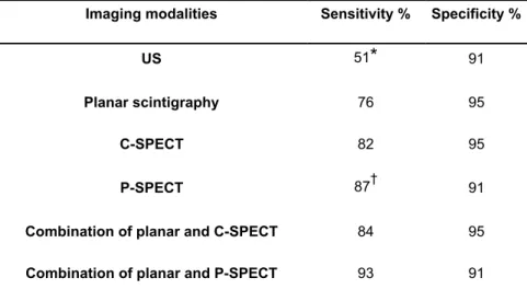
The sensitivities of US and scintigraphic procedures are reported in Table 1. US showed the lowest sensitivity compared with all scintigraphic procedures (p < 0.05), detecting only 28/55 lesions (51%). Of the 27 FN for US imaging, 21 glands (78%) were ≤ 0.5 g and 7 glands were ectopic (64% of all ectopic glands).
Planar scintigraphy identified 42/55 lesions (76%) whereas C-SPECT and P-SPECT detected respectively 45/55 (82%) and 48/55 (87%) of the abnormal glands. Of the 13 FN for planar scintigraphy, 10 concerned glands ≤ 0.5 g and 10 of these lesions were solitary.
Imaging by C-SPECT and especially by P-SPECT improved the detection rate with detection of 4/13 and9/13 of these lesions respectively. The 5 adenomas detected only by P-SPECT ranged from 0.3 to 1.9 g (4 glands weighing ≤ 0.6 g) and measured from 13 to 27 mm in diameter (median: 18 mm). Four lesions were not detected by planar and tomographic (C-SPECT and P-(C-SPECT) images. Three of these lesions were hyperplastic lesions with weights
of 0.1-0.2 g, associated with identified others lesions. The fourth lesion not detected by scintigraphy was an inferior adenoma of 3 g located in the continuity of the thyroid lobe and considered as thyroidal by planar scintigaphy, C-SPECT and P-SPECT.
All ectopic lesions were detected by both planar scintigraphy and C-SPECT, whereas P-SPECT was negative in 2/11 cases. In the first case, a deep mediastinal lesion was not in the field of view of the pinhole, and in the second, a misalignment of the pinhole collimator relative to the thyroid was responsible for the non-visualization of the pathological gland. Except for these 2 cases, the use of parallel hole collimator (C-SPECT) instead of pinhole collimator (P-SPECT) did not yield any additional information.
Despite a lower sensitivity compared with SPECT, planar scintigraphy and US detected one additional lesion each and gave additional information about thyroid morphology.
US and planar imaging showed respectively 14 and 8 FP. The number of FP was higher with P-SPECT than for C-SPECT acquisition (14 vs 8). Among these 14 FP, 5 showed a CS = 2 and 9 a CS = 1. The FP with a CS = 2 were visualized in 4 patients with concomitant nodular goiter and in one patient treated by amiodarone with poor thyroid uptake and rapid washout of 99mTc-MIBI.
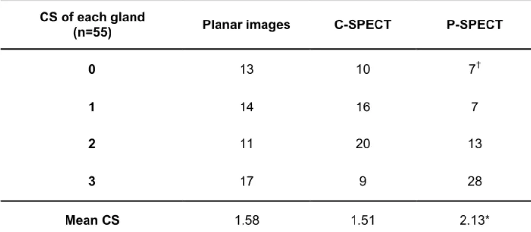
The diagnostic confidence score (CS) assigned to each gland with the different scintigraphic modalities is reported in Table 2. P-SPECT showed the highest mean CS among the 55 pathologic glands compared with planar and C-SPECT (p < 0.001). P-SPECT contributed to significantly reduce the number of negative or uncertain results (CS = 0 or 1, p < 0.01).
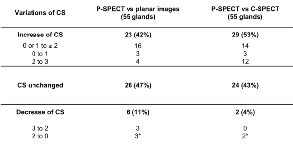
Table 3 summarizes the variations of CS on a per gland basis with SPECT. P-SPECT increased CS for 42% of parathyroid lesions when compared with planar imaging and
for 53% of parathyroid lesions when compared with C-SPECT. Fig. 1 illustrates the case of a right inferior orthotopic adenoma (0.4 g, 13_7_8 mm3) showing a very doubtful uptake on planar scintigraphy which appears clearly on SPECT and especially P-SPECT.
However, the CS of 6 glands was inferior with P-SPECT compared with planar imaging. In 3/6 cases, the reduction of CS was mild (from 3 to 2) and did not modify final diagnosis. In one case, a focus was assigned to thyroidal origin because of its location in the continuity of the inferior thyroid lobe on P-SPECT and C-SPECT but was correctly identified on dual tracer planar images. In the other two cases, the glands were not in the field of view of the pinhole collimator.
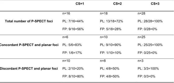
When the diagnosis is based on the CS value and a comparison between planar imaging and P-SPECT, the number of P-SPECT FP could be markedly reduced. Table 4 shows the CS assigned to each focus detected by P-SPECT and the results of comparison between P-SPECT and planar images. The probability of a parathyroid lesion visualization increased with CS (in particular, all the foci with a CS =3 corresponded to a parathyroid lesion) and in the case of an agreement between P-SPECT and planar images (95% vs 43% in case of discordance). In contrast, the probability of a P-SPECT FP was higher in the case of discordance between planar and P-SPECT, specially with CS =1 (80%).
DISCUSSION
Parathyroidectomy significantly improves the quality of life of patients suffering from pHPT and even those with mild elevated PTH level [5]. Consequently, more and more patients with mild pHPT are operated on. As glands responsible for mild pHPT are generally
small, conventional preoperative localization studies should be improved to allow minimally invasive surgery.
A pinhole collimator is routinely used to increase spatial resolution and improve sensitivity of planar parathyroid scintigraphy [12]. SPECT could certainly be optimized by the use of these collimators. Several technical studies performed on thyroid phantoms showed the feasibility of such explorations [15-16, 18, 21]. Clinical applications are rare because of the difficulties to use this method routinely.
Only two studies had evaluated P-SPECT in patients suffering from HPT. Profanter et al. [14] compared 99mTcO4- /201Tl pinhole substraction SPECT and US in 15 patients and
showed that P-SPECT was significantly more accurate than US in detection of pathological parathyroid glands. P-SPECT showed a sensitivity of 80% and a specificity of 93% with only one case of FP reported. Spanu et al. [13] showed that P-SPECT was a highly sensitive method, identifying more lesions than planar scans in 67 patients. Interestingly, P-SPECT was the only positive procedure in 8.9% of these patients. For the 48 patients explored for pHPT, the sensitivity of P-SPECT reached 98% (vs 88% for planar scans). Sensitivities of P-SPECT and planar scans observed in our patients were slightly lower (87% and 76%) but glands were smaller in our series (60% ≤ 0.5 g vs 30% in the series of Spanu). Spanu and collaborators used the filtered back-projection algorithm of Feldkamp, Davis and Kress which was shown to produce more artifacts compared to ML-EM based reconstruction for pinhole geometry [17]. Surprisingly, no FP results relative to artifacts were reported in their study. Unlike Spanu et al., we decided to consider accumulation in thyroidal structures or nodules as FP findings. FP were frequent with P-SPECT in our patients with a high prevalence of thyroid nodules (43%). With a higher spatial resolution compared with parallel collimation, irregularities of dystrophic thyroid lobes and thyroid nodules were easily detected due to the
better definition of thyroid contours and could be falsely interpreted as parathyroid lesions especially in case of heterogeneous and faint thyroid uptake. A faint focal uptake (CS = 1) unilateral or bilateral, was frequently observed in the upper and inside face of thyroid lobes. This corresponds probably to a thickening resulting from fusion of the ultimobranchial body into the principal medial thyroid process (Zuckerkandl's tuberculum) [22]. The higher level of FP due to the use of pinhole collimator for SPECT acquisition represents a potential drawback of SPECT interpretation. Nevertheless, FP could be reduced with a comparison between P-SPECT and planar imaging. The FP probability was only 10% in case of agreement with a CS = 2 and reached 80% in cases of discordance with a CS = 1. In practice, we concluded to a high probability of FP when a doubtful focus (CS = 1) of P-SPECT was not confirmed by planar views and particularly when one another evident focus was detected elsewhere. If no another focus was detected, the possibility of a small parathyroid lesion detected only by P-SPECT remained possible and a comparison with US was recommended.
Our study demonstrated a potential improvement of sensitivity of P-SPECT compared with conventional scintigraphic procedures. P-SPECT was able to confirm clearly doubtful results of C-SPECT or planar views. P-SPECT may thus replace C-SPECT, except in cases of major ectopy that could be previously detected on planar scintigraphy. A morphologic analysis of the thyroid (thyroid scintigraphy and US) remained useful. In particular, an US examination may reduce FP due to thyroid nodules [23] and FN caused by adenoma of a cystic nature or associated with concomitant hyperfunctionning thyroid nodules, which may be undetected by scintigraphy [11].
To our knowledge, this is the first study that compares SPECT acquisition with a parallel and a pinhole collimator for the same patient. For ethical reasons, we decided to perform the 2 studies after a single injection of 99mTc-MIBI, however this leads to a different
time delay between injection and acquisition for the 2 imaging procedures evaluated. C-SPECT was always performed before P-C-SPECT in order to minimize the time delay of the collimator change over 30 minutes and to not change the procedure performed routinely in our institution. This delay may have an impact on CS as it may depend on relative uptake and retention of 99mTc-MIBI by parathyroid and thyroid. Nevertheless, if many authors recommend an early acquisition (15-30 minutes) in order to avoid rapid washout of adenomas [1, 24], a significant difference between a 30 min and 60 min p.i SPECT imaging has not been yet demonstrated. As a consequence,it would be useful in additional comparative studies to alternate the 30 and 60 minutes starting times for C-SPECT and P-SPECT.
From a practical point of view, owing to the presence of the patient's shoulders, the use of a tilted geometry is essential in order to optimize the distance between the thyroid and the pinhole collimator [15]. We should emphasize that this geometry is not possible with the current generation of gamma cameras, and could limit clinical P-SPECT development in the future.
Furthermore, in our study, the algorithm used for P-SPECT is a straightforward implementation of the OSEM method for the pinhole geometry without any compensation for resolution recovery. This correction has been recently evaluated by two studies (using either a multi-ray approach [25] or a point-spread function based resolution recovery [26]) and was found to improve markedly the trade-off between spatial resolution and noise. This correction should be added in future algorithm development.
CONCLUSION
The use of a pinhole collimator for SPECT in pHPT increases the sensitivity of scintigraphy and is very useful to confirm doubtful results of conventional scintigraphy. P-SPECT gives
additional topographic information of benefit to the surgeon. Combined with double phase
99mTc-MIBI/99mTcO
4-, planar scintigraphy and neck US, P-SPECT appears to be a highly
accurate procedure to select patients for minimally invasive surgery.
ACKNOWLEDGEMENTS
The authors are very grateful to Pr. Michel Defrise from the Vrije Universiteit Brusselfor providing the reconstruction algorithm dedicated to pinhole geometry. We would like to thank also Dr. Samantha Warren for her very useful suggestions.
REFERENCES
1. Mariani G, Gulec SA, Rubello D, Boni G, Puccini M, Pelizzo MR, et al. Preoperative localization and radioguided parathyroid surgery. J Nucl Med 2003;44:1443-58.
2. Henry JF, Iacobone M, Mirallie E, Deveze A, Pili S. Indications and results of video-assisted parathyroidectomy by a lateral approach in patients with primary hyperparathyroidism. Surgery 2001;130:999-1004.
3. Arbab AS, Koizumi K, Toyama K, Arai T, Arak T. Ion transport systems in the uptake of 99Tcm-tetrofosmin, 99Tcm-MIBI and 201Tl in a tumour cell line. Nucl Med Commun 1997;18:235-40.
4. Carpentier A, Jeannotte S, Verreault J, Lefebvre B, Bisson G, Mongeau CJ, et al. Preoperative localization of parathyroid lesions in hyperparathyroidism: relationship between technetium-99m-MIBI uptake and oxyphil cell content. J Nucl Med 1998;39:1441-4.
5. Caillard C, Sebag F, Mathonnet M, Gibelin H, Brunaud L, Loudot C, et al. Prospective evaluation of quality of life (SF-36v2) and nonspecific symptoms before and after cure of primary hyperparathyroidism (1-year follow-up). Surgery 2007;141:153-9. 6. Moka D, Voth E, Dietlein M, Larena-Avellaneda A, Schicha H. Technetium 99m-MIBI-SPECT: A highly sensitive diagnostic tool for localization of parathyroid adenomas. Surgery 2000;128:29-35.
7. Biertho LD, Kim C, Wu HS, Unger P, Inabnet WB. Relationship between sestamibi uptake, parathyroid hormone assay, and nuclear morphology in primary hyperparathyroidism. J Am Coll Surg 2004;199:229-33.
8. Sharma J, Mazzaglia P, Milas M, Berber E, Schuster DM, Halkar R, et al. Radionuclide imaging for hyperparathyroidism (HPT): which is the best technetium-99m sestamibi modality ? Surgery 2006;140:856-63.
9. Lorberboym M, Minski I, Macadziob S, Nikolov G, Schachter P. Incremental diagnostic value of preoperative 99mTc-MIBI SPECT in patients with a parathyroid adenoma. J Nucl Med 2003;44:904-8.
10. Billotey C, Sarfati E, Aurengo A, Duet M, Mundler O, Toubert ME, et al. Advantages of SPECT in technetium-99m-sestamibi parathyroid scintigraphy. J Nucl Med 1996;11:1773-8.
11. Ansquer C, Mirallie E, Sadot S, Couturier O, Valette F, Kraeber-Bodéré F. Comparison of 99mTc-MIBI / 99mTc dual planar scintigraphy, 99mTc-MIBI SPECT and ultrasound in preoperative localization of parathyroid lesions for primary hyperparathyroididsm [abstract]. Eur J Nucl Med Mol Imaging 2005;31(suppl): S263. 12. Arveschoug AK, Bertelsen H, Vammen B. Presurgical localization of abnormal parathyroid glands using a single injection of Tc-99m sestamibi: comparison of high-resolution parallel-hole and pinhole collimators, and interobserver and intraobserver variation. Clin Nucl Med 2002;27:249-54.
13. Spanu A, Falchi A, Manca A, Marongiu P, Cossu A, Pisu N, et al. The usefulness of neck pinhole SPECT as a complementary tool to planar scintigraphy in primary and secondary hyperparathyroidism. J Nucl Med 2004;45:40-8.
14. Profanter C, Gabriel M, Wetscher GJ, Gadenstatter M, Mittermair R, Moncayo R, et al. Accuracy of preoperative pinhole subtraction single photon emission computed tomography for patients with primary and recurrent hyperparathyroidism in an endemic goiter area. Surg Today 2004;34:493-7.
15. Seret A, Defrise M, Blocklet D. 180 degree pinhole SPET with a tilted detector and OS-EM reconstruction: phantom studies and potential clinical applications. Eur J Nucl Mol Imaging 2001;28:1836-41.
16. Wanet PM, Sand A, Abramovici J. Physical and clinical evaluation of high-resolution thyroid pinhole tomography. J Nucl Med 1996;37:2017-20.
17. Vanhove C, Defrise M, Franken PR, Everaert H, Deconinck F, Bossuyt A. Interest of the ordered subsets expectation maximization (OS-EM) algorithm in pinhole single-photon emission tomography reconstruction: a phantom study. Eur J Nucl Med Mol Imaging 2000;27:140-6.
18. Carlier T, Bodet-Milin C, Oudoux A, Defrise M, Seret A, Couturier O, et al. Technical feasibility of pinhole SPECT acquisition in primary hyperparathyroidism: phantoms and patients studies [abstract]. J Nucl Med 2006;47(suppl):378P.
19. Smith MF, Jaszczak RJ. Penetration and angle-dependent sensitivity for pinhole collimation. Med Phys 1997;24:1701-9.
20. http://lib.stat.cmu.edu/R/CRAN, accessed on March 28th, 2007
21. Palmer J, Wollmer P. Pihnole emission computed tomography: method and experimental evaluation. Phys Med Biol 1990;35:339-50.
22. Chevallier JM, Martelli H, Wind P. Surgical discovery of parathyroid glands and the recurrent laryngeal nerve. Application of well known embryological concepts in the operating room. Ann Chir 1995;49:296-304.
23. Freudenberg LS, Frilling A, Sheu SY, Görges R. Optimizing preoperative imaging in primary hyperparathyroidism. Langenbecks Arch Surg 2006;391:551-6.
24. Rubello D, Gross MD, Mariani G, Al-Nahhas A. Scintigraphic techniques in primary hyperparathyroidism: from pre-operative localisation to intra-operative imaging. Eur J Nucl Med Mol Imaging 2007;926-33.
25. Vanhove C, Andreyev A, Defrise M, Nuyts J, Bossuyt A. Resolution recovery in pinhole SPECT based on multi-ray projections: a phantom study. Eur J Nucl Med Mol Imaging 2007;34:170-80.
26. Sohlberg A, Watabe H, Zeniya T, Iida H. Comparison of multi-ray and point-spread function based resolution recovery methods in pinhole SPECT reconstruction. Nucl Med Commun 2006;27:823-7.
Fig. 1. Patient without nodular thyroid disease (A) suffering from pHPT (PTH = 110 pg/mL)
caused by a right inferior orthotopic adenoma. Very doubtful focus indicated by the arrows on
99mTc-MIBI planar scintigraphy (B, CS = 1) and evident focus on C-SPECT (C, CS=2) and
Table 1. Sensitivities and specificities of US and scintigraphic procedures, alone or
combined.
Imaging modalities Sensitivity % Specificity %
US 51* 91
Planar scintigraphy 76 95
C-SPECT 82 95
P-SPECT 87† 91
Combination of planar and C-SPECT 84 95 Combination of planar and P-SPECT 93 91
*Significantly lower than other imaging modalities: p < 0.05 (Mac Nemar test) † Including the 2 parathyroid lesions outside the field of view of the pinhole collimator
Table 2. Diagnostic confidence score (CS) of imaging modalities
CS of each gland
(n=55) Planar images C-SPECT P-SPECT
0 13 10 7†
1 14 16 7
2 11 20 13
3 17 9 28
Mean CS 1.58 1.51 2.13* †Including the 2 parathyroid lesions outside the field of view of the pinhole collimator * Mean CS significantly higher than the others mean CS: p < 0.001 (Wilcoxon test)
Table 3. Variations of diagnostic confidence score (CS) for each lesion with the
additional use of P-SPECT
Variations of CS P-SPECT vs planar images(55 glands) P-SPECT vs C-SPECT(55 glands) Increase of CS 23 (42%) 29 (53%) 0 or 1 to ≥ 2 0 to 1 2 to 3 16 3 4 14 3 12 CS unchanged 26 (47%) 24 (43%) Decrease of CS 6 (11%) 2 (4%) 3 to 2 2 to 0 3 3* 0 2*
Table 4. Probability occurrence of parathyroid lesion (PL) or false positive (FP) for
P-SPECT according to diagnostic confidence score (CS) and planar scans
CS=1 CS=2 CS=3
Total number of P-SPECT foci
n=16 PL: 7/16=44% FP: 9/16=56% n=18 PL: 13/18=72% FP: 5/18=28% n=28 PL: 28/28=100% FP: 0/28=0%
Concordant P-SPECT and planar foci n=6 PL: 5/6=83% FP: 1/6=17% n=10 PL: 9/10=90% FP: 1/10=10% n=25 PL: 25/25=100% FP: 0/25=0%
Discordant P-SPECT and planar foci n=10 PL: 2/10=20% FP: 8/10=80% n=8 PL: 4/8=50% FP: 4/8=50% n=3 PL: 3/3=100% FP: 0/3=0%
C D