Acute experimental glomerulonephritis induced by the glomerular
deposition of circulating polymeric IGA-concanavalin A complexes
Davin J-C[1], Dechenne C[2], Lombet J[1], Rentier B[3], Foidart J.B[2], Mahieu P.R[2]
[1] Department of Pediatrics,[2] Department of Nephrology,[3] Department of Experimental Virology, State University of Liege, Belgium
Summary
The perfusion of polymeric or secretory IgA-Concanavalin A complexes into the aorta of rats led to a mannose-dependent binding of both IgA and lectin to the glomerular capillary wall, as shown by double immunolocalization experiments, by quantitative analysis of the amount of radiolabeled complexes bound per g of kidney, and by blocking experiments with the corresponding carbohydrate. Rats injected with amounts of those complexes as low as 500 μg developed, one hour later, a focal and segmental proliferative glomerulonephritis characterized by the deposition of injected complexes and of rat C3 and rat fibrin/ fibrinogen in most glomeruli ; focal thrombosis and small areas of necrosis in 10 to 15% of glomeruli, confined to the periphery of a single lobule of the tuft and segmental
infiltration of these glomeruli by polymorphonuclear leucocytes and platelets. At the same time, many mesangial cells exhibited a hyperactive appearance, and red blood cells were noted in tubular lumens. In contrast, rats similarly injected with either monomeric IgA-ConA complexes, multimeric or secretory IgA-peanut agglutinin complexes or polymeric or monomeric IgA aggregates of comparable apparent molecular weight did not develop obvious glomerular lesions within one hour. The data indicate that preformed polymeric IgA-ConA complexes can specifically bind to glomerular structures in vivo and trigger acute glomerular lesions locally, analogous to those observed in some glomerular diseases associated with a cryoglobulinemia.
Introduction
Experimental and clinical investigations suggest that immunopathologically - mediated glomerulo-nephritis are the consequence of two major mechanisms (Dixon et al. 1971): the first occurs when antigens (of exogenous or endogenous origin) combine with serum antibodies to form circulating immune complexes, which secondarily deposit in the glomerulus; the second is triggered by local interactions between specific antibodies and antigenic determinants already present within the glomerular structure. Numerous clinical and histopathological observations indicate that both Berger's disease and Henoch-Schönlein purpura nephritis (IgA-as-sociated nephropathies) are most probably related to the former pathogenetic mechanism, the glomerular deposition of circulating IgA immune complexes (Clarkson 1987). However, contrarily to what occurs classically in acute glomerulonephritis mediated by IgG immune complexes and C3, the bouts of macroscopic haematuria in IgA-asso-ciated nephropathies frequently appear before the host is capable of mounting a T cell-derived antibody response against the infectious agents involved (Clarkson 1987). It is therefore likely that other mechanisms such as a polyclonal B cell activation (Goldman et al. 1987) must trigger the IgA antibody production and the episodes of haematuria in those nephropathies.
Lectins are carbohydrate-binding proteins that exhibit two critical properties: specificity for particular carbohydrates and bivalency or polyvalency (Barondes 1984). They have been found as endogenous components of many pathogenic bacteria (Christensen et al. 1985), viruses (Meager and Hughes 1977) or alimentary antigens (Freed DLF 1987) implicated in IgA-associated nephropathies. Through their carbohydrate-binding properties, some lectins are mitogenic substances which can directly activate B cells, and subsequently induce the secretion of immunoglobulins and of a wide variety of antibodies in vitro (Lis and Sharon 1981). However, the occurrence and the precise role of such a B cell activation during the acute phases of IgA nephropathies remain a matter of controversy (Sakai 1987). The IgA1 subtype of IgA contains both N-glycosidically and O-glycosidi-cally linked oligosaccharides on its heavy chain (Baenziger and Kornfeld 1974), which allows firm interactions with exogenous or endogenous lectins in vitro (Roque-Barreira and Campos-Neto 1985; Stockert et al. 1982). In addition, it has been shown that IgA1 purified from the serum of patients presenting with IgA-associated
nephropathies exhibits a higher degree of polymerization (Clarkson 1987) and a significantly higher binding capacity to Con-canavalin A (ConA) and to peanut agglutinin (PNA), two lectins specifically recognizing terminal oc-mannosyl (or α-glucosyl) and β-galactosyl residues respectively, than to normal IgA1 (Davin et al. 1987b). Accordingly, one may wonder whether exogenous proteins exhibiting a lectin-like activity might not bind in vivo, as in vitro, directly to some of those "altered" IgA molecules. These interactions may lead to the formation of circulating non-immune complexes which could secondarily deposit into the glomeruli and trigger a local inflammatory reaction. Taken together, these considerations prompted us to test the possibility that preformed IgA-lectin complexes could accumulate into the glomeruli in vivo and induce glomerulonephritic changes in rats. To that purpose, we have first prepared IgA-lectin complexes by mixing in vitro equal amounts of either monomeric, polymeric or secretory human IgA with either ConA or PNA. These different complexes were then separated from free IgA or lectins by gel filtration chromatography and were thereafter injected in the aorta of rats. The examination of their kidneys one hour later by
immunofluorescence, photonic and transmission electron microscopy indicates that only polymeric IgA-ConA complexes can elicit an acute glomerular inflammatory reaction, as in the vasculitis seen in the Arthus reaction (Peltier 1979).
Materials and methods
Female Sprague-Dawley rats, weighing 200 to 250 g, were used for all the experiments. They were purchased from Iffa Credo Corporation (Lyon, France).
Three types of human IgA preparations were tested: (a) human monoclonal monomeric IgA (mIgA) was purified from a multi-meric IgA preparation (Gamma Co, Liege, Belgium) by gel filtration chromatography on an Ultrogel AcA 22 column (1 x 80 cm) (LKB, Bromma, Sweden) equilibrated and eluted with phosphate-buffered saline, ph 7.4 (PBS). The column was previously calibrated with human IgG, human fibronectin and human IgM purchased from Sigma Chemical Corporation (St-Louis, Missouri); (b) human polymeric IgA (pIgA) was purchased from Tago Co. (Burlingam, California); (c) human secretory IgA (sIgA) was from Sigma. Their apparent Mol.Wt. was determined by gel filtration chromatography on an Ultrogel AcA 22 column as described above. The mIgA molecules were eluted as standard IgG, whereas pIgA exhibited an apparent Mol.Wt. of about 800 Kd. Finally, the apparent Mol.Wt. of the human sIgA preparation used ranged from 380 to 800 Kd. Two lectins were purchased from Sigma: Concanavalin A (ConA) which specifically binds to α-D-mannosyl and α-D-glu-cosyl residues, and peanut agglutinin (PNA) which specifically recognizes β-galactosyl residues. The apparent Mol.Wt. of both lectins, determined as described above, was about 400 Kd. Al-pha-methyl-mannoside and beta-methyl-galactoside were also from Sigma. All the other reagents used were of analytical grade.
Proteins were labelled with125I or with131I by the chlora-mine-T method of McConahey and Dixon (1966). Labelled IgA-ConA or IgA-PNA complexes were prepared by mixing for 1 h at 37° C in one ml of PBS, pH 7.4, containing 2 mM CaC12, equal amounts of125I-ConA or of125I-PNA with either mIgA, pIgA or sIgA labelled with131I. At the end of the incubations, complexes were separated from free IgA or lectins by gel filtration chromatography through the Ultrogel AcA22 column. The presence of ConA, PNA or IgA in the one-ml fractions collected was searched for by measuring their125I and13 ^-radioactivities in a gamma spectrometer exhibiting a counter efficiency of 83% (Rack Gamma II, Turku, Finland). Control experiments were conducted by mixing for 1 h at 37° C radiolabeled IgA and lectins in PBS containing either 100 mM α-methyl-mannoside or 100 mM β-methyl-galactoside. Unlabeled IgA-ConA or IgA-PNA complexes were similarly prepared, but in this case, the IgA content of eluted fractions was measured by Elisa and their lectin content by RIA, both assays being performed as previously described (Davin et al. 1987a; Malaise et al. 1987).
Monomeric or polymeric IgA were radiolabeled with125I to a specific activity of about 0.1 mCi/mg by the chloramine-T method of McConahey and Dixon (1966). Aggregation was performed by heating 10mg/ml of125I-IgA for 150 min at 63° C in 0.15 M Tris-buffered saline, pH 7.4. Soluble aggregates of pIgA or mIgA (apparent Mol.Wt.: ≈106 daltons at least) were isolated from non-aggregated IgA by gel filtration chromatography on the Ultrogel AcA 22 column. Aggregates of unlabelled pIgA or mIgA were similarly prepared and purified.
On the day of the study, rats were anesthetized with Inactin® (Byk, Konstanz, FRG) and the abdominal aorta was cannu-lated with a plastic catheter, placed just above the renal arteries. Before their injection, all the PBS or saline solutions were sterilized by Millipore filtration and cleared at 6000 rpm for 15 min in a Sorvall RC2-B centrifuge.They were then infused at a rate of 0.10 ml/min (total volume: about 0.5 ml). Animals were sacrificed one hour later and their kidneys were excised for morphological or
quantitative studies. Four groups of rats were studied by immunofluorescence, photonic and/or transmission electron microscopy:
Group 1: were injected with 100, 200 or 500 μg of either mIgA, pIgA or sIgA (n = 3 to 4 for each
experimental condition). In addition, three animals received 200 or 500 μg of monomeric or polymeric IgA aggregates.
Group 2: were perfused either with 100, 200 or 500 μg of ConA or PNA only or with 500 μg of ConA
or PNA followed in
5 min by 1 ml of 1M α-methyl-mannoside or of 1M β-methyl-galactoside (n = 3 for each experimental condition).
Group 3: were perfused with 100, 200 or 500 μg of the various IgA-lectin complexes exhibiting an
apparent Mol.Wt. of 106 daltons at least (n = 3 for each subgroup).
Group 4: (n = 4) underwent the same surgical protocol as the other groups, but received one ml of PBS
or saline solution only.
Two further groups of rats were used for the quantitative determination of renal binding of either radiolabeled IgA aggregates or radiolabeled IgA-lectins complexes. The first group was perfused with 100 or 500 μg of aggregated IgA (either monomeric or polymeric) labelled with125Iodine, followed by a perfusion of either 1 ml of 1M α-methyl-mannoside or 1 ml of 1M β-methyl-galactoside or by no perfusion (n = 3 for each subgroup).
The second group was perfused with 100, 200 or 500 μg of the various IgA-lectin complexes prepared by mixing either125I-IgA with cold lectins or125I-labelled lectins with cold IgA, as described above. To investigate the competitive removal of IgA-ConA from the glomeruli by its natural ligand, i.e. a-methyl-mannoside, we also perfused rats with 500 μg of125I-labelled ConA-IgA complexes followed in 5 min by 1 ml of isoosmotic solutions of either 1M α-methyl-mannoside or 1M β-methyl-galactoside (n
= 3 to 5 for each subgroup).
In all these experiments, paired label injections were performed using equal amounts of131I-bovine serum albumin (BSA) and of125I-labelled complexes or aggregates. The kidneys of injected animals were removed one hour later and homogenized in a Waring blender. The homogenate was washed three times in PBS and the final pellet was counted in the gamma scintillation counter. Correction for counts contributed by nonspecifically trapped BSA was made by a classical formula (Wilson and Dixon 1970), and the μg of protein accumulated per g of kidney was then calculated from the specific activity of each tracer used.
For light microscopy, the kidneys were fixed in Bouin's solution and embedded in paraffin; 4 μ sections were then stained with haematoxylin and eosin, periodic acid Schiff or Masson's trich-rome. For immunofluorescence kidney fragments were snap frozen in liquid nitrogen. Some frozen sections were first incubated for 30 min at room temperature with a rabbit anti-human α chain antiserum (Behring-Werke AG, Marburg, West Germany). After washing with PBS, they were thereafter stained with fluorescein isothiocyanate (FITC)-conjugated goat anti-rabbit IgG antibodies (Nordic, Tilburg, The Netherlands). Other sections were directly stained with FITC-conjugated antibodies specific for rat IgA, rat IgG, rat C3 and rat fibrin/fibrin-ogen (Nordic). Finally, the binding of lectins to kidney
structures was studied using ConA or PNA labelled with rhodamine (Nordic) and injected into the aorta either in a free or in a complexed form; in this latter case, IgA-ConA and IgA-PNA complexes were prepared as described above. Deposits seen by direct or indirect immunofluorescence were each time graded semiquantitatively from 0 to 3 + by two independent observers.
Kidneys to be examined by transmission electron microscopy were fixed by immersion in Karnovsky's fixative and then post-fixed for 1 h in 1% buffered osmic acid. After dehydration through a series of alcohols, tissues were embedded in Epon 812. Thin sections (500 to 700 A) were stained with uranyl acetate and lead citrate and were then examined in a Siemens 101 electron microscope.
Fig. 1. Gel filtration chromatography on an Ultrogel AcA 22 column of PNA-IgA complexes prepared by mixing for 1 h at 37° C equal amounts of unlabelled lectin and human monoclonal polymeric IgA (MW ≈ 800 Kd) in a phosphate-buffered saline solution, ph 7.4, in presence (b) or not (a) of α-methyl-galactoside 100 mM. The IgA and PNA concentration was measured in each fraction by Elisa (Davin et al. 1987 a) and RIA respectively (Malaise et al. 1987). The column was first calibrated using human IgM, fibronectin or IgG as standards. Note in (a) only the presence of a peak containing both IgA and PNA and corresponding to an apparent M.W. higher than that of IgM
Results
An example of elution profile of unlabelled IgA-lectin complexes is shown in Fig. 1. It must be noted that, after mixing with PNA, IgA fractions exhibiting an apparent Mol.Wt. of at least 900 Kd were obtained (Fig. 1 a). This high Mol.Wt. material really corresponded to IgA-PNA complexes since: (a) it was not observed after mixing IgA and PNA in the presence of 100 mM β-methyl-galactoside (Fig. lb) and (b) PNA antigens were detected by RIA in the same fractions (Fig. 1 a). Comparable elution profiles were obtained after the addition of ConA to the different IgA preparations tested (data not shown).
An example of elution profile of labelled IgA-ConA complexes is illustrated in Fig. 2. In this
representative experiment, equal amounts (100 μg) of 125I-ConA and of 131I-pIgA were mixed for 1 h at 37° C before gel filtration chromatography on the Ultrogel AcA 22 column.
Fig. 2. Gel filtration chromatography on an Ultrogel AcA 22 column of ConA-IgA complexes prepared by mixing for 1 h at 37° C equal amounts of125I-Concanavalin A (125I-ConA) and human monoclonal polymeric131I-IgA (131I-pIgA) (MW ≈800 Kd) in phosphate-buffered saline, ph 7.4, in presence (b) or
not (a) of α-methyl-mannoside 100 mM. In each fraction, the radioactivity of125I-ConA and131I-pIgA has been determined in a gamma spectrometer exhibiting a counter efficiency of 83% (Rack Gamma II, Turku, Finland). The column was first calibrated using human IgM, fibronectin or IgG as standards. Note in (a) only the presence of a peak containing both IgA and ConA and corresponding to an apparent M.W. higher than that of IgM
Here again, a peak containing both 125I- and 13 I-radioactivities
was eluted in the void volume and exhibited an apparent Mol.Wt. of 900 Kd at least (Fig. 2 a). This peak was not observed when similar experiments were conducted in the presence of 100 mM α-methyl-mannoside (Fig. 2b). Comparable elu-tion profiles were found after mixing 125I-PNA with 131I-IgA molecules. The yield of labelled ConA-IgA complexes ranged from 5 to 11 % (n = 7), whereas that of labelled PNA-IgA complexes ranged from 12 to 25% (n = 8).
On immunofluorescence microscopy in Group 1 glomerular deposits were not seen in rats having received 100, 200 or 500 μg of either human mIgA, pIgA or sIgA one h before sacrifice (data not shown). However, the animals injected with 500 μg of aggregates of human polymeric IgA displayed weak mesangial IgA deposits without concomittant deposition of either rat IgA, rat IgG, rat C3 or rat fibrin/fibrinogen (Table 1).
As expected on the basis of previous studies (Golbus and Wilson 1979; Holthöfer 1983) in Group 2 animals PNA did not bind to glomerular structures whereas rats perfused with ConA exhibited a nearly continuous deposition of this lectin along the glomerular capillary wall (data not shown), this binding being strongly reduced by a subsequent infusion of 1 ml of an isoosmotic solution of α-methyl-mannoside, - but not of β-methyl-galactoside - (data not shown). No glomerular deposits of rat IgA, rat IgG, rat C3 or rat fibrin/ fibrinogen were observed in ConA- or in PNA-injected animals (Table 2). In Group 3, rats perfused with 100, 200 or 500 μg of IgA-PNA complexes did not present with obvious mesangial glomerular deposits (Table 3). In contrast, the intraaortic infusion of IgA-ConA complexes was followed by the deposition of those complexes in the capillary lumens of most glomeruli (80% at least), the pattern of staining being identical for both IgA and ConA, as shown by double
immunolocalization experiments (Fig. 3 a).
IgA aggre gates
Number of rats Amount injected (μg) Immunofluorescence microscopy (glomerular deposits)a
Human Rat
IgA IgA C3 IgG Fibrin
Mono meric 3 200 0 0 0 0 0 Mono meric 3 500 + 0 0 0 0 Poly meric 3 200 + 0 0 0 0 Poly meric 3 500 + + 0 ± 0 0 a
The intensity of fluorescence was arbitrarily estimated using a scale from 0 to 3 +. The glomerular deposits exhibited a mesangial granular pattern
Table 2. Immunofluorescence microscopy of kidneys from rats injected with lectins (group 2)
Lecti n inject ed
Number of rats Amount injected (μg) Immunofluorescence microscopy (glomerular deposits)a
Lectin Rat
IgA C3 IgG Fibrin
PNA 3 100 0 0 0 0 0 3 200 0 0 0 0 0 3 500 0 0 0 0 0 CON A 3 100 + 0 0 0 0 3 200 + 0 0 0 0 3 500 2 + 0 0 0 0 a
The intensity of fluorescence was arbitrarily estimated using a scale from 0 to 3 +. The glomerular deposits exhibited a nearly continuous pattern along the glomerular capillary wall, as already observed in a previous study (Golbus and Wilson 1979)
Table 3. Immunofluorescence microscopy of kidneys from rats injected with IgA-lectin complexes (group 3)
Complexes injected Number
of rats IgA form Amount injected (μg)
Immunofluorescence microscopy (glomerular deposits)a
Lectin Human Rat
IgA IgA C3 IgG Fibrin
IgA-Con A 3 Monom eric 100 ± ± 0 0 0 0 3 Monom eric 200 ± ± 0 0 0 0 3 Monom eric 500 + + 0 0 0 0 IgA-Con A 3 Polyme ric 100 + + 0 ± 0 0 3 Polyme ric 200 2 + 2 + ± + ± ± 3 Polyme ric 500 3 + 3 + + 2 + + 2 + IgA-PNA 3 Monom 500 0 0 0 0 0 0
eric 3 Polyme ric 200 0 0 0 0 0 0 3 Polyme ric 500 + + 0 0 0 0 a
The intensity of fluorescence was arbitrarily estimated using a scale from 0 to 3 +
In addition, all the rats injected with 500 μg of pIgA-ConA complexes displayed a glomerular
accumulation of rat C3 and rat fibrin/fibrinogen (Table 3). The pattern of staining of those endogenous plasma components was generally similar to that of IgA-ConA complexes. Finally, in two out of 3 rats having received 500 μg of pIgA-ConA complexes, weak granular deposits of rat IgG and rat IgA were observed in about 15% of the glomeruli containing pIgA-ConA complexes as well. Their pattern of distribution was also comparable to that of those complexes (data not illustrated). None of the animals in Group 4 exhibited any glomerular deposits.
The quantities of 125I-ConA or of 125I-IgA specifically bound to rat kidneys 60 min after perfusion of either 125I-pIgA-ConA complexes or 125I- ConA-pIgA complexes increased according to the dose injected (Table 4). Perfusion of 500 μg of labelled ConA-pIgA resulted in a binding of 69.2 μg of ConA/g of kidney and of 42.6 μg of pIgA/g of kidney, which was about 10-fold greater than the amount of pIgA aggregates specifically bound to the perfused kidneys under otherwise identical experimental conditions (111.8 μg of IgA-ConA vs. 12.1 μg of pIgA per g) (Table 5). In control rats, the perfusion of 500 μg of radiolabelled pIgA-ConA complexes was followed by perfusion with α-methyl-mannoside or with β-methyl-galactoside. The former carbohydrate, - but not the latter -, induced a strong decrease in the amount of IgA-lectin complexes specifically bound to perfused renal tissue (Table 4). Interestingly, the amount of 125I-labelled pIgA aggregates specifically bound to rat kidneys was also reduced by a subsequent infusion of α-methyl-mannoside, - but not of β-methyl-galactoside - (Table 5).
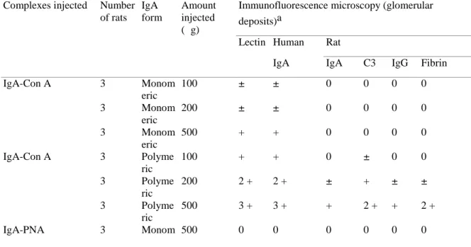
Fig. 3. (a) Immunofluorescence microscopy of a glomerulus from a rat injected intra-aortically with 500 μg of human pIgA-ConA complexes. Note the presence of IgA deposits in a vascular and
mesangial pattern after staining with a rabbit anti-human α chain antiserum and FITC-conjugated goat anti-rabbit IgG antibodies (original magnification, x 300). Deposits of rhodamin-conjugated ConA exhibited exactly the same staining pattern (data not shown), (b-f) Light microscopy of glomeruli from
a rat injected with 500 μg of human pIgA-ConA complexes. Note the presence of a fibrinoid thrombus
(arrow) confined to the periphery of a single lobule of the glomerular tuft (b), segmental and focal
mesangial enlargements occluding some capillary lumens (c), a polymorphonuclear cells infiltration
(arrows) (d) the hypercellularity of a glomerulus (e), and the almost normal aspect of another in which
one polymorphonuclear leucocyte only is seen and in which the capillary lumens remain empty (f) (Tri. original magnification, b, c, e, f x 200; d x 400
Table 4. Amounts of125I-Con A (*Con A) or of125I-pIgA (*p!gA) bound to perfused renal tissue
Experimental conditions Amount infused (μg) 125I-Con A bound" (μg/g of kidney)
125
I-pIgA bounda (μg/g of kidney)
*Con A-pIgA 100 12.1+ 4.6b Not done
200 28.9+ 6.2 Not done
500 69.2+14.7 Not done
Con A-*pIgA 100 Not done 7.6 + 2.4
200 Not done 19.2 + 4.1
500 Not done 42.6 + 9.5
*Con A-pIgA followed by α-methyl-mannoside
500 14.6+ 2.9 Not done
Con A-*pIgA followed by α-methyl-mannoside
500 Not done 9.2 + 3.1
a
Correction for counts contributed by nonspecifically trapped proteins was made by the simultaneous injection of an equal amount of131I-BSA (see the Material and Methods section)b Values are means + SEM (n = 3 to 5)
Table 5. Amounts of125I-labelled IgA aggregates bound to perfused renal tissue
Experimental conditions IgA form Amount infused (μg) 125I-IgA bounda(μg/g of
kidney)
125I-IgA aggregates Monomeric 100 1.2 + 0.2b
Monomeric 500 2.1 + 1.4
125I-IgA aggregates Polymeric 100 2.9 + 0.4
Polymeric 500 12.1 + 1.8
125
I-IgA aggregates followed by α-methyl-mannoside
Polymeric 500 3.2+1.4
125I-IgA aggregates followed by /3-methyl-galactoside
Polymeric 500 14.2 + 2.8
a
Correction for counts contributed by nonspecifically trapped proteins was made by the simultaneous injection of an equal amounts of131I-BSA (Wilson and Dixon 1970)b Values are means + SEM (n = 3)
No histological lesions were observed in the glomeruli of rats from groups 1, 2 and 4. In contrast, in the rats injected with 500 μg of either pIgA-ConA complexes or sIgA-ConA complexes (group 3), ten to 15 per cent of glomeruli exhibited obvious lesions, which were never noted in the animals having received either 100 μg of pIgA-ConA complexes, 500 μg of pIgA-PNA complexes or 500 μg of pIg A (or mIgA) aggregates. These lesions were characterized consistently by the presence of: (a) hyaline thrombi and fibrinoid material occluding some capillary lumens (Fig. 3 b); (b) segmental and focal mesangial enlargements (Fig. 3 c); (c) a mild polymorphonuclear leucocyte infiltration (Fig. 3 d); (d) some degree of hypercel-lularity (Fig. 3e). These appearances were therefore quite different from those observed in the majority of glomeruli from the same rats. Indeed, in those latter glomeruli, no obvious hypercellularity nor mesangial enlargement was noted, and their capillary lumens appeared "empty" (Fig. 3f).
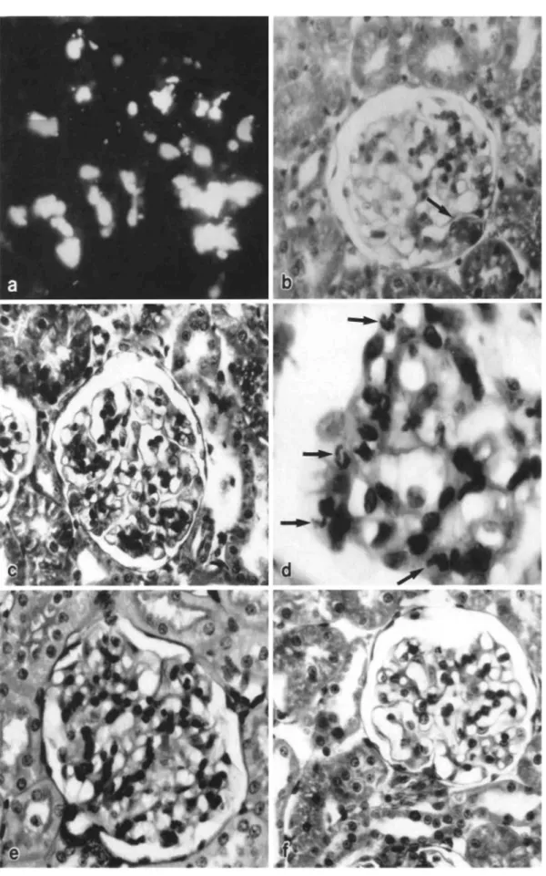
Electron microscopy examination of kidneys from animals of groups 1, 2 and 4 did not reveal any obvious ultrastructural alterations. Similarly, no major lesions were noted in the rats from group 4 injected with the various IgA-PNA complexes. In contrast, the involved glomeruli from animals injected with 200 or 500 μg of either pIgA-ConA or sIgA-ConA complexes presented the following ultrastructural features: (a) the presence of fibrinoid thrombi and degranulated polymorphonuclear leucocytes in the capillary lumens (Fig. 4); (b) the existence of platelets in viscous metamorphosis in
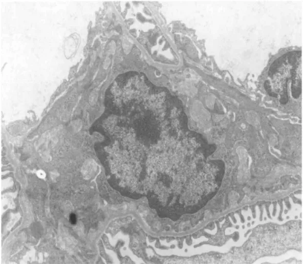
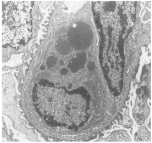
the same capillary lumens (Fig. 4); (c) an infiltration of some glomeruli by mononuclear cells containing many large vacuoles filled with fibrinoid material (Fig. 5); (d) a "hyperactive" aspect of mesangial cells, which exhibited an increased number of polyribosomes, a well-developped rough endoplasmic reticulum and a basement membrane-like matrix overload (Fig. 6); (e) a "phagocytic aspect" of some endothelial cells, which contained large cytopasmic vacuoles and a well-developped rough endoplasmic reticulum (Fig. 7); (f) red blood cells in some tubular lumens (Fig. 8). No deposits of osmiophilic material were either seen within the basement membrane or the mesangium. No clear cristalloid structures were observed in the protein thrombi.
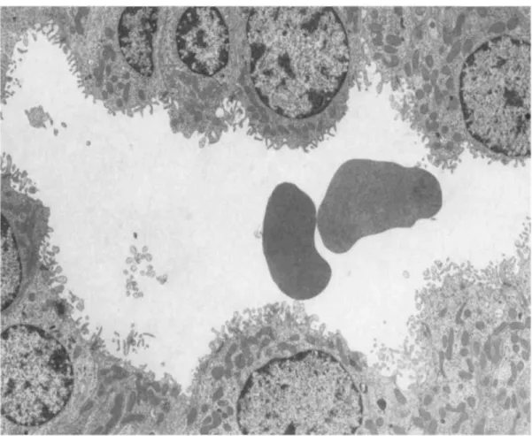
Fig. 4. Electron microscopy of a glomerular capillary loop from a rat intra-aortically injected with 500
μg of human pIgA-ConA complexes. Note the presence of a degranulated polymorphonuclear leucocyte
(*), platelets (►) and of fibrinoid material (*) (Ur.Pb. original magnification, x 3000)
Discussion
We have shown that non-immune complexes containing human IgA and ConA have the capacity to bind to glomerular structures readily after their intra-aortic injection into Sprague-Dawley rats. Indeed, double immunolocalization experiments show granular deposits of both human IgA and ConA in most glomeruli, these deposits being mainly localized in the capillary loops. In contrast, kidneys similarly perfused with human IgA aggregates of comparable size (≈106 daltons at least) only displayed weak
glomerular IgA deposition.
Fig. 5. Electron microscopy of a mononuclear phagocyte in a glomerular capillary loop from a rat intra-aortically injected with 500 μg of human pIgA-ConA complexes. Note the presence of numerous vacuoles containing fibrinoid material (►) (Ur.Pb. original magnification, x 6000)
This suggests that the glomerular accumulation of IgA-ConA complexes is not related to their molecular size alone. It is well known that ConA can bind to mannosyl and/or glucosyl residues (Lis and Sharon 1973) present in the structural glycoproteins (Spiro 1967) of the glomerular capillary wall (Bretton and Bariety 1974). This property has been subsequently utilized to "plant" an antigen in the glomeruli of rats and to induce an experimental glomerulonephritis by in situ formation of immune complexes (Golbus and Wilson 1979). Proof that the glomerular accumulation of IgA-ConA complexes depends, at least in part, on similar interactions is afforded by blocking experiments with the
corresponding carbohydrate. We have noted that the intraaortic perfusion of αmethylmannoside, -but not of β-methyl-galactoside -, significantly decreased the amount of both radiolabeled IgA and ConA specifically bound to the perfused renal tissue. This also implies that even after their interaction with IgA molecules, some mannosyl-bind-ing sites of ConA remain available for bridging formation with the glomerular capillary wall, which is not surprising since ConA molecules are tetrameric and accordingly possess sixteen manno-syl-binding sites (Kalb and Lustig 1968). Previous studies have demonstrated that, in contrast with ConA, the PNA lectin cannot bind to rat glomerular structures in vitro (Holthδfer 1983). We have made the same observation in vivo and, in addition, we have found that rats injected with 500 μg of preformed pIgA-PNA complexes exhibiting, as pIgA-ConA
complexes, an apparent Mol.Wt. of ≈106 daltons only displayed weak glomerular deposits of both IgA and PNA. This result adds another proof that the glomerular binding of pIgA-lectin complexes is not
related to their size alone, but also to the glycoprotein-binding properties of the lectin present in those complexes.
It has been shown that mIgA-ConA complexes prepared in vitro did not accumulate in kidneys to the same extent as pIgA-ConA complexes of similar molecular size. The kidney uptake of soluble pIgA aggregates was also significantly higher in our experimental conditions than that of soluble mIgA aggregates exhibiting comparable Mol.Wt. These results confirm (Egido 1987) that the presence of pIgA in IgA aggregates is important for their deposition at sites susceptible to injury. In addition, we have noted that the uptake of pIgA aggregates by the perfused renal tissue, like that of pIgA-ConA complexes, was significantly reduced by a subsequent perfusion of α-methyl-man-noside, - but not of β-methyl-galactoside. These data therefore support the possibility that the renal accumulation of pIgA aggregates, and probably also of some pIgA-ConA complexes may involve lectin-like interactions between terminal mannosyl residues of pIgA molecules and endogenous tissue moieties with ConA-like activity, as has been already suggested for IgG antibody molecules bearing terminal mannosyl residues (Winkelhalke and Nicolson 1976). If such it is the case, one can speculate that the different ability of polymeric and monomeric IgA aggregates to accumulate into the glomeruli might be due, at least in part, to the greater number of accessible mannosyl residues in pIgA than in mIgA molecules (Davin et al., manuscript submitted for publication).
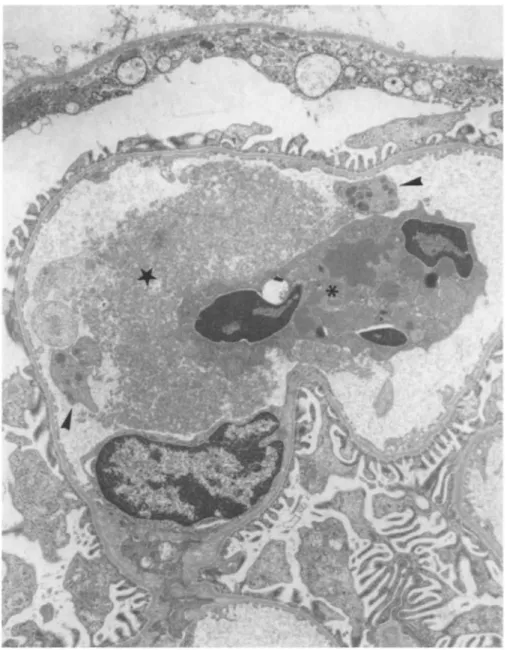
Fig. 6. Electron microscopy of a mesangial area from a rat injected intra-aortically with 500 μg of human pIgA-ConA complexes. Note the hyperactive aspect of the mesangial cell containing a well-developed rough endoplasmic reticulum and numerous nodules of basement membrane-like material (Ur.Pb. original magnification, x 6000)
We have noted that the intra-aortic infusion of pIgA-ConA complexes was followed within one hour by morphological changes in rat kidneys, depending on the dose of preformed complexes bound per g of renal tissue, as has been already noted in experimental immune complex-mediated glomerular diseases (Dixon et al. 1971). By contrast, no histological effects resulted within one hour when similar amounts of monomeric or polymeric IgA aggregates, of pIgA-PNA complexes or of mIgA-ConA complexes were injected into the aorta of Sprague-Dawley rats. Fries et al. (1988) have recently observed that the
injection of ConA-ferritin complexes into the renal arteries of' Sprague-Dawley rats is not followed by the development of a glomerulonephritis, despite the deposition of ferritin on the glomerular
endothelial cell surface and in the lamina rara interna. These data and our observations therefore support the concept that pIgA-ConA complexes can specifically induce, as immune-complexes, acute glomerular lesions in rats.
No haematuria nor proteinuria were detected in our rats within one hour (the interval between the injection of pIgA-ConA complexes and the killing of the animals). However, it has been shown that red blood cells were consistently present in the tubular lumens of rats having received 500 μg of pIgA-ConA complexes, but not in the tubular lumens of the other groups of rats (Table 6). This strongly suggests that the injection of 500 μg of pIgA-ConA complexes at least is necessary for inducing some alterations in rat glomerular function.
The main glomerular changes triggered by the infusion of pIgA-ConA complexes into the aorta of rats were characterized by: (a) the deposition of pIgA and ConA, of rat fibrin/fibrinogen and of rat C3 in most glomeruli; (b) the infiltration of 15% of glomeruli by polymorphonuclear leucocytes, platelets and mononuclear cells; (c) focal thromboses, as well as small areas of necrosis located at the periphery of the glomerular tufts.
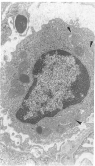
Fig. 7. Electron microscopy of a capillary loop from a Sprague-Dawley rat injected with 500 μg of human pIgA-ConA complexes. Note the phagocytic appearance of an endothelial cell displaying large cytoplasmic vacuoles numerous polyribosomes and a well-developed rough endoplasmic reticulum (Ur.Pb. original magnification, x 6000)
The intravenous injection of small amounts of antigen in hyperimmunized animals induces the formation of insoluble immune complexes triggering an intravascular coagulation syndrome (Vas-salli and McCluskey 1971). This syndrome is accompanied by fibrin deposition in the capillary beds of the spleen, the liver and the glomerulus. In addition, focal thrombosis is then observed in glomeruli by photonic, immunofluorescence and transmission electron microscopy (Hepstinstall 1966). These data suggest that pIgA-ConA complexes behave as insoluble or poorly-soluble immune complexes and can induce an "intravascular coagulation" mainly affecting the glomeruli after their intra-aortic injection. Indeed, fibrin/fibrino-gen deposits were not found in the spleen and the liver of our rats (Davin et al.,
unpublished data).
The presence of C3 in glomerular lesions has been reported in many cases of human or experimental glomerulonephritis including IgA nephropathies (Sinniah 1987; Emancipator 1987). We have found that glomerular C3 deposits associated with a polymorphonuclear leucocytes infiltration of glomeruli were noted consistently only in rats injected with 500 μg of pIgA-ConA complexes. It is therefore likely that a complement activation occurs in those animals. Some lectins can activate the complement system through the alternate pathway (Freed 1987). However, the injection of ConA alone was not followed by a glomerular deposition of C3. Previous studies have shown that IgA-im-mune complexes containing polymeric IgA activate the complement system in vitro more intensively than those containing monomeric IgA (Rits et al. 1987). We have noted that the intra-aortic injection of either pIgA or mIgA aggregates did not induce a major glomerular deposition of C3 in our experimental conditions. Taken together, these data suggest that the glomerular accumulation of C3 in rats having received pIgA-ConA complexes most probably results from an activation of the complement system by the complexes themselves rather than by each partner of these complexes. The mechanisms involved are now under study, as well as the possible role of such an activation in the induction of glomeruli lesions.
In addition to rat fibrin/fibrinogen and to rat C3, glomeruli from some rats given pIgA-ConA
complexes contained weak granular deposits of rat IgA and rat IgG. Such deposits were never found in the animals injected with preformed mIgA-ConA or PNA-IgA complexes.
Fig. 8. Electron microscopy of a kidney from a rat intraortically injected with 500 μg of human pIgA-ConA complexes. Note the presence of red blood cells in tubular lumens (Ur.Pb. original
magnification, x 2400)
It has been shown (Golbus and Wilson 1979) than ConA perfusion into the aorta of rats followed by the injection of normal rat serum did not induce the formation of granular deposits of rat IgG or rat IgA in glomeruli, despite the presence of 0 to 2+ coarse granular deposits of ConA along the glomerular capillary wall. It is therefore likely that the glomerular accumulation of those plasma components in our rats is not related to the glycoprotein-binding properties of ConA, but most probably results from
their non-specific "trapping" into the capillary loops due to focal thrombosis. Indeed, it must be recalled that glomerular deposits of rat IgG and of rat IgA were mainly present in the area of necrosis located at the periphery of glomerular tufts. In addition to polymorphonuclear leucocytes, the glomeruli from rats injected with pIgA-ConA complexes, - but not with pIgA aggregates -, contained many platelets in viscous metamorphosis, as well as mononuclear cells.
Table 6. Renal deposition of IgA-Con A complexes and presence of red blood cells (RBC) in tubular lumens
Groups of rats Number of rats IgA form Amount (μg) injected Number of rats
with RBC in tubular lumens
IgA-Con A 4 Monomeric 100 0
4 Monomeric 500 1
4 Polymeric 100 0
4 Polymeric 500 4
IgA aggregates 4 Monomeric 100 0
4 Monomeric 500 0
4 Polymeric 100 0
4 Polymeric 500 0
Platelets in glomerular capillary lumens have also been described in experimental immune complex-mediated glomerulonephritis in which ConA is used either as an antigen (Johnson et al. 1987) or as a mean to "plant" ferritin on the endothelial side of glomerular basement membranes (Fries et al. 1988), before the subsequent injection of anti-ConA or of anti-ferritin antibodies, respectively. It is
conceivable that platelets accumulated into the glomeruli of our rats might participate in the local inflammatory reaction by an in situ release of mediators such as PAF or 12-HETE which are capable of modulating some glomerular functions (Camussi and Brentjens 1988). However, the possibility remains that platelets infiltrating the capillary tuft early in the course of the disease process might also participate in the removal of some pIgA-ConA complexes, as has been already suggested for ferritin-anti-ferritin antibodies assembled proximally on the endothelium or in the lamina rara interna of glomeruli from Sprague-Dawley rats (Fries et al. 1988). The eventual spontaneous reversibility of the glomerular lesions induced by pIgA-ConA complexes is now under investigation.
Electron microscopic examination of kidneys from rats injected with 500 μg of pIgA-ConA complexes showed the presence of mononuclear phagocytes filled with large vacuoles containing amorphous material consistently, as well as endothelial cells displaying a phagocytic appearance. These data support the possibility that the mononuclear phagocytes infiltrating the glomeruli may be involved in the local damage, as has been suggested in some human (Monga et al. 1979) or other experimental glomerulonephritis (Schreiner et al. 1978). In addition, a mesangial involvement in the rat model of glomerular disease described is suggested by the ultrastructural aspect of mesangial cells. The lack of electron dense deposits in the mesangium might result from the short delay between the injection of pIgA-ConA complexes and the sacrifice of animals. Indeed, this delay could be insufficient to allow complexes to rearrange locally and condense in order to become large enough to be detectable by transmission electron microscopy (Mannik and Gauthier 1984).
What is the most probable human counterpart of the acute glomerular changes induced by the
deposition of circulating pIgA-ConA complexes? Although clear cristalloid structures were not obvious in the protein thrombi present in the capillary lumens, many light, immunofluorescence and electron microscopy findings support the possibility that the glomerular lesions triggered by pIgA-ConA complexes are analogous to those observed in patients with cryoglobulinemia. Three types of cryoglobulins have been identified in man (Ponti-celli and D'Amico 1988). Type I are single monoclonal immunoglobulins (myeloma proteins and macroglobulins), whereas types II and III are mixed cryoglobulins, composed of at least two immunoglobulins. In both types II and III
cryoglobulins, a polyclonal IgG is bound to another immunoglobulin, which acts like an anti-IgG rheumatoid factor (Ponticelli and D'Amico 1988). Type III cryoglobulins are frequently associated with various diseases including Henoch-Schδnlein purpura (Garcia-Fuentes et al. 1977). It has been shown that IgAt purified from the serum of patients presenting with IgA-associated nephropathies exhibits a higher degree of polymerization (Clarkson 1987) and a higher lectin binding capacity (Davin et al. 1987b), than normal IgA1. This latter result probably reflects some degree of alteration in the oligosaccharide chains of IgA1. Although a lower concentration of sialic acid in the IgG, which could make the immunoglobulin hydrophobic and thus enhance its insolubility (Zinne-man et al. 1968), has
been found in cryoprecipi-tates, it has been demonstrated that the isolated components from mixed cryoglobulins cannot precipitate by themselves, but that both immunoglobulins must be present for cryoprecipitation (Mcintosh et al. 1971). Lectins have been found as endogenous components of many pathogenic bacteria or of viruses (Christensen et al. 1985; Meager and Hughes 1977). Henoch-Schönlein purpura nephritis usually shortly follow upper respiratory infections by infectious agents bearing lectin-like activity. One can thus postulate a sequence of events in which, during the course of an infection, exogenous proteins with lectin-like properties can bind into the circulation to accessible sugar residues of some "abnormal" pIgA molecules, and form large non immune-complexes which secondarily deposit into the glomeruli, as mixed cryoglobulins. In addition, we have observed that this deposition is mediated by carbohydrates-dependent mechanisms and by polymeric IgA. Since pIgA is also necessary for the induction of glomerular lesions, it seems reasonable to suggest that pIgA may be involved in the pathogenesis of these lesions.
Acknowledgements
We are indebted to Mrs Y. Pirard and A. Desoroux for their technical assistance and to Mrs M.-A. Delavignette and M. Beyer for their help in the preparation of the manuscript.
References
Baenziger J, Kornfeld S (1974) Structure of the carbohydrate units of IgA1 immunoglobulin. II. Structure of the O-glyco-sidically linked oligosaccharide units. J Biol Chem 22:7270—7281 Barondes SH (1984) Soluble lectins: a new class of extracellular proteins. Science 223:1159-1263 Bretton R, Bariety J (1974) Ultrastructural localization of Con-canavalin A in normal rat kidney-glomeruli and arterioles. J Ultrastruct Res 48:396-403
Camussi G, Brentjens JR (1988) Inflammatory mediators: in vitro assessment on cultured glomerular cells. In: Davison AM (ed) Nephrology. Bailliere Tindall, London, Philadelphia, Toronto, Sidney, Tokyo, pp 392-401
Christensen GD, Simpson WA, Beachey EH (1985) Microbial adherence infection. In: Mandell GL, Douglas RG, Bennet JE (ed) Principles and Practice of Infectious Diseases. John Wiley and sons, New York, pp 6—22
Clarkson AR (1987) Clinical and laboratory features of IgA Nephropathy. In: Clarkson AR (ed) IgA nephropathy. Martinus Nijhoff Publishing, Boston, Dordrecht, Lancaster, pp 204—213
Davin J-C, Foidart J-B, Mahieu PR (1987 a) Relation between biological IgA abnormalities and mesangial IgA deposits in isolated hematuria in childhood. Clin Nephrol 28:73-80
Davin J-C, Malaise M, Foidart J-B, Mahieu PR (1987b) Evidence that carbohydrates of the Fc domain of IgA are involved in the formation of large IgA-immune complexes in IgA-associated nephropathies. Xth Int. Congr. of Nephrology, London 1987, abstracts, p 322
Dixon FJ, Wilson CB, Marquardt H (1971) Les glomeruloneph-rites immunologiques experimentales. In: Hamburger J, Crosnier J, Funk-Brentano J-L (eds) Actualites Nephrolo-giques de l'Hopital Necker, Flammarion Medecine-Sciences, Paris, pp 7-16
Egido J (1987) The role of polymeric IgA in the pathogenesis of IgA nephropathy. In: Clarkson AR (ed) IgA nephropathy. Martinus Nijhoff Publishing, Boston, pp 157-175
Emancipator SN, Ovary Z, Lamm ME (1987) The role of me-sangial complement in the hematuria of experimental IgA nephropathy. Lab Invest 57:269-276
Freed DLF (1987) Dietary Lectins and Disease. In: Brostoff J, Challacombe SJ (eds) Food Allergy and Intolerance. Bal-liere Tindall, London, pp 375—400
Fries JWV, Hendrick Donna L, Rennke HG (1988) Determinants of immune complex-mediated glomerulonephrites. Kidney Int 34:333-345
Garcia-Fuentes M, Chantler C, Williams DG (1977) Cryoglobulinemia in Henoch-Schδnlein purpura. Br Med J 2:163-165
Golbus SM, Wilson CB (1979) Experimental glomerulonephritis induced by in situ formation of immune complexes in glomerular capillary wall. Kidney Int 16:148-157
Heptinstall RH (1966) Schonlein-Henoch Syndrome: Lung hemorrhage and glomerulonephritis. In: Heptinstall RH (ed) Pathology of the Kidney. Little, Brown and Company, Boston, pp 335-344 Holthöfer H (1983) Lectin binding sites in kidney. A comparative study of 14 animal species. J Histochem Cytochem 31:531-537
Johnson RJ, Klebanoff SJ, Ochi RF, Adler S, Baker P, Sparks L, Couser WG (1987) Participation of the myeloperoxidase-H202-halide system in immune complex nephritis. Kidney Int 32:342-349 Kalb AJ, Lustig A (1968) The molecular weight of Concanava-lin A. Biochem Biophys Acta 168:366-367
Lis H, Sharon N (1973) The biochemistry of plant lectins (phy-tohemagglutinins). Ann Rev Biochem 42:541-574
Lis H, Sharon N (1981) Lectins in higher plants. In: Marcus A (ed) Proteins and Nucleic acids. The biochemistry of Plants, Vol VI. New York: Academic Press
McConahey PJ, Dixon FJ (1966) A method of trace iodination of protein for immunologic studies. Int Arch Allergy Appl Immunol 29:135-138
Mcintosh RM, Kulvinkas C, Kaufman DB (1971) Cryoglobulins. II. The biological and clinical properties of cryopro-teins in acute poststreptococcal glomerulonephritis. Int Arch Allergy Appl Immunol 41:700-715
Malaise MG, Franchimont P, Bouillenne C, Houssier C, Mahieu PR (1987) Increased concanavalin A-binding capacity of immunoglobulin G purified from sera of patients with rheumatoid arthritis. Clin Exp Immunol 68:543-551
Meager A, Hughes RC (1977) Virus receptors: In: Cuatrecasas J, Greaves H (eds) Receptors and Recognition. Chapman & Hall, London, pp 143-152
Monga G, Mazzucco G, Barbiano Di Belgiojoso G, Busnach G (1979) The presence and possible role of monocyte infiltration in human chronic proliferative glomerulonephritis. Am J Pathol 94:271-281 Peltier AP (1979) Le complement. In: Bach J-F (ed) Immunologic. Flammarion Medecine-Sciences, Paris, pp 235-265
Ponticelli C, D'Amico G (1988) Essential mixed cryoglobulinemia. In: Schrier RW, Gottschalk CW (eds) Diseases of the kidney. Little, Brown and Company, Boston/Toronto, pp 2377-2392
Rits M, Kints JP, Bazin H, Vaerman JP (1987) Rat C3 conversion by rat anti-2, 4, dinitrophenyl (DNP) hapten IgA immune precipitates. Scan J Immunol 25:359-366
Roque-Barreira M, Campos-Neto A (1985) Jacalin: an IgA-binding lectin. J Immunol 134:1740-1745 Sakai H (1987) Lymphocyte function in IgA nephropathy. In: Clarkson AR (ed) IgA Nephropathy: Martinus Nijhoff Publishing, Boston, Dordrecht, Lancaster, pp 176-187
immunological glomerulonephritis. J Exp Med 147:369-384
Sinniah R (1987) The pathology of IgA* nephropathy. In: Clark-son AR (ed) IgA nephropathy. Martinus Nijhoff Publishing, Boston, pp 66-96
Spiro RJ (1967) Studies on the renal glomerular basement membrane. Preparation and Chemical composition. J Biol Chem 242:1915-1922
Stockert RJ, Kressner MS, Collins JC, Steanlieb I, Morell AG (1982) IgA interaction with the asialoglycoprotein receptor. Proc Natl Acad Sci USA 79:6229-6235
Vassalli P, McCluskey RT (1971) Role du processus de coagulation dans les affections glomerulaires d'origine immunolo-gique. In: Hamburger J, Crosnier J, Funk-Brentano JL (eds) Actualites
nephrologiques de l'Hôpital Necker. Flammarion Medecine-Sciences, Paris, pp 65—72 Wilson CB, Dixon FJ (1970) Antigen quantitation in experimental immune complex glomerulonephritis: I. Acute serum sickness. J Immunol 105:279-290
Winkelhalke JL, Nicolson GL (1976) Aglycosylantibody. Effects of exoglycosidase treatments on autochtonous antibody survival time in the circulation. J Biol Chem 251:1074-1080


![Cis-trans isomerization in the S[subscript 1] state of acetylene: Identification of cis-well vibrational levels](data:image/gif;base64,R0lGODlhAQABAIAAAP///wAAACH5BAEAAAAALAAAAAABAAEAAAICRAEAOw==)