HAL Id: hal-02898244
https://hal.archives-ouvertes.fr/hal-02898244
Submitted on 2 Oct 2020HAL is a multi-disciplinary open access archive for the deposit and dissemination of sci-entific research documents, whether they are pub-lished or not. The documents may come from teaching and research institutions in France or abroad, or from public or private research centers.
L’archive ouverte pluridisciplinaire HAL, est destinée au dépôt et à la diffusion de documents scientifiques de niveau recherche, publiés ou non, émanant des établissements d’enseignement et de recherche français ou étrangers, des laboratoires publics ou privés.
STRUCTURAL AND CHEMICAL INVESTIGATION
OF THERMALLY EVAPORATED Ti-Ni-Zr FILMS
Valerie Brien, A. Dauscher, Sébastien J. Weber, J Ledeuil, F. Machizaud
To cite this version:
Valerie Brien, A. Dauscher, Sébastien J. Weber, J Ledeuil, F. Machizaud. STRUCTURAL AND CHEMICAL INVESTIGATION OF THERMALLY EVAPORATED Ti-Ni-Zr FILMS. Bulgarian Journal of Physics, 2002, 29, pp.142 - 150. �hal-02898244�
STRUCTURAL AND CHEMICAL INVESTIGATION
OF THERMALLY EVAPORATED Ti-Ni-Zr FILMS
V. BRIEN
Laboratoire de Science et Génie des Matériaux et de Métallurgie UMR 7584, CNRS-INPL-UHP
Parc de Saurupt, ENSMN, 54042 NANCY Cedex, France
A. DAUSCHER, S. WEBER
Laboratoire de Physique des Matériaux UMR 7556, CNRS-INPL-UHP
Parc de Saurupt, ENSMN, 54042 NANCY Cedex, France
J. B. LEDEUIL and F. MACHIZAUD
Laboratoire de Science et Génie des Matériaux et de Métallurgie UMR 7584, CNRS-INPL-UHP
Parc de Saurupt, ENSMN, 54042 NANCY Cedex, France
Abstract. The structure and chemical composition of Ti-Ni-Zr thin
films grown by thermal evaporation were examined by grazing X-ray diffraction, transmission electron microscopy and secondary neutral mass spectroscopy. The influence of the deposition temperature and substrate nature was studied. Although small sizes of crystallites in the films (1–10 nm) made the analysis of the phases in present difficult because of the spreading of the diffraction peaks, the knowledge of the chemical composition of the films helped to conclude that strong deple-tion in Zr in the films was observed when the deposideple-tion temperature was high.
PACS number: 68.55.-a
1. Introduction
Discovered in the eighties the quasicrystals belong to a new class of materials. They present particular chemical and physical properties: hardness and fragility
Structural and Chemical Investigation of Thermally Evaporated Ti-Ni-Zr Films
with fragile-ductile transition with temperature, low friction tribological prop-erties, good behaviour facing oxidation and corrosion [1–2]. Their structure is well-ordered, but aperiodic, exhibiting classically forbidden rotation sym-metries (e. g. fivefold, tenfold rotation axes, etc.). The crystals (obviously of periodic structure) whose local order and chemistry are close to those of the quasicrystals are called approximants of the corresponding quasicrystalline phase.
Recent works of Kelton et al [3] and Nicula et al [4] have shown that the Ti-Ni-Zr system presents a quasicrystalline phase for the Ti41.5Ni17Zr41.5 com-position. In the bulk phase, compounds have been found to store in a reversible way prodigious amounts of hydrogen [5]. The optimization of energy storage implies the maximization of the surface/volume ratio of the constitutive ma-terial of hydrogen batteries. Then preparation in the film form becomes an interesting challenge. It is essential to find a thin film deposition process to easily control the deposited stoichiometry and to achieve the quasicrystalline phase. Literature previous works dealing with quasicrystalline films synthesis show that different deposition methods can be considered. Publications [7– 13] present results concerning films obtained by cathodic sputtering, pulsed laser deposition, vacuum deposition, sequential or simultaneous evaporation or electron-ion sputtering. Most of the authors tackled the elaboration of Al-based quasicrystalline films, none Ti-based ones (except in our work [6]). Some au-thors suggest post-deposition annealing treatments of deposited matter to obtain the phases [14-16]. Unfortunately, such treatments can provoke unstitching of the films from the substrates, namely due to the relative difference of the ther-mal expansion coefficients between the substrate and the film. Our previous study [6] showed the feasibility of the quasicrystal synthesis in the shape of a film by another process: pulsed laser deposition (PLD). The choice of thermal evaporation was led by the simplicity of its implementation keeping in mind that no post-deposition treatment should be performed. The goal of this work is to see if thermal evaporation (TE) process can be as successful. Different experimental conditions have been tested. Indeed, different substrate tempera-tures ranging from 160◦C to 350◦C have been applied. The synthesized films were systematically analyzed from a structural, morphological and chemical aspect. The results were discussed and compared in discussion, notably with the results obtained by Brien et al [6].
2. Experimental Details
2.1. Sample PreparationThermal evaporation was performed in an Edwards AUTO 306 vacuum coater using tungsten filaments. The films were produced from small pieces of matter
(50 mg) placed on a tungsten filament. These pieces were cut out of an ingot obtained by melting, and mixing of high purity elements (99.97% for Ni, 99.7% for Ti and Zr) under helium atmosphere in an induction furnace (radio-frequency melting). An ingot of nominal composition Ti41.5Ni17Zr41.5 was taken for the preparation of the evaporated films. The chemical composition and distribution of the elements were checked by electron probe microanalysis (EPMA). Such a checking led us to show that good homogenization of the ingots could be achieved by performing three melts followed by undercooling in the induction furnace. The films were deposited on glass or sapphire substrates oriented along their [0001] direction. The thermal evaporation setup allows the heating of the substrate thanks to Joule effect up to 320◦C. A resistance located behind the holders produces it. The temperature of the substrate was controlled by means of a thermocouple located behind the substrate holders. Three deposition temperatures have been tested: 150, 270 and 320◦C. The vacuum condition before deposition was 10−7mbar.
2.2. Techniques of Characterization
The chemical depth profiles of the films were obtained by secondary neutral mass spectroscopy (SNMS). All the chemical compositions are given within an experimental error of 5 at.%. The thickness of the films was estimated by performing profilometry on the SNMS craters and was found to be in the range of 15–30 nm. Grazing incidence X-Ray diffraction (GXRD) using the CoKα1 radiation and a curved detector (INEL CPS120) was performed at 0.5◦ incidence for the identification of the phases. The carbon coated copper electron microscopy grids were systematically placed just next to the film substrate on the substrate holder to perform transmission electron diffraction and imaging. Transmission electron microscopy (TEM) observations were carried out on a PHILIPS CM200 microscope; an accelerating voltage of 200 kV was used.
3. Results
3.1. Structural Analysis
As the nature of substrate (glass, sapphire) has been found to produce no notice-able difference on results, the GXRD patterns obtained from the films deposited on glass substrates at the three different temperatures tested are given in Fig. 1. Vertical strikes, indicating the expected positions of the main peaks: (2/3 0/0 1/2), (2/4 0/0 0/0), (3/4 0/1 0/0) and (4/6 0/0 0/0) of the icosahedral structure have been drawn on the X-ray diffractograms. They were respec-tively labeled 2, 1, 4 and 3. The GXRD patterns all exhibit a broad maximum. Whatever the substrate temperature is, it is located around2θ = 45◦. Its broad-ness suggests the films could either be amorphous, either be constituted of very
Structural and Chemical Investigation of Thermally Evaporated Ti-Ni-Zr Films
small-sized crystallites, or be a mixture of both. To lift up the ambiguity, TEM analyses were performed. TEM diffraction patterns are reported in Fig. 2a–
c. The corresponding dark field images using the first diffused paraxial ring
were also recorded (Fig. 2d–e). TEM dark field images of the film prepared at 320◦C could not be done because of the prohibitive exposure times. There-fore, a bright field image was recorded instead (Fig. 2f). All the films were nanocrystallized. The average size of the crystallites was estimated from the TEM images and was found to be around 1 nm whatever the deposition temper-ature was, supporting the fact that broad diffraction peaks have been observed in GXRD. 20 30 40 50 60 70 80 c 2 4 a b 2ÿ (deg) a.u. a.u. a.u.
Fig. 1. GXRD patterns of thin films obtained by thermal evaporation on
glass substrates (amorphous signal of glass substrate was subtracted) (a) film deposited at 150◦C; (b) film deposited at 270◦C; (c) film de-posited at 320◦C.
Positions of the main peaks of the icosahedral structure have been set
Because of the broadness of both the GXRD diffraction peaks and the TEM-diffraction rings, it is rather difficult to identify the phase of the nanocrystallites. It can however be estimated that the maxima of the broad signal in GXRD are localized around the (2/3 0/0 1/2), (2/4 0/0 0/0) and (3/4 0/1 0/0) peaks of the icosahedral structure (Fig. 1).
Fig. 2. TEM diffraction patterns and images of the films prepared by
thermal evaporation at different temperatures
(a) to (c) diffraction patterns; (d) and (e) dark field images obtained on the first diffused paraxial ring and (f) bright field image showing the size of the crystallites. Substrate temperatures are labelled
In the same way, the first TEM ring (Fig. 2c, labeled 1) is localized around the position of the set of the three rings of the icosahedral ones: (1/4 0/1 0/0), (2/3 0/0 1/2), and (2/4 0/0 0/0). Similarly, the second (labeled 2) and the
Structural and Chemical Investigation of Thermally Evaporated Ti-Ni-Zr Films
third (labeled 3) rings were found to be very near the positions of the (4/5 0/0 1/0) and (4/6 0/0 0/0) reflections. This would tell the evaporated films could be made of nanocrystallites of the icosahedral phase or of a close approximant.
3.2. Chemical Analysis
The nature of the substrate (glass, sapphire) was found like for the structural results to produce no influence on the results obtained for the chemical analysis. The SNMS depth profiles analyses were carried out on the samples prepared at the different deposition temperature to look at the influence of the deposition temperature and at several locations of every sample to check the composition homogeneity of the films. The obtained profiles are exemplified in Fig. 3, as a function of the deposition temperature (150 and 270◦C) and location on the same sample. Both profiles recorded on the film prepared at 270◦C were on a distance of 10 mm. concentration (at. ) concentration (at. ) depth (nm) depth (nm) 0 10 20 30 40 50 60 70 0 10 15 0 10 20 30 40 50 60 70 0 10 15 0 10 20 30 40 50 60 70 0 10 15 20 25 30 Zr Ti Ni O 5 5 5 150 C 270 C – A 270 C – B a c b
Fig. 3. SNMS graphs giving the composition profiles of the thermally evaporated
films versus depth. Substrate is on the right
(a) film deposited at 150◦C; (b) and (c) film deposited at 270◦C; (b) profile recorded in point A; (c) profile recorded in point B. A and B are 10 mm distant from each other
On all samples, the chemical composition of the film is not constant along the film thickness. All the films exhibit a superficial zone of about 5 to 10 nm in thickness where oxidation seems to have occurred after deposition. Indeed, this zone is marked by a nickel depletion combined with strong oxygen enrichment, suggesting the presence of titanium or zirconium oxides on the surface.
The composition homogeneity can be checked in the sample prepared at 270◦C. The recorded profiles are on a distance of 10 mm (Fig. 3b and 3c). The trends of the in-depth changes in the composition are on the whole same. Small differences are observed with the substrate near the interface.
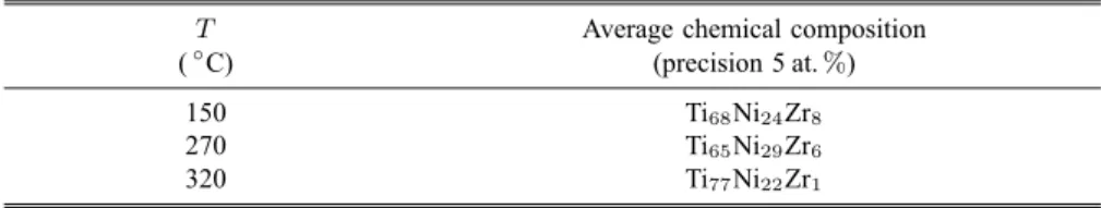
Table 1. Average chemical composition (at.%) obtained from SNMS analyses on films versus the substrate temperature. Nominal composition of evaporated matter was Ti41.5Ni17Zr41.5
T Average chemical composition (◦C) (precision 5 at.%)
150 Ti68Ni24Zr8
270 Ti65Ni29Zr6
320 Ti77Ni22Zr1
The average chemical compositions taken in the non-oxidized zone of the films were evaluated on the films prepared at different temperatures. They are given in Table 1. One can realize that whatever the substrate temperature used, the thermal evaporation produces a big gap between the composition of the matter to be evaporated and that of the resulting films. A large enrichment of Ti to the detriment of Zr can be seen. The Zr depletion is particularly important close to the interface. The films deposited at 320◦C do contain almost no Zr (cf. Table 1). Such an evaporation-condensation technique is a very sensitive method to the values of the respective saturated vapor pressures of the elements [17]. The Ni content in the films is also higher than the value expected.
4. Discussion
The TEM observations performed on carbon-coated grids were generally found to be consistent with the GXRD results performed on glass substrates. One can exclude then the important substrate effects. This point was also observed when similar films were grown by PLD [6].
Although the small crystallites constituting the films produce diffraction signals located around those of the quasicrystalline expected phase, and so, present close order to the searched one, their chemical composition is far from Ti41.5Ni17Zr41.5. Indeed, relative vapour saturation tensions differences of dif-ferent phases in the ingot have a drastic effect on condensation. Further works should have use ingots of different composition to compensate the loss of Zr in the films. The obtained compositions seem to be logical with the values of the vapour saturation tensions of Zr and Ti. This implies that work on the link between initial nominal composition and the one of the films is neces-sary. However, the authors think a complementary and careful work has to be
Structural and Chemical Investigation of Thermally Evaporated Ti-Ni-Zr Films
done on the link between the nature of the phases present in the matter to be evaporated and the obtained chemistry in the films. Ingots used here are not quasicrystalline but composed of different phases. This lack of homogeneity in the initial matter in order to be evaporated must influence the behaviour during evaporation although it is a quick thermal phenomenon.
The technique has proven to be rather good to provide a homogeneous sam-ple. Even if the film thickness is out of control in contrary to results of [6], the simplicity of the technique makes it very attractive for industrial consid-erations. Moreover, one can imagine equipping the current evaporation setup with a regular supplying of matter to be able to grow thicker samples.
5. Conclusions
Ti-Ni-Zr films of 15 to 50 nm in thickness have been prepared by TE process. Different substrate temperatures ranging between 160◦C and 350◦C have been applied. The structure, nature and chemical composition of the films have been studied and compared. The Ti-Ni-Zr icosahedral phase is not formed under the tested conditions but films seem structurally close regarding different diffraction patterns although their chemical composition is so far. Conclusive decisions for a further work have been suggested.
Acknowledgements
The authors would like to thank J.-M. Dubois for having suggested this study. We are most grateful to F. Brillancourt from LCTMR laboratory (CNRS France) for her help to prepare the ingots and to A. Percheron (LCTMR) for her constant interest in this work.
References
1. D. Schechtman, D. Blech, D. Gratias and J. W. Cahn. J. of Phys. Rev. Lett. 53 (1984) 1951.
2. C. Janot. Quasicrystals: A Primer. (Second edn, Oxford Science Publication, Ox-ford University Press, OxOx-ford, 1994).
3. K. Kelton and P. Gibbons. Mat. Res. Soc. Bull. 22 (1997) 69.
4. R. Nicula, A. Jianu, A. Birirs, D. Lupu, R. Manaila, A. Devenyi, C. Kumpf and E. Kurpel. J. of Eur. Phys. J. B. 3 (1998) 1.
5. A. Viano, E. Majzoub, R. Stroud, M. Kramer, S. Misture, P. Gibbons and K. Kelton. Phil. Mag. A 78 (1998) 131.
6. V. Brien, A. Dauscher, P. Weisbecker and F. Machizaud. J. of Applied Physics A
76 (2003), 187.
7. N. Ichikawa, O. Matsumoto, T. Hara, T. Kitahara, T. Yamauchi, T. Matsuda, T. Takeuchi and U. Mizutani. Jpn. J. Appl. Phys. 33 (1994) 736.
8. R. Teghil, L. D’Alessio, M. A. Simone, M. Zaccagnino, D. Ferro and D. Sordelet. J. of Appl. Surf. Sci. 168 (2000) 267.
9. E. Emeric, P. Gas, G. Clugnet and C. Bergman. J. of Mat. Sci. Eng. 50 (2000) 285.
10. F. Giroud. Ph.D. Thesis, Université J. Fourier, Grenoble, 1998.
11. C. Bergman, E. Emeric, P. Donnadieu, J. Dubois and P. Gas. In: C. Janot, R. Mosseri (Eds), Proc. Fifth Int. Conf. on Quasicrystals, ICQ5, Avignon, France, May 22–26, 1995, p. 774.
12. E. Emeric, C. Bergman, J. Dubois, G. Clugnet and P. Gas. In: S. Takeuchi, T. Fujiwara (Eds), Proc. Sixth Int. Conf. on Quasicrystals, ICQ6, Tokyo, Japan, May 26–30, 1997, p. 281
13. J. Joulaud. Ph.D. Thesis, Université Paris VI, France, 1997. 14. C. Chien and M. Lu. Phys. Rev. B 45 (1992) 12793. 15. T. Klein and O. Symbko. Appl. Phys. Lett. 64 (1994) 431.
16. A. Yoshida, K. Kimura and S. Takeuchi. Jpn J. Appl. Phys. 34 (1995) 1606. 17. A. Richardt and A.-M. Durand. In: Le vide, les couches minces, les couches dures,
In Fine (Ed.), Imb Press, Vesoul, 1994, ISBN: 2-8404-030-0, p. 195 18. J. W. Cahn, D. Shechman and D. Gratias. J. Mater. Res. 1 (1986) 13.
19. H. Krebs, O. Bremert, M. Störmer and T. Luo. J. of Appl. Surf. Sci. 86 (1995) 90. 20. S. Sibirtsev, V. Chebotnikov, V. Molokanov and Yu. Kovneristyi. JETP Lett. 47
(1988) 744.
21. V. Molokanov and V. Chebotnikov. J. Non Crystal. Solids 789 (1990) 117. 22. X. Zhang, R. Stroud, J. Libbert and K. Kelton. Phil. Mag. B 70 (1994) 927. 23. A. Viano, R. Stroud, P. Gibbons, A. McDowell, M. Conradi and K. Kelton. Phys.
Rev. B 51 (1995) 12026.
24. A. Viano, A. McDowell, M. Conradi, R. Stroud, P. Gibbons and K. Kelton. In:
Quasicrystals. C. Janot, R. Mosseri (Eds), World Scientific Publishing Singapore