Occurrence of enterotoxins, exfoliative toxins
and toxic shock syndrome toxin-1 genes in
Staphylococcus aureus
and CoNS isolated from
clinical and food samples in Algeria
1,3
Rachid Achek,
2Zafer Cantekin,
3Mustapha Oumouna,
3Amira Mahdi,
1Taha-Mossadak Hamdi
1 High National Veterinary School, Algiers, Algeria; 2 Department of Microbiology, Faculty of Veterinary Medicine, Mustafa Kemal
University, Hatay, Turkey; 3 Faculty of sciences, Yahia Farès University, Médéa, Algeria.
and see (SEE) genes (Balaban and Rasooly2000; Le Loir et al 2003);Staphylococcal pathogenicity islands for sec (SEC) and tst (TSST-1) gene (Dinges et al 2000; Argudin et al 2010). These aspects of mobile carriage accentuate horizontal trans-mission among isolates as well as modify their virulent ability and their evolution state (Becker et al 2003; Argudin et al 2010; Grumann et al 2014).
Coagulase positive Staphylococci (CoPS) especially Staphylococcus aureus were considered as the most pathogenic species (Hennekinne et al 2010), while, Coagulase Negative Staphylococci (CoNS) had gained little attention and thought to be non-pathogenic (Kloos and Schleifer 1975). However, recently, CoNS ability to carry multiple pathogenic factors such as enterotoxins and TSST-1 toxin have been demonstrated (da Cunha et al 2007; Zell et al 2008; Vasconcelos et al 2011;Batista et al 2013).Toxin-associated genes carried by CoNS can be transmitted by cohabitation with other pathogenic Staphylococci (Ławrynowicz-Paciorek et al 2007; Vasconcelos and da Cunha 2010). Nevertheless, CoNS can contaminate foodstuffs and their clinical importance and toxigenic capacity cannot be ignored (Kloos and Bannerman 1995; Otto 2009).
Algerian studies were focused on Staphylococci antibiotic re-sistance (Ramdani-Bouguessa et al 2006; Rebiahi et al 2011;
Introduction
Staphylococci are responsible for nosocomial/community ac-quired infections and foodborne illnesses worldwide (Le Loir et al 2003). This bacterium remains a persistent opportunistic of the respiratory tract and skin, either for human or animals (Monecke et al 2011) and constitutes a contamination source in food or health-care facilities (Kluytmans et al 1997; Von Eiff et al 2001).
Staphylococci produce a variety of toxins responsible for food-borne illness or toxic affections in patients with serious health problems (Hennekinne et al 2010). Consumption of contami-nated meals with enterotoxins causes vomiting, diarrhoea and enteritis within six hours (Le Loir et al 2003; Seo and Bohach 2007). In addition, Staphylococci cause severe systemic affec-tions (pneumonia, endocarditis), associated with variety of ex-otoxins such as toxic shock syndrome toxin-1 (TSST-1), exfo-liative toxins A and B, and Panton-Valentine leucocidin (PVL) (Balaban and Rasooly 2000; Dinges et al 2000).
Moreover, Staphylococci have a wide ability to acquire toxin-encoding genes. Most of these genes are located and transmit-ted via mobile genetic elements. For example, plasmids for seb (SEB), sed (SED) and etb (exfoliative toxin B) genes (Bayles and Iandolo 1989; Argudin et al 2010); prophages for sea (SEA)
Abstract. The aim of this study was to determine the occurrence of toxin-genes carried by strains of Staphylococcus aureus and Coagulase
Negative Staphylococci (CoNS) in Algeria. The present study performed two set multiplex PCR assay using specific primers for screening of 8 genes encoding for classical enterotoxins (SEs) (sea to see), Exfoliative Toxins (eta, etb) and Toxic Shock Syndrome Toxin-1 (tst). We analyzed 51 strains isolated from food samples and 45 strains originated from clinical cases. We observed that more than half of food strains (52.94%) possessed at least one of SEs genes; where S. aureus appears to be potentially enterotoxigenic than CoNS (68.18% vs 41.37%). From all the SEs genes amplified (27), sed gene (19;70.37%) was the most frequently detected. In clinical isolates, only 6 (13%) S. aureus harboured at least one SEs genes. However, 55.55% of clinical isolates (S. aureus or CoNS) possessed tst gene for Toxic Shock Syndrome Toxin-1. There were no foods or clinical isolates detected to possess exfoliative toxins genes (eta, etb). In conclusion, this study showed high frequency of SEs genes in food isolates, and tst gene in clinical isolates; our findings provide updated data on the Staphylococci toxins carriage in Algeria.
Key Words: Staphylococcus aureus; Enterotoxins; PCR; CoNS; TSST-1.
Copyright: This is an open-access article distributed under the terms of the Creative Commons Attribution License, which permits unrestricted
use, distribution, and reproduction in any medium, provided the original author and source are credited.
Ouchenane et al 2011) and rarely on virulence factors such as:TSST-1 and PVL toxins (Ramdani-Bouguessa et al 2006).The aim of the present study was to determine by PCR the frequency of genes encoding staphylococcal enterotoxins, exfoliative tox-ins, and toxic shock syndrome toxin-1 genes, in Staphylococci strains isolated from food and clinical samples in Algeria.
Materials and methods
Bacterial StrainsA total of 96 Staphylococcus spp. isolates were used. Fifty one (53.13%) isolates were obtained from food matrix, samples in-cluding raw milk, minced beef meat, sausages and creamy cake were collected from retail market (supermarkets, conventional markets, or bazaars) in large cities at two regions of Médéa and Ain defla provinces, Algeria.
Forty five (46.88%) strains were isolated from clinical samples, clinical samples included: pus, sperm, urine, vaginal discharge, wound, catheter tips and secretions in general were aseptically collected in different services of Mohamed Boudiaf hospital, Médéa, Algeria. Clinical samples were taken to the microbiol-ogy laboratory of the same hospital for analysis.
Detection and enumeration of coagulase-positive Staphylococci in food matrix was done according to ISO 6888-1:1999/A1:2003. Baird Parker agar plates were incubated for 24 to 48 h at 37 °C. Microbiological analysis of clinical samples was done accord-ing to Quinn et al (1994). For coagulase-positive Staphylococci (CoPS) confirmation, catalase and coagulase tests were used (Rabbit plasma, Oxoid, UK). Staphylococcus aureus identifi-cation for both food and clinical isolates was made by Pastorex staph plus (Bio-Rad, France 2016).
DNA extraction
All isolates were purified by inoculation in Trypticase Soya Broth (TSB), and then transferred to Trypticase Soya Agar (TSA) plates for 18h incubation at 37°C. For bacterial cell ly-ses, lysozyme (100mg/ml) and proteinase-K (10mg/ml)were used. Nucleic acid extraction was performed with Phenol/chlo-roform extraction method according to Sambrook and Russel (2001). DNA pellet was re-suspended in Diethyl pyrocarbonate (DEPC) treated water and stored at –20 °C until PCR analyses.
PCR Procedure
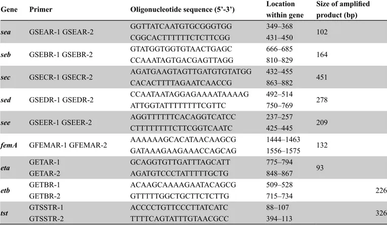
Specific primers were used for the amplification of the sea, seb, sec, sed, see (SEs) genes; eta, etb (Exfoliative Toxins A, B as-sociated genes), and tst (Toxic Shock Syndrome Toxin-1 gene) (Table 1). The femA primers were used for confirmation of S. aureus among studied strains. PCR procedures were applied ac-cording to Mehrotra et al (2000). For the PCR analysis, positive control DNAs were obtained from Mustafa Kemal University, Faculty of Veterinary Medicine, Microbiology Department. Two sets of multiplex PCR procedures were performed accord-ing to Mehrotra et al (2000) with minor modifications. Briefly, for SEs, amplification mix containing 200µM deoxynucleoside triphosphates; 5 µl of 10X reaction buffer (100 mMTris-HCl [pH 8.3], 500 mMKCl); 1.5 mM MgCl2; 20 pmol of each sea, seb, sec, see, and femA primers; 40 pmol of sed primer; 2.5 U of Taq DNA polymerase (Thermo scientific), was added to 3 µl of template DNA. For others toxins genes, PCR reaction had the same constituents as in SEs except for the MgCl2 concen-tration (2.0 mM) and the primers, which were used at 50 pmol for eta and 20 pmol for etb and tst. The final volume was ad-justed to 25 µl by adding sterile ultrapure water. Amplification (CFX 96 thermal cycler, Bio-Rad) programme was as follows:
Gene Primer Oligonucleotide sequence (5’-3’) Locationwithin gene Size of amplifiedproduct (bp)
sea GSEAR-1 GSEAR-2 GGTTATCAATGTGCGGGTGG 349–368 102 CGGCACTTTTTTCTCTTCGG 431–450
seb GSEBR-1 GSEBR-2 GTATGGTGGTGTAACTGAGC 666–685 164 CCAAATAGTGACGAGTTAGG 810–829
sec GSECR-1 GSECR-2 AGATGAAGTAGTTGATGTGTATGG 432–455 451 CACACTTTTAGAATCAACCG 863–882
sed GSEDR-1 GSEDR-2 CCAATAATAGGAGAAAATAAAAG 492–514 278 ATTGGTATTTTTTTTCGTTC 750–769
see GSEER-1 GSEER-2 AGGTTTTTTCACAGGTCATCC 237–257 209 CTTTTTTTTCTTCGGTCAATC 425–445
femA GFEMAR-1 GFEMAR-2 AAAAAAGCACATAACAAGCG 1444–1463 132 GATAAAGAAGAAACCAGCAG 1556–1575
eta GETAR-1 GCAGGTGTTGATTTAGCATT 775–794 93 GETAR-2 AGATGTCCCTATTTTTGCTG 848–867
etb GETBR-1 ACAAGCAAAAGAATACAGCG 509–528 226 GETBR-2 GTTTTTGGCTGCTTCTCTTG 715–734
tst GTSSTR-1 ACCCCTGTTCCCTTATCATC 88–107 326 GTSSTR-2 TTTTCAGTATTTGTAACGCC 394–113
initial denaturation at 94°C for 3 min followed by 29 cycles of amplification (denaturation at 94°C for 1.5 min, annealing at 54°C for 1.5 min, and extension at 72°C for 1 min). A final extension step (72°C for 7 min) was performed after the com-pletion of cycles.
PCR products, were loaded into 2% agarose gel (BioMax) stained with 5 µl of RedSafeTM nucleic acid (iNtROn Biotechnology, Korea) and visualized using a UV transluminator (EC3, UVP Bioimaging systems, Inc).
Statistical analysis
Fisher’s exact test was used to compare the frequency of each enterotoxin, exfoliative toxin and TSST-1 toxin genes. P value of <0.05 was considered as statistically significant.
Results
In the present study, the amplification of SEs, other toxin-asso-ciated genes and femA was successfully obtained. Table 2 and Table 3 show the PCR results for the detection of classic en-terotoxins encoding genes; exfoliative toxins and toxic shock syndrome toxin-1 encoding genes of all Staphylococci isolates. According to phenotypic methods and femA gene detection, 61 isolates (63.54%) (22 food, 39 clinical) were confirmed as Staphylococcus aureus.
Among food isolates (n=51), 27 (52.94%) had at least one gene encoding for SEs production. The detection of sed gene was the most predominant (70.37%) being found in 19 isolates (11 in S. aureus and 8 in CoNS strains). However, three isolates carried sec and see genes. Also, sea gene was confirmed in only two S. aureus isolates, while seb gene was not detected.
Regarding to food-matrix origins, raw milk and minced beef meat were the most common matrix which contained Staphylococci harbouring sed gene (10 and 6 isolates, respectively) (Table 2). In addition, three see genes were detected in raw milk isolates only. However, the two S. aureus isolates possessing the sea gene were exclusively isolated from creamy cake samples. In all food isolates, S. aureus was more enterotoxigenic than CoNS (68.18% vs41.37% respectively) but not significantly different (P=0.089), this difference was remarkably observed for sed and sea genes(P=0.145, P=0.181). However, the frequency of sec and see genes in CoNS (6.9%) was higher but not significant compared to S. aureus isolates (4.54%) (P=1.000). The exfo-liative encoding genes (eta, etb) and tst gene were not detected in any food isolates.
Regarding clinical isolates, the rate of staphylococcal-harbour-ing enterotoxin genes isolated from clinical samples was sig-nificantly lower (6/13.33%) compared to that of food isolates (27/55.94%) (P<0.001). All clinical CoNS isolates were nega-tive for SEs genes. Among the six SEs genes posinega-tive isolates, three hospital acquired isolates possessed the sed gene and one isolate harboured seb gene. Exceptionally, one community ac-quired S.aureus isolate had the seb gene.
The exfoliative encoding genes (eta, etb) were not found in any clinical isolates. Nevertheless, a high percentage of the tst gene (55.55%) was detected, with predominance of pus samples (9 isolates) (Table 3). For all clinical CoNS isolates, the tst gene was found only in one community acquired strain.
Table 4 lists the genotype profiles for all isolates harbouring one or multiple toxin genes. Among all profiles observed, the most commonly identified gene profiles were those containing a single Table 2. Distribution of the enterotoxins, Exfoliative toxins and Toxic Shock Syndrome Toxin-1 genes for food isolates.
Target genes
Amplicon size (bp) Food isolates (n = 51) P value Samples origin
S. aureus CoNS (n = 22; 43.14%) (n = 29; 56.86%) femA(132) 22 - ND (10) Raw milk (2) Creamy cake (2) Sausages (8)Minced beef meat
sea(102) 2 (9.09%) - 0.181 (2) Creamy cake
seb(164) - - ND
-sec(451) 1(4.55%) 2(6.90%) 1
(1)Creamy cake (2) Minced beef meat
sed(278) 11(50%) 8(27.59%) 0.145
(10)Raw milk (1)Creamy cake
(2) Sausages (6)Minced beef meat
see (208) 1(4.55%) 2(6.90%) 1 (3)Raw milk
eta(93) - - ND
-etb(266) - - ND
-toxin gene (sea, seb, sec, sed, see and tst).Twenty six (27.08%) isolates had one type of enterotoxins gene, and 23 (23.95%) possessed only the tst gene. Besides, the sed and tst genotypes were the most observed with 18 (%) and 23 (%) strains, respec-tively. Combinations genes had been found in four genotypes (sed-sea, sed-sec, sed-tst, sed-seb-tst); each multi-toxin geno-type had only one strain.
The present results showed that sea, seb, sea, sec, sed-seb-tst and sed-tst genotypes had occurred in S. aureus iso-lates. The remaining genotypes included both S. aureus and CoNS isolates.
Discussion
In Algeria, few studies have focused on the toxigenic potential of staphylococcal strains and their occurrence in food poisoning
Target genes
Amplicon size (bp) Clinical isolates (n = 45) P value Samples origin
Hospital acquired Community acquired
Isolates (n = 18) Isolates (n = 27)
S. aureus S. aureus CoNS
(n = 18; 40%) (n = 21; 46.67%) (n = 06; 13.33%)
femA(132) 18 21 - ND
-sea(102) - - - ND
-seb(164) 1(5.56%) 1(4.76%) - 1 (1)Sperm
(1) Vaginal discharge
sec(451) 1(5.56%) - - 1 (1) Throat samples
sed(278) 3(16.67%) - - 1 (1) Blood culture (1) Urinary probe (1)Vaginal discharge see (208) - - - ND -eta(93) - - - ND -etb(266) - - - ND -tst(326) 9(50%) 15(71.43%) 1(16.67%) 0.558 (9) Pus (5) Urine (5) Sperm (3) Vaginal discharge (1) Joint fluid (1 )Surgical wound (1) Urinary probe Table 3. Distribution of the enterotoxins, Exfoliative toxins and Toxic Shock Syndrome
Genotypic profiles
Food isolates (n = 51) Clinicalisolates (n = 45)
S. aureus CoNS S.aureus CoNS
(n = 22) (43.14%) (n = 29) (56.86%) (n = 39) (86.67%) (n = 06) (13.33%) sea 1 - - -seb - - 1 -sec - 2 1 -sed 9 8 - 1 see 1 2 - -sed-sea 1 - - -sed-sec 1 - - -sed-seb- tst - - 1 -sed -tst - - 1 -tst - - 22 1
or hospital/community acquired infections. Algerian reports had studied PVL and TSST-1 genes carriage in MRSA (Ramdani-Bouguessa et al 2006; Bittar et al 2009; Ouchenane et al 2010). Antri et al (2010) studied the Staphylococcus aureus enterotoxin genes carriage isolated in hospital and recorded a low frequency for classical enterotoxin genes (8.6%). For the knowledge of the authors, no data has been published about the frequency of tox-ins genes carriage in Staphylococci isolated form food matrix. Our results showed that the staphylococcal isolates harbour-ing genes for classical enterotoxins were responsible for more than half of all cases of food contamination. These results are in accordance with many previous works (Chapaval et al 2006; Pereira et al 2009; Carfora et al 2015). In the present study, sed was the most frequent toxin-encoding gene (19 of 51 isolates, 37.25%) isolated from food. This gene was mainly detected in Staphylococci isolated from raw milk and minced beef meat. These results are in agreement with other studies, such as those of Pu et al (2011) and Bianchi et al (2013) who reported that 15% (23/152) and 25% (120/481) of retail meat isolates and dairy product isolates had amplified the sed gene. In contrast, Zouharova and Rysanek (2008) reported that a low percentage of S.aureus isolates encoding the sed gene (2/70; 2.9%) in raw milk, whereas Balaban and Rasooly (2000) and Normanno et al (2005) found no evidence that the SED enterotoxin was in-volved in Staphylococcal food poisoning (SFP).
In our study, no isolates taken from raw milk encoded the sea gene. The only detected sea gene came from isolates which originated from cream cake samples. In contrast, Chapaval et al (2006) and Rall et al (2008) reported that sea was the most com-mon toxin gene detected in raw milk isolates, finding 67.78% (61 out 90 isolates) and 28.9% (11 out 38 isolates), respectively. Omoe et al (2005), Chiang et al (2008) and Tang et al (2011) de-tected the sea gene mainly in Staphylococci isolated from food matrix. They reported that it was responsible for 5.8%, 29.2% and 50% of cases of SFP, respectively. However, Pereira et al (2009) did not detect the sea gene in 20 S. aureus isolated from raw milk, which is in accordance with our results.
In terms of the frequency of seb gene detection, our results are in accordance with those of Cremonesi et al (2005) and Bianchi et al (2013), who reported that all of Staphylococcal strains isolated from milk and dairy products were negative for the seb gene. In our study, see gene was harboured only by three Staphylococci strains isolated from raw milk; Rall et al(2008) detected the see gene in 5.26% (2 out of 38 isolates) S. aureus isolated from raw milk, while Zouharova and Rysanek (2008) did not. For sec gene carriage, we reported two strains isolated from minced beef meat, while Pu et al (2011) did not detect any sec gene in 152 S. aureus isolates.
Regarding clinical isolates, the rate of staphylococcal-harbour-ing enterotoxin genes observed in our study was lower than that reported in earlier clinical investigations (Becker et al 2003; Nashev et al 2007; Chiang et al 2008). Our clinical samples consisted mainly of samples obtained from patients with hos-pital- and community-acquired infections. Thus, the discord-ance may be explained by differences in the origin of the spec-imens, which included samples from food poisoning cases, or potential contamination by carriage sources of Staphylococci (nasal cavities and hands). However, some authors (Naffa et al 2006; da Cunha et al 2007 and Demir et al 2011) reported a high
percentage of staphylococcal-positive SE-encoding genes ob-tained from samples population similar to ours: 23/100 (23%), 56/120 (47.5%) and 66/120 (55%), respectively. Moreover, con-sidering that our results are in concordance with a local report of Antri et al (2010), we can consider that geographical loca-tion might explain the large difference between our findings and those reported earlier.
Irrespective of the clinical sampling origin, several studies re-ported a high frequency of the sea gene in Staphylococci iso-lates (Naffa et al 2006; Nashev et al 2007; Demir et al 2011). However, it should be pointed out that in the present study, sea gene was not detected in any of the clinical isolates. The frequen-cy of see gene found in our study was in accordance with that of all the above studies, except the study by Becker et al (2003). The occurrence enterotoxin-encoding genes detected in CoNS isolated from food samples was 41.37% (12 out of 29 CoNS iso-lates), which was higher than reported in other studies (Blaiotta et al 2004; da Cunha et al 2006) but similar to another (Fijałkowski et al 2016). In our study, the most frequently detected entero-toxin gene was sed, and neither sea nor seb were detected in the CoNS isolates. Da Cuhna et al (2006) reported that 15% (3 out 20 isolates) of CoNS isolated from foods harboured the sea gene. Other studies also detected enterotoxin genes in CoNS strains from both dairy and meat products (Vernozy-Rozand et al1996; Rodriguez et al 1996).
In the present study we did not detect eta or etb genes in any of the tested samples. Some studies reported that S. aureus pro-duced one or both exfoliative toxins (Hayakawa et al 2000; Becker et al 2003; Demir et al 2011). Jarraud et al (2001) did not detect eta and etb genes in 58 S. aureus isolates responsible for suppurative diseases.
In our study, tst gene was not detected in the food origin isolates, this result corroborate the Fijałkowski et al (2016) works. In contrast, we observed a high frequency of the tst gene (25/45; 55.55%) in clinical isolates, this result being similar to that found by Chiang et al (2008), who reported a frequency of 59.1% (87 out of 147 isolates). This was higher than that detected in earlier studies of Becker et al 2003; da Cunha et al 2007 and Demir et al 2011, who reported frequencies of (87/429; 20.3%), (11/104; 10.58%) and (17/120; 14.17%), respectively.
The TSST-1 toxin is a causative agent of systemic infections, such as the staphylococcal toxic shock syndrome (Dinges et al 2000) but is rarely implicated in SFP. A number of studies re-ported that TSST-1, was frequently detected in S. aureus clinical isolates but rarely in food isolates (Lappin and Ferguson 2009; Tsen et al 1998; El-Ghodban et al 2006).
In the present study, most genotypes contained a single toxin gene, regardless of the origin of the sample, with tst and sed as the most common genes. This result is in accordance with those of several other studies (Nashev et al 2007; Zouharova and Rysanek2008; Rall et al 2010), although the sea genotype was predominant in these studies. However, in other studies (Bianchi et al 2013; Pu et al 2011) the sed genotype was the most common genotype detected. In the present study, in clini-cal samples, the frequency of the tst genotype was in agreement with that found in an earlier study (Chiang et al 2008). In the current study, frequency of toxin gene combinations was low, being observed in only four isolates. The tst gene was de-tected in combination with enterotoxin genes in two genotypes
(sed-seb-tst and sed-tst). A number of previous studies reported that Staphylococci strains harboured the tst gene, either alone or in combination with SE-encoding genes (Becker et al 2003; Chapaval et al 2006; da Cunha et al 2007; Chiang et al 2008; Demir et al 2011). Others studies recorded a tst combination with sec, but not with sed or seb (Hwang et al 2007).
The toxins production by Staphylococci strains is complex and involves gene carriage and gene promoters, such as multiple global regulators of virulence (e.g. agr, sarA, rot and sigB). Previous studies suggested that toxin-encoding genes (SEs, TSST-1 and exfoliative toxins) could be located on a mobile genetic carrier, which would provide potential support for horizontal transfer or genotype combination (Omoe et al 2005; Chiang et al 2008, Grumann et al 2014). Jarraud et al (2001) suggested that SEs and TSST-1 share common structural and biological properties and those that are derived from a common ancestor.
To conclude, this study reports the occurrence of the toxin genes in staphylococcal isolates from food and clinical samples in Algeria. Using a multiplex PCR method, a high frequency of SEs genes in food isolates and tst gene in clinical isolates were recorded. The pathogenic potential of CoNS points to the need for a greater surveillance in the area of hygiene and pub-lic health. There is an urgent need to establish legal standard-ized methods to be able to verify and quantify the degree of Staphylococci enterotoxins contamination in foods in Algeria. Further research is needed in order to investigate the contami-nation routes of food consumed in Algeria and the distribution of newly described Staphylococcal toxin genes.
Acknowledgments
Authors expressed their grateful acknowledgement to Dr.Yasar Ergün and other staff members of Veterinary Faculty, Mustafa Kemal University, Hatay, Turkey for their help and supportive assistance during the preparation of this work.
References
Antri K, Rouzic N, Boubekri I, Dauwalder O, Beloufa A, Ziane H, et al. Forte prévalence des infections communautaires et nosocomi-ales à Staphylococus aureus résistant à la méticilline et portant le gène de la leucocidine de Panton-Valentine dans l’Algerois. J pat-bio 2010;58:15–20.
Argudin MA, Mendoza MC, Rodicio MR. Food poisoning and Staphylococcus aureus enterotoxins. Toxins2010;2:1751–1773. Balaban N, Rasooly A. Staphylococcal enterotoxins. Int J Food Microbiol
2000;61:1–10.
Batista JEC, Ferreira EL, de Oliveira Nascimento DC, Ventura RF, de Oliveira WLM, Leal NC, Lima-Filho JV. Antimicrobial resistance and detection of the mecA gene besides enterotoxin-encoding genes among coagulase-negative Staphylococci isolated from clam meat of Anomalocardiabrasiliana. Foodborne Pathog Dis2013;10:1044–1049. Bayles KW, Iandolo JJ. Genetic and molecular analyses of the gene en-coding staphylococcal enterotoxin D. J Bacteriol 1989;171:799–806. Becker K, Friedrich AW, Lubritz G, Weilert M, Peters G, Von Eiff C.
Prevalence of genes encoding pyrogenic toxin superantigens and ex-foliative toxins among strains of Staphylococcus aureus isolated from blood and nasal specimens. J Clin Microbiol 2003;41:1434–1439. Bianchi DM, GallinaS, Bellio A, Chiesa F, Civera T, Decastelli L.
Enterotoxin gene profiles of Staphylococcus aureus isolated from milk and dairy products in Italy. Lett Appl Microbiol 2013;58:190–196.
Bittar F, Ouchenane Z, Smati F, Raoult D, Rolain JM. MALDI-TOF-MS for rapid detection of staphylococcal Panton-Valentine leuko-cidin. Int J Antimicrob Agents 2009;34(5):467–470.
Blaiotta G, Ercolini D, Pennacchia C, Fusco V, Casaburi A, Pepe O, Villani F. PCR detection of staphylococcalenterotoxingenes in Staphylococcusspp. Strains isolated from meat and dairy products. Evidence for new variants of seG and seI in S. aureus AB- 8802. J Applied Microbiol 2004;97:719–730.
Carfora V, Caprioli A, Marri N, Sagrafoli D, Boselli C, Giacinti G, et al. Enterotoxin genes, enterotoxin production, and methicillin resist-ance in Staphylococcus aureus isolated from milk and dairy prod-ucts in Central Italy.Int Dairy J 2015;42:12–15.
Chapaval L, Moon DH, Gomes JE, Duarte FR, Tsai SM. Use of PCR to detect classical enterotoxins genes (ent) and toxic Shock syn-drome toxin-1 gene (tst) in Staphylococcus aureus isolated from crude milk and determination of toxin productivities of S. aureus isolates harboring these genes. Arq Inst Biol 2006;73(2):165–169. Chiang YC, Liao WW, Fan CM, Pai WY, Chiou CS, Tsen HY. PCR de-tection of Staphylococcal enterotoxins (SEs) N, O, P, Q, R, U, and survey of SE types in Staphylococcus aureus isolates from food-poisoning cases in Taiwan. Int J Food Microbiol 2008;121:66–73 Cremonesi P, Luzzana M, Brasca M, Morandi S, Lodi R, Vimercati C,
et al. Development of a multiplex PCR assay for the identification of Staphylococcus aureusenterotoxigenic strains isolated from milk and dairy products. Mol Cell Probes 2005;19:299–305.
DaCunhaM de LRS, Calsolari-Regina AO, Araújo-Júnior JP. Detection of Enterotoxin and Toxic Shock Syndrome Toxin 1 Genes in Staphylococcus, with Emphasis on Coagulase-Negative Staphylococci. Microbiol Immunol 2007;51(4):381–390.
DaCunha M de LRS, Peresi E, Oliveira-Calsolari RA, Araújo-Júnior JP. Detection of enterotoxins genes in coagulase-negative Staphylococci isolated from foods. Braz J Microbiol 2006;37:70–74.
Demir C, Aslantaş Ö, Duran N, Ocak, Özer B. Investigation of toxin genes in Staphylococcus aureus strains isolated in Mustafa Kemal University Hospital. Turk J Med Sci2011;41(2):343–352.
Dinges MM, Orwin PM, Schlievert PM. Exotoxins of Staphylococcus aureus. Clin Microbiol Rev 2000;13:16–34.
El-Ghodban A, Ghenghesh KS, Marialigeti K, Esahli H, Tawil A. PCR detection of toxic shock syndrome toxin of Staphylococcus aureus from Tripoli, Libya. J Med Microbiol 2006;55:179–182.
Fijałkowski K, Peitler D, KarakulskaJ.Staphylococci isolated from ready-to-eat meat – Identification, antibiotic resistance and toxin gene profile.Int J Food Microbiol 2016;238:113–120.
Grumann D, Nübel U, Bröker BM. Staphylococcus aureus toxins – Their functions and genetics. Infect Genet Evol 2014;21:583–592. Hayakawa Y, Akagi M, Hayashi M, Shimano T, Komae H, Funaki
O, et al. Antibody response to toxic shock syndrome toxin-1 of Staphylococcus aureusin dairy cows. Vet Microbiol 2000;72: 321–327. Hennekinne JA, Ostyn A, Guillier F, Herbin S, Prufer AL, Dragacci S.
How should staphylococcal food poisoning outbreaks be character-ized.Toxins 2010;2:2106–2116.
Hwang SY, Kim SH, Jang EJ, Kwon NH, Park YK, Koo HC, et al. Novel multiplex PCR for the detection of the Staphylococcus au-reussuperantigen and its application to raw meat isolates in Korea. Int J Food Microbiol 2007;117: 99–105.
ISO 6888-1/A1: 2003. Microbiology of food and animal feeding stuffs -Horizontal method for the enumeration of coagulase-positive staphy-lococci (Staphylococcus aureus and other species)-Part 1: Technique using Baird-Parker agar medium.
Jarraud S, Peyrat MA, Lim A, Tristan A, Bes M, Mougel C, Etienne J, Vandenesch F, Bonneville M, Lina G. A Highly Prevalent Operon of Enterotoxin Gene, Forms a Putative Nursery of Superantigens in Staphylococcus aureus. J Immunol 2001;166:669–677.
Kloos WE, Bannerman TL. [Staphylococcus and Micrococcus]. In: [Manual of Clinical Microbiology]. Murray PR, Baron EJ, Pfaller MA, Tenover FC, Yolken, RH. (ed.), pp.282–298. American Society Microbiology, Washington,1995.
Kloos WE, Schleifer KH. Simplified scheme for routine identification of human Staphylococcus species. J ClinMicrobiol 1975;1:82–88. Kluytmans J, Van Belkum A, Verbrugh H. Nasal carriage of Staphylococcus
aureus: epidemiology, underlying mechanisms, and associated risks, Reviews. J ClinMicrobiol 1997;10:505–520.
Lappin E, Ferguson AJ. Gram-positive toxic shock syndromes. Lancet Infect Dis 2009;9: 281–290.
Ławrynowicz-Paciorek M, Kochman M, Piekarska K, Grochowska A, Windyga B. The distribution of enterotoxin and enterotoxin-like genes in Staphylococcus aureus strains isolated from nasal carri-ers and food samples. Int J Food Microbiol 2007;117: 319–323. Le Loir Y, Baron F, Gautier M. Staphylococcus aureus and food
poi-soning. Genet Mol Res2003;2:63–76.
Mehrotra M, Wang G, Johnson WM. Multiplex PCR for detection of genes for Staphylococcus aureus Enterotoxins, Exfoliative Toxins, Toxic Shock Syndrome Toxin 1, and Methicillin Resistance. J Clin Microbiol 2000;38(3):1032–1035.
Monecke S, Ehricht R, Slickers P, Wernery R, Johnson B, Jose S, Wernery U. Microarray-based genotyping of Staphylococcus aureus isolates from camels. Vet Microbiol 2011;150:309–314.
Naffa RG, Bdour SM, Migdadi HM, Shehabi AA. Enterotoxicity and genetic variation among clinical Staphylococcus aureus isolates in Jordan. J Med Microbiol 2006;55:183–187.
Nashev D, Toshkova K, Bizeva L, Akineden Ö, Lämmler C, Zschöck M. Distribution of enterotoxin genes among carriage- and infection-associated isolates of Staphylococcus aureus. FEMS Microbiol Lett 2007;45:681–685.
Normanno G, Firinu A, Virgilio S, Mula G, Dambrosio A, Poggiu A, et al. Coagulase-positive Staphylococci and Staphylococcus aureus in foods products marketed in Italy. Food Microbiol 2005;98:73–79. Omoe K, Hu DL, Takahashi-Omoe H, Nakane A, Shinagawa K.
Comprehensive analysis of classical and newly described staphy-lococcal superantigenic toxin genes in Staphylococcus aureus iso-lates. FEMS Microbiol Lett 2005;246:191–198.
Otto M. Staphylococcus epidermidis– the ‘accidental’ pathogen. Nat Rev Microbiol 2009;7:555–567.
Ouchenane Z, Smati F, Rolain JM, Raoult D. Molecular characterization of methicillin-resistant Staphylococcus aureus isolates in Algeria. Pathol Biol 2011;59:129–132
Pereira V, Lopes C, Castro A, Silva J, Gibbs P, Teixeira P. Characterization for enterotoxin production, virulence factors, and antibiotic sus-ceptibility of Staphylococcus aureusisolates from various foods in Portugal. Food Microbiol 2009;26:278–282.
Pu S, Wang F, Ge B. Characterization of Toxin Genes and Antimicrobial Susceptibility of Staphylococcus aureus Isolates from Louisiana Retail Meats. Foodborne Pathog Dis 2011;8(2):299–306.
Quinn PJ, Carter ME, Markey BK, Carter GR. Clinical Veterinary Microbiology. Mosby-Year Book Europe Limited, Lynton House (Eds). London, England,1994.
Rall VLM, Sforcin JM, Augustini VCM, Watanabe MT, Fernandes JrA, Rall R, et al. Detection of enterotoxin genes of staphylococcus sp isolated from nasal cavities and hands of food handlers. Braz J Microbiol 2010;41:59–65.
Rall VLM, Vieira FP, Rall R, Vieitis RL, FernandesJrA, Candeias JMG, Cardoso KFG, Araújo-Júnior JP. PCR detection of staphylococcal enterotoxin genes in Staphylococcus aureus strains isolated from raw and pasteurized milk. Vet Microbiol2008;132:408–413. Ramdani-Bouguessa N, Bes M, Meugnier H, Forey F, Reverdy ME, Lina
G, et al. Detection of Methicillin-Resistant Staphylococcus aureus Strains Resistant to Multiple Antibiotics and Carrying the Panton-Valentine Leukocidin Genes in an Algiers Hospital. Antimicrob Agents Chemother 2006;50(3):1083–1085.
Rebiahi SA, Abdelouahid DE, Rahmoun M, Abdelali S, Azzaoui H. Emergence of vancomycin-resistant Staphylococcus aureus identi-fied in the Tlemcen university hospital (North-West Algeria). Med Mal Infect 2011;41:646–651
Rodriguez M, Nunez F, Cordoba JJ, Bermudez E, Asensio MA. Gram-Positive, Catalase-Positive Cocci from Dry Cured Iberian Ham and Their Enterotoxigenic Potential. Appl Environ Microbiol 1996;62(6): 1897–1902.
Sambrook J, Russell W. Molecular cloning: a laboratory manual. (3rd-Eds.) A8.9-A8.10. Cold Spring Harbor Press, New York,2001. Seo KS, Bohach GA,[Staphylococcus aureus]. In: [Food Microbiology:
Fundamentals and Frontiers]. Doyle MP, Beuchat LR. (ed.), pp.518. Washington, DC: ASM Press,2007.
Tang J, Tang C, Chen J, Du Y, Yang XN, Wang C, et al. Phenotypic Characterization and Prevalence of Enterotoxin Genes in Staphylococcus aureus Isolates from Outbreaks of Illness in Chengdu City. Foodborne Pathog Dis 2011;8(12):1317–1320.
Tsen HY, Yu GK, Wang KC, Wang SJ, Chang MY, Lin LY. Comparison of the enterotoxigenic types, toxic shock syndrome toxin 1 (TSST-1) strains and antibiotic susceptibilities for enterotoxigenicStaph-ylococcus aureus strains isolated from food and clinical samples. Food Microbiol 1998;15:33–41.
Vasconcelos NG, da Cunha M. Staphylococcal enterotoxins: mo-lecular aspects and detection methods. J Public Health Epidemiol 2010;2(3):29–42.
Vasconcelos NG, Pereira VC, Araújo-Júnior JP, da Cunha M. Molecular detection of entérotoxines E, G, H and I in Staphylococcus aureus and coagulase-negative Staphylococci isolated from clinical sam-ples of newborns in Brazil. J Appl Microbio l2011;111:749–762. Vernozy-Rozand C, Mazuya C, Prevostb G, Lapeyrec C, Besd M,
Brund Y, Fleuretted J. Enterotoxin production by coagulase-nega-tive staphylococci isolated from goats’ milk and cheese. Int J Food Microbiol 1996;30:271–280.
Von Eiff C, Becker K, Machka K, Stammer H, Peters G. Nasal car-riage as a source of Staphylococcus aureusbacteremia. N Engl J Med 2001;344:6–11.
Zell C, Resch M, Rosenstein R, Albrecht T, Hertel C, Gotz F. Characterization of toxin production of coagulase-negative staph-ylococci isolated from foods and starter cultures. Int J Food Microbiol2008;127:246–251.
Zouharova M, Rysanek D. Multiplex PCR and RPLA Identification of Staphylococcus aureusEnterotoxigenic Strains from Bulk Tank Milk. Zoonoses Public Health 2008;55:313–319.
Authors
•Rachid Achek, High National Veterinary School, Issad Abbes Avenue, Oued Smar, Algiers, Algeria, email: achekrachid@ gmail.com
•Zafer Cantekin, Department of Microbiology, Faculty of Veterinary Medicine, Mustafa Kemal University, Tayfur Sokmen Campus 31000 Hatay, Turkey, email: [email protected].
•Mustapha Oumouna, Faculty of sciences, YahiaFarès University, Urban pole, Médéa, Algeria, email: [email protected].
•Amira Mahdi, Faculty of sciences, YahiaFarès University, Urban pole, Médéa, Algeria, email:[email protected]
Citation
Achek R, Cantekin Z, Oumouna M, Mahdi A, Hamdi TM. Occurrence of enterotoxins, exfoliative toxins and toxic shock syndrome toxin-1 genes in Staphylococcus aureus and CoNS isolated from clinical and food samples in Algeria. HVM Bioflux 2018;10(2):85-92.
Editor Stefan C. Vesa Received 24 February 2018 Accepted 4 May 2018 Published Online June 2018
Funding None reported Conflicts/
Competing
Interests None reported
•Taha-Mossadak Hamdi, High National Veterinary School, Issad Abbes Avenue, Oued Smar, Algiers, Algeria, email: [email protected]