HAL Id: dumas-02954622
https://dumas.ccsd.cnrs.fr/dumas-02954622
Submitted on 1 Oct 2020
HAL is a multi-disciplinary open access
archive for the deposit and dissemination of sci-entific research documents, whether they are pub-lished or not. The documents may come from teaching and research institutions in France or abroad, or from public or private research centers.
L’archive ouverte pluridisciplinaire HAL, est destinée au dépôt et à la diffusion de documents scientifiques de niveau recherche, publiés ou non, émanant des établissements d’enseignement et de recherche français ou étrangers, des laboratoires publics ou privés.
Quantitative measurement of total lung vascular volume
by computed tomography in COPD patients: correlation
with clinical events and biological data
Cevdet Ozcelik
To cite this version:
Cevdet Ozcelik. Quantitative measurement of total lung vascular volume by computed tomography in COPD patients: correlation with clinical events and biological data. Human health and pathology. 2018. �dumas-02954622�
AVERTISSEMENT
Ce document est le fruit d'un long travail approuvé par le
jury de soutenance et mis à disposition de l'ensemble de la
communauté universitaire élargie.
Il n’a pas été réévalué depuis la date de soutenance.
Il est soumis à la propriété intellectuelle de l'auteur. Ceci
implique une obligation de citation et de référencement
lors de l’utilisation de ce document.
D’autre part, toute contrefaçon, plagiat, reproduction illicite
encourt une poursuite pénale.
Contact au SID de Grenoble :
bump-theses@univ-grenoble-alpes.fr
LIENS
LIENS
Code de la Propriété Intellectuelle. articles L 122. 4
Code de la Propriété Intellectuelle. articles L 335.2- L 335.10
http://www.cfcopies.com/juridique/droit-auteur
UNIVERSITÉ GRENOBLE ALPES UFR DE MÉDECINE DE GRENOBLE
Année : 2018
MESURE QUANTITATIVE DU VOLUME VASCULAIRE PULMONAIRE
TOTAL PAR TOMODENSITOMETRIE CHEZ LES PATIENTS BPCO,
CORRELATION AVEC LES EVENEMENTS CLINIQUES ET LES
DONNEES BIOLOGIQUES
THÈSE
PRÉSENTÉE POUR L’OBTENTION DU TITRE DE DOCTEUR EN MÉDECINE DIPLÔME D’ÉTAT
Cevdet OZCELIK
THÈSE SOUTENUE PUBLIQUEMENT À LA FACULTÉ DE MÉDECINE DE GRENOBLE LE 2 OCTOBRE 2018
DEVANT LE JURY COMPOSÉ DE Président du jury :
Monsieur le Professeur Gilbert FERRETTI Membres :
Monsieur le Professeur Sam BAYAT Monsieur le Professeur Jean-Louis PEPIN Monsieur le Docteur Adrien JANKOWSKI
Monsieur le Docteur Julien COHEN (directeur de thèse)
L’UFR de Médecine de Grenoble n’entend donner aucune approbation ni improbation aux opinions émises dans les thèses ; ces opinions sont considérées comme propres à leurs auteurs.
Sommaire
• REMERCIEMENTS ... 8
• LISTE DES ABBREVIATIONS ... 10
• RESUME EN FRANÇAIS ... 11
• RESUME EN ANGLAIS ... 12
• ARTICLE ... 13
• FIGURES ET TABLEAUX ... 25
• REFERENCES ... 31
•
CONCLUSION ... 34
Remerciements
A mon président de thèse le Pr FERRETTI Gilbert.
Merci de me faire l’honneur de présider ma thèse.
A monsieur le Pr Jean Louis PEPIN.
Je vous remercie de l’intérêt que vous témoignez en acceptant de juger ce travail et pour votre expertise.
A monsieur le Pr Sam BAYAT.
Je vous remercie pour votre implication et vos conseils apportés pour la réalisation de ce travail, ainsi que vos corrections pour la rédaction de l’article.
A monsieur le Dr Adrien JANKOWSKI.
Je te remercie d’avoir accepté de participer à ce jury de thèse.
A mon directeur de thèse monsieur le Dr Julien COHEN.
Merci de m’avoir proposé ce sujet intéressant. J’espère de tout cœur qu’il aboutira à une publication.
Remerciements
Merci à Lucie pour m’avoir offert ces deux merveilles, pour m’avoir supporté toutes ces années
d’externat et d’internat pas toujours faciles et pour m’avoir motivé à avancer cette thèse.
Merci à mes parents et ma famille pour toute leur aide pendant ces longues années d’études et
de concours, et ces dernières années pour l’aide avec les enfants.
Merci à mes beaux-parents, Huguette, Magalie, Manon et Alexis pour ces super weekends
passés dans le Vercors, en hiver comme en été.
Merci à tous mes co-internes et aux assistants pour ces interminables staffs à discuter de dossiers
compliqués. J’y ai beaucoup appris et ils ont grandement contribué à ma formation radiologique.
Merci à toute l’équipe de l’hôpital Sud pour m’avoir accueilli. Merci à Pierre et Jean Noel pour m’avoir offert cette opportunité.
Liste des abréviations
ABPM: Ambulatory Blood Pressure Monitoring COPD: Chronic obstructive pulmonary disease CRP: C reactive protein
HsCRP: high sensitive C reactive protein CT: computed tomography
FEV1: forced expiratory volume in one second FVC: forced vital capacity
GOLD: global initiative for chronic obstructive lung disease ICU : intensive care unit
IL 6 : inter-leukine 6
LVEF: left ventricular ejection fraction MRI: magnetic resonance imaging NoV: number of vessels
NoV5: number of vessels <5mm² cross section
Nt-pro-BNP: N-terminal pro-brain natriuretic peptide Pa O²: pressure of artery oxygen
PFT: pulmonary function test PH: pulmonary hypertension TVV: total lung vascular volume
TVVn: normalised total lung vascular volume TNFα: tumor necrosis factor α
Résumé
OBJECTIF
L’objectif était d’étudier par tomodensitométrie la relation entre les mesures quantitatives vasculaires et les données cliniques, les marqueurs sanguins de l'inflammation et du stress oxydatif chez les patients BPCO (bronchite pulmonaire chronique obstructive).
MATÉRIELS ET MÉTHODES
Le volume vasculaire pulmonaire total (TVV), la TVV normalisée à la surface corporelle (TVVn), le nombre total de vaisseaux (NoV) et le nombre de vaisseaux dont la section transversale est inférieure à 5 mm² (NoV5) ont été mesurés avec un logiciel sur des tomodensitométries thoraciques non injectées. Notre cohorte comprenait 62 patients atteints de BPCO (63,8 ans [59 ; 69,6] ; 74 % d'hommes) dont la répartition était : GOLD I n=8 (12,9 %), GOLD II n=39 (62,9 %), GOLD III n=10 (16,1 %) et GOLD IV n=5 (8,1 %). Les données cliniques concernant l'hospitalisation toutes causes confondues, l'infection pulmonaire, l'exacerbation et l'hospitalisation en unité de soins intensifs ont été enregistrées jusqu'à 5 ans après la tomodensitométrie thoracique.La fonction pulmonaire respiratoire (EFR), les gaz du sang artériel et la protéine C réactive plasmatique haute sensibilité (hsCRP), le fibrinogène, les thiols et la capacité antioxydante sérique totale ont été mesurés.
RÉSULTATS
TVVn avait une corrélation significative avec l'infection pulmonaire (OR = 1,06[1,01 ; 1,12] p = 0,02) et l'hospitalisation (OR = 1,06[1,01 ; 1,12] p = 0,01), HsCRP (β = 3,12 (0,84) p = 0,001) et une corrélation inverse avec la capacité antioxydante sérique totale (β = -90,21 (41,38) p = 0,03). Il n’y avait aucune corrélation entre la NoV et la NoV5 et les données cliniques.
CONCLUSION
Notre étude suggère que les mesures quantitatives par tomodensitométrie, en particulier la TVVn, peuvent aider à prédire les complications cliniques chez les patients BPCO et méritent d'être étudiées davantage.
Abstract
OBJECTIVES
To investigate the relationship between quantitative CT measurements of pulmonary vessel volume and number with clinical outcome, serum biomarkers of inflammation and oxidative stress, and lung function, in COPD patients.
MATERIALS AND METHODS
Total lung vascular volume (TVV), TVV normalized to body surface area (TVVn), total number of vessels (NoV) and number of vessels <5 mm² cross section (NoV5) were measured using a custom software on unenhanced thoracic CT scans. Our cohort included 62 COPD patients (63.8 years [59 ; 69.6]; 74% male) with the following distribution: GOLD I n=8 (12.9%), GOLD II n=39 (62.9%), GOLD III n=10 (16.1%) and GOLD IV n=5 (8.1%). Clinical outcome data for all-cause hospitalization, lung infection, exacerbation, and intensive care unit hospitalization, were recorded up to 5 years following chest CT. Lung function, arterial blood gas and high sensitivity plasma C-reactive protein (hsCRP), fibrinogen, thiols and serum antioxidant capacity were measured.
RESULTS
TVVn significantly correlated with pulmonary infection (OR = 1.06 [1.01; 1.12] p = 0.02) and hospitalization (OR = 1.06 [1.01; 1.12] p = 0.01), HsCRP (β = 3.12 (0.84) p = 0.001) and inversely correlated with total serum antioxidant capacity (β = -90.21 (41.38) p = 0.03). There was no correlation between NoV and NoV5 with clinical outcome.
CONCLUSION
Our study suggests that quantitative CT measurements, particularly TVVn may help predict clinical outcome in COPD patients, and merit further investigation as a surrogate imaging biomarker.
QUANTITATIVE MEASUREMENT OF TOTAL LUNG VASCULAR
VOLUME BY COMPUTED TOMOGRAPHY IN COPD PATIENTS
:
CORRELATION WITH CLINICAL EVENTS AND BIOMARKERS
Cevdet OZCELIK a, Sam BAYAT b, Jean Louis PEPIN b, Gilbert FERRETTI a, Meriem
BENMERAD b, Ludovic BROCHE c, Julien COHEN a
a Clinique Universitaire de Radiologie et Imagerie Médicale (CURIM), Centre Hospitalier Universitaire Grenoble
Alpes, CS 10217 38043 Grenoble Cedex 9, France.
b Laboratoire d’Exploration Fonctionnelle Respiratoire, Centre Hospitalier Universitaire Grenoble Alpes, CS 10217
38043 Grenoble Cedex 9, France.
c Laboratoire Rayonnement Synchrotron et Recherche médicale (RSRM) - EA 7442 Université Grenoble Alpes.
Grenoble. France.
Keywords :
COPD
Total lung vascular volume Vascular phenotype Inflammation Oxydative stress
Introduction
Chronic Obstructive Pulmonary Bronchitis (COPD) is a progressive and irreversible impairment of breathing capacity. It is one of the leading causes of global mortality [1]. Morphologically, COPD is characterized by both progressive parenchymal destruction leading to emphysema, and significant remodelling of small airways [2-3]
It is increasingly recognized that changes in pulmonary vasculature, which have been demonstrated by post-mortem morphological analysis on autopsy [4-5] as well as imaging study [6-7], may accompany or even precede emphysematous changes in the lung parenchyma. Such vascular lesions are manifested by a remodelling of the pulmonary arterioles through proliferation of smooth muscle cells and vasoconstriction in the early moderate stages of COPD [8]. Although such phenomena may occur as a result of alveolar hypoxia, and may lead to pulmonary hypertension with time and disease progression [9], other mechanisms such as oxidative stress due to both tobacco exposure and recruitment and activation of inflammatory cells in the lungs [10] or repeated exacerbations or lung infection may be involved [11].
CT imaging offers the possibility to quantitatively assess the pulmonary vasculature. Distal vascular changes have been documented in recent studies, demonstrating an inverse correlation between the cross-sectional surfaces of small pulmonary vessels and emphysema [12]. However, there are a proportion of COPD patients without correlation between emphysema and decreased cross-sectional area of small pulmonary vessels, involving other phenomena as endothelial dysfunction [13], suggesting different lesional phenotypes.
Currently, the significance of changes in pulmonary vascular morphology in COPD patients and whether they are associated with clinical outcome, such as exacerbation or hospitalization, is not known. In this study, we hypothesized that changes in pulmonary vascular morphology, namely
the total volume of the vascular network and the number of pulmonary vessels may be associated with clinical outcomes such as exacerbation, pulmonary infection, all-cause hospitalization or intensive care unit (ICU) hospitalization. We further assessed whether changes in morphologic indices of the pulmonary vascular tree were related to inflammation, oxidative stress, lung function and gas exchange.
Materials and methods
Study population
121 COPD patients selected from a cohort of COPD patients followed at the Grenoble University Hospital, were prospectively enrolled in the study.
The inclusion criteria for the initial cohort were : age > 18 years and FEV1/FVC <70%. Patients with ongoing infection or plasma CRP > 100 mg/l; acute decompensation of chronic heart failure or left ventricular ejection fraction (LVEF) < 45%; tobacco smoke exposure < 10 cigarettes/day; active neoplasia; antioxidant treatment (n-acetyl-cysteine, selenium, vitamin C or E); pregnancy or breastfeeding; inability to provide informed consent; participation in another clinical research study; lack of health insurance coverage; deprived of civil liberty or claustrophobia were excluded from the study.
All patients gave written informed consent. The study was a registered under AFSSAPS 2006-A00491-50.
Study design
During the clinical follow-up, clinical events were recorded up to 5 years after CT scan when possible. Those events were all-cause hospitalization, hospitalization in ICU, lung infection, and exacerbation. Lund infection and exacerbation have been diagnosed by the physician.
CT Imaging
All patients underwent chest CTs with the following protocol: 120kV, slice thickness 0,625 mm spaced by 0,625 mm intervals.
We used a custom thoracic vessel segmentation software developed by us (Broche L. 2017). The images were processed by a single radiologist with 5 years of experience (C.O). The pulmonary vasculature was segmented automatically as follows: vascular arterial or venous reference points were manually selected in the mediastinal window. Three points of reference were positioned in the upper, middle and lower third of each lung. The software then mapped the complete vascular system, including pulmonary arteries and veins using a region growing algorithm. Following segmentation, the total lung vascular volume (TVV) was computed and reported in ml. A 3D rendering of the segmented vessels was examined in order to ensure quality. The TVV was subsequently normalized to body surface area in order to account for between-subject differences in body size.
Additionally, the software provided the number of in-plane vessels observed in 3 reference axial slices selected in a standardized manner [12] : 1 cm above the aortic arch for the apical slice, 1 cm below the carina for the middle slice, and 1 cm below the lower right pulmonary vein for the caudal slice. Vessels oblique to the axial image plane were excluded in order to avoid overestimation of the cross-sectional area [14-15]. The total number of vessels (NoV) and the number of vessels with a cross section < 5 mm² (NoV5) within the 3 slices were reported.
Emphysema was assessed using a commercial software (Software ADW 4.6 GE Healthcare, Milwaukee, WI, USA). An attenuation threshold of -950HU was selected according to previous studies in the literature [12-15]. Data were reported as percentage of lung total volume.
The diameters of the pulmonary artery trunk and the aorta were measured on the same section using manual calipers (Software ADW 4.6 GE Healthcare, Milwaukee, WI, USA).
Image processing
Images were processed with a custom made software based on the python programming language (Python Software Foundation; Python Language Reference, version 2.7). Segmentation of the aerated lung volume from the CT images was performed using a region growing algorithm [16] which include all voxel spatially connected with a density value ranging from -1023 to -224 HU. With a morphological operation the blood vessels and bronchi structures were included into the image mask (morphological closing with a 5 mm diameter sphere followed by an erosion with a 2 mm diameter sphere). Within the lung mask the blood vessels were extracted with an active contour segmentation [17] initialized by manually selected seeds points within the structure. The segmented vessels isosurfaces were smoothed by a Laplacian filter to remove potential pixelated effect.
Vessels number and surface were measured in three separate axial cross sections manually positioned (described above). Only vessels cross section going through the reference planes with a circularity higher than 0.8 were selected for measurement. Surface was computed by fitting the vessels contour with an ellipse.
Blood analysis
Blood biomarkers were collected: hsCRP, NT-pro-BNP, fibrinogen, protein, and blood gases. Blood oxidative stress markers were evaluated by 2 biomarkers:
- Total serum antioxidant power (SAP) based on the reduction of a chromogenic sensitive to redox equilibrium plasma (Randox). This setting takes into account all the antioxidants and pro-oxidants plasma parameters and measures the antioxidant capacity of serum.
- Plasma thiols: colorimetric measurement thanks to a Hitachi automaton. Plasma thiols are essentially represented by those of the albumin, which is the most powerful extracellular antioxidant.
Lung function
Pulmonary function tests (PFT) were performed before or after the CT examinations. Lung function testing was performed using a BodyBox system (Medisoft, Sorinnes, BE). The forced expiratory volume in 1 second (FEV1), forced vital capacity (FVC), FEV1/FVC ratio, residual volume (RV), and total lung capacity (TLC) were recorded and expressed as percentage of predicted values.
Other physiological measurements were performed using: measurements of heart rate and transcutaneous oxygen saturation.
Statistical analysis
For the primary objective, we analysed the relationship between CT vascular parameters (TVV, TVVn, NoV and NoV5) and clinical events (all-cause hospitalization, ICU hospitalization, exacerbation, pulmonary infection).
For the secondary objective, we compared those same CT parameters to blood analysis parameters (hsCRP, Nt-pro-BNP, fibrinogen, plasma thiols, protein, thiols/protein ratio, serum antioxidant power, Pa O2, Pa CO2, blood pH, bicarbonates, IL6, TNF-), GOLD stage and pulmonary functional tests.
Univariate analysis (primary and secondary objectives)
For quantitative variables, we used the Mann and Whitney or Kruskal-Wallis tests. For qualitative variables, Fisher exact test was used. A simple imputation was performed for missing data and replaced by the median for each categorical variable.
Multivariate analysis
For the selection of multivariate variables, we used a logistic regression step for each clinical events, thought 3 lists separately. The collinearity between the variables and the least collinear variables of the 3 lists were tested. Stepwise logistic regression for clinical group used the following variables: age, sex, total lung vascular volume (per 10 units), total vessels number.
Results
Population characteristics
Out of the 121 patients in the cohort, 47 patients were excluded for lack of as they did not have chest CT data at upon inclusion or during follow-up. Seventy four patients underwent chest CT imaging from April 2007 to January 2016. Blood samples were obtained inclusion.
The demographic, clinical, blood analysis and pulmonary function data of patients are shown in Table 1. Median age was 63.8 years [59 ; 69.6], with a body mass index of 25.8 kg/m² [21.1 ; 28.7], and tobacco smoking history of 40 pack-years [20 ; 53]. Disease severity based on the GOLD classification was as follows: GOLD I: 8%, GOLD II : 39%, GOLD III: 10%, and GOLD IV: 5%.
Data on the main outcome measures of the study are summarized in Table 2. Exacerbation was the most frequent adverse clinical outcome (51.6%) followed by all-cause hospitalisation (50%).
Primary objective: study of vascular parameters and clinical events of COPD patients
Univariate analysis
TVV and TVVn were significantly higher for hospitalized patients (375 ml [319,3 ; 481,6] vs 315 ml [228.5 ; 389.8], p=0.01) and (200.7 ml [180.3 ; 261.1] vs 165 ml [138.5 ;212.4], p= 0.02) respectively.
Concerning lung infections, TVV and TVVn significantly increased for patients who had lung infection (237.3 ml [187; 275.7] vs 185.2 ml [150.6; 212.4] p = 0.05) and 458 [345 ; 489.5] vs 339 ml [246.4 ; 389.8], p=0.02 respectively. .
There is no significant difference concerning TVV and TVVn for the other clinical event (p>0.05). NoV and NoV5 didn’t show any significant correlation with clinical events in our study (p>0.05).
Multivariate analysis
Multivariate analysis were performed for correlations with a univariate p≤0.3.
A stepwise logistic regression was used for each clinical events group with the variables: age, sex, total pulmonary vascular volume (per 10 units) and number of vessels.
The higher the TVVn, the higher was the risk of lung infections and hospitalization (respectively OR=1.06 p=0.02 and OR=1.06 p=0.01).
Secondary objective: relationships between lung vascular volume and biological data
Univariate analysis
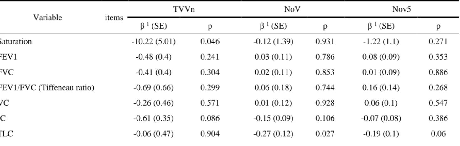
TVVn was correlated to hsCRP (regression coefficient (β) =3.64 (0.89), p = 0.01), inversely correlated) to serum antioxidant power (β=-102.77 (48.03), p = 0.036) (table 5) and to blood oxygen saturation (β =-10.22 (5.01), p = 0.046 (Table 6).
There was no a significative correlation concerning the other serum biomarkers (blood PaO², fibrinogen, plasma tiols), nor respiratory functional data (Tiffeneau ratio, FEV1), nor morphological data (emphysema, pulmonary artery diameter/aortic diameter ratio) (Table 7).
Multivariate analysis
There was an inverse correlation of TVVn with oxygen saturation (β = -9.44 (3.46), p = 0.01), and with the total anti-oxidant power (β = -90.21 (41.38), p = 0.03).
There was a significative independent correlation between TVVn and hsCRP (β = 3.12 (0.84) p = 0.001) (table 8).
Discussion
This study was undertaken to explore the relation between changes in pulmonary vascular morphology and 5-year post-CT clinical outcome in a cohort of patients with mild to moderate COPD. Our main findings were that body surface area-normalized total lung vascular volume (TVVn) was significantly correlated to all-cause hospitalisation and lung infection, as well as to some markers of systemic inflammation such as high-sensitivity CRP, while it was inversely correlated to total serum anti-oxidant capacity.
One of the most frequent co-morbidities of COPD is pulmonary vascular disease with a prevalence of pulmonary hypertension between 30% and 70 % [18]. However, the true prevalence of pulmonary hypertension in mild to moderate COPD is hard to estimate due to lack of large scale epidemiological studies and to the fact that pulmonary artery catheterisation is an invasive procedure. Moreover, up to two-thirds of COPD patients with normal resting pulmonary hemodynamics at rest may have abnormal pulmonary vascular pressure elevations during exercise [19]. When present, pulmonary hypertension is associated with exercise intolerance and increased risk of hospitalisation and mortality [20].
In the present study, we applied an automated analysis of lung CT images to measure the total volume of the segmented pulmonary vasculature. Our finding that an increased TVVn was associated with increased risk of lung infection and all-cause hospitalisation, may be explained by the progressive remodelling and increased resistance of small pulmonary arterioles, leading to
upstream distension of larger arterial branches. These larger more proximal vessels may have contributed more to the total vascular volume than the small peripheral vessels. The different mechanisms that may lead to increased arteriolar resistance, due to constriction and remodelling include hypoxemia and hypercapnia, polycythemia and acidosis, and mechanisms which lead to pruning of the vascular tree, comprising lung parenchymal destruction due to emphysema and inflammation related to cigarette smoke exposure and recurrent lung infections [18, 20]. In the present study, systemic inflammation as reflected by high sensitivity CRP and a decreased blood antioxidant capacity were significantly correlated with TVVn, while the estimated number of small pulmonary vessels was significantly correlated only with high-sensitivity CRP (Table 5). We found no significant correlation between lung vascular measurements and neither lung functional nor gas exchange parameters, emphysematous changes in the parenchyma as quantified by LAA%, nor AP/Ao ratio.
Previous studies have found that extra parenchymal vascular engorgement as assessed by a AP/Ao ratio > 1 is associated with severe exacerbations of COPD [21]. However, a single measurement of the pulmonary arterial trunk may be less sensitive to changes in the total volume of the pulmonary vasculature measured in 3D [22-23], which may explain the lack of significant correlation between this metric and TVV in the present study.
We did not find a significant correlation between the number of small pulmonary vessels and clinical outcome. Several previous studies have found that a 2-dimensional measurement of small vessel cross-sections (%CSA< 5 mm2) expressed as percentage of the total lung cross section
significantly correlates negatively with emphysema and positively with FEV1 [24-12] as well as with acute COPD exacerbation [25]. One hypothesis is that in our population of mild to moderate COPD patients, remodelling of the peripheral pulmonary blood vessels may have been the predominant mechanism rather than pruning of the vessels due to parenchymal destruction and
emphysema. Another potential explanation is that our study included only a small number of severe COPD patients (GOLD IV 8%) similar to the overall COPD population [26] and the prevalence of pulmonary vascular remodelling and hypertension may have been limited in these patients. Moreover, %CSA< 5 mm2 reported in previous studies in the literature is an aggregate
metric which may be driven both by the cross-section of small vessels as well as the cross-sectional area of the lung. The latter may be significantly affected by hyperinflation and emphysema, which may explain the strong correlations between this parameter and %LAA in other studies. This may explain why some studies have failed to find a consistent relation between %CSA< 5 mm2 and
%LAA in all COPD patients [15].
We found a significant correlation between TVVn and systemic inflammation (HsCRP) as well as an inverse correlation with total serum anti-oxidant capacity, a marker of coping mechanisms against oxidative stress. The oxidative stress produced by tobacco smoke exposure is thought to have a central role in COPD progression [27]. Inflammation is believed to be partly responsible for vascular endothelial dysfunction resulting in vasoconstriction and proliferation of endothelial smooth muscle cells leading to pulmonary vascular remodelling and pruning. Systemic inflammation and oxidative stress are also thought to play a significant role in cardiovascular comorbidities [28-29], neurodegenerative diseases [30-31], metabolic syndrome [32-33], cancer [34] in COPD patients. Inflammation and oxidative stress are also closely associated with lung infection [10], and may therefore represent pathways through which patients with increased risk of hospitalisation and lung infection may be characterized by a an increase in TVVn.
According to our study, vascular parameters were independent to other historical CT phenotypes data for COPD patients such as emphysema (β = 0.64 (0.72), p=0.379) or FEV1 (β = -0.48 (0.4), p=0.241). Vascular parameters may therefore be a key factor to take into account in COPD patients, although this observation remains to be confirmed by a larger scale prospective study.
Our study had several limitations. First, this was a retrospective observational study on a small population of mainly mild to moderate COPD patients. Our study would have benefited from the inclusion of healthy controls and smokers without COPD in order to better characterize the natural history of the changes in pulmonary vascular morphology. Moreover, the natural variations in pulmonary vasculature are currently unknown in healthy subjects. It would be therefore interesting to complete this study by conducting comparative studies of TVVn in healthy subjects, smokers without obstructive disease (stage 0 COPD) and smokers with COPD, in order to investigate vascular disease phenotypes in smokers with or without COPD. The metric used for the assessment of small pulmonary vessels included only the number of vessels with a cross-section < 5 mm2.
Although this measure is relevant to pruning of the pulmonary vasculature, the total cross-section of small vessels normalized to the total vascular volume could potentially have better characterized pulmonary vascular remodelling.
Conclusion
We investigated the relationship between changes in pulmonary vascular morphology and 5-year post-CT clinical outcome in a cohort of patients with mild to moderate COPD. Our data show that TVVn is associated with a small but significant additional risk of lung infection and all-cause hospitalization, is correlated to high-sensitivity CRP, a marker of systemic inflammation, and is inversely correlated to total serum antioxidant capacity. Moreover, the number of small pulmonary blood vessels was significantly correlated to hsCRP highlighting the role of systemic inflammation in the pruning of the peripheral pulmonary vasculature. Together, these data suggest that the non-invasive quantitative assessment of the pulmonary vasculature by multi-detector CT imaging may help characterize patient phenotypes at risk of infection and hospitalization, and merits larger controlled studies.
Figures and tables
Qualitative variables are presented in number and percentage (n (%)) and quantitative variables in median and interquartile range.
Table 1: Population characteristics
Variables n (%)
Exacerbation 32 (51.6)
All-cause hospitalization 31 (50)
Lung infection 13 (21)
ICU hospitalization 7 (11.3)
Lung infection and exacerbation have been diagnosed by the physician. All-cause hospitalization and all-cause ICU hospitalization
Table 2: Clinical events
Variables Items n (%) ; median [Q1 ; Q3]
Missing (%) Age (years) 63.8 [59 ; 69.6]
Sex male 46 (74.2) Body mass index (kg/m²) 25.8 [21.1 ; 28.7]
Tabacco pack-year 40 [20 ; 53] 1 (1.6)
Diabetes 7 (11.3)
High blood pressure ABPM 24h >130/80mmHg
30 (48.4)
Dyslipidaemia 27 (43.5)
HsCRP (if normal CRP) (mg/l) 2.8 [1 ; 7.4] 2 (3.2) Fibrinogen (g/l) 3.6 [3 ; 4.1]
COPD Stage GOLD 1 8 (12.9) GOLD 2 39 (62.9) GOLD 3 10 (16.1) GOLD 4 5 (8.1)
Heart failure (None) 61 (100) 1 (1.6) Nt pro BNP (pg/mL) 71.5 [40 ; 125]
IL6 (UI) 7 [2 ; 19] 37 (59.7) TNFa (UI) 10 [2 ; 10] 37 (59.7) Emphysema (-950UH, %) 9.5 [2.3 ; 16.5] 1 (1.6) Plasma thiols (UI) 423 [394 ; 455]
Quantitative values were presented as follow : median [Q1 ;Q3] The Mann and Whitney or Kruskal-Wallis tests used
Table 3: Correlation between vascular parameters (TVV, TVVn, NoV, NoV5) and all-cause hospitalisation, exacerbation, ICU hospitalization, and lung infection.
Clinical events Variable OR [IC] p
Lung infection TVVn (per 10 units) 1.06[ 1.01 ; 1.12] 0.02
All-cause hospitalization TVVn (per 10 units) 1.06[ 1.01 ; 1.12] 0.01
Table 4: Multivariate study of TVVn and lung infection and all-cause hospitalization
Variable
All-cause hospitalization ICU hospitalization Exacerbation Lung infection
No (n=31) Yes (n=31) p No (n=49 ) Yes (n=13 ) p No (n=30) Yes (n=32) p No (n=55) Yes (n=7) p
TVV ml 315 [228.5 ; 389.8] 375 [319.3 ; 481.6] 0.01 347 [268 ; 418.9] 375 [258.5 ; 398] 0.67 367.5 [246.3 ; 392.6] 340.5 [279.3 ; 438.5] 0.93 339 [246.4 ; 389.8] 458 [345 ; 489.5] 0.02 TVVn ml 165.8 [138.5 ; 212.4] 200.7 [180.3 ; 261.1] 0.02 189 [155.5 ; 231.9] 200.7 [152.3 ; 261.1] 0.66 191.4 [155.7 ; 231.9] 188 [153.9 ; 242.2] 0.83 185.2 [150.6 ; 212.4] 237.3 [187 ; 275.7] 0.05 NoV 39 [33 ; 52] 46 [39 ; 61] 0.13 46 [34 ; 55] 42 [37 ; 56] 0.81 45.5 [35 ; 52] 45 [35.5 ; 59.5] 0.68 43 [35 ; 52] 54 [42 ; 62] 0.09 NoV5 32 [25 ; 46] 39 [35 ; 47] 0.14 38 [27 ; 46] 37 [29 ; 45] 1.00 38.5 [29 ; 46] 37.5 [28.5 ; 46.5] 0.94 37 [29 ; 44] 45 [37 ; 52] 0.16
Variable items
TVVn NoV Nov5
β 1 (SE) p β 1 (SE) p β 1 (SE) p
hsCRP 3.64 (0.89) <0.001 0.79 (0.25) 0.002 0.44 (0.21) 0.04 Nt pro BNP 0.04 (0.02) 0.043 0.01 (0) 0.253 0.01 (0) 0.142 Fibrinogen 7.5 (9.84) 0.449 3.49 (2.61) 0.186 1.03 (2.12) 0.628 Plasma thiols -0.23 (0.15) 0.144 -0.03 (0.04) 0.443 -0.04 (0.03) 0.212 Protein -1.56 (1.12) 0.171 -0.19 (0.3) 0.543 -0.13 (0.24) 0.587 Thiols/protein ratio -6.51 (12.57) 0.606 -1.35 (3.37) 0.69 -2.82 (2.69) 0.298 Total antioxidant serum
power -102.77 (48.03) 0.036 -14.87 (13.22) 0.265 -7.01 (10.68) 0.514 PaO2 -2.02 (1.06) 0.062 0.05 (0.29) 0.852 -0.12 (0.23) 0.621 PaCO2 0.26 (2.04) 0.897 0.13 (0.55) 0.818 -0.2 (0.44) 0.654 pH -86.72 (340.73) 0.80 -34.93 (91.28) 0.703 -32.48 (73.23) 0.659 Bicarbonates -0.41 (2.14) 0.85 -0.26 (0.57) 0.658 -0.34 (0.46) 0.46
1 : correlation factor β : increase of one unit of variable is associated with increase in the TVVn
Table 5: correlation between vascular parameters (TVVn, NoV, NoV5) with biomarkers
Variable items
TVVn NoV Nov5
β 1 (SE) p β 1 (SE) p β 1 (SE) p
Saturation -10.22 (5.01) 0.046 -0.12 (1.39) 0.931 -1.22 (1.1) 0.271
FEV1 -0.48 (0.4) 0.241 0.03 (0.11) 0.786 0.08 (0.09) 0.353
FVC -0.41 (0.4) 0.304 0.02 (0.11) 0.853 0.01 (0.09) 0.886
FEV1/FVC (Tiffeneau ratio) -0.69 (0.66) 0.299 0.06 (0.18) 0.744 0.16 (0.14) 0.268
VC -0.26 (0.46) 0.571 0.01 (0.12) 0.928 0.06 (0.1) 0.547
IC -0.61 (0.35) 0.086 -0.15 (0.09) 0.106 -0.07 (0.08) 0.386
TLC -0.06 (0.47) 0.904 -0.27 (0.12) 0.027 -0.19 (0.1) 0.06
1 : correlation factor β : increase of one unit of variable is associated with increase in the TVVn
Table 6: correlation between vascular parameters (TVVn, NoV, NoV5) with functional data (PFT)
1 : correlation factor β : increase of one unit of variable is associated with increase in the TVVn
² : pulmonary artery diameter/aorta diameter ratio
Table 7: correlation between vascular parameters (TVVn, NoV, NoV5) with CT morphological data
Table 8: Multivariate correlation between vascular volume and biological data
Variable items
TVVn NoV Nov5
β 1 (SE) p β 1 (SE) p β 1 (SE) p
Emphysema (-950UH) 0.64 (0.72) 0.379 -0.03 (0.2) 0.896 -0.18 (0.16) 0.255
PA diameter (mm) 1.37 (1.88) 0.47 -0.14 (0.51) 0.789 0.27 (0.41) 0.509
PA/Ao ratio² 29.73 (69.54) 0.671 1.85 (18.67) 0.921 10.64 (14.92) 0.478
TVVn
Variables β (SE) P Variables used in the model
HsCRP 3.12 (0.84) 0.001 IC, hsCRP, Nt pro BNP, anti-oxidant serum power,
Protein, 0² Saturation, FEV1/FVC ratio.
0² saturation -9.44 (3.46) 0.01
References
[1] Vestbo J, Hurd SS, Agustí AG, Jones PW, Vogelmeier C, Anzueto A, et al. Global Strategy for the Diagnosis, Management, and Prevention of Chronic Obstructive Pulmonary Disease. Am J Respir Crit Care Med. 15Feb2013;187(4):347 65.
[2] Petty TL. Building a National Strategy for the Prevention and Management of and Research in Chronic Obstructive Pulmonary DiseaseNational Heart, Lung, and Blood Institute Workshop Summary. JAMA: The Journal of the American Medical Association. 1997 Jan 15;277(3):246. [3] McDonough JE, Yuan R, Suzuki M, Seyednejad N, Elliott WM, Sanchez PG, et al. Small-Airway Obstruction and Emphysema in Chronic Obstructive Pulmonary Disease. New England Journal of Medicine. 2011 Oct 27;365(17):1567–75.
[4] Magee F, Wright JL, Wiggs BR, Paré PD, Hogg JC. Pulmonary vascular structure and function in chronic obstructive pulmonary disease. Thorax. 1 Mar1988;43(3):183 9.
[5] Santos S, Peinado VI, Ramírez J, Melgosa T, Roca J, Rodriguez-Roisin R, et al. Characterization of pulmonary vascular remodelling in smokers and patients with mild COPD. European Respiratory Journal. 1Apr 2002;19(4):632 8.
[6] Alford SK, van Beek EJR, McLennan G, Hoffman EA. Heterogeneity of pulmonary perfusion as a mechanistic image-based phenotype in emphysema susceptible smokers. Proceedings of the National Academy of Sciences. 2010 Apr 20;107(16):7485–90.
[7] Estépar RSJ, Kinney GL, Black-Shinn JL, Bowler RP, Kindlmann GL, Ross JC, et al. Computed Tomographic Measures of Pulmonary Vascular Morphology in Smokers and Their Clinical Implications. American Journal of Respiratory and Critical Care Medicine. 2013 Jul 15;188(2):231–9.
[8] Barberà JA, Peinado VI, Santos S. Pulmonary hypertension in chronic obstructive pulmonary disease. European Respiratory Journal. May 2003;21(5):892 905.
[9] Naeije R, Barberà JA. Pulmonary hypertension associated with COPD. 2001;5(6):4.
[10] Repine JE, Bast A, Lankhorst I. Oxidative Stress in Chronic Obstructive Pulmonary Disease. 1997;156:17.
[11] Mallia P. Mechanisms and Experimental Models of Chronic Obstructive Pulmonary Disease Exacerbations. Proceedings of the American Thoracic Society. 2005 Nov 1;2(4):361–6.
[12] Matsuoka S, Washko GR, Dransfield MT, Yamashiro T, San Jose Estepar R, Diaz A, et al. Quantitative CT Measurement of Cross-sectional Area of Small Pulmonary Vessel in COPD. Academic Radiology. Jan 2010;17(1):93 9.
[13] Lang RM, Bierig M, Devereux RB, Flachskampf FA, Foster E, Pellikka PA, et al. Recommendations for Chamber Quantification: A Report from the American Society of Echocardiography’s Guidelines and Standards Committee and the Chamber Quantification
Writing Group, Developed in Conjunction with the European Association of Echocardiography, a Branch of the European Society of Cardiology. Journal of the American Society of Echocardiography. 2005 Dec;18(12):1440–63.
[14] Coste F, Dournes G, Dromer C, Blanchard E, Freund-Michel V, Girodet P-O, et al. CT evaluation of small pulmonary vessels area in patients with COPD with severe pulmonary hypertension. Thorax. sept 2016;71(9):830 7.
[15] Mashimo S, Chubachi S, Tsutsumi A, Kameyama N, Sasaki M, Jinzaki M, et al. Relationship between diminution of small pulmonary vessels and emphysema in chronic obstructive pulmonary disease. Clinical Imaging. nov 2017;46:85 90.
[16] Kybic J, Unser M: Fast parametric elastic image registration. IEEE transactions on image processing : a publication of the IEEE Signal Processing Society 2003, 12(11):1427-1442.
[17] Leventon, M.E. et al. "Statistical Shape Influence in Geodesic Active Contours", CVPR 2000 [18] O.A. Minai, A. Chaouat, S. Adnot, Pulmonary hypertension in COPD: epidemiology, significance, and management: pulmonary vascular disease: the global perspective, Chest 137(6 Suppl) (2010) 39S-51S.
[19] M. Oswald-Mammosser, M. Apprill, P. Bachez, M. Ehrhart, E. Weitzenblum, Pulmonary hemodynamics in chronic obstructive pulmonary disease of the emphysematous type, Respiration; international review of thoracic diseases 58(5-6) (1991) 304-10.
[20] F.N. Rahaghi, E.J. van Beek, G.R. Washko, Cardiopulmonary coupling in chronic obstructive pulmonary disease: the role of imaging, J Thorac Imaging 29(2) (2014) 80-91.
[21] J.M. Wells, G.R. Washko, M.K. Han, N. Abbas, H. Nath, A.J. Mamary, E. Regan, W.C. Bailey, F.J. Martinez, E. Westfall, T.H. Beaty, D. Curran-Everett, J.L. Curtis, J.E. Hokanson, D.A. Lynch, B.J. Make, J.D. Crapo, E.K. Silverman, R.P. Bowler, M.T. Dransfield, C.O. Investigators, E.S. Investigators, Pulmonary arterial enlargement and acute exacerbations of COPD, The New England journal of medicine 367(10) (2012) 913-21.
[22] Chaouat A, Bugnet A-S, Kadaoui N, Schott R, Enache I, Ducoloné A, et al. Severe Pulmonary Hypertension and Chronic Obstructive Pulmonary Disease. American Journal of Respiratory and Critical Care Medicine. 2005 Jul 15;172(2):189–94
[23] Corson N, Armato SG, Labby ZE, Straus C, Starkey A, Gomberg-Maitland M. CT-Based Pulmonary Artery Measurements for the Assessment of Pulmonary Hypertension. Academic Radiology. 2014 Apr;21(4):523–30.
[24] Y. Matsuura, N. Kawata, N. Yanagawa, T. Sugiura, Y. Sakurai, M. Sato, K. Iesato, J. Terada, S. Sakao, Y. Tada, N. Tanabe, Y. Suzuki, K. Tatsumi, Quantitative assessment of cross-sectional area of small pulmonary vessels in patients with COPD using inspiratory and
[25] K. Yoshimura, Y. Suzuki, T. Uto, J. Sato, S. Imokawa, T. Suda, Morphological changes in small pulmonary vessels are associated with severe acute exacerbation in chronic obstructive pulmonary disease, Int J Chron Obstruct Pulmon Dis 11 (2016) 1435-45.
[26] Naeije R, Barberà JA. Pulmonary hypertension associated with COPD. 2001;5(6):4
[27] Kirkham PA, Barnes PJ. Oxidative Stress in COPD. Chest. 2013 Jul;144(1):266–73.
[28] Haverkate E, Thompson SG, Pyke SD, Gallimore JR, Group MBP. Production of C-reactive protein and risk of coronary events in stable and unstable angina. The Lancet. Feb 1997;349(9050):462 6.
[29] Thunström E, Glantz H, Fu M, Yucel-Lindberg T, Petzold M, Lindberg K, et al. Increased Inflammatory Activity in Nonobese Patients with Coronary Artery Disease and Obstructive Sleep Apnea. Sleep. march 2015;38(3):463 71.
[30] Lyras L, Cairns NJ, Jenner A, Jenner P, Halliwell B. An Assessment of Oxidative Damage to Proteins, Lipids, and DNA in Brain from Patients with Alzheimer’s Disease. Journal of Neurochemistry. 2002 Nov 18;68(5):2061–9.
[31] Aguirre T, Matthijs G, Robberecht W, Tilkin P, Cassiman J-J. Mutational analysis of the Cu/Zn superoxide dismutase gene in 23 familial and 69 sporadic cases of amyotrophic lateral sclerosis in Belgium. European Journal of Human Genetics. 1999 Jul;7(5):599–602.
[32] Shin S-K, Cho H-W, Song S-E, Song D-K. Catalase and nonalcoholic fatty liver disease. Pflügers Archiv - European Journal of Physiology [Internet]. 2018 Aug 17 [cited 2018 Aug 24]; Available from: http://link.springer.com/10.1007/s00424-018-2195-z
[33] Almeida-Suhett CP, Graham A, Chen Y, Deuster P. Behavioral changes in male mice fed a high-fat diet are associated with IL-1β expression in specific brain regions. Physiology & Behavior. 2017 Feb;169:130–40.
[34] Woraruthai T, Charoenlap C, Hongsaprabhas C, Mutirangura A, Honsawek S. Alu hypermethylation and high oxidative stress in patients with musculoskeletal tumors. PeerJ. 2018 Aug 16;6:e5492.