Open Archive TOULOUSE Archive Ouverte (OATAO)
OATAO is an open access repository that collects the work of Toulouse researchers and
makes it freely available over the web where possible.
This is an author-deposited version published in :
http://oatao.univ-toulouse.fr/
Eprints ID : 18544
To link to this article : DOI:10.1016/j.otsr.2015.06.026
URL :
https://doi.org/10.1016/j.otsr.2015.06.026
To cite this version : Bonnevialle, Nicolas Radial head replacement
in adults with recent fractures. (2016) Orthopaedics & Traumatology:
Surgery & Research, vol. 102 (n° 1 suppl). pp. S69-S79. ISSN
1877-0568
Any correspondence concerning this service should be sent to the repository
administrator:
[email protected]
Radial
head replacement in adults with recent fractures
N.
Bonnevialle
∗Service d’orthopédie et de traumatologie, hôpital Riquet, place Baylac, 31059 Toulouse cedex 09, France
Keywords: Radial head Fracture Prosthesis
a b s t r a c t
Radial head fractures are fairly common (20% of all traumatic elbow injuries). Non-operative treatment is indicated in non-displaced fractures, and direct stable internal fixation allowing early elbow mobilisation in most other cases. For severely comminuted fractures precluding stable fixation, replacement of the radial head was introduced in the 1970s as a better alternative to simple radial head resection, which can induce instability of the elbow and/or forearm, most notably in patients who have complex fractures with concomitant lesions to other structures. With contemporary implants (modular or monoblock, with or without a mobile cup), mechanical stability is close to that provided by the native radial head, although appropriate treatment of concomitant lesions remains crucial (e.g., re-attachment of the radial collateral ligament, or distal radio-ulnar stabilisation in patients with Essex-Lopresti fracture). The key techni-cal points are selection of implant size and determination of the optimal implantation height. The two most common complications are capitellar overloading due to excessively high implantation of the pros-thetic head, which causes stiffness and pain, and loosening of the stem. These complications may require removal of the implant at a distance from the injury. Studies have demonstrated satisfactory clinical outcomes in 60% to 80% of cases.
1. Introduction
Radial head fractures are common lesions found in nearly 20% of elbow injuries, typically after a fall on the outstretched hand with the wrist extended and the forearm pronated[1].
Simple isolated non-displaced radial head fractures are treated non-operatively. In contrast, whenever possible, displaced frac-tures should be managed by anatomic reduction, stable fixation, and repair of any concomitant lesions, which are present in nearly 80% of cases[2–4]. Severe comminution, however, precludes radial head reconstruction. In this situation, radial head replacement can be considered, as a means of preventing elbow instability.
Radial head replacement was introduced in the 1970s as a better alternative to simple radial head resection. This procedure was initially designed to prevent ectopic ossification [5]. Since then, improvements in the knowledge of elbow biomechanics have allowed advances in the design of radial head implants and in the materials used to construct them.
The objective of this work is to describe the biomechanical role of the radial head, to discuss the role for radial head replacement in the treatment of radial head fractures in adults, and to assess the
∗ 113, avenue de Muret, 31300 Toulouse, France. Tel./fax: +33 5 61 77 21 04. E-mail addresses:[email protected],[email protected]
outcomes and main complications of this procedure in the light of recently published data.
2. Anatomy and biomechanics of the radial head
The radial head is part of the lateral compartment of the elbow. It articulates with the capitellum to form the humero-radial joint. In addition, the circumference of the radial head articulates with the radial notch of the ulna, forming the proximal radio-ulnar joint. Finally, the radial head is a key component of the forearm scaffold.
The radial head is not round; instead, it has an ellipsoid and conical shape, with a proximal-to-distal increase in size[6]. The longest axis (20–23 mm on average) is perpendicular to the radial notch of the ulna in neutral rotation. As a consequence, the annular ligament that encircles the radial head becomes slack when the forearm is in supination[6–8](Fig. 1). The neck of the radius is about 13 mm long and forms an angle of about 17◦with the radial shaft[6,8].
The blood supply to the radial head is most abundant periph-erally, where it derives from metaphyseal arterial branches. These arteries form a terminal network, a configuration associated with a risk of post-traumatic necrosis and non-union[9].
The medial cortex is reinforced by trabeculae that converge towards the radial (bicipital) tuberosity. As a result, traumatic force
Fig. 1. a: in the neutral position, the annular ligament is not under tension and the proximal radio-ulnar joint is visible. In (b) supination and (c) pronation, the longest diameter of the radial head aligns with the radial notch of the ulna, the annular ligament is under tension, and the joint space is closed. LD: long diameter; SD: short diameter.
From F. Duparc[9].
applied in valgus often causes lateral depression of the epiphysis and metaphysis.
2.1. Radial head and pronation/supination
Two factors allow pronation/supination of the forearm. One is the crankshaft shape of the radius composed of a pronating dia-physeal curve surmounted by a proximal supinating counter-curve formed by the neck and head of the radius. The other is the stabil-ity of the two-bone forearm scaffold, provided proximally by the radial head and annular ligament, quadrate ligament of Denucé, and interosseous membrane; and distally by the radio-ulnar ligament and triangular fibrocartilage complex.
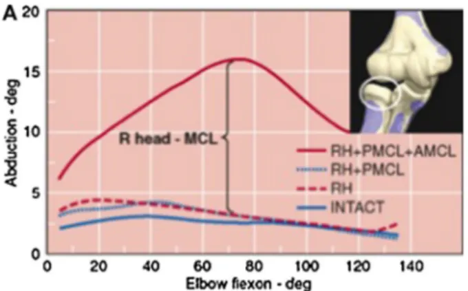
Fig. 2. Findings from the biomechanical study by Morrey et al.[10]showing the extent of valgus instability of the elbow at various degrees of flexion after removal of the radial head (RH) combined with division of the medial (ulnar) collateral ligament (MCL) or of only its anterior (AMCL) or posterior (PMCL) portion.
2.2. Radial head and stability of the elbow
The ulnar collateral ligament (medial collateral ligament) is a primary stabiliser of the elbow in resisting valgus stress. Its ante-rior portion attached to the medial margin of the coronoid process makes a major contribution to this role, as demonstrated in biome-chanical studies by Morrey et al.[10]. Thus, isolated lesions of the radial head with an intact ulnar collateral ligament do not cause valgus instability (Fig. 2). Sectioning the anterior and posterior por-tions of the ulnar collateral ligament results in only 6◦to 8◦of joint distraction provided the radial head is intact. However, section of the ligament combined with a radial head fracture causes valgus subluxation of the elbow (Fig. 3)[10].
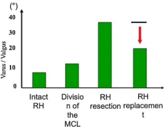
Valgus stability after radial head replacement with a metal implant is similar to that provided by the native radial head. In contrast, silicone implants do not provide adequate biomechanical resistance (Fig. 4)[11,12].
The radial collateral ligament (lateral collateral ligament) con-tributes to the lateral stability of the humero-ulnar joint, preventing rotational instability in supination and in varus, most notably via its posterior portion attached to the ulna.
Fig. 3. Findings from the biomechanical study by Morrey et al.[10]showing the contribution to valgus stability of the radial head (RH), medial (ulnar) collateral ligament (MCL), and anterior (AMCL) and posterior (PMCL) portions of the medial collateral ligament.
Fig. 4. Evaluation of valgus/varus stability with different procedures. RH: radial head.
Congruity of the humero-ulnar joint is also crucial, for both rotational and lateral stability. Resection of more than 50% of the olecranon or coronoid process causes instability[13].
Dynamic stabilisation by the muscles is more controversial. The flexors and pronators contribute to valgus stability of the elbow and the anconeus muscle to varus stability. The muscles attached to the lateral epicondyle of the humerus are involved in postero-lateral stabilisation of the elbow[13].
3. Clinical features
Radial head fractures are often caused by an indirect trauma produced by a fall onto an outstretched hand (FOOSH injury), with the elbow extended and the forearm pronated. Despite the overall oedema of the elbow, care should be taken to feel the bony land-marks of the elbow (olecranon, medial and lateral epicondyles, and radial head). Marked posterior prominence of the olecranon sug-gests dislocation of the elbow, which may be part of a terrible triad injury (radial head fracture, coronoid fracture, and postero-lateral dislocation of the elbow) or combined with of a fracture of both forearm bones.
Great care should be taken to identify concomitant lesions in patients with radial head fractures. Thus, visible medial bruising may indicate injury to the ulnar collateral ligament (Fig. 5); whereas pain and oedema of the forearm and wrist suggest damage to the interosseous membrane (Essex-Lopresti fracture).
4. Imaging workup
Standard radiographs of the elbow in the antero-posterior and lateral projections should be obtained, as well as a radial
Fig. 5. Medial bruising suggestive of damage to the ulnar (medial) collateral liga-ment in addition to the radial head fracture.
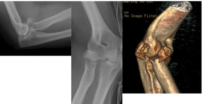
head-capitellum view (modified lateral view with the tube angled 45◦ towards the radial head, as described by Greenspan) [14]. Scrupulous attention should be directed to the detection of any concomitant lesions, such as a coronoid fracture. Displacement and comminution of the fracture are often underestimated on standard radiographs. Computed tomography (CT) provides a more accurate evaluation (Fig. 6).
An antero-posterior radiograph of the wrist, or ideally of both wrists, is used to assess the radio-ulnar index (ulnar variance). A positive index with the ulnar surface projecting more distally indi-cates ascension of the radius related to an Essex-Lopresti fracture. Marked displacement of the radial head fracture and pain and/or oedema of the wrist further support this diagnosis (Fig. 7).
Ultrasonography and magnetic resonance imaging (MRI) are helpful for diagnosing rupture of the interosseous membrane when the antero-posterior radiograph of the wrist is normal. A useful sign of inter-osseous membrane injury is herniation of the muscles into the inter-osseous space when moving the probe over the forearm in supination[15](Fig. 8).
MRI is effective in detecting capitellar cartilage injuries.
5. Classifications
The first widely accepted classification was developed by Mason in 1954 based on 100 radial head fractures treated operatively or non-operatively and re-evaluated after more than 2 years[16]. Three fracture types that require different therapeutic approaches were distinguished: Type I, non-displaced marginal fissure or fracture; Type II, displaced marginal fracture with separation or impaction; and Type III, displaced comminuted fracture involving the entire radial head (Fig. 9). Subsequently, Broberg and Morrey added a type IV defined as a radial head fracture combined with elbow dislocation[17].
Hochkiss modified Mason’s classification by adding a number of clinical criteria. Type II is defined as a displaced fracture of the radial head or neck combined with mechanical blocking of joint motion or with loss of joint congruity. Type III is characterised by comminution, which precludes internal fixation and requires either resection or prosthetic replacement of the radial head (Table 1)[18]. A limitation of these classifications is their poor intra-observer and inter-observer reproducibility. Another key weakness is their failure to consider concomitant lesions, which are present in nearly 80% of multi-fragment fractures (particularly Type III fractures)
[2–4].
The Mayo Clinic classification, in contrast, considers all con-comitant lesions and therefore deserves preference[19](Fig. 10). The radial head fracture is described as in Mason’s classification, and letters are added to indicate the concomitant lesions, in upper case if the lesion is treated and lower case otherwise. For instance, a Type III Lcm fracture is a Mason Type III fracture of the radial head with a repaired tear in the radial collateral ligament (L), an untreated coronoid fracture (c), and an untreated tear in the ulnar collateral ligament (m).
Table 1
Mason classification as modified by Hotchkiss[18].
Type I Fracture of the radial head or neck, with little or no displacement Range of pronation/supination limited only by pain and oedema Articular fracture < 2 mm or marginal fracture of the radial head Type II Displaced fracture (> 2 mm) or fracture of the neck with angulation
Motion range limitation due to mechanical impingements or loss of joint congruity
No severe comminution (internal fixation feasible) Type III Severe comminuted fracture of the radial head or neck
Reconstruction not feasible according to criteria assessed intra-operatively or radiologically
Fig. 6. Standard radiographs and 3D computed tomography reconstruction providing an evaluation of the extent of comminution.
6. Therapeutic strategy and indications
Type I fractures are managed non-operatively and Type II frac-tures by reduction and stable internal fixation[2,20]. Radial head replacement is considered only in Type III fractures for which anatomic reduction and stable internal fixation allowing early mobilisation would be difficult or impossible to achieve. Thus, radial head replacement is an alternative to a challenging internal fixation procedure or radial head excision.
Fig. 7. Antero-posterior radiograph of the wrist with the forearm pronated: positive ulnar variance indicating patent instability of the forearm scaffold as a component of an Essex-Lopresti fracture.
6.1. Isolated multi-fragment fracture of the radial head
The absence of any concomitant lesions is very difficult to estab-lish with absolute certainty, even based on the above-described clinical and imaging findings. In most cases, the radial head frac-ture is only apparently isolated. Consequently, when the radial head cannot be reconstructed, resection arthroplasty is not advisable. In Type III multi-fragment fractures, resection arthroplasty is followed by proximal migration in nearly 50% of cases, cubitus valgus in 30% of cases, and medium-term humero-ulnar osteoarthritis in 50% of cases[21]. Radial head replacement is thus the immediate solution when stable internal fixation is not feasible.
6.2. Terrible triad of the elbow
Terrible triad injury consists of postero-lateral elbow dislo-cation, a radial head fracture, and a coronoid fracture. Simple reduction is often unstable and surgery is therefore warranted. The goal of surgery is to reconstruct the lateral column of the elbow, at all costs, to avoid the development of postero-lateral instability
[13]. The lateral column includes the radial head and the radial col-lateral ligament. In terrible triad injury, there is no role for radial head excision when internal fixation is not feasible. Instead, the treatment should combine radial head replacement, internal fix-ation of the coronoid, and re-attachment of the radial collateral ligament (to the lateral pillar, using bone anchors in most cases or, more rarely, trans-osseous suture fixation). If the elbow remains unstable after completion of these procedures, a medial approach is used to re-attach the ulnar collateral ligament[22].
6.3. Proximal fracture of both forearm bones
The radial head fracture may be accompanied with a fracture of the proximal ulna[23]. Stabilisation of the ulnar fracture requires strong plate fixation capable of withstanding the major stresses that tend to dislocate the elbow. When internal fixation of the radial head is not feasible, radial head replacement should be performed to reconstruct the radial column, as healing of the ulnar fracture is difficult to obtain in the absence of radial column reconstruction
Fig. 8. “Muscular hernia sign” described by Soubeyrand et al. and indicating a tear in the inter-osseous membrane[15].
6.4. Can radial head replacement be used in all age groups?
Multi-fragment radial head fractures can be caused by a high-energy trauma in a young individual. Anatomic reduction followed by stable internal fixation is the best option in this situation. However, it would be unwise to insist on this strategy when perfect reduction and/or stable fixation cannot be achieved, as post-operative immobilisation would be necessary. Radial head replacement allowing early mobilisation is preferable.
In published studies, mean age of patients treated with radial head replacement was about 50–60 years, and the lowest end of the age range was 16 years[24–29]. However, younger age is associated with a significant increase in the risk of revision surgery, for which the main reason is loosening of the implant[24].
7. Types of radial head implants
7.1. Swanson-type SilasticTMimplants
SilasticTM implants became popular in the 1970s based on
data published by Swanson. These semi-rigid cementless implants were sometimes used as temporary spacers to prevent ascension
of the radius in patients with instability of the forearm scaffold
[30]. Advantages of using a yielding material such as silicone elastomer are good clinical tolerance despite possibly imperfect implant position and greater ease of removal compared to rigid implants. However, in patients with ulnar collateral ligament injuries, SilasticTM implants cannot act as secondary valgus
sta-bilisers[10]. Finally, high rates of osteolysis (triggered by silicone particles: siliconitis) and of implant fractures have been reported. Because these complications require removal of the implant at a more or less early date, SilasticTMradial head implants are no longer
recommended[31].
7.2. Metal implants
Long-term outcome data are available for several contemporary models of rigid radial head implants. They can be classified based on three main criteria:
• intra-prosthetic mobility; • modularity;
• stem fixation (cemented, cementless, or floating in the intra-medullary canal) (Fig. 11).
Type I
Type II
Type III
Fig. 10. Mayo Clinic classification[19].
Bipolar implants were developed after the Judet prosthesis to improve proximal radio-ulnar congruity via self-alignment of the cup (with a decrease in local stresses)[32]. In vitro, bipolar implants provide less stability than do monoblock implants, although this difference has not been demonstrated clinically[33]. Furthermore, there have been reports of cup disassembly related to insufficient retention within the implant, in patients with residual elbow insta-bility[26](Fig. 12).
Modular implants offer two advantages:
• the optimal head size can be chosen;
• the height of the head and neck can be adjusted to match the height of the resection, which is a crucial technical point.
Some implant types also allow adjustment of the head-neck angle to achieve the indispensable goal of replicating the patient’s native anatomy as closely as possible. This possibility is particularly valuable when anchoring a long straight stem into the shaft, as the axis of these stems differs from that of the shorter radial neck.
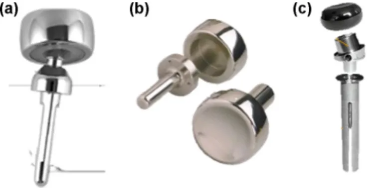
Fig. 11. a: example of a modular mobile-cup implant (bipolar); b: example of a modular pyrolytic carbon implant with a fixed head-neck angle; c: example of a monoblock implant.
Fixation within the intra-medullary canal is usually achieved using either a cemented stem, which is often smooth, or a cement-less press-fit stem. Primary stability of press-fit stems is difficult to obtain, as micro-fractures are common, particularly when the stem diameter is oversized by 1 mm or more[34]. Furthermore, stress shielding has been reported as a cause of early proximal peri-prosthetic osteolysis[34].
It has been suggested that the implant can be left loose in the proximal radial shaft, with no attempt to achieve primary stability by press-fit implantation. The goal is to allow self-alignment of the
Fig. 12. Detachment of the implant head due to residual postero-lateral elbow instability.
prosthesis on the capitellum. The development of radio-lucencies around the stem do not seem clinically relevant, and satisfactory medium-term outcomes have been reported[35].
Promising early outcomes have been reported with implants made of pyrolytic carbon[36]. This material is well tolerated when placed in contact with the capitellar cartilage. However, longer-term data are needed to validate pyrocarbon implants.
There is now a recommendation not to use Swanson-type implants. No clinical studies have established that any of the other implant types is superior over the others.
8. Operative technique: tips and tricks
The Kocher lateral approach between the extensor carpi ulnaris and anconeus is the most widely used. Another option is the Kaplan approach, which is located more anteriorly, between the extensor carpi radialis longus and the extensor digitorum communis.
The annular ligament is spared to allow its suture if needed at the end of the procedure. Careful attention is directed to sparing the radial collateral ligament (particularly its ulnar portion) if this structure was not damaged at the time of the trauma.
Pronation of the forearm places the inter-osseous nerve at a safe distance when approaching the neck of the radius.
When primary internal fixation of the olecranon via the poste-rior approach is performed also in a patient with proximal fractures of both forearm bones, a single posterior skin incision can be per-formed. The radial head is then approached either via a lateral arthrotomy or through the olecranon fracture.
The capitellar cartilage should be evaluated during the arthro-tomy. Although the condition of the capitellar cartilage has no bearing on the therapeutic conditions, it is among the factors that govern the long-term prognosis.
The technique for implanting a radial head prosthesis, with or without ancillary instruments, may appear simple. Nevertheless, two fundamental principles must be applied[20]: the implant must replicate the thickness and diameter of the native radial head, and it must be positioned at the same height. Low implant position may result in residual elbow instability, whereas high implant position places excessive stress on the condyle, which rapidly causes joint damage with pain and motion range limitation at the elbow, par-ticularly in patients with trauma-induced osteochondral lesions of the capitellum[37].
Reconstitution on the operating table of the native head, as if putting a puzzle together, confirms that all the intra-articular frag-ments have been removed and provides guidance about the implant diameter that best replicates the native radial head (Fig. 13).
A fluoroscopic evaluation after placement of the trial prosthesis is recommended to confirm that the best size has been chosen.
Fig. 13. Intra-operative selection of the head size that best replicates the patient’s native anatomy.
When there is doubt between two sizes, the smallest size should be selected.
Replicating the height of the native radial head is the second technical challenge. The match with the native geometry must be as close as possible. When the forearm scaffold is unstable (Essex-Lopresti fracture), reduction and pin fixation of the distal radio-ulnar joint should be performed before determining the size and height of the implant, to avoid errors due to ascension of the radius relative to the ulna.
A number of anatomical landmarks help to determine the opti-mal implant height on fluoroscopy images (Fig. 14):
• alignment of the implant on the ulnar notch, with no overshoot, on the lateral view;
• and symmetric appearance of the medial and lateral sides of the humero-ulnar joint space.
Tests using trial implants are indispensable. The radial neck can be recut at the level of the radial notch of the ulna to lower an excessively high implant. If the implant is too low, the length of the prosthetic neck can be increased by choosing an appropriate modular stem.
The radius is a very slender bone and should therefore be pre-pared with great care to avoid creating micro-fractures of the neck. When a cemented stem is used, low-viscosity cement may be preferable. The shaft can be occluded by a fragment of the head or an artificial plug to allow cement fixation under pressure and to avoid distal cement leakage.
Finally, radial head replacement does not necessarily ensure sta-bility of the elbow and forearm scaffold if the concomitant lesions are left untreated.
To prevent elbow instability, it is important to re-attach the radial collateral ligament complex, which is often detached proxi-mally, as well as the muscles inserted on the lateral epicondyle. Either bone anchors or trans-osseous suture fixation can be used. This step is particularly crucial in terrible triad injury, together with fixation of the coronoid (if allowed by the bone fragment). If the elbow remains unstable, an additional medial approach is per-formed to re-attach the ulnar collateral ligament. When this step is insufficient, external fixation is required (Fig. 15).
Fig. 14. Implant position criteria determined by intra-operative fluoroscopy. The lateral and medial sides of the humero-ulnar joint space should be symmetrical. The implant aligns with the radial notch of the ulna.
Fig. 15. External fixation to stabilise the elbow in a patient with residual postero-lateral instability after treatment of terrible triad injury consisting of radial head replacement, re-attachment of the radial collateral ligament, fixation of the coronoid, and re-attachment of the ulnar collateral ligament.
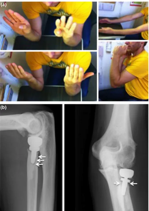
To prevent forearm scaffold instability due to an Essex-Lopresti fracture, the recommended procedure is pinning in supination to stabilise the distal radio-ulnar joint. If needed, repair of the trian-gular fibrocartilage complex is recommended to allow healing of the inter-osseous membrane. The diagnosis of Essex-Lopresti frac-ture rests on pre-operative imaging (ultrasonography or MRI) and, above all, on the intra-operative radius joystick test described by Soubeyrand et al.[38](Fig. 16). The instability can also be visualised on fluoroscopy images centred on the distal radio-ulnar joint, which show piston motion of the radius during dynamic manoeuvres.
During the post-operative period, early mobilisation within a safe range is recommended for the first 6 weeks. The elbow should not be fully extended. After repair of the radial collat-eral ligament complex, the forearm should be kept in pronation. In patients with elbow instability, great care should be taken to
remain within the safe range of motion, which should be deter-mined intra-operatively.
9. Outcomes and complications
9.1. Outcomes
After radial head replacement for a recent fracture, the outcome is satisfactory in 60% to 80% of cases (Table 2). Burkhart et al.[40]
reported that the clinical outcomes remained satisfactory in the long term (after more than 8 years), despite the development of humero-ulnar osteoarthritis.
In a recent study of 105 cementless monoblock implants re-evaluated after more than 6 years, Duckworth et al.[24]found a
Fig. 16. a and b: Joystick test described by Soubeyrand et al. as a means of detecting tears in the inter-osseous membrane. The shoulder is in abduction and internal rotation and the forearm is pronated. The neck of the radius is grasped using a forceps and pulled laterally. The test is negative (a) when the radius remains in place opposite the capitellum. Lateral translation of the radius indicates a positive test (b).
Table 2
Main case-series studies of radial head replacement to treat recent fractures.
Reference n Type of implant Flex/Ext (◦) Pron/Sup (◦) Grip strength vs. contralateral upper limb
Satisfactory objective outcome (%)
Mean follow-up (years)
Judet et al.[25] 5 Bip 138/-6 86/82 100 4
Alnot et al.[28] 18 Bip 126/-18 77/79 75% 100 1.5
Dotzis et al.[39] 12 Bip 140/-14 87/84 90% 83 5
Doornberg et al.[35] 27 Mod not bip 131/-20 73/57 82 3.5
Flinkkilä et al.[27] 42 Mod not bip 136/-20 75/73 85% 62 4
Allavena et al.[26] 14 Bip 113/-29 71/73 71 4
Burkhart et al.[40] 9 Bip 124/-21 64/64 100 8.8
Popovic et al.[41] 51 Bip 130/-14 68/71 85% 76 8.4
Grewal et al.[42] 26 Mod not bip 138/-25 71/56 85% 61 2
Moro et al.[43] 25 Mono 140/-8 78/68 68 3.5
Lamas et al.[44] 47 Mod not bip 140/-6 75/67 89 4
Bip: bipolar; Mod: modular; Mono: monoblock; Flex: flexion; Ext: extension; Pro: pronation; Sup: supination.
Fig. 17. a and b: young patient 4 years after treatment of a complex radial head fracture by implantation of a cemented bipolar prosthesis. Although the clinical outcome is good (a), the implant is loose and the stem is tilted in an area of diaphyseal osteolysis (b).
Fig. 18. a and b: severe capitellar erosion by a radial head implant that was positioned too high (a) Intra-operative appearance at removal of the implant (b).
28% revision rate. Independent risk factors for failure were young age and the use of silicone implants.
However, in the large multicentre study of 315 radial head implants of any type conducted by the French Society for the Shoulder and Elbow (SOFEC), revision because of clinical fail-ure was required in only 26 (8%) cases. Reasons for failfail-ure were loosening, instability, painful stiffness due to overstuffing, and/or radio-capitellar osteoarthritis[45].
9.2. Complications
9.2.1. Aseptic loosening
Stem loosening does not seem to correlate with clinical symp-toms. A radiolucent line is often visible surrounding the stem even in patients who remain completely asymptomatic (Fig. 17). Typical features of pain related to aseptic loosening consist of a mechani-cal time pattern and location at the forearm with radiation to the wrist.
A radiolucent line is a consistent finding when the stem is inten-tionally left loose in the shaft[35]. Radiological signs of loosening usually develop fairly early (within 12 months) with cementless press-fit stems and may develop later on with cemented implants, particularly those having a bipolar design[1,27].
In patients with functional impairment, implant removal rather than exchange is recommended, particularly if the concomitant lesions caused by the trauma have had time to heal.
9.2.2. Elbow instability
Elbow instability after radial head replacement is often ascrib-able to incomplete treatment of all the concomitant lesions. An example is failure to re-attach the radial and ulnar collateral lig-aments and to perform internal fixation of the coronoid process in patients with terrible triad injury[22].
The type of implant may influence elbow stability. In theory, fixed-cup implants provide a greater degree of elbow stability than do bipolar mobile-cup implants[33]. If the radial ligament complex is deficient, postero-lateral subluxation of a mobile cup may occur, as demonstrated by O’Driscoll. In this situation, there is a risk of cup disassembly[26].
In every case, the treatment of residual elbow instability after radial head replacement rests on the repair of the concomitant lig-ament and bone lesions, as opposed to revision of the implant itself, which should be left in place if it is properly positioned. External fixation to neutralise dislocating forces may be useful to protect the healing process.
9.2.3. Overstuffing with radio-capitellar pressure elevation
Excessive pressure applied to the humeral condyle by the radial head causes pain and stiffness of the elbow. The most common cause is malposition of the implant, which is usually too high
[1,22,37]. However, even when the implant is optimally positioned, persistent forearm scaffold instability (Essex-Lopresti fracture)
with failure of the intra-osseous membrane to heal can result in radio-capitellar pressure elevation.
The trauma responsible for the radial head fracture fairly often causes osteochondral damage to the capitellum, which worsens in the event of overstuffing. In patients with radiological erosion of the capitellum, implant removal relieves the pain (Fig. 18).
9.2.4. Osteoarthritis
Capitellar osteoarthritis is ascribable not only to cartilage lesions caused by the initial trauma, but also to increased stress on the radio-capitellar compartment and/or to persistent postero-lateral instability.
The osteoarthritis is usually progressive and extends to the humero-ulnar compartment. Removal of the implant seems to accelerate the pace of osteoarthritis progression.
10. Conclusion
There is a sound rationale for including radial head replacement into the therapeutic strategy for multi-fragment radial head frac-tures for which reliable internal fixation is not feasible (type III fractures).
Resection arthroplasty on an emergency basis is best avoided, as ligament damage is very common and can result in instability of the elbow and/or forearm scaffold. Silicone implants should not be used. Among the other types of implant, none has been proven superior over the others.
Great care is essential to determine the optimal height of the implant and to avoid overstuffing responsible for pain and/or stiff-ness or, on the contrary, for residual instability.
Comprehensive management of the concomitant lesions is indispensable to ensure stability of the elbow.
Disclosure of interest
The author declares that he has no competing interest.
Acknowledgements
We thank Prof. P. Mansat, Prof. F. Duparc, Dr M. Soubeyrand, Dr C. Allavena, and Dr S. Delclaux for their contribution to the scientific content and illustrations of this lecture.
References
[1]Van Riet RP, Van Glabbeek F, Morrey BF. Radial head fracture. In: Morrey BF, editor. The elbow and its disorders. 4th ed. Philadelphia, PA: Saunders Elsevier; 2009. p. 359–81.
[2]Duckworth AD, Wickramasinghe NR, Clement ND, Court-Brown CM, McQueen MM. Long-term outcomes of isolated stable radial head fractures. J Bone Joint Surg Am 2014;96:1716–23.
[3]Mason ML. Some observations on fractures of the head of the radius with a review of one hundred cases. Br J Surg 1954;42(172):123–32.
[4]Van Riet RP, Morrey BF, O’Driscoll SW, Van Glabbeek F. Associated injuries complicating radial head fractures: a demographic study. Clin Orthop Relat Res 2005;441:351–5.
[5]Speed K. Ferrule caps for the head of the radius. Surg Gynecol Obstet 1941;73:845–50.
[6]Captier G, Canovas F, Mercier N, Thomas E, Bonnel F. Biometry of the radial head: biomechanical implications in pronation and supination. Surg Radiol Anat 2002;24:295–301.
[7]Koslowsky TC, Germund I, Beyer F, Mader K, Krieglstein CF, Koebke J. Morpho-metric parameters of the radial head: an anatomical study. Surg Radiol Anat 2007;29:225–30.
[8]Van Riet RP, Van Glabbeek F, Neale PG, Bortier H, An KN, O’Driscoll SW. The noncircular shape of the radial head. J Hand Surg Am 2003;28:972–8.
[9]Duparc F, Tobenas AC, Dujardin F, Thomine JM. Fractures de la tête radiale : données anatomiques et biomécaniques. In: Gazielly DF, Goutallier D, editors. Fracture de la tête radiale. Montpellier: Sauramps; 1999. p. 15–25.
[10]Morrey BF, Tanaka S, An KN. Valgus stability of the elbow. A definition of pri-mary and secondary constraints. Clin Orthop Relat Res 1991;265:187–95.
[11]King GJ, Zarzour ZD, Rath DA, Dunning CE, Patterson SD, Johnson JA. Metallic radial head arthroplasty improves valgus stability of the elbow. Clin Orthop Relat Res 1999;368:114–25.
[12]Pomianowski S, Morrey BF, Neale PG, Park MJ, O’Driscoll SW, An KN. Contribu-tion of monoblock and bipolar radial head prostheses to valgus stability of the elbow. J Bone Joint Surg Am 2001;83-A:1829–34.
[13]Mansat P. Instabilité traumatique du coude de l’adulte. In: In conférence d’enseignement 2002. Cahier d’enseignement de la Sofcot no79. Paris:
Expan-sion scientifique; 2002. p. 141–62.
[14]Greenspan A, Norman A. The radial head, capitellum view: useful technique in elbow trauma. AJR Am J Roentgenol 1982;138:1186–8.
[15]Soubeyrand M, Lafont C, Oberlin C, France W, Maulat I, Degeorges R. The “mus-cular hernia sign”: an original ultrasonographic sign to detect lesions of the forearm’s interosseous membrane. Surg Radiol Anat 2006;28:372–8.
[16]Mason ML. Some observations on fractures of the head of the radius with a review of one hundred cases. Br J Surg 1954;42:123–32.
[17]Broberg MA, Morrey BF. Results of treatment of fracture-dis- locations of the elbow. Clin Orthop Relat Res 1987;216:109–19.
[18]Hotchkiss RN. Displaced fractures of the radial head: internal fixation or exci-sion? J Am Acad Orthop Surg 1997;5:1–10.
[19]Van Riet RP, Morrey BF. Documentation of associated injuries occurring with radial head fracture. Clin Orthop Relat Res 2008;466:130–4.
[20]Judet T. Fractures de la tête radiale chez l’adulte. In: In conférence d’enseignement 2005. Cahier d’enseignement de la Sofcot no87. Paris:
Expan-sion scientifique; 2005. p. 77–93.
[21]Mikíc ZD, Vukadinovíc SM. Late results in fractures of the radial head treated by excision. Clin Orthop Relat Res 1983;181:220–8.
[22]Chemama B, Bonnevialle N, Peter O, Mansat P, Bonnevialle P. Terrible triad injury of the elbow: how to improve outcomes? Orthop Traumatol Surg Res 2010;96:147–54.
[23]Beaufils P, Audren JL, Lortat-Jacob A, Benoit J, Perreau M, Ramadier JO. Complex injuries of the upper end of the 2 forearm bones. Rev Chir Orthop Reparatrice Appar Mot 1983;69:303–16.
[24]Duckworth AD, Wickramasinghe NR, Clement ND, Court-Brown CM, McQueen MM. Radial head replacement for acute complex fractures: what are the rate and risks factors for revision or removal? Clin Orthop Relat Res 2014;472:2136–43.
[25]Judet T, Garreau de Loubresse C, Piriou P, Charnley G. A floating prosthesis for radial-head fractures. J Bone Joint Surg Br 1996;78:244–9.
[26]Allavena C, Delclaux S, Bonnevialle N, Rongières M, Bonnevialle P, Mansat P. Outcomes of bipolar radial head prosthesis to treat complex radial head
fractures in 22 patients with a mean follow-up of 50 months. Orthop Traumatol Surg Res 2014;100:703–9.
[27]Flinkkilä T, Kaisto T, Sirniö K, Hyvönen P, Leppilahti J. Short- to mid-term results of metallic press-fit radial head arthroplasty in unstable injuries of the elbow. J Bone Joint Surg Br 2012;94:805–10.
[28]Alnot JY, Katz V, Hardy P, GUEPAR. Guepar radial head prosthesis for recent and old fractures: a series of 22 cases. Rev Chir Orthop Reparatrice Appar Mot 2003;89:304–9.
[29]Shore BJ, Mozzon JB, MacDermid JC, Faber KJ, King GJ. Chronic posttraumatic elbow disorders treated with metallic radial head arthroplasty. J Bone Joint Surg Am 2008;90:271–80.
[30]Petitjean C, Thomazeau H, Dréano T, Huten D, Ropars M. Middle-term results of a Silastic prosthesis used as a temporary spacer for unreconstructable radial head fractures. Chir Main 2013;32:373–9.
[31]Worsing Jr RA, Engber WD, Lange TA. Reactive synovitis from particulate silas-tic. J Bone Joint Surg Am 1982;64:581–5.
[32]Yian E, Steens W, Lingenfelter E, Schneeberger AG. Malpositioning of radial head prostheses: an in vitro study. J Shoulder Elbow Surg 2008;17: 663–70.
[33]Chanlalit C, Shukla DR, Fitzsimmons JS, An KN, O’Driscoll SW. The biomechan-ical effect of prosthetic design on radiocapitellar stability in a terrible triad model. J Orthop Trauma 2012;26:539–44.
[34]Chanlalit C, Shukla DR, Fitzsimmons JS, An KN, O’Driscoll SW. Stress shielding around radial head prostheses. J Hand Surg Am 2012;37:2118–25.
[35]Doornberg JN, Parisien R, van Duijn PJ, Ring D. Radial head arthroplasty with a modular metal spacer to treat acute traumatic elbow instability. J Bone Joint Surg Am 2007;89:1075–80.
[36]Sarris IK, Kyrkos MJ, Galanis NN, Papavasiliou KA, Sayegh FE, Kapetanos GA. Radial head replacement with the MoPyC pyrocarbon prosthesis. J Shoulder Elbow Surg 2012;21:1222–8.
[37]Van Glabbeek F, Van Riet RP, Baumfeld JA, Neale PG, O’Driscoll SW, Morrey BF, et al. Detrimental effects of overstuffing or understuffing with a radial head replacement in the medial collateral-ligament deficient elbow. J Bone Joint Surg Am 2004;86-A:2629–35.
[38]Soubeyrand M, Ciais G, Wassermann V, Kalouche I, Biau D, Dumontier C, et al. The intra-operative radius joystick test to diagnose complete disruption of the interosseous membrane. J Bone Joint Surg Br 2011;93:1389–94.
[39]Dotzis A, Cochu G, Mabit C, Charissoux JL, Arnaud JP. Comminuted fractures of the radial head treated by the Judet floating radial head prosthesis. J Bone Joint Surg Br 2006;88:760–4.
[40]Burkhart KJ, Mattyasovszky SG, Runkel M, Schwarz C, Küchle R, Hessmann MH, et al. Mid- to long-term results after bipolar radial head arthroplasty. J Shoulder Elbow Surg 2010;19:965–72.
[41]Popovic N, Lemaire R, Georis P, Gillet P. Midterm results with a bipolar radial head prosthesis: radiographic evidence of loosening at the bone-cement inter-face. J Bone Joint Surg Am 2007;89:2469–76.
[42]Grewal R, MacDermid JC, Faber KJ, Drosdowech DS, King GJ. Comminuted radial head fractures treated with a modular metallic radial head arthroplasty. Study of outcomes. J Bone Joint Surg Am 2006;88:2192–200.
[43]Moro JK, Werier J, MacDermid JC, Patterson SD, King GJ. Arthroplasty with a metal radial head for unreconstructible fractures of the radial head. J Bone Joint Surg Am 2001;83-A:1201–11.
[44]Lamas C, Castellanos J, Proubasta I, Dominguez E. Comminuted radial head fractures treated with pyrocarbon prosthetic replacement. Hand (N Y) 2011;6:27–33.
[45]Delclaux S, Allavena C, Lebon J, Bonnevialle N, Mansat P. Complications des prothèses de tête radiale. In: Coulet B, editor. Prothèse de la tête radiale. Montpellier: Sauramps; 2014. p. 99–109.
![Fig. 8. “Muscular hernia sign” described by Soubeyrand et al. and indicating a tear in the inter-osseous membrane [15].](https://thumb-eu.123doks.com/thumbv2/123doknet/3160792.90099/6.918.129.819.82.475/muscular-hernia-described-soubeyrand-indicating-inter-osseous-membrane.webp)