LETTERS TO THE EDITOR
DISTRIBUTION OF FIBRILLATION POTENTIALS IN RADICULOPATHIES The report by Dillingham et al.1 questions the general
electromyographic (EMG) wisdom that fibrillation poten-tials develop and resolve earliest in proximal muscles in a damaged root distribution. The data presented do not al-low the authors to make such a judgment. Apparently, patients were selected for entry into the study based on EMG features of cervical radiculopathy requiring denerva-tion in at least two muscles in the diagnosed root distribu-tion, but we are not informed of the criteria by which specific root level diagnoses were made.2 A number of
patients were entered into the study based on the presence of paraspinal fibrillation only (‘‘indeterminate levels’’). Some patients demonstrated multilevel paraspinal fibrilla-tion potentials and evidence of bilateral cervical radicu-lopathies (‘‘multiple radicuradicu-lopathies’’), further confusing the issue of the timing of the onset of the radiculopathy, and the segmental source of the fibrillation potentials. Five of the patients included in the analysis were said to have fibrillation potentials in a cervical root distribution 5 years after onset of the symptoms, adding doubt to the cause of the paraspinal fibrillation and the timing of onset. Clinical criteria used for the inclusion of patients with cervical ra-diculopathy were not reviewed in the methods section and there was no information about the underlying structural causes of the radiculopathies or their anatomic correlation with the root levels diagnosed by EMG.2Regardless of the
quality of the statistical analysis, the lack of precision in the data on which the statistics were based prevents any con-clusions from being drawn.
Kerry H. Levin, MD
Department of Neurology, Cleveland Clinic Foundation, Cleveland, Ohio 44195, USA
1. Dillingham TR, Pezzin LE, Lauder TD. Cervical paraspinal muscle abnormalities and symptom duration: a multivariate analysis. Muscle Nerve 1998;21:640–642.
2. Levin KH, Maggiano HJ, Wilbourn AJ. Cervical
radiculopa-thies: comparison of surgical and EMG localization of single-root lesions. Neurology 1996;46:1022–1025.
Reply
We appreciate the comments by Dr. Levin on our study examining the relationship between symptom duration and the probability of finding denervation potentials in upper limb muscles among patients with cervical radicu-lopathy.1 Dr. Levin raises questions about our ability to
reach conclusions based on our sample selection criteria and the lack of information about the underlying cause of the radiculopathy. Although these are important issues for other investigations, we believe that Dr. Levin’s concerns are not relevant given the focus of our study.
The proposition that paraspinal and other limb muscles demonstrate a defined time course of denervation and reinnervation presupposes that the radiculopathy can be electrodiagnostically confirmed. There are cases in which a radiculopathy is suggested either clinically or by imaging studies, yet electromyographic (EMG) muscle as-sessment produces normal results. Conversely, an EMG study may be suggestive of a radiculopathy even in the absence of structural findings. Because our purpose was not to evaluate structural causes of radiculopathy but rather to examine the relationship between denervation and symptom duration, the inclusion criteria of electrodi-agnostically confirmed cervical radiculopathies was most appropriate. As stated in our article, root level classifica-tion was based upon the minimum number of root levels necessary to account for all abnormal muscles.
Dr. Levin’s contention that a lack of precision in the data prevents any conclusions from being drawn is incor-rect. The large number of cases studied, capturing a broad time spectrum, ensured adequate power to detect statisti-cally significant relationships. As Dr. Levin pointed out, 5 patients reported symptom durations of many years. Sim-ply discarding these patients from the analyses would mis-represent the sample. The absence of a significant associa-tion between symptom duraassocia-tion and the probability of denervation potentials for any muscle examined suggests that this relationship does not exist.
Of note, similar nonsignificant findings were found in our analyses of lumbosacral radiculopathies.2As discussed
Muscle Nerve 22: 287–292 CCC 0148-639X/99/020287-06 © 1999 John Wiley & Sons, Inc.
in our article, however, our results are based on a retro-spective study using data from a single institution. These findings should be verified in a prospective multicenter trial.
Timothy R. Dillingham, MD1
Liliana E. Pezzin, PhD2
Tamara D. Lauder, MD3
1Department of Physical Medicine and Rehabilitation,
The Johns Hopkins University, Baltimore, Maryland
2Department of Emergency Medicine, The Johns Hopkins
University, Baltimore, Maryland
3Department of Physical Medicine and Rehabilitation,
The Johns Hopkins University, Baltimore, Maryland
1. Dillingham TR, Pezzin LE, Lauder TD. Cervical paraspinal muscle abnormalities and symptom duration: a multivariate analysis. Muscle Nerve 1998;21:640–642.
2. Dillingham TR, Pezzin LE, Lauder TD. Relationship between muscle abnormalities and symptom duration in lumbosacral radiculopathies. Am J Phys Med Rehabil 1998;77:103–107. – – – – – – – – – – – – – – – – – – – – – – – – – – – – – – – – – – – HYPOGLOSSAL VERTEBRAL
ENTRAPMENT SYNDROME
The intracranial vertebral artery can compress the extra-medullary portion of the hypoglossal nerve, a condition
termed hypoglossal vertebral entrapment syndrome (HVES).5,9We performed a neurophysiological study in a
patient with this syndrome.
In 1990 a 61-year-old woman noted loss of bulk of the left side of her tongue. Neurological examination revealed an isolated palsy of the left hypoglossal nerve. Cranial and skull base computed tomography and magnetic resonance (MR) imaging and hematological and biochemical screen-ing tests were normal. Three years later, neurological ex-amination confirmed isolated palsy of the left hypoglossal nerve, with marked atrophy and abundant fasciculations of the tongue. MR imaging was again normal, except that the intracranial left vertebral artery was lodged between the medulla and the proximal portion of the left hypoglossal nerve, with mild impingement on the latter and torsion of the medulla toward the left side. MR angiography showed an elongated and tortuous left intracranial vertebral ar-tery. Motor and somatosensory evoked potentials and brain stem auditory evoked potentials were normal. Con-centric needle electromyography showed fibrillation in the left side of the tongue at rest. The tracing showed a dis-crete motor unit firing pattern when the patient pro-truded her tongue (Fig. 1C). Conduction of the hypoglos-sal nerves was evaluated with surface electrode recording and electric stimulation in the submandibular region and
FIGURE 1. Transcranial magnetic stimulations at vertex at rest (A) and with activation (B) show increased latency in left (third line from the top) as compared to right (first line) hypoglossal nerve. The other lines refer to recordings of right (second line) and left (fourth line) facial muscles. EMG of right (top) and left (bottom) side of the tongue during voluntary activity (C) shows a discrete motor unit firing pattern in the left half of the tongue. Magnetic stimulation at inion (D) shows minimal differences of latency of the evoked potential in right (top line) and left (third line from the top) side of the tongue. The second and fourth lines refer to recordings from right (second line) and left (bottom line) nasal muscles.
magnetic transcranial stimulation at the vertex and inion.1,7 Recordings of facial muscles during magnetic
stimulation were also taken for comparison.4
With stimulation at the vertex, response latency was increased on the affected side (Fig. 1A,B). The latency difference between affected and unaffected sides was con-siderably reduced when stimuli were delivered at the inion (Fig. 1D), consistent with axonal damage or conduction delay after emergence of the hypoglossal nerve. Response latencies were symmetric with submandibular stimulation. Four years later the neurological examination and neuro-imaging findings were unchanged.
Isolated unilateral hypoglossal palsy is rare and can be caused by skull base and neck tumors, trauma, surgery, infectious mononucleosis, schwannoma, and percutane-ous procedures. It is less widely known that it can also be caused by neurovascular pathology involving the extracra-nial internal carotid artery3,6,8,10or the intracranial
verte-bral artery.5,9MR imaging in our patient showed contact
of an elongated and tortuous intracranial vertebral artery with the clinically affected hypoglossal nerve, and impinge-ment and dislocation of the nerve itself and adjacent brain stem. The study of hypoglossal nerve conduction sup-ported the MR imaging findings by pointing to a lesion in the cisternal portion of the hypoglossal nerve. In fact, since vertebrobasilar dolichoectasia is a frequent finding in asymptomatic patients, transcranial magnetic stimula-tion may help to establish a causal relastimula-tionship among cranial neuropathies and abnormalities of the vessels course in the posterior cranial fossa.
F. Salvi, MD, PhD1
Mario Mascalchi, MD, PhD2
R. Plasmati, MD1
V. Tugnoli, MD3
D. De Grandis, MD3
1Divisione di Neurologia, Ospedale Bellaria, Bologna, Italy 2Sezione di Radiodiagnostica, Dipartimento di Fisiopatologia
Clinica, Universita´ di Firenze, Viale Morgagni 85, 50134 Florence, Italy
3Divisione di Neurologia, Ospedale S. Anna, Ferrara, Italy
1. Benecke R, Meyer BV, Schonle P, Conrad B: Transcranial magnetic stimulation of the human brain: responses in muscles supplied by cranial nerves. Exp Brain Res 1998;71: 623–632.
2. Carney AL, Anderson EM: Hypoglossal carotid entrapment syndrome. Adv Neurol 1981;30:223–247.
3. Farrel FW, Ellenberger C Jr: Transient hemilingual paralysis: selective compression of the twelfth nerve and jugular bulb by a saccular carotid artery aneurysm. Neurology 1972;22:1061– 1064.
4. Ghezzi A, Callea L, Zaffaroni M, Zibetti A, Montanini R: Clini-cal application of magnetic transcranial stimulation in facial nerve lesions. In: Lissens MA, editor. Clinical Applications of Magnetic Transcranial Stimulation. Leuven: Peters Press; 1992. p 234–246.
5. Giuffrida S, De Luca S, Tomarchio L, Milone P, Restivo D, Le Pira F, Fabbri G, Cristaudo C: Isolated peripheral hypoglossal nerve palsy associated with arterial hypertension caused by neurovascular compression. Riv Neuroradiol 1997;10:369– 372.
6. Goodman JM, Zink WL, Cooper DF: Hemilingual paralysis caused by spontaneous carotid artery dissection. Arch Neurol 1983;40:653–654.
7. Muellbacher W, Mathis J, Hess CW: Electrophysiological as-sessment of central and peripheral motor routes to the lin-gual muscles. J Neurol Neurosurg Psychiatry 1994;57:309– 315.
8. Nusynowitz RN, Stricof DD: Pseudoaneurysm of the cervical internal carotid artery with associated hypoglossal nerve palsy. Neuroradiology 1990;32:229–231.
9. Rollnik JD, Sindern E, Mosler F, Spring B, Malin JP: Isolated peripheral hypoglossal palsy caused by a kinking of the left vertebral artery (hypoglossal vertebral entrapment syn-drome). Eur Neurol 1996;36:324–325.
10. Scotti G, Melancon D, Olivier A: Hypoglossal paralysis due to compression by a tortuous internal carotid artery in the neck. Neuroradiology 1978;14:263–265.
– – – – – – – – – – – – – – – – – – – – – – – – – – – – – – – – – – –
MITOCHONDRIAL MYOPATHY
MIMICKING FIBROMYALGIA SYNDROME Fibromyalgia syndrome (FS) is a common, painful clinical disorder, the diagnostic criteria for which include wide-spread muscle pain and the presence of tender points at specific anatomical sites.1The etiology of this condition is
still unclear.
We here report on a 45-year-old woman whose past medical history was uneventful until age 40, when she first complained of diffuse muscle pain. Treatment with either nonsteroidal anti-inflammatory drugs or tranquilizers did not relieve her symptoms. Family history was unremark-able. When examined by us, the patient complained of widespread muscle pain, which worsened upon compres-sion of several muscles, including trapezius, vastus lateralis, and deltoid muscles. Also, there was mild proximal muscle weakness. No clinical signs or symptoms commonly asso-ciated with mitochondrial myopathies were found. In par-ticular, neither ptosis nor external ophthalmoparesis was clinically observed.
Routine laboratory tests performed included erythro-cyte sedimentation rate, blood count, electrolytes, creati-nine, alanine-aminotransferase, creatine kinase, thyroid function, rheumatoid factor, and tests for antinuclear an-tibodies. The results of all these tests were normal. A blood test for lactate and pyruvate, performed before and after exercise, yielded the highest value within our group of normal controls. Electromyography (EMG) did not show spontaneous activity at rest in any examined muscles, but motor unit potentials of small amplitude and reduced du-ration were recorded in proximal muscles.
To further investigate the patient’s clinical profile, a skeletal muscle biopsy was obtained from the right trape-zius muscle. It was snap frozen in liquid nitrogen-cooled isopentane. Transverse cryostat sections were stained with routine histochemical methods.4 Light microscopy
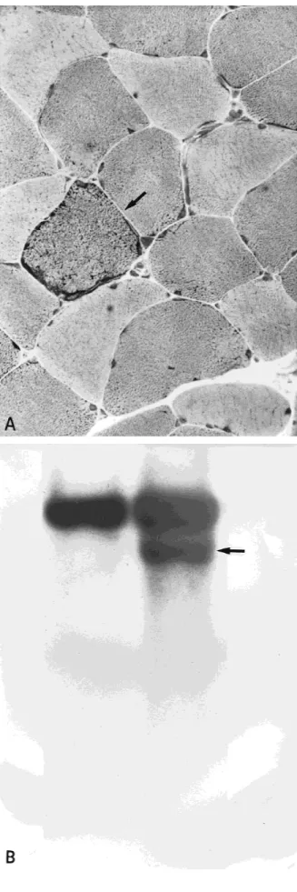
showed some variability in fiber size with occasional inter-nal nuclei. With the hematoxylin-eosin and modified Go-mori-trichrome stainings, characteristic ‘‘ragged red fi-bers’’ (RRF) were observed (Fig. 1A). Furthermore, an increase in red staining material in the subsarcolemmal region of many fibers was also observed. The oxidative
enzyme reactions showed increased staining for succinate dehydrogenase (SDH) in RRF, while the same muscle fi-bers resulted negative for cytochrome c oxidase (COX) staining. Electron microscopy revealed the aggregation of abnormal mitochondria in most of the muscle fibers.
The above-listed features are characteristically ob-served in patients with mitochondrial encephalomyopa-thies. On the basis of the results of histological studies, Southern blot analysis was performed on our patient’s muscle DNA using whole mitochondrial DNA (mtDNA) as a probe. Quantitation of deleted species was performed by densitometry. In combination with wild-type molecules, we observed another mtDNA species of about 12.5 kb (Fig. 1B). The latter corresponds to a single, large-scale mtDNA deletion of about 4 kb, which accounted for 45% of total mitochondrial genomes. No other rearrangements were detected by our analyses.
The presence of RRF in muscle biopsies of patients with FS has been previously described.2,3,5However, these
pathological findings have been considered nonspecific. In our study, we show that RRF in the muscle biopsy of our patient were associated with a single mtDNA deletion. This led us to conclude with certainty that our patient was suf-fering from a mitochondrial disorder. We believe that a muscle biopsy in patients with FS could be useful in ruling out other disorders in which muscle pain is present, such as a mitochondrial myopathy. If RRF are found, mtDNA analyses should be considered, as a mitochondrial disor-der could surface, along with new pathogenetic or thera-peutical implications. Marcello Villanova, MD, PhD,1 Enrico Selvi, MD2 Alessandro Malandrini, MD,1 Carlo Casali, MD, PhD,3 Filippo M Santorelli, MD,3 Renato De Stefano, MD,2 Roberto Marcolongo, MD2
1Institute of Neurological Sciences, University of Siena, 53100
Siena, Italy
2Institute of Rheumatology, University of Siena, Siena, Italy 3Institute of Neurological Sciences, University ‘‘La Sapienza,’’
Rome, Italy
1. American College of Rheumatology; Criteria for the classifi-cation of fibromyalgia. Arthritis Rheum 1990;33:160–172. 2. Bengtsson A, Henriksson KG, Larsson J: Muscle biopsy in
primary fibromyalgia. Scand J Rheumatol 1986;15:1–6. 3. Drewes AM, Andreasen A, Scroder HD, Hogsaa B, Jennum P:
Pathology of skeletal muscle in fibromyalgia: a histo-immuno-chemical and ultrastructural study. Br J Rheumatol 1993;32: 479–483.
4. Dubowitz V, Sewry CA, Fitzsimons RB: Muscle Biopsy: A Prac-tical Approach, 2nd ed. London; Baillie`re Tindall; 1985. 5. Lindh M, Johansson G, Hedberg M, Henning GB, Grimbly G:
Muscle fibre characteristics, capillaries and enzymes in
pa-<
FIGURE 1. (A) Cryostat section stained with hematoxylin and
eosin showing a characteristic ‘‘ragged red fiber.’’ (B) Southern blot hybridization analysis of DNA from a healthy control (left) and our patient (right). Note the presence of another mtDNA species of smaller size (arrow), which represents a deletion in the muscle of our patient.
tients with fibromyalgia and control. Scand J Rheumatol 1995; 24:34–37.
– – – – – – – – – – – – – – – – – – – – – – – – – – – – – – – – – – –
BIFOCALS AND CERVICAL RADICULOPATHY:
A CLINICAL REMINDER
In 1972, Johnson and Wolfe described the relationship of cervical radiculopathy, spondylosis, and bifocals.1They
re-ported 25 patients in whom radicular symptoms coincided with the onset of bifocal use or the change from bifocals to trifocals. Since that publication, no specific report has dealt with the role of eyewear in the occurrence of cervical radiculopathy. We have seen 2 patients in the last month with radicular symptoms following the onset of bifocal use. Our experience may serve as a useful clinical reminder to readers of the journal.
The first patient, a 45-year-old computer programmer, began wearing bifocals and within 1 month noted neck pain and accompanying referred paresthesias to the third digit in the left upper limb. He had no history of trauma. Physical examination revealed weakness in the C7 myo-tome. The triceps muscle stretch reflex was slightly de-creased. Cervical spine plain films revealed mild degenera-tive changes at C6–7. Magnetic resonance imaging (MRI) of the cervical spine demonstrated degenerative changes with foraminal encroachment on the C7 nerve root. Elec-tromyography (EMG) revealed membrane instability in the C7 myotome.
The second patient, a 46-year-old woman, reported 4 weeks of cervical discomfort and pain in a left C6 derma-tomal distribution. She gave no history of injury, but re-ported periods of cervical extension while cross-stitching. Bifocal use had started 6 weeks prior to presentation. Physical examination revealed slight weakness in the C6 myotome. Cervical spine plain films and MRI revealed de-generative changes at C5–6. EMG demonstrated mild membrane instability in a C6 myotomal distribution.
Both patients were placed in formal physical therapy consisting of heavy intermittent cervical traction and cer-vicothoracic stabilization exercises. A tapering course of oral prednisone was also used. The reading segment of the patients’ bifocals was placed at the top. Within 4 weeks, both individuals had significantly reduced symptomatolo-gy and markedly improved physical examinations.
Age-related changes in the cervical spine have been well reported in the medical literature, with 50% of pa-tients in the fifth decade and 90% of those in the seventh decade having such changes.2Most people with these
find-ings are asymptomatic. However, the biomechanics of the cervical spine can cause an individual who may be predis-posed to nerve root compromise due to spondylitic changes to manifest clinical signs and symptoms of nerve root irritation. Johnson and Wolfe described at length the effect that bifocals can have on the position of the cervical spine, which can in turn affect the cervical nerve root.
Careful history and physical examination in a patient who presents without an obvious inciting event for a cervical radiculopathy/radiculitis can reveal findings similar to those discussed above. Treatment as outlined for our 2 patients should be beneficial. Paramount to this treatment program may be placement of the reading segment at the top of the lens of the bifocals.
Richard T. Kozakiewicz, MD William J. Hennessey, MD
Pennsylvania Physical Medicine, Inc., Berkshire Center, Greensburg, Pennsylvania 15601, USA
1. Johnson EW, Wolfe CV: Bifocal spectacles in the etiology of cervical radiculopathy. Arch Phys Med Rehabil 1972;53: 201–205.
2. Resnick D, editor. Bone and Joint Imaging, 2nd ed. Philadel-phia: WB Saunders; 1996. p 360–368.
– – – – – – – – – – – – – – – – – – – – – – – – – – – – – – – – – – –
SINGLE MOTOR UNIT H REFLEXES RECORDED IN THENAR MUSCLES AT REST
While estimating the thenar motor unit number by the adapted multiple point stimulation method (AMPS)8in 36
amyotrophic lateral sclerosis (ALS) patients, surface-recorded single motor unit H reflexes (MUPH) were
evoked in 3 patients. We have taken the opportunity to evoke in the same motor unit a direct motor response (M) and a single MUPHto test the hypothesis that, with
increas-ing stimulation intensity, the reflex responses are sup-pressed due to a collision between ascending and descend-ing impulses in the motor fibers. The surface recorddescend-ing and stimulating electrode placements have been described previously.8
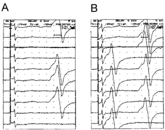
Each thenar single MUPHwas evoked by subthreshold
stimulation for motor axon (Fig. 1A). When the stimulus intensity was slightly increased, either a direct motor unit potential (MUP) or a single MUPHwas recorded. In the
case illustrated by Figure 1B, a direct MUP was recorded in 157/200 stimuli and a single MUPHwas observed in 43/
200 stimuli; but direct and reflex responses were never observed together after one stimulus. When the stimulus intensity was further increased, other direct MUPs were evoked; the H reflex, however, continued to be inhibited despite the fact that a greater number of sensory afferent fibers was recruited by peripheral stimulation.
The physiological explanation of the disappearance of the H reflex when stimulus intensity is raised remains de-bated. Several hypotheses are currently proposed. (a) There may be a collision, just distal to the alpha motoneu-rons, between the centrifugal H-reflex impulses and action potentials propagating antidromically in the motor nerves.5(b) Anterior horn cells may be incapable of
gen-erating an H reflex from monosynaptic Ia inputs because they are in a refractory state after F-wave generation.3(c)
Motoneurons are submitted to Renshaw or Ib inhibi-tion.2,6,7
For discussion, two situations have to be distinguished: MUPH evoked by subthreshold (Fig. 1A) and
near-threshold (Fig. 1B) stimulation of motor axons. When evoked by subthreshold stimulation (Fig. 1A), the pattern might suggest active inhibition, since the same stimulus evoked H reflexes in only some trials. As no direct motor response was evoked, Renshaw inhibition and antidromic refractoriness of motoneurons can be excluded. Thus, only transient reduction in Ib inhibition might explain the intermittent evocation of MUPH. However, Ib inhibition is
rarely proposed to explain the relationship between stimu-lus intensity and disappearance of H reflexes. In fact, Ib fiber conduction velocity is usually lower than that of Ia fibers; moreover, the Ib inhibitory pathway involves an in-terneuron that would introduce an additional delay. Fi-nally, Ib inhibition has been proposed to induce disap-pearance of H reflexes solely after stimuli of maximum strength.7Thus, the absence of a motor response in Figure
1A does not reflect Ib inhibition, but depends on an Ia input that is not strong enough to bring motoneurons to discharge.
With near-threshold stimulation, either a direct MUP or a MUPHwith an identical shape may be evoked (Fig.
1B). MUPHwas suppressed as soon as the motor unit was
directly recruited by peripheral stimulation; with increase of the stimulus intensity, it was neither recruited again nor replaced by an F wave. These results are not in favor of active inhibition. In fact, Renshaw cell inhibition requires discharge of 20–40% of motoneurons.1Thus, Renshaw
in-hibition should not in principle be activated by the dis-charge of a single motor unit. Moreover, the motoneurons supplying the small muscles of the digits, at least in the cat, have no Renshaw cells.4 Ib inhibition is ruled out on the
basis of the arguments discussed above. Finally, if active inhibition or antidromic refractoriness of motoneurons was responsible, either a direct MUP and a single MUPHor
a direct MUP and a single MUPF should sometimes be
evoked together after one stimulus. After several hundred trials, we have never recorded this combination of early and late responses. We therefore believe that a collision mechanism explains the H-reflex suppression that occurs with elevation of stimulus intensity.
Franc¸ois-Charles Wang, MD Paul J. Delwaide, MD, PhD
University Department of Neurology, Hoˆpital de la Citadelle, Bld du 12° de Ligne 1, B-4000 Lie`ge, Belgium
1. Bussel B, Pierrot-Deseilligny E: Inhibition of human moto-neurons, probably of Renshaw origin, elicited by an ortho-dromic motor discharge. J Physiol 1977;269:319–339. 2. Eccles JC: The inhibitory control of spinal reflex action.
Elec-troencephalogr Clin Neurophysiol 1967;25(suppl):20–34. 3. Gottlieb GL, Agarwal GC: Extinction of the Hoffmann reflex
by antidromic conduction. Electroencephalogr Clin Neuro-physiol 1976;41:19–24.
4. Illert M, Wietelman D: Recurrent inhibition in the cat fore-limb. Pflugers Arch 1988;411(suppl 1):R136.
5. Magladery JW, McDougal DB: Electrophysiological studies of nerve and reflex activity in normal man. I. Identification of certain reflexes in the electromyogram and the conduction velocity of peripheral nerve fibres. Bull Johns Hopkins Hosp 1950;86:265–290.
6. Renshaw B: Influence of the discharge of motoneurons upon excitation of neighboring motoneurones. J Neurophysiol 1941;4:167–183.
7. Trontelj JV: A study of the H-reflex by single fibre EMG. J Neurol Neurosurg Psychiatry 1973;36:951–959.
8. Wang FC, Delwaide PJ: Number and relative size of thenar motor units estimated by an adapted multiple point stimula-tion method. Muscle Nerve 1995;18:969–979.
FIGURE 1. Surface-recorded single thenar motor unit H reflex (MUPH) in a amyotrophic lateral sclerosis patient. Identical shape
and size of early and late motor responses were observed on several occasions with an all-or-nothing relationship, which indi-cated that only one motor unit was involved in these responses. (A) At 21.5 mA, a response was evoked in some trials with a 27-ms latency without a direct motor response. An early sensory potential was systematically recorded indicating afferent sensory fiber activation. Thus, this late motor response presented char-acteristics of an H reflex and clearly differed from an F wave, which is seen only if the motor unit involved in the late response is previously directly recruited by peripheral stimulation. (B) At 27 mA, the single MUPHwas systematically suppressed as early as