Université de Montréal
Lymphatic vessel function in atherosclerosis
par
Andreea Milasan
Département de Médecine, Institut de Cardiologie de Montréal Faculté de Médecine
Mémoire présenté à la Faculté des études supérieures en vue de l’obtention du grade de Maîtrise (MSc)
en Sciences Biomédicales
Octobre, 2015
Université de Montréal Faculté des études supérieures
Cette thèse intitulée
Lymphatic vessel function in atherosclerosis
présentée par Andreea Milasan
a été évaluée par un jury composé des personnes suivantes: Bruce G. Allen Président-rapporteur Catherine Martel Directrice de recherche Jean-Francois Gauchat Examinateur externe Mémoire accepté le : 26 Février 2016
Résumé
L'athérosclérose est une maladie inflammatoire chronique caractérisée par l'accumulation de cholestérol dans la paroi artérielle et associée à une réponse immunitaire anormale dans laquelle les macrophages jouent un rôle important. Récemment, il a été démontré que les vaisseaux lymphatiques jouent un rôle primordial dans le transport inverse du cholestérol (Martel et al. JCI 2013). L’objectif global de mon stage de maîtrise a été de mieux caractériser la dysfonction lymphatique associée à l’athérosclérose, en étudiant de plus près l’origine physiologique et temporelle de ce mauvais fonctionnement. Notre approche a été d’étudier, depuis l’initiation de l’athérosclérose jusqu’à la progression d’une lésion athérosclérotique tardive, la physiologie des deux constituants principaux qui forment les vaisseaux lymphatiques : les capillaires et collecteurs lymphatiques. En utilisant comme modèle principal des souris Ldlr-/-; hApoB100+/+, nous avons pu démontrer que la dysfonction
lymphatique est présente avant même l’apparition de l’athérosclérose, et que cette dysfonction est principalement associée avec un défaut au niveau des vaisseaux collecteurs, limitant ainsi le transport de la lymphe des tissus périphériques vers le sang. De plus, nous avons démontré pour la première fois l’expression du récepteur au LDL par les cellules endothéliales lymphatiques. Nos travaux subséquents démontrent que ce défaut de propulsion de la lymphe pourrait être attribuable à l’absence du récepteur au LDL, et que la dysfonction lymphatique observée précocement dans l’athérosclérose peut être limitée par des injections systémiques de VEGF (vascular endothelial growth factor) –C. Ces résultats suggèrent que la caractérisation fonctionnelle de la capacité de pompage des vaisseaux collecteurs serait une condition préalable à la compréhension de l'interaction entre la fonction du système lymphatique et la progression de l'athérosclérose. Ultimement, nos travaux nous ont amené à considérer de nouvelles cibles thérapeutiques potentielles dans la prévention et le traitement de l’athérosclérose.
Mots-clés : athérosclérose, vaisseaux lymphatiques, cellules endothéliales lymphatiques, cellules musculaires lisses lymphatiques, lipoprotéines, transport cellulaire, inflammation.
Abstract
Atherosclerosis is driven by the accumulation of cholesterol in the arterial wall, which triggers an inappropriate immune response in which macrophages play an important role. It has now been shown that the lymphatic vessels play an important role in reverse cholesterol transport (Martel et al. JCI 2013). The overall objective of my Master internship was to better characterize lymphatic dysfunction associated with atherosclerosis, studying closely the physiological and temporal origin of this pathological feature. Our approach was to study, from the initiation of atherosclerosis to the progression of the atherosclerotic lesion, the physiology of the two main components that form the lymphatic vessels: the lymphatic capillaries and collectors. Using a mouse model that closely resembles human atherosclerosis (Ldlr-/-; hApoB100+/+) we have demonstrated that lymphatic dysfunction is present before the
onset of atherosclerosis, and that this dysfunction is primarily associated with a defect in the collecting vessels, thereby limiting the lymph transport from peripheral tissues to the blood. In addition, we have clearly demonstrated, for the first time to our knowledge, the presence of the LDL receptor on lymphatic endothelial cells. Our subsequent work shows that this reduction in lymph flow could be due to the absence of the LDL receptor, and that lymphatic transport can be restored by systemic injections of VEGF (vascular endothelial growth factor) –C. These results suggest that the functional characterization of the pumping capacity of the collecting vessels would be a prerequisite for the understanding of the interactions between the function of the lymphatic system and the progression of atherosclerosis. Altogether, our work unveils new potential therapeutic targets for the prevention and treatment of atherosclerosis.
Keywords: atherosclerosis, lymphatic vessels, lymphatic endothelial cells, lymphatic smooth muscle cells, lipoproteins, cellular transport, inflammation.
Table of contents
Résumé ... iii
Abstract ... iv
Table of contents ... v
List of figures ... vii
List of abbreviations ... viii
Acknowledgements ... xi
1. INTRODUCTION ... 1
1.1 Atherosclerosis ... 3
1.1.1 Pathogenesis of atherosclerosis: a multifactorial process ... 3
1.1.2Lipid metabolism and its role in atherosclerosis ... 4
1.2 The lymphatic system ... 8
1.2.1 General anatomy and functions of the lymphatic system ... 8
1.2.2The physiology of contraction ... 11
1.2.3Lymph composition in lymphatic function ... 13
1.3 Lymphatic vessels in inflammation and cardiovascular diseases ... 15
1.3.1Mouse models of lymphatic dysfunction ... 15
1.3.2Lymphatic function in atherosclerosis ... 16
2. RESULTS ... 19
2.1 General mémoire objective ... 19
2.2 Presentation of the article... 19
3. DISCUSSION ... 51
3.1 Pathophysiology of the lymphatic collecting vessels ... 52
3.2 Potential treatment using a selective agonist of VEGFR-3 ... 53
3.3 LDLR modulation and PCSK9 as a potential therapeutic target ... 55
4. CONCLUSION ... 57
List of figures
Figure 1. Structure of the artery wall. ... 4 Figure 2. Organization of the lymphatic vasculature. ... 10 Figure 3. Examples of lymphangion coordination. ... 12
List of abbreviations
ApoA-1 Apolipoprotein A-1
ApoE Apolipoprotein E
Ca2+ Calcium ion
CaM Calmodulin
CETP Cholesterylester transfer protein eNOS Endothelial nitric oxide synthase
Erk1/2 Extracellular-signal-regulated kinase 1/2
FITC Fluorescein isothiocyanate hApoB100 Human apolipoprotein B100 HDL High density lipoprotein
IDL Intermediate-density lipoprotein LDL Low density lipoprotein
LDLR Low density lipoprotein receptor LEC Lymphatic endothelial cell
LV Lymphovenous
LYVE-1 Lymphatic vessel hyaluronan receptor 1 MLC20 Myosin light chain of 20 kDa
MLCK Myosin light chain kinase MLCP Myosin light chain phosphatase
MP Microparticle
mRCT Macrophage reverse cholesterol transport
MV Microvesicle
NO Nitric oxide
oxLDL Oxidized LDL
P70S6K P70S6 kinase
PCSK9 Proprotein convertase subtilisin/kexin type 9 Pcsk9-/- Mouse strain deficient in PCSK9
PI3K Phosphatidylinositol 3-kinase
PLCγ1 Phospholipase C gamma 1
PROX-1 Prospero homeobox 1
RCT Reverse cholesterol transport SMC Smooth muscle cell
TPC Total plasma cholesterol
VEGF-C Vascular endothelial growth factor-C
VEGFR-3 Vascular endothelial growth factor receptor-3 VLDL Very low-density lipoprotein
This mémoire is dedicated to my parents.
Acknowledgements
First and foremost, I would like to thank my wonderful supervisor Dr. Catherine Martel for her guidance, insightful ideas, constant encouragements and support. I am very grateful for all our accomplishments to date, and for your help and interest in seeing me succeed not only academically, but in all my endeavours as well. I could not dream of a better mentor.
To my dear lab mate, François Dallaire, thank you for all your help and enthusiastic nature. I am really happy that we have formed such an awesome team in such little time!
To all my friends, thank you for always being there for me. Your friendship makes my life a wonderful experience, and has helped me stay focused on my graduate studies.
Special thanks to Gabriel Moreau. You are amazing and I cannot imagine what I would do without you.
Most importantly, I want to thank my amazing parents Nina Milasan and Eugen Milasan, and my younger brother Andrei Milasan for giving me the strength to reach for the stars and chase my dreams. Thank you for always being there for me. Without the inspiration, drive and support you gave me, I would not be the person I am today.
Last but certainly not least, I would like to thank the jury committee members, Drs. Jean-François Gauchat and Bruce G. Allen, for taking their precious time to read and correct this mémoire.
1. INTRODUCTION
Cardiovascular diseases (CVDs) are the number one cause of death worldwide. CVDs are characterized by disorders of the heart and blood vessels, and so, most deaths are from heart attacks caused by sudden blood clots in the heart’s arteries1. Atherosclerosis is the
principal cause of coronary artery disease (CAD), affecting large- and medium-sized arteries. Although atherosclerosis is a chronic inflammatory disease that remains asymptomatic for many years, its development is progressive and cumulative2. An advanced plaque can rupture
depending on its vulnerability, often leading to the formation of a circulating thrombus that will slow down, or completely stops blood flow through heart arteries causing death of the surrounding tissues3. All these events can produce devastating results, such as stroke, or even
worse, death.
Macrophages and cholesterol are the two main constituents driving the inflammatory response that characterizes atherosclerosis. Lipoproteins such as low-density lipoprotein (LDL) and high-density lipoprotein (HDL) play an important role in atherosclerotic lesion modulation. LDL particles get trapped in the artery wall of the vessel where they are more prone to free radical oxidation than while circulating in the bloodstream4. Oxidized LDL
(OxLDL) further aggravate plaque formation through recruitment of pro-inflammatory factors5. On the other hand, HDL is a key element in atherosclerosis modulation because of its
role in reverse cholesterol transport, a process that helps with cholesterol removal from plaque, and its delivery to the liver and/or the intestines for eventual excretion6. Therefore, to
halt atherosclerosis progression, emphasis has been put on increasing the levels of HDL directly, or by inhibiting cholesterylester transfer protein (CETP), which normally transfers cholesterol from HDL to very low-density lipoprotein (VLDL) or LDL6. Unfortunately, the
clinical outcomes revealed disappointing negative results7-9, which made the scientific
community re-think the way they approach the development of therapies that aim to promote the movement of cholesterol out of the artery wall. The identification and validation of the pathogenic mechanisms underlying this disease became a prerequisite.
New findings offer novel insight into the path that cholesterol trapped in peripheral tissues follows during cholesterol mobilization for eventual excretion outside of the body. The
lymphatic system was identified as a novel prerequisite player in the removal of cholesterol from the atherosclerotic lesion (Martel et al. JCI 2013)10. It has been shown that without a
functional lymphatic network, cholesterol gets trapped in the artery wall and potentially aggravates the disease. It is suggested that the lymphatic system cleans up arteries by promoting cholesterol transport out of atherosclerotic lesions10. The lymphatic vessels are
composed of two main components, namely the absorptive lymphatic capillaries, responsible for the uptake of cells, molecules and fluid, and the collecting vessels, characterized by pumping units (lymphangions) that are propelling the lymphatic content toward the blood circulation in a unidirectional manner11. The relative roles of the lymphatic capillaries and
collectors in the onset of the atherosclerotic disease are still unclear.
This present work will help better delineate the specific functional roles of the lymphatic system throughout the atherosclerotic process.
1.1 Atherosclerosis
1.1.1 Pathogenesis of atherosclerosis: a multifactorial process
Atherosclerosis is the principal cause of mortality worldwide and is at the origin of most cardiovascular diseases12. It is a chronic inflammatory disease that affects large- and
medium-sized arteries, and is now considered a multifactorial disease that involves the interplay between genetic, intrinsic and environmental factors. Atherosclerosis occurs when the arteries become clogged with fatty deposits, also called plaque, and they lose their elasticity while decreasing the lumen space, leading to reduced blood flow, or even worse, a total blockade of the vessel.
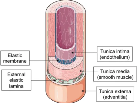
The artery wall is comprised of three different layers, which from the inside outwards, are: the intima, the media and the adventitia (Fig. 1). The intima is the thinnest and innermost layer, and it is at this level that atherosclerosis develops. It is composed of a single layer of endothelial cells displaying various properties including metabolic activities, thromboresistance, immune functions and elasticity13. The media is the thickest of the three
layers. It is the main constituent of the artery and consists of smooth muscle cells (SMCs) surrounded by an extracellular matrix composed of elastic and fibrous proteins (collagen and elastin). In general, the media is avascular, except its outer regions, which are perfused by the vasa vasorum of the adventitia (tunica externa)13. The latter is the external layer of the blood vessel wall that consists of loosely organized connective tissue rich in collagen and elastic fibers, as well as fibroblasts and adipocytes. The adventitia ensures the anchorage of the arteries to the surrounding structures and is sometimes also traversed by longitudinal smooth muscle fibers13.
Figure 1. Structure of the artery wall.
Illustration showing the different layers of a blood vessel wall. Adapted from Servier Medical Art http://www.servier.com/slidekit/?item=16
Atherosclerotic plaque formation is a continuous process and can extend around the entire circumference of the vessel. Plaque development is slow, gradual and remains asymptomatic for a long time5. Monocyte-derived macrophages and resident macrophages
accumulate in the aortic intima14; they engulf lipids and become foam cells, and produce a
wide-ranging spectrum of inflammatory mediators15-17. The physiological mechanism through
which cholesterol homeostasis is obtained in the artery wall, characterized by its mobilization from foam cells and subsequent transfer from the peripheral tissues to the liver and the intestines for further excretion, is called macrophage reverse cholesterol transport (mRCT)18.
The removal of cholesterol from foam cells and plaques is essential for the reduction of atherosclerosis burden and plaque rupture19.
1.1.2 Lipid metabolism and its role in atherosclerosis
Triglycerides, cholesterol and different lipoproteins are well known to be involved in the pathogenesis of atherosclerosis20. A high ratio of HDL: LDL in the body is associated with
a lower risk of CAD21, and both entities play a pivotal, yet opposite role, in the modulation of
atherosclerosis.
LDL is directly involved in the development of atherosclerosis, mostly due to its accumulation in the endothelium of the blood vessel. The required receptor for the endocytosis of cholesterol-rich LDL is found mainly at the surface of hepatocytes and is called the LDL receptor (LDLR). If not enough of these receptors are present, cholesterol uptake by the cells is reduced, leading to increased cholesterol in the blood vessels22. A commonly known genetic
disorder, familial hypercholesterolemia, is characterized by the inability of LDL particles to be removed from the body and therefore leads to a severe increase in the risk of cardiovascular disease23. LDL is composed of apolipoproteins, phospholipids, triglycerides and cholesterol.
Its most important and atherogenic component is apolipoprotein B (ApoB), more precisely apoB100, which is synthetized by the liver. It is this outer phospholipid layer of the LDL particles that binds to LDLR24. ApoB100 is also present in other lipoproteins such as VLDL,
intermediate-density lipoproteins (IDL) and chylomicrons. However, the main protein of IDL and chylomicrons is apolipoprotein E (ApoE), which is primarily produced by the liver and macrophages, and also mediates cholesterol metabolism25. The initial retention of LDL in the
vessel wall, followed by its oxidation, is the primary event in the early stages of an atherosclerotic lesion development, and this oxLDL promotes further recruitment and retention of monocytes that become macrophages. The protein portion of LDL will then be modified, which will lead to its impaired recognition by the LDLR and instead, increase its recognition by scavenger receptors and oxLDL receptor that are not regulated by cholesterol content in the cell26,27. Overall, this leads to overaccumulation of cholesterol, and both of these
derivatives will go on to activate endothelial cells, SMCs and macrophages, which in the end will facilitate vasoconstriction, thrombosis and platelet aggregation. Furthermore, the uptake of oxLDL by these monocyte-derived macrophages will lead to the formation of foam cells in the subendothelial space, and in the end, all these support the formation of the atherosclerotic lesion28.
Interestingly, LDLR is tightly regulated by a proprotein convertase subtilisin/kexin type-9 (PCSK9), which binds to it and targets it for lysosomal degradation in cells, leading to decreased hepatic clearance of plasma LDL-cholesterol (LDL-C). In addition to modulating
cholesterol transport and metabolism, PCSK9 also promotes intestinal overproduction of triglyceride-rich apoB lipoproteins29. However, since PCSK9 interferes with the clearance of
LDL-C from the blood, its loss-of-function mutations are associated with up to 85% lower plasma LDL-C levels30 and offer significant protection from coronary heart disease, such as
atherosclerosis31. PCSK9 is now considered a great target for cholesterol-lowering therapies32.
In the largest monotherapy trial using a PCSK9 inhibitor to date, Evolocumab reduced LDL-C from baseline by 55% to 57% more than placebo and 38% to 40% more than ezetimibe, which lowers cholesterol absorption from the intestine and was well tolerated in patients with hypercholesterolemia33. Furthermore, as statin intolerance has been a major limitation in the use of statins, a first and only study of a new class of LDL-C-lowering agents in patients selected with a rigorously documented intolerance to statins showed promising results. Alirocumab reduces LDL-C levels by approximately 50% when used as monotherapy, and has so far shown a safety profile comparable with ezetimibe or placebo34. On July 24, 2015 the
first PCSK9 inhibitor, Alirocumab was approved by the FDA as a second line of treatment for adults with high cholesterol not controlled by diet and statin treatment. On August 2015, the
FDA approved a second PCSK9 inhibitor, Evolocumab, for patients who are unable to get their LDL cholesterol under control with current treatment options. Moreover, PCSK9 has also been shown to affect the regulation of epithelial Na+ channel (ENaC) trafficking and therefore
modulate epithelial Na+ absorption that is critical for blood pressure control35. Therefore,
PCSK9 inhibition could be effective by its direct modulation of LDL-C uptake by the liver, but it could also potentially be associated with pleiotropic effects. So far, PCSK9 inhibition has an excellent safety profile and promises to provide a well-tolerated and effective therapeutic measure against coronary heart disease (CHD)36.
Another key element in atherosclerosis is HDL, which is the smallest of the lipoprotein particles, but it is the densest because it contains the highest proportion of proteins to lipids. Its most abundant apolipoprotein is apolipoprotein A-1 (ApoA-1)37. Most protective effects of
HDL are mediated by its cell surface receptors. HDL counteracts the proatherogenic activity of LDL by mobilizing cholesterol from the arterial intima and delivering it to the scavenger receptor class B1 (SR-B1), present on the surface of the liver, for further elimination into the bile38. In humans, one of the most relevant pathways is the indirect one, which is mediated by
CETP, a protein that exchanges triglycerides of VLDL for cholesteryl esters of HDL and leads to the processing of VLDLs to LDL, which will then be removed from circulation by the LDLR pathway39. Furthermore, it has been shown that HDL can function as an acceptor,
transporter and inactivator of oxLDL, and is also responsible for the inhibition of monocyte adhesion in the intima of the blood vessel, preserves endothelium-dependent vascular activity through its effect on endothelial nitric oxide synthase (eNOS) and prevents thrombosis40.
Therefore, HDL particles are important because unlike the larger particles, such as LDL, they transfer cholesterol away from cells, artery walls and tissues, through the bloodstream, back to both LDL particles, as well as back to the liver for excretion.
The inverse relationship between HDL-cholesterol levels and CHD incited a lot of interest in pharmacological agents that elevate plasma levels of HDL. Statins are first-line drugs for the treatment of dyslipidemias and CAD prevention due to their ability to lower plasma LDL-C levels41. This treatment focuses mainly on endogenous LDL-C, and results in
neglect of other important aspects of lipoprotein metabolism42. Significant side effects such as
severe muscular pain are also displayed in most patients43. As statins offer little to no effect on
increasing HDL levels, the attempt was then focused on other ways to increase these levels, such as directly through the inhibition of CETP, which normally transfers cholesterol from HDL, to VLDL or LDL44. Examples include HDL-raising drugs such as torcetrapib, which
ended up showing an increase in cardiovascular risk45, as well as dalcetrapib, that simply
lacked effectiveness46. Niacin, or Vitamin B3, are other effective HDL-raising agents currently on the market that first emerged as dyslipidemia treatment47. As a result, new ways to
approach treatments, particularly those aimed at preventing plaque development in atherosclerosis, are currently under close investigation, and as we will soon see in the sections to come, there are promising venues ahead.
1.2 The lymphatic system
1.2.1 General anatomy and functions of the lymphatic system
The lymphatic system is part of the circulatory system and plays a vital role in host defense and adaptive immunity. Although the roles of lymphatic vessels in tissue maintenance and disease are now well known, their origins are still a subject of debate. As far back as 1902, Florence Sabin, based upon results obtained by India ink injection experiments in pigs, proposed that isolated primitive lymph sacs, which are precursors of the lymph vessels, originated from endothelial cells that bud from the cardinal vein during early development48.
Interestingly, Klotz et al. found that the lymphatic endothelial cells (LECs) that form lymphatic vessels in the heart originate both from embryonic veins and from other non-venous sources, like the yolk sac49. Therefore, contrary to previous belief, it has been proven that a
significant part of the dermal lymphatic vasculature forms independently of sprouting from veins50. Many different transcription factors regulate lymphatic development, one of the most
crucial being Prospero-related homeobox-1 (Prox1)51. Cells that are Prox1+ depend on a
paracrine factor called VEGF-C to be able to spread away from the embryonic veins. The vascular endothelial growth factor receptor-3 (VEGFR-3) serves as a receptor for lymphatic-specific VEGFs, VEGF-C and VEGF-D. VEGF-C is important for normal development of the lymphatic vessels and Prox1 drives VEGFR-3 expression that enables LECs to respond to VEGFR-3 ligands52. In zebra fish, it has been shown that VEGF-D can compensate for the
absence of VEGF-C, making the latter dispensible53. Separation of lymphatic fluid from blood
requires platelet aggregation54. Platelets regulate the blood/lymphatic vessel separation by
inhibiting the proliferation, migration, and tube formation of LECs, upon activation by C-type lectin-like receptor 2 (CLEC-2)/podoplanin interaction. Interaction of podoplanin present on LECs with CLEC-2 present on platelets triggers a signalling cascade leading to platelet aggregation and the formation of fibrin-containing platelet thrombi that protect both the lymphovenous junction (LV) and the thoracic duct from backward flow55.
Characterized by a network of vessels that carry a clear fluid called lymph, the lymphatic system is now recognized as working in close collaboration with the cardiovascular
system, but unlike the circulatory system, it is not a closed system56. One of its main roles is to
maintain fluid homeostasis in the body with the help of its thin-walled and blind-ended lymphatic capillaries57. These initial lymphatics help absorb the ultrafiltrates from peripheral
tissues, they are highly permeable and are constituted of specialized, discontinuous ‘button-like’ junctions between endothelial cells58. Lymphatic capillaries are characterized by the
absence of SMCs and at the surface of the LECs they specifically express lymphatic vessel endothelial hyaluronan receptor (LYVE-1). Following its absorption by the lymphatic capillaries, lymph will move on to converge into larger pre-collecting vessels, to subsequently reach the collecting lymphatic vessels57,59. Lymphatic capillaries have a sparse and
discontinuous basement membrane and they lack pericytes, which are the contractile cells that wrap around the endothelial cells of blood capillaries and venules throughout the body. Anchoring filaments attach LECs to the extracellular matrix and prevent vessel collapse under conditions of increased interstitial pressure. Both “buttons” and “zippers” are composed of adherens and tight junction–associated proteins like VE-cadherin, zonula occludens-1, occludin, and claudin-5 (Fig. 2). The main difference between them resides in their organization60. Collecting lymphatic vessels are covered with continuous basement membrane
and SMCs, and are characterized by the expression of podoplanin. Therefore, collecting vessels are contractile lymphatics that propel lymph in a unidirectional manner, with the help of intraluminal bi-leaflet valves, as well as smooth muscle walls. The functional unit of a collecting lymphatic vessel is called a lymphangion, representing the segment between two valves. All the lymph collected from the entire left side of the body, the digestive tract and the right side of the lower part of the body flows into a single major vessel, the thoracic duct. The latter empties into the left subclavian vein. The lymph in the right side of the head, neck, and chest is collected by the right lymph duct and empties into the right subclavian vein. The two subclavian veins will then drain into the blood circulation through the LV, right near their junction with the internal jugular veins59.
Figure 2. Organization of the lymphatic vasculature.
(A) The lymphatic vasculature is responsible for the absorption of fluid, macromolecules, and cells from the interstitium. (B) Mechanism of lymph formation in capillaries. Interstitial components penetrate lymphatic capillaries via openings between LECs. The specialized structure of such openings prevents the return of lymph back to the interstitium. Anchoring filaments attach LECs to the ECM and prevent vessel collapse under conditions of increased interstitial pressure (black arrow). (C) Junctional organization of LECs in lymphatic capillaries and collecting vessels. Both “buttons” and “zippers” share a repertoire of adherens and tight junction–associated proteins, like VE-cadherin, zonula occludens, occludin, and claudin-5. The main difference between them resides in their organization58. (D) Mechanism of lymph
propulsion in collecting vessels. Coordinated opening and closure of lymphatic valves is important for efficient lymph transport. SMCs covering each lymphangion possess intrinsic contractile activity. Schulte-Merker S, et al. Lymphatic vascular morphogenesis in development, physiology, and disease. J Cell Biol 2011; 193(4): 607-18.
1.2.2 The physiology of contraction
Largely aqueous with relatively low concentrations of proteins and cells compared to blood, lymph is propelled through the collecting lymphatics via two primary mechanisms: 1) intrinsic, characterized by active pumping as a result of lymphatic muscle cell contraction, the lymphangions and the valves; 2) extrinsic, compression mechanisms such as the movement of skeletal muscle or other tissues surrounding the lymphatics, as well as respiration. Coordinated opening and closure of lymphatic valves is important for efficient lymph transport. As previously discussed, lymphangions are wrapped in a disorderly manner by lymphatic smooth muscle cells61. Unlike vascular smooth muscle, lymphatic muscle is characterized by both a
rapid, phasic contractile activity that drives the intrinsic lymphatic pumping, as well as a slower, tonic form of contractions seen in blood SMCs. The uniqueness of lymphatic contractility is due to the lymphatic muscle being composed of both smooth and striated muscle62. However, lymphatic valve function, which operates in an open–close manner, is
passive and responds to the pressure difference between pre- and post-valve lymphagions63. In
contrast, lymphangion contraction is an active process that requires the generation of force by SMCs, which are mainly dependant on myocyte intracellular Ca2+ levels. In this regard, a
subset of muscle cells acts as pacemaker cells, initiating and driving propagation of the Ca2+
wave to other cells downstream, inducing a series of action potential-like spikes of calcium that cause the synchronized contractions of lymphangions64. Contraction by the lymphangions
is an alternate process, and at the same time, it is normal for two or more adjacent lymphangions to contract and relax at the same time (Fig. 3)65.
Figure 3. Examples of lymphangion coordination.
(A) Contractions might alternate in adjacent lymphangions. (B) It is possible that two or more adjacent lymphangions contract and relax together. Adapted from: Munn LL. Mechanobiology of lymphatic contractions. Semin Cell Dev Biol 2015; 38: 67-74.
Several mechanisms trigger and modulate vessel phasic or tonic contraction by influencing lympatic SMCs cytoplasmic Ca2+ levels. Modulation of the 20-kDa myosin light
chain (MLC20) is an important factor of lymphatic contractile strength. MLC20
phosphorylation is regulated by the activities of myosin light-chain kinase (MLCK) and myosin light-chain kinase phosphatase (MLCP). Lymphatic muscle contraction is initiated by an increase in cytosolic Ca2+ levels resulting in Ca2+ binding to the universal intracellular Ca2+
receptor protein, calmodulin (CaM), which will activate the catalytic subunit of MLCK. MLCK then phosphorylates MLC20, leading to lymphatic vessel contraction. MLC20
phosphorylation is reversed by MLCP, resulting in lymphatic muscle relaxation66. The
best-known and most studied mechanism for interfering with SMC contraction is nitric oxide (NO), which acts at multiple points of the Ca2+ contraction pathway. NO produced by eNOS
regulates systemic blood pressure, vascular remodelling and angiogenesis67. Shear stress is the
most important physiological stimulus of its production and phosphorylation of eNOS by Akt represents a Ca2+-independent regulatory mechanism for activation of eNOS. When it comes
to the lymphatic system, once produced, NO can diffuse to the SMCs enveloping the lymphatic vessels and affect pumping. It works by modulating Ca2+ release and uptake68, as
well as the enzymes responsible for force production69,70. NO decreases vascular tone through
release from internal stores. Conversely, NO can activate several other channels to increase Ca2+ efflux71. NO also induces relaxation through its direct inhibitory effect on the MLC72.
Overall, NO is a vasodilator that opposes the Ca2+ response of the lymphatic vessels, leading
to dilatation and decreased contraction frequency73. Hence, the lymphatic system is under the
constant influence of various factors that modulate its contractility, controlling its crucial roles both as conduit and pump.
1.2.3 Lymph composition in lymphatic function
Lymph composition is changed during its flow from the periphery, with its protein concentration increasing along the lymphatic vessels74. Since lymph is derived from the
interstitial fluid, its composition constantly changes depending on its interaction between the surrounding cells and blood. Blood and the interstitial fluid are in dynamic equilibrium with each other, meaning that water and solutes can pass between the two by diffusion across gaps in capillary walls called intercellular clefts. Although peripheral lymph lipoproteins have been characterized in animals, information about their composition is limited, and their ultrastructure is nearly unexplored. Studies analyzing human lymph have started to emerge, and they have confirmed that lymph composition is different than that of plasma or serum10.
Back in 2000, Nanjee et al. demonstrated that the concentration of small pre-beta HDLs in human tissue fluids is determined only in part by their transfer across capillary endothelium from plasma. They showed that the local production of pre-beta HDL in the periphery, by remodelling of spheroidal HDLs in tissue fluids, is just as important29. Continuing down this
path, in 2001, Nanjee et al. examined the composition, as well as the ultrastructures of different subclasses of normal human peripheral lymph lipoproteins. Total cholesterol concentration in lymph HDL was 30% greater in lymph than could be explained by the transendothelial transfer of HDL from plasma, which provided direct confirmation that HDL acquire cholesterol in the extravascular compartment30.
Extracellular vesicles (EVs) are plasma membrane-derived vesicles released from cells upon activation or during apoptosis. Cellular EVs in body fluids constitute a heterogeneous population, differing in cellular origin, numbers, size, antigenic composition and functional properties75. Despite being considered simple cellular debris for the longest time, it is now
players in many physiopathological processes such as thrombosis, autoimmune diseases and inflammation76. Due to their diversity, EVs are considered important biomarkers and thus,
their precise detection in several biological fluids, such as lymph, is important to better understand all their different functional activities77. As they are an important vector of
information exchange between cells of different origins, in the course of their various interactions they cause structural and functional changes, especially at the level of the vascular wall, in the endothelium78. Although EVs are present in the peripheral blood of healthy
individuals, marked elevations occur in many disease states76, as they have been shown to be a
major component of atherosclerotic plaques, especially platelet-derived EVs75. Therefore, in a pathological setting, particularly atherosclerosis, it is important to understand why and how the presence of EVs is affected, by studying lymph composition.
We now know that a broad array of cytokines, proteins, growth factors, lipoproteins and maybe even extracellular vesicles are contained within lymphatic fluid, which play an important role in metabolism, proliferation, as well as immunoregulation76. We have yet to see
1.3 Lymphatic vessels in inflammation and cardiovascular
diseases
1.3.1 Mouse models of lymphatic dysfunction
Several models with impaired lymphatic function have started to emerge. Primary congenital lymphedema (Milroy disease) is a rare autosomal dominant condition caused by mutations in the vegfr-3 gene79. Primary human lymphedema is characterized by a chronic and
disfiguring swelling of the extremities. A popular mouse model to study the physiological regulation of lymph flow and to assess the therapeutic potential of VEGF-C to stimulate lymphatic revascularization has been put forth by Alitalo et al80. Chy mice have an
inactivating VEGFR-3 mutation in their germ line and swelling of the limbs because of incomplete development of lymphatic vessels within the dermis. Furthermore, promising therapeutic results were observed when they used virus-mediated VEGF-C gene therapy, as they were able to generate functional lymphatic vessels in lymphedema mice81. Another
example is the Prox1+/- mouse, which accumulates fat as a consequence of lymphatic vascular
leakage, and has been used as a new model for adult-onset obesity and lymphatic vascular disease. Functional inactivation of a single allele of the Prox1 gene led to adult-onset obesity due to abnormal lymph leakage from mispatterned and ruptured lymphatic vessels82. Another
interesting mouse model includes the microsurgical ablation of the lymphatic vessels in the tail of the mouse, which results in lymph stagnation, lymph vessel dilation (with a marked increase in tail volume), accumulation of fibroblasts, fat, and skin cells, impaired clearance of immune cells from the tail, and profound accumulation of inflammatory cells83. It is a useful
model that closely imitates some key features of acquired lymphedema in humans, and in combination with a diverse array of mouse genetics, it could lead the way to the better understanding of the molecular basis of lymphedema. All these different mouse models have not only helped us to improve our understanding of different lymphatic-related pathologic conditions and their relationship with inflammation, but also to re-evaluate the functional roles of the lymphatic vascular network.
1.3.2 Lymphatic function in atherosclerosis
Despite the well-defined roles of the lymphatic system in preserving fluid balance throughout the body by returning plasma proteins from interstitial spaces back to the blood circulation, its function in heart disease has lately been getting much attention. Alterations in the intestinal lymphatic network are well-established features of human and experimental inflammatory bowel disease (IBD)84. As lymphatic vessels play an essential role in intestinal lipid uptake, impairment of lymphatic vessel function leads to enhanced adipose tissue accumulation in patients with lymphedema and in genetic mouse models of lymphatic dysfunction85. Interestingly, Blum et al. showed that adipose tissue expansion due to a high-fat
diet leads to functional impairment of the lymphatic vasculature, mainly at the level of the collecting vessels. In the heart, evidence shows that blocking cardiac lymph flow may contribute to several forms of cardiac injury including cardiac lymphedema and poor heart performance in animal and human heart studies86. It was back in 1981 that Lemole observed
intimal vessel thickness following lymphatic vessel blockage. He suggested that the accumulation of interstitial fluid in the artery wall could be due to a phenomenon called lymphostasis, which may contribute to the development of atherosclerosis due to factors present in the intimal edema87. Thirty years later, fundamental studies and clinical studies
started to emerge in this regard. Studies analyzing the morphology of lymphatic vessels in the artery wall allowed for insights between their associations with atherosclerosis. In animal models, lymphatic vessels have been observed in the adventitia of the artery wall88. In fact, Xu
et al. associated the presence of lymphatic vessels within the adventitia of the artery wall an important factor for the draining of local inflammatory cells and cytokines from peripheral tissues89. In a clinical setting, Drozdz et al. took interest in the presence of lymphatics in the
adventitia of the internal carotid artery in humans and showed that the number of adventitial lymphatics increases with the severity of atherosclerosis measured as intimal thickness90.
Furthermore, they showed that arteries with a dense network of lymphatic vessels seem to be naturally protected against atherosclerosis when compared to those without such a network91.
Martel et al. introduced a new integrated model of mRCT in which they clearly showed that without a functional lymphatic network, cholesterol cannot be properly conducted out of the artery wall. They used a surgical model of aortic transplant from a hypercholesterolemic ApoE
deficient (ApoE-/-) donor to a hypercholesterolemic ApoE-/- receiver in which ApoE vector was injected to induce cholesterol efflux, and showed that the pattern of the newly regrown lymphatic vessels post-transplant is influenced by the blood flow through the transplant10. The
lymphatic vessels thus formed appeared to be atheroprotective: in conditions where lymphatic vessels had fully grown post-transplant, the cholesterol contained in the transplanted artery was able to exit the atherosclerotic lesion. By contrast, partial inhibition of lymphatic regrowth using VEGFR-3 antibody was reflected by retention of cholesterol in the artery wall of the transplanted aortic segment10. Along the same path, Vuorio et al. published that lymphatic
impairment worsened the atherosclerotic plaque formation in atherogenic Ldlr-/-/ApoB100/100
mice crossed with transgenic mice bearing localized lymphatic insufficiency (sVEGFR-3 or Chy), and analyzed the effects of the absence of lymphatics on lipoprotein metabolism and atherosclerosis92. The group observed a positive correlation between atheroma formation and
the absence of lymphatic vessels, contrary to previous studies that showed opposite effects. This study further questions whether lymphatic vessels are beneficial or detrimental. Furthermore, these Ldlr-/-/ApoB100/100 mice crossed with transgenic mice bearing localized
lymphatic insufficiency (sVEGFR-3 or Chy) have increased cholesterol levels leading to accelerated atherogenesis, which suggests that lymphatic vessels play a crucial role in the maintenance of proper lipoprotein metabolism and overall vascular homeostasis92.
Based on the newly described integrated model for mRCT10, it is believed that within
the atherosclerotic lesion, cholesterol ester (CE) is exported from macrophages and uploaded onto cholesterol acceptors like HDL, which will then cross the medial layer of the artery to reach the adventitia. From there, it will enter the adventitial lymphatic capillaries and get propelled through the afferent collecting lymphatic to the draining lymph node. It passes through the efferent collecting lymphatic to enter the bloodstream at the level of the subclavian vein10. The next steps are well studied and imply an uptake of the circulating
cholesterol by the liver, via SR-B1 receptor93, where most cholesterol is transformed into bile
acids and secreted into the bile for excretion. In parallel, CE from HDL can also be transferred in VLDL and LDL, via CETP94, a pathway that is not present in mice, and internalized by the
therapies towards improving lymphatic function, which should facilitate cholesterol clearance and limit inflammation, all in the hopes of preventing or reversing atherosclerosis.
2. RESULTS
2.1 General mémoire objective
The general objective of my M.Sc. project was to better delineate the functional roles of the lymphatic system before atherosclerosis onset and its progression. More specifically, I have studied the effect of in vivo modulation of the LDLR on lymphatic function.
Based on published data by Dr. Martel10, we were able to pursue her work with newly
formed hypotheses of our own. This new data I have helped acquire is now the stepping-stone to many upcoming projects currently being developed in our laboratory. Our first manuscript is presented in section 4.2.
2.2 Presentation of the article
In this article, we built on previously published results by Dr. Martel10, as well as the
literature, to study the origin of the lymphatic dysfunction seen in atherosclerosis. The objective was to see at which level of the lymphatic vessel functional impairment originates and at what point during atherosclerosis development the defect becomes prominent.
We characterized thoroughly in different mouse models the morphology and functionality of the lymphatic capillaries, and it gave us no reason to doubt that lymphatic dysfunction must surely originate at a higher level, namely the collecting vessels. Our next step was to better grasp the cellular composition of these vessels, and that is how we came to show that the LDLR is present even at the surface of LECs. As LDLR, whose expression is down-regulated by physiologic levels of PCSK9, is a major player in atherosclerosis, we aimed to investigate whether and how PCSK9 and LDLR modulation plays a role in lymphatic function.
We subsequently observed that in a mouse model with an over-accumulation of LDLR, lymphatic function increased throughout age, when compared to wild type. In contrast, even before development of plaque, in an Ldlr-/-; hApoB100+/+ mouse model (also called ATX), an
therapeutic targets such at VEGF-C, which we demonstrate may exert a rescue effect when treating young Ldlr-/-; hApoB100+/+ mice, before atherosclerosis onset.
Overall, our results suggest that:
Lymphatic transport tends to improve with age in Pcsk9-/- mice compared to WT
LDLR is expressed on LECs and associated with lymphatic vessel function
Collecting lymphatic vessels may be responsible for the impairment in lymphatic function in Ldlr-/-; hApoB100+/+ mice
VEGF-C systemic treatment abrogates the lymphatic dysfunction that is observed before atherosclerosis onset in Ldlr-/-; hApoB100+/+
Participation of each author of the article:
AM: Project conceptualisation, troubleshooting, methods validation, experiments, data analysis and manuscript writing
FD: Experiments and data analysis
GM: Resources (Pcsk9-/- mice), scientific consultation
CM: Project conceptualisation, troubleshooting, methods validation, experiments, data analysis and manuscript writing
Article submitted to Scientific Reports
Effects of LDL Receptor Modulation on Lymphatic Function
Andreea Milasan,1,2 François Dallaire,2 Gaétan Mayer,1,2 and Catherine Martel,1,2,*
1 Department of Medicine, Faculty of Medicine, Université de Montréal, Montreal, Quebec,
Canada
2Montreal Heart Institute, Montreal, Quebec, Canada
*Corresponding author:
Catherine Martel, PhD
Montreal Heart Institute Research Center Department of Medicine, Faculty of Medicine Université de Montréal
5000, Belanger street, Room S3560 Montreal, Quebec (Canada)
H1T 1C8
ABSTRACT
Atherosclerosis is driven by the accumulation of immune cells and cholesterol in the arterial wall. Although recent studies have shown that lymphatic vessels play an important role in macrophage reverse cholesterol transport, the mechanisms regulating this pathological feature remain unknown. In the current report, we aim to better characterize this lymphatic dysfunction associated with atherosclerosis by studying the physiological and temporal origins of this impairment. We observed that mice deficient in Pcsk9 had improved lymphatic function throughout age when compared to WT mice for up to six months, while displaying enhanced expression of LDLR on lymphatic endothelial cells. Then, using a mouse model (Ldlr-/-; hApoB100+/+) that closely resembles human atherosclerosis we show that lymphatic
dysfunction is present before atherosclerosis lesion formation, and that this dysfunction is primarily associated with a defect in the lymphatic collecting vessels. Systemic treatment with a selective VEGFR-3 agonist (VEGF-C 152s) rescued this impairment in a cholesterol-independent manner. Taken together, our results suggest for the first time that the absence of PCSK9 is associated with improved lymphatic function and unveil new potential therapeutic targets for the prevention and treatment of atherosclerosis.
Introduction
Low-density lipoprotein receptors (LDLR) are important players in atherosclerotic lesion development and progression mostly through lipoprotein metabolism regulation1, as
demonstrated in patients with familial hypercholesterolemia2. This genetic disorder is most
often caused by reduced function of LDLR, apolipoprotein B (apoB) or gain-of-function mutations in protein convertase subtilisin kexin type 9 (PCSK9), resulting in a severe elevation of the plasma levels of LDL3. Circulating LDL-cholesterol (LDL-C) accumulates in
the artery wall of blood vessels and leads to premature atherosclerosis4. ApoB100 is the
apolipoprotein and ligand of LDLR found in lipoproteins synthesized by the liver, and is the sole protein of LDL5. LDLR is present on the outer surface of many types of cells6.
Hepatocytes are important cells bearing LDLR, as the liver is responsible for removing most excess cholesterol from the body, through LDL uptake5. PCSK9 is a well-established
down-regulator of LDLR7, which acts by binding to the receptor causing its lysosomal degradation
in cells. Since PCSK9 interferes with the clearance of LDL-C from the blood, its loss-of-function mutations are associated with up to 85% lower plasma LDL-C levels8 and offer
significant protection from coronary artery disease (CAD), like atherosclerosis9. On the
contrary, high-density lipoprotein (HDL) reduces cardiovascular risk, mainly due to its role in macrophage reverse cholesterol transport (mRCT), promoting cholesterol removal from plaque and its eventual excretion by the liver and/or the intestines10,11. In order to halt
atherosclerosis progression and decrease the prevalence of CAD, emphasis has logically been put on improving this physiological process. However, most of the different ways used to increase HDL levels did not demonstrate any clinical benefits, and did not lead to improved mRCT or decreased CAD12-14. Consequently, such conclusions made the scientific community
re-think the way they approach the development of therapies aimed to increase mRCT and favourably modulate atherosclerosis.
PCSK9 inhibition has an excellent safety profile in clinical trials and promises to provide a well-tolerated and effective therapeutic measure against CHD15. PCSK9 inhibition could act
via its direct modulation of LDL-C uptake by the liver, but it could also potentially be associated with pleiotropic effects. Recent findings show that PCSK9 deficiency increases
CD36 levels in the liver and adipose tissue16 and reduces liver metastasis by its ability to lower
cholesterol levels17. Moreover, PCSK9 has been shown to affect the regulation of epithelial
Na+ channel (ENaC) trafficking and therefore modulate epithelial Na+ absorption, which is
critical for blood pressure control18. Lastly, PCSK9 was shown to be expressed endogenously
both at the mRNA and protein level in murine peritoneal macrophages19. Giunzioni et al.
showed for the first time that this PCSK9 expression directly influences atherosclerotic plaque composition with no changes in serum cholesterol levels, which suggests a direct effect of macrophage PCSK9 in inflammation and plaque development20.
An additional new therapeutic target in atherosclerosis has recently arisen. In a recent study, the lymphatic system has been identified as a novel prerequisite player in the removal of cholesterol from the atherosclerotic lesion by mRCT21. It has been reported that without a
functional lymphatic network, cholesterol cannot leave the artery wall and might potentially aggravate the disease. Accordingly, it is now suggested that cholesterol leaves tissues and reaches the bloodstream by first entering lymphatic vessels (LVs), putting forward a new integrated model for mRCT21,22. The blood vasculature and the lymphatic systems are parallel
and interdependent networks23. In contrast to the blood vascular network, the lymphatic
vascular network is an open, unidirectional and low-pressure vascular system. Lymphatic development and regulation are dependent mostly upon VEGF-C/D and its receptor VEGFR-324. The LVs are composed of two different entities, bearing distinct but complementary roles.
Lymph is first absorbed through thin-walled and blind-ended initial lymphatics (also called lymphatic capillaries), which are highly permeable and are constituted of specialized, discontinuous "button-like" junctions between endothelial cells25. Expression of lymphatic
vessel hyaluronan receptor 1 (LYVE-1) on the LEC and absence of smooth muscle cells (SMC), are characteristics of the lymphatic capillaries25. Following, lymph moves from the
lymphatic capillaries into collecting vessels, the entities responsible of maintaining lymph flow through contraction of units called lymphangions. Collecting vessels resemble small veins, but have bi-leaflet valves between the contractile units in order to prevent back flow. They are also characterized by a basement membrane, down-regulation of LYVE-1 expression, podoplanin expression, continuous "zipper-like" cell-cell junctions and a discontinuous SMC layer26. Lymph is propelled away from the periphery most of the time
against a hydrostatic pressure gradient primarily via the phasic and synchronized contractions of the lymphangions, mediated by the intrinsic contractility of SMC, the contraction of surrounding skeletal muscles, and arterial pulsations27-31.
Since lymphatic function is now linked to atherosclerosis, and the LDLR plays a central role in cardiovascular disease and exhibits modulation following PCSK9 inhibition treatment, studying the implication of PCSK9 on lymphatic function has become of great interest. Herein, we sought to better characterize the interplay between lymphatic function and the onset and progression of atherosclerosis by exploring the possible link between lymphatic function and LDLR modulation in Pcsk9-/- and Ldlr-/-; hApoB100+/+ mice. We investigate
whether and how the absence of PCSK9 and, subsequently, increased LDLR protein expression has a beneficial effect on lymphatic function, and further portrays the premises of the chronological sequence of atherosclerosis-associated lymphatic dysfunction.
Results
Lymphatic vessel function is enhanced in Pcsk9-/- mice.
As PCSK9 targets and mechanisms of action are now known not to be solely confined to the liver, we sought to investigate its effect on lymphatic function. We herein hypothesized that knocking out Pcsk9 may positively modulate lymphatic transport. To address this, we first assessed the capacity of popliteal lymphatic vessels to carry Evans blue (EB) dye from the initial lymphatics located in the dermis of the foot pad up to the collecting lymphatics and the corresponding draining popliteal lymph node (LN) (Fig. 1a, upper panel; Supplementary Fig. S1 online). Intradermal injection of EB dye revealed that the dye intensity was greater within the dominant32 collecting vessel of Pcsk9-/- mice, with no or little interruption on its path (Fig.
1b, lower panel, green arrow vs. red arrows). This observation suggests that lymphatic collecting vessel function was improved in Pcsk9-/- mice when compared to
atherosclerosis-prone Ldlr-/-; hApoB100+/+ and even WT mice. In addition, less extravasated EB dye was
detected in the surrounding adipose tissue of Pcsk9-/- mice when compared to that of Ldlr-/-;
As a complementary measure of lymphatic function, we evaluated the efficiency of lymphatic vessels to transport dendritic cells from the peripheral tissue to the corresponding draining LNs, using the well-described FITC painting assay33. Similar to the improved EB dye
transport, the migration of skin dendritic cells (CD45+CD11c+FITC+) in Pcsk9-/- mice (Fig. 1b)
was improved when compared to WT mice at 6 months. Analysis of the back skin from that same region (Supplementary Fig. S2a online) revealed that the adipose tissue layer tends to be thinner in 3-month-old Pcsk9-/- mice when compared to WT mice (Supplementary Fig. S2b
online), but not in 6- (Supplementary Fig. S2c online) and 12- (Supplementary Fig. S2d online) month-old Pcsk9-/- mice.
Initial lymphatic vessel morphology and number are unchanged in Pcsk9-/-.
As hypercholesterolemia-associated lymphatic dysfunction in 16-week-old apoE−/− mice
is associated with initial lymphatic hyperplasia34, we conversely sought to investigate whether
the absence of PCSK9 would be associated with morphological changes within initial lymphatics. We first assessed initial lymphatic vessel (Lyve-1+) morphology and density (Fig.
2a) by looking at their diameter (Fig. 2b), their number (Fig. 2c) and the total surface area occupied by the vessels (Fig. 2d) in the mouse ear dermis. No significant changes were observed in the athero-protected mouse model when compared to WT mice in an age-dependent manner. In order to confirm our finding in the artery wall, we investigated the presence of initial lymphatic in the adventitia of the aortic sinus (Fig. 2e), a blood vessel layer where lymphatic vessels have been consistently observed35.Once again, no difference was
seen at any age when looking at the morphology or density of Lyve-1+ vessels (Fig. 2f-h).
LDLR is expressed on lymphatic endothelial cells of popliteal collecting vessel.
Based on our findings that lymphatic function is improved in Pcsk9-/- mice without
apparent modulation in initial lymphatic morphology or growth, we sought to investigate whether collecting LVs could thus be responsible for the observed related lymphatic gain-of-function observed in this Pcsk9-/- mouse model. To begin with, we hypothesized that LDLR
per se could act as an important modulator in lymphatic collecting vessel function, as it would be drastically increased on LVs compared to WT mice. Our initial step was to detect the
presence of LDLR on collecting LVs and then test its modulation by PCSK9. Therefore, for the first time to our knowledge, we have shown that LDLR was expressed on popliteal lymphatic collecting vessels (Fig. 3a). Immunofluorescence was performed and demonstrated the presence of LDLR on podoplanin+ LECs, but to a lesser extent smooth muscle cells (SMC,
Fig. 3a). Immunofluorescence (Fig. 3b) and western blots performed with equal amount of protein loading (Fig. 3c) revealed that Pcsk9-/- mice displayed a LDLR overaccumulation
phenotype when compared to WT and Ldlr-/- mice.
Lymphatic dysfunction appeared before atherosclerosis onset and decreased during its progression in Ldlr-/-; hApoB100+/+ mice.
As our data are bridging lymphatic function to LDLR modulation on lymphatic collecting vessel ECs through PCSK9, we next conversely investigated whether decreased LDLR levels per se could be a premise to lymphatic collecting vessel dysfunction. Atherosclerosis-prone Ldlr-/-; hApoB100+/+ mice are severely dyslipidemic, exhibit premature blood endothelial
dysfunction, oxidative stress and inflammation at 3-month-old36-38.They spontaneously
develop aortic atherosclerotic lesions after 4 months while on regular chow diet. Before that age, they do not have atherosclerotic lesions, but as they get older, they become increasingly atherosclerotic (Supplementary Fig. S3a, S3c, S3d online). Resembling the human atherosclerotic phenotype due in part by the addition of the human apoB100 transgene, these mice are a suitable model to study in further detail factors that could lead to atherosclerosis or at least impact its progression. Lymphatic transport assays revealed a significant (p<0.001) lymphatic function impairment in 3-month-old Ldlr-/-; hApoB100+/+ mice (Fig. 4a) that are not
yet bearing atherosclerotic lesions (Supplementary Fig. S3a, S3c, S3d) or macrophages in the artery wall (Supplementary Fig. S3b and S3e online). As these mice are highly dyslipidemic, we wanted to test whether this severely increased plasma cholesterol content per se would be reflected by an enhanced lymphatic transport defect. To test this, we have measured lymphatic function in both young (3-month-old) preatherosclerotic Ldlr-/-; hApoB100+/+ mice and Ldlr
-/-mice. As total cholesterol content of Ldlr-/- mice is known to be half the concentration
retrieved in the Ldlr-/-; hApoB100+/+ mice (~200 mg/ml vs. ~700 mg/ml)37,39,40, one could
expect to see an even more impaired lymphatic function in the latter group. However, as a premise of the effect of LDLR per se on lymphatic function as a proatherosclerotic factor, we
observed no significant difference between the two groups (Fig. 4a). Furthermore, lymphatic dysfunction became even more apparent (p<0.05, at 12 mo) throughout atherosclerosis progression (Fig. 4b). Just like in apoE-/- mice in which dermal lymphatic vessel diameter
increases in conjunction with dyslipidemia34, aging Ldlr-/-; hApoB100+/+ mice displayed
enlarged initial lymphatic vessels (Fig. 4e, p<0.0001, between 6- to 12-month), increased total Lyve-1+ area (Fig. 4f, p<0.001, at 12-month) and decreased Lyve-1+ vessels number. As
mentioned previously, in contrast to Pcsk9-/- mice (Fig. 1a), Ldlr-/-; hApoB100+/+ mice
demonstrated interrupted EB dye presence (red arrows), as well as extravasated dye from these vessels (yellow arrows). This further supported our showcased lymphatic impairment following FITC painting, and was also a first indication of collecting vessel malfunction. As Pcsk9-/- mice abundant in LDLR display improved lymphatic transport and as 3 month-old
pre-atherosclerotic Ldlr-/-; hApoB100+/+ mice lacking LDLR already showed a lymphatic
dysfunction that was exacerbated in parallel to lesion formation, these results reinforce the idea that the LDLR per se could play a direct role on lymphatic function.
VEGF-C 152s treatment rescues lymphatic function in young preatherosclerotic Ldlr-/-; hApoB100+/+ mice
VEGF-C was proven to reverse hypercholesterolemia-associated lymphatic dysfunction in apoE-/- mice, and to stimulate lymphatic pumping ex vivo in a model of rat mesenteric
lymphatics by a VEGF receptor-3-dependent mechanism27. Therefore, as our results pointed
out that LDLR mediated-lymphatic function modulation, at least through PCSK9 variation, would most likely be due to an effect on lymphatic collecting vessels, we then investigated whether and how lymphatic dysfunction can be restored in young Ldlr-/-; hApoB100+/+ mice.
VEGF-C 152s is a point mutant that only binds to and activates signaling through VEGFR-3, and unlike wild type VEGF-C, is unable to bind VEGFR-2. VEGF-C treatment has previously been shown to restore lymphatic function in mice with established atherosclerosis41, and we
herein aimed to assess whether treatment with VEGF-C 152s could rescue lymphatic function before the onset of atherosclerosis in 3-month-old pre-atherosclerotic Ldlr-/-; hApoB100+/+
mice. Following treatment, a significant (p<0.001) increase in lymphatic cellular transport was observed (Fig. 5c), despite no difference seen in dextran-Cy5 absorption by the initial lymphatics (Fig. 5d). Furthermore, when assessing the number of Lyve-1+ vessels in the