Université de Montréal
Using Whole-Exome Sequencing Data in an
Exome-Wide Association Study Approach to Identify Genetic
Risk Factors Influencing Acute Lymphoblastic Leukemia
Response: A Focus on Asparaginase Complications &
Vincristine-Induced Peripheral Neuropathy
par
RACHID ABAJI
Département de Pharmacologie et de Physiologie
Faculté de médicine
Thèse présentée en vue de l’obtention du grade de
Doctorat en Pharmacologie
(option Phamacogénomique)
Décembre 2018
© RACHID ABAJI, 2018Université de Montréal
Département de Pharmacologie et de Physiologie
Faculté de médicine
Cette thèse intitulée :
Using Whole-Exome Sequencing Data in an
Exome-Wide Association Study Approach to Identify Genetic
Risk Factors Influencing Acute Lymphoblastic Leukemia
Response: A Focus on Asparaginase Complications &
Vincristine-Induced Peripheral Neuropathy
Présentée par :
Rachid Abaji
A été évaluée par un jury composé des personnes suivantes
Dr. Noël J-M Raynal
Président-rapporteur
Dre. Maja Krajinovic
Directeur de recherche
Dr. Simon De Denus
Membre du jury
Dre. Isabelle Laverdière
Examinateur externe
Dr. Réné Cardinal
i
I. Resumé
Le traitement de la leucémie lymphoblastique aiguë (LLA) de l’enfant, une affection d'origine maligne des cellules progénitrices lymphoïdes, s’est considérablement amélioré au cours des dernières décennies. En effet, le taux de succès du traitement a dépassé 90% dans des conditions favorables. Cependant, des toxicités liées au traitement peuvent être fatales et entrainer l’interruption ou la cessation du traitement. L'allergie, la pancréatite et la thrombose sont des complications fréquentes du traitement de la LLA et sont associées à l'utilisation de l'asparaginase (ASNase), tandis qu’une toxicité fréquente due à la vincristine (VCR) induit la neuropathie périphérique (VIPN). Étant donné que l’ajustement du schéma posologique afin d’augmenter l'efficacité et diminuer la toxicité est un processus sensible, ceci demeure un défi majeur dans plusieurs protocoles de traitement. La pharmacogénétique étudie comment des altérations de la composante génétique peuvent influer sur la variabilité interindividuelle observée dans la réponse au traitement. Une meilleure compréhension de la base moléculaire de cette variabilité pourrait améliorer considérablement les résultats du traitement, en permettant la personnalisation de ce dernier en fonction du profil génétique du patient.
Des études récentes suggèrent l’avantage d’appliquer l’analyse de l’exome à la découverte de variants associés à des traits humains complexes ainsi qu’à des phénotypes de réactions médicamenteuses. L'objectif de notre travail était d'utiliser les données de séquençage pour réaliser des études d'association à l'échelle de l'exome, y compris des étapes de filtrage et de validation, afin d'identifier de nouveaux variants génétiques susceptibles de moduler le risque de développer des complications associées à ASNase et à VIPN.
ii
Douze SNP étaient associés à des complications due à l’ASNase dans la cohorte initiale, dont 3 étaient associés à une allergie, 3 à une pancréatite et 6 à une thrombose. Parmi ceux-ci, les variants rs3809849, rs11556218 et rs34708521 des gènes MYBBP1A,
IL16 et SPEF2 respectivement ont été associés à des complications multiples et leur
association à une pancréatite a été répliquée dans une cohorte de validation indépendante. En ce qui concerne la VCR, trois variantes ont été associées à la modulation du risque de VIPN: rs2781377 dans SYNE2, rs10513762 dans MRPL47 et rs3803357 dans BAHD1. Nous démontrons également le puissant effet combiné de la présence de plusieurs variants de risque pour chacune des toxicités étudiées et fournissons des modèles de prédiction du risque pour la pancréatite et le VIPN basés sur la méthode d’évaluation du risque génétique pondérée et qui ont été validés à l’interne.
De plus, étant donné une association du polymorphisme du gène MYBBP1A avec de multiples issus de traitement, nous avons cherché à comprendre comment cette altération génétique se traduit par des variabilités de réponse aux traitements à l’ASNase. En utilisant la technique CRISPR-CAS9 pour induire l'inactivation de gènes dans des lignées cellulaires cancéreuses PANC1 (pancréatiques) nous avons testé la différence de viabilité entre les cellules inactivées et les cellules du type sauvage à la suite de la suppression du gène et du traitement par ASNase. Nos résultats suggèrent un rôle fonctionnel de ce gène dans la modulation de la viabilité, de la capacité de prolifération et de la morphologie des cellules knock-out, ainsi que dans leur sensibilité à l'ASNase, et plaident en outre pour que le gène influence l’issus du traitement de la LLA par ASNase.
iii
Le présent travail démontre que l’utilisation de l’approche de séquençage de l’exome entier dans le contexte d’une étude d’association à l’échelle de l’exome est une stratégie valide « sans hypothèse » pour identifier de nouveaux marqueurs génétiques modulant l’effet du traitement de la LLA de l’enfant, et souligne l’importance de l'effet synergique de la combinaison des locus à risque.
Mots clés:
Leucémie lymphoblastique aiguë; effets indésirables des médicaments; asparaginase; étude d'association; pan-exomique; neuropathie; pancréatite; pharmacogénétique; vincristine; séquençage de l'exome entier.
iv
II. Abstract
Treatment of childhood acute lymphoblastic leukemia (ALL), a malignant disorder of lymphoid progenitor cells has improved significantly over the past decades and treatment success rates have surpassed 90% in favorable settings. However, treatment-related toxicities can be life-threatening and cause treatment interruption or cessation. Allergy, pancreatitis and thrombosis are common complications of ALL treatment associated with the use of asparaginase (ASNase), while vincristine-induced peripheral neuropathy (VIPN) is a frequent toxicity of vincristine (VCR). It is a sensitive process and a constant struggle to adjust the dosing regimen to ensure maximum efficacy and minimum toxicity. Pharmacogenetics studies show alterations in the genetic component between individuals can influence the observed variability in treatment response. A better understanding of the molecular basis of this variability in drug effect could significantly improve treatment outcome by allowing the personalization of ALL treatment based on the genetic profile of the patient.
Emerging reports suggest the benefit of applying exome analysis to uncover variants associated with complex human traits as well as drug response phenotypes. Our objective in this work was to use available whole-exome sequencing data to perform exome-wide association studies followed by stepwise filtering and validation processes to identify novel variants with a potential to modulate the risk of developing ASNase complications and VIPN.
v
Twelve SNPs were associated with ASNase complications in the discovery cohort including 3 associated with allergy, 3 with pancreatitis and 6 with thrombosis. Of those, rs3809849 in MYBBP1A, rs11556218 in IL16 and rs34708521 in SPEF2 genes were associated with multiple complications and their association with pancreatitis was replicated in an independent validation cohort. As for VCR, three variants were associated with modulating the risk of VIPN: rs2781377 in SYNE2, rs10513762 in MRPL47 and rs3803357 in BAHD1. We also demonstrate a strong combined effect of harbouring multiple risk variants for each of the studied toxicities, and provide internally-validated risk-prediction models based on the weighted genetic risk score method for pancreatitis and VIPN.
Furthermore, given the association of the polymorphism in MYBBP1A gene with multiple treatment outcomes, we aimed at understanding how this genetic alteration translates into differences in ASNase treatment response through cell-based functional analysis. Using CRISPR-CAS9 technology we produced gene knockout of PANC1 (pancreatic) cancer cell-lines and tested the difference in viability between the knockouts and wild-type cells following gene deletion and ASNase treatment. Our results suggest a functional role of this gene in modulating the viability, proliferation capacity and the morphology of the knockout cells as well as their sensitivity to ASNase and further advocates the implication of the gene in influencing the outcome of ALL treatment with ASNase.
vi
The present work demonstrates that using whole-exome sequencing data in the context of exome-wide association study is a successful “hypothesis-free” strategy for identifying novel genetic markers modulating the effect of childhood ALL treatment and highlights the importance of the synergistic effect of combining risk loci.
Keywords:
Acute lymphoblastic leukemia; adverse drug reactions; asparaginase; association study; exome-wide; neuropathy; pancreatitis; pharmacogenetics; vincristine; whole-exome sequencing.
vii
III. Table of Contents
I.
Resumé ... i
II.
Abstract ... iv
III.
Table of Contents ... vii
IV.
List of Figures ... xv
V.
List of Tables ... xviii
VI.
Abbreviations and Acronyms ... xx
VII.
Dedication ... xxvi
VIII.
Acknowledgement ... xxvii
IX.
Preface ... xxix
Section A
... 1
Chapter 1
... 11. General Introduction ... 2
1.1. Definition & Statistics ... 2
1.2. Prognostic Factors ... 5
1.2.1. Age & WBC count ... 6
1.2.2. Immunophenotype ... 6
1.2.3. Cytogenetics & Molecular Genetics ... 7
1.2.4. Early Treatment Response: ... 11
1.3. ALL Treatment and Outcome ... 12
viii
1.3.2. Contemporary Therapy ... 16
1.3.3. Treatment of Refractory/Relapsed ALL ... 24
1.3.4. Targeted Therapy and Precision Medicine ... 25
1.3.5. Short and Long Term Toxic Effects of Treatment ... 26
1.3.6. Overview on selected ALL treatment-related toxicities important for this thesis. ... 28
1.3.6.1. Chemotherapy-Induced Acute Pancreatitis ... 28
1.3.6.2. Chemotherapy-Induced Peripheral Neuropathy ... 29
1.4. Pharmacogenomics and Pharmacogenetics ... 31
1.4.1. Genetic-association approaches ... 32
1.4.2. Genotyping vs. Sequencing methods ... 34
1.4.2.1. Genotyping ... 34
1.4.2.2. Sequencing ... 36
1.4.2.3. Whole-Exome Sequencing ... 41
1.4.3. Genome Editing Techniques ... 42
1.5. Study Hypotheses ... 44 1.6. Research Objectives ... 45 1.7. References ... 46
Chapter 2
... 56 2.1. Abstract: ... 59 2.2. Introduction ... 60 2.3. TPMT Pharmacogenetics ... 622.3.1. Acute Lymphoblastic Leukemia (ALL) ... 66
2.3.2. Inflammatory bowel disease (IBD) ... 71
ix
2.4. Cost-Effectiveness ... 76
2.5. Impact on Clinical Practice ... 77
2.6. TPMT in the New Era of Sequencing ... 79
2.7. Conclusion ... 80 2.8. Perspective ... 82 2.9. Acknowledgments ... 83 2.10. Disclosure ... 83 2.11. References ... 84
Section B
... 98Chapter 3
... 98 3.1. Abstract ... 101 3.2. General Introduction ... 1023.2.1. Asparaginase and Acute Lymphoblastic Leukemia ... 102
3.2.2. Mechanism of Action, Resistance & Formulations ... 104
3.2.3. Pharmacogenetics of Asparaginase ... 108
3.2.3.1. Hypersensitivity Reactions, Pancreatitis & Thrombosis ... 108
3.2.3.2. Other Less Common Toxicities ... 117
3.2.3.3. Relapse ... 118
3.2.4. MicroRNA ... 120
3.3. General Conclusion ... 121
3.4. Declarations ... 123
3.5. Rreferences ... 124
x
Chapter 4
... 134 4.1. Abstract ... 138 4.2. Introduction ... 139 4.3. Results ... 142 4.3.1. Asparaginase-related complications ... 142 4.3.2. Association Study ... 143 4.3.3. Replication Analysis ... 1454.3.4. Combined Effect Model ... 146
4.3.5. Risk Prediction ... 147
4.4. Discussion ... 149
4.5. Patients and methods ... 154
4.5.1. Study population and endpoints in the analysis ... 154
4.5.2. Whole Exome Sequencing (WES) ... 155
4.5.3. Validation of top-ranking EWAS signals by Genotyping ... 156
4.5.4. Risk Prediction ... 157
4.6. Authorship Contributions ... 158
4.7. Acknowledgements ... 158
4.8. Disclosure of Conflicts of Interest ... 158
4.9. Funding ... 158
4.10. References ... 159
4.11. Tables ... 164
4.12. Table and Figure Legends ... 168
xi
4.14. Supplemental Material ... 178
4.15. Unpublished Data ... 192
4.15.1. rs3809849 in MYBBP1A gene and risk of osteonecrosis ... 193
4.15.2. rs11556218 in IL16 gene and event-free survival ... 196
4.15.3. rs11556218 in IL16 gene and drug sensitivity ... 198
Chapter 5
... 2005.1. Abstract ... 203
5.2. Introduction ... 205
5.3. Materials and Methods ... 206
5.3.1. PANC1 cell line acquisition and maintenance ... 206
5.3.2. MYBBP1A Knock-out PANC1 cell line production ... 207
5.3.2.1. Cas9 cloning into the PANC1 cell genome ... 207
5.3.2.2. Knocking-out of MYBBP1A ... 207
5.3.3. Proliferation capacity assay ... 208
5.3.4. IC50 determination assay ... 209
5.3.5. Colony formation assay ... 210
5.3.6. Flow Cytometry analysis ... 211
5.3.7. Epithelial-Mesenchymal Transition characterization ... 212
5.4. Results ... 213
5.4.1. MYBBP1A Knock-out PANC1 cell line production & characterization ... 213
5.4.2. Proliferation capacity ... 214
5.4.3. IC50 determination and in-vitro sensitivity ... 214
5.4.4. Colony formation ... 215
xii
5.4.6. Epithelial-Mesenchymal Transition ... 217
5.5. Discussion ... 218
5.6. Authorship Contributions ... 225
5.7. Disclosure of Conflicts of Interest ... 225
5.8. Figure Legends ... 226 5.9. Figures ... 229 5.10. References ... 234 5.11. Supplemental Material ... 237 5.11.1. Supplemental Figures ... 238 5.11.2. Supplemental Methods ... 242
Section C
... 244Chapter 6
... 244 6.1. Abstract ... 247 6.2. Introduction ... 2486.3. Patients and Methods ... 251
6.3.1. Study population and endpoints in the analysis ... 251
6.3.2. Methods ... 253
6.3.2.1. Risk Prediction using weighted Genetic Risk Score ... 254
6.4. Results ... 254
6.4.1. Vincristine-induced peripheral neuropathy ... 254
6.4.2. Association Study ... 255
6.4.3. Combined Effect Model ... 256
xiii 6.4.5. Risk Prediction ... 257 6.5. Discussion ... 259 6.6. Conclusion ... 264 6.7. Summary Points ... 265 6.8. Acknowledgements ... 266 6.9. Authorship Contributions ... 267
6.10. Disclosure of Conflicts of Interest ... 267
6.11. Ethical conduct of research ... 267
6.12. References ... 268 6.13. Reference Annotations ... 273 6.14. Tables ... 275 6.15. Figure Legends ... 277 6.16. Figures ... 279 6.17. Supplemental Material ... 282
Section D
... 291Chapter 7
... 291 7. General Discussion ... 292 7.1. Discussion of Section A ... 292 7.2. Discussion of Section B ... 2947.2.1. MYBBP1A & Pancreatitis ... 295
7.2.2. IL16 & Pancreatitis ... 299
7.2.3. rs3809849, rs11556218 & Pancreatitis ... 301
xiv
7.2.4.1. rs3809849 and chemotherapy-induced osteonecrosis ... 303
7.2.4.2. rs11556218 and drug sensitivity, and ALL prognosis ... 306
7.3. Discussion of Section C ... 308
7.4. Limitations ... 311
7.5. Prospective Studies ... 316
7.5.1. Pancreatitis ... 316
7.5.2. Vincristine-induced peripheral neuropathy ... 317
7.6. Conclusion ... 319
7.7. References ... 321
Annex I
... I Copyright Authorisations ... IAnnex II
... X List of other scientific contributions ... XAnnex III
... XII List of Scholarships & Prizes ... XIIxv
IV. List of Figures
Chapter 1
Figure 1. Differentiation of hematopoietic stem cells. ... 3 Figure 2. The relative frequency of major B-ALLs and T-lineage subtypes of ALL. ... 9
...Erreur ! Signet non défini.
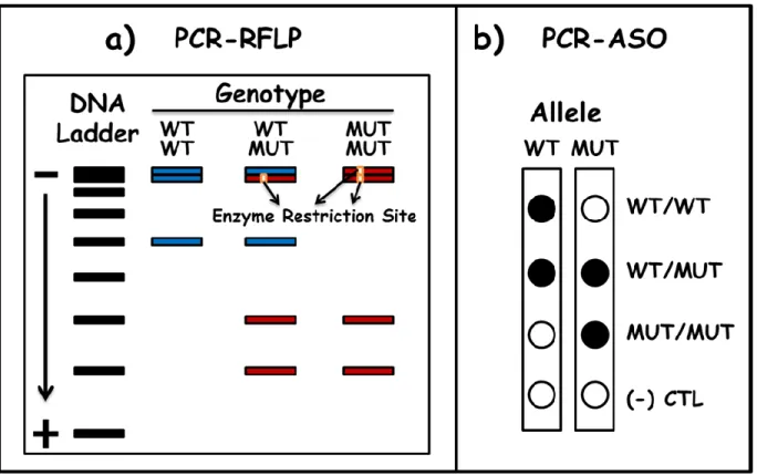
Figure 3. Visual illustration of Polymerase Chain Reaction (PCR)-based genotyping techniques.
... 35
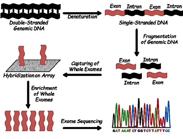
Figure 4. Visual illustration of the workflow of the array-based exome-enrichment and
whole-exome Sequencing approach. ... 40
Chapter 2
Figure 1. Metabolic pathways involved in the mechanism of action of thiopurines. ... 96
Chapter 3
Figure 1. Mechanism of action of Asparginase. ... 133
Chapter 4
Figure 1. The selection process following the exome-wide association study... 172 Figure 2. Top-ranking EWAS signals common for several asparaginase-related toxicities ... 173 Figure 3. Association of rs3809849 in MYBBP1A gene with ASNase-related toxicities and with
event free- and overall survival. ... 174
Figure 4. Combined-effect model. ... 175 Figure 5. Replication analysis in the independent validation cohort. ... 176
xvi
Figure 6. Distribution of patients with pancreatitis among risk groups established using wGRS
from the comprehensive combined-effect model in QcALL & DFCI cohort. ... 177
Supplemental figure S1. Comprehensive combined-effect model of all SNPs significantly
associated with pancreatitis. ... 188
Supplemental figure S2. Combined-effect analysis in Combined-cohort for the 3 SNPs and 5
SNPs significantly associated with pancreatitis. ... 190
Unpublished Data Figure U1. Association of the genotype of rs3809849 polymorphism in the
MYBBP1A gene with the risk of osteonecrosis during ALL treatment. ... 193
Unpublished Data Figure U2. Association of rs11556218 in IL16 gene with white blood cell
count at presentation & event-free survival. ... 196
Unpublished Data Figure U3. Cell viability assay in lymphoblastoid cell-lines in relation to
rs11556218 IL16 gene polymorphism. ... 198
Chapter 5
Figure 1. Production of Cas9 expressing, & MYBBP1A gene knock-out PANC1 cell lines and
evaluation of cell proliferation capacity. ... 229
Figure 2. In-vitro sensitivity to asparaginase and vincristine in relation to MYBBP1A gene
Knock-out. ... 230
Figure 3. Effect of MYBBP1A gene knock-out from PANC1 cells on their colony formation
capacity and response to treatment with asparaginase. ... 231
Figure 4. Impact of MYBBP1A gene deletion on PANC1 cellular functions: a) Cell-Cycle and b)
Apoptosis/Necrosis, and response to asparaginase exposure. ... 232
Figure 5. Relative expression of markers associated with epithelial-mesenchymal transition in
PANC1 cells a) in response to MYBBP1A gene deletion and b) asparaginase exposure. ... 233
Supplemental Figure S1. Result of the mismatch cleavage assay performed on the cell
population used for clonal selection. ... 238
xvii
Supplemental Figure S3. In-vitro sensitivity to asparaginase and vincristine in relation to
MYBBP1A gene Knock-out 96 hours post incubation with a) asparaginase (ASNase) or b)
vincristine (VCR). ... 240
Supplemental Figure S4. Association of MYBBP1A gene expression levels with survival
probability in different types of cancer. ... 241
Chapter 6
Figure 1. Confirmatory step following the exome-wide association study. ... 279 Figure 2. Performance of the wGRS based combined-effect model in the discovery cohort and
classification efficiency in both the discovery and replication cohorts... 280
Figure 3. Performance of the comprehensive combined-effect model in predicting the risk of
VIPN and classifying patients into risk groups. ... 281
Supplemental Figure S1. Meta-analysis of the top-ranking associations combining both cohorts.
... 286
Supplemental Figure S2. Association between the top-ranking hits and the expression of their
respective genes based on the genotype. ... 287
Chapter 7
Discussion Figure 1. Additive effect of carrying the minor alleles of rs11556218 in IL16 gene
and rs3809849 in MYBBP1A gene on the risk of pancreatitis. ... 302
Discussion Figure 2. Association of rs3803357 in BAHD1 gene with Overall Survival in the a)
xviii
V. List of Tables
Chapter 1
Table 1. Summary of outcomes derived from most recent front-line trials for children and
adolescents newly diagnosed with acute lymphoblastic leukemia. ... 13
Table 2. Asparaginase exposure in Dana Farber Cancer Institute protocols. ... 15
Table 3. Therapy on DFCI ALL Consortium Protocols: 1981–2000. ... 19
Table 4. Therapy on AIEOP-BFM ALL 2000 protocol. ... 22
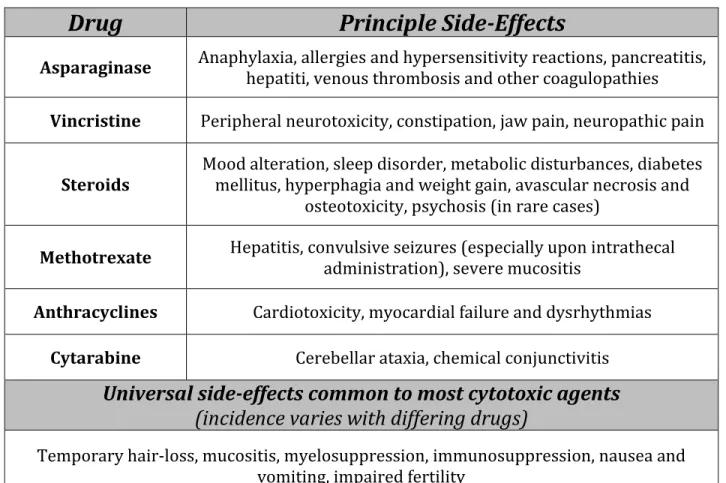
Table 5. Class-specific and universal side-effects of chemotherapeutic agents. ... 27
Chapter 2
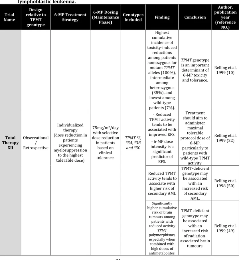
Table 1. Summary of selected studies which investigated the influence of TPMT pharmacogenetics on thiopurine treatment response in childhood acute lymphoblastic leukemia. ... 91Table 2. Summary of selected studies which investigated the influence of TPMT pharmacogenetics on thiopurine treatment response in inflammatory bowel disease. ... 95
Chapter 3
Table 1. This table summarizes the prominent studies in the literature which investigated the pharmacogenetics of asparaginase and highlights the most important finding. ... 130Chapter 4
Table 1. Characteristics of the discovery and the replication cohort. ... 164xix
Table 2. Top-ranking signals from the exome-wide association study confirmed by genotyping.
... 165
Table 3. Performance of the comprehensive genetic model in predicting the risk of pancreatitis.
... 167
Supplemental Table S1. Association of WES data with asparaginase-related complications ... 179 Supplemental Table S2. Function and differential protein expression of genes selected for
further investigation. ... 183
Supplemental Table S3. Multivariate analysis for primary and secondary associations confirmed
by genotyping ... 186
Supplemental Table S4. Combined-cohort analysis performed for SNPs with reproducible
associations with pancreatitis. ... 187
Unpublished Data Table U1. Stratification analysis of the association of rs3809849
polymorphism in the MYBBP1A gene with the risk of osteonecrosis based in clinical subgroups in the combined-DFCI cohort. ... 194
Unpublished Data Table U2. Multi-variant analysis for the risk of osteonecrosis associated
adjusting for rs3809849_MYBBP1A polymorphism genotype along with clinical factors. ... 195
Chapter 6
Table 1. Characteristics of the discovery and the replication cohorts. ... 275 Table 2. Top-ranking signals from the exome-wide association study confirmed by genotyping.
... 276
Supplemental Table S1. Association of WES data with vincristine-induced peripheral
neuropathy. ... 283
Supplemental Table S2. Association of the risk alleles with the reduction in the VCR dose
administered. ... 284
Supplemental Table S3. Association of the risk alleles with the number of episodes of
high-grade VIPN. ... 285
Supplemental Table S4. Analysis of the association between the risk alleles and lower-grade
xx
VI. Abbreviations and Acronyms
6-MP: 6-mercaptopurine
6-TGN: 6-Thioguanine Nucleotides
ACTG1: Actin Gamma 1
ADAMTS17: A Disintegrin-Like And Metalloprotease (Reprolysin Type) With Thrombospondin Type 1 Motif, 17
ADRs: Adverse Drug Reactions
AhR: Aryl Hydrocarbon Receptor
AIEOP: Associazione Italiana di Ematologiae Oncologia Pediatrica (Italian Association of
Pediatric Haematology and Oncology)
AKT: AKT Serine/Threonine Kinase 1
ALL: Acute Lymphoblastic Leukemia ASN: Asparagine
ASNase: Asparaginase
ASO: Allele-Specific Oligonucleotides
ASS1: ArginoSuccinate Synthase 1 ATF5: Activating Transcription Factor 5
AUC: Area Under the Curve AZA: Azathioprine
BAHD1: Bromo adjacent homology domain containing protein 1
xxi
BFM: Berlin-Frankfurt-Münster BSA: Body Surface Area
Cas9: CRISPR Associated Protein 9 CI: Confidence Interval
CIPN: chemotherapy-induced peripheral neuropathy CMT: Charcot-Marie-Tooth
CNS: Central Nervous System COG: Children's Oncology Group
CPIC: Clinical Pharmacogenetics Implementation Consortium
CRISPR: Clustered Regularly Interspaced Short Palindromic Repeats
CRLF2: Cytokine Receptor Like Factor 2
CTCAE: Common Terminology Criteria for Adverse Events
CYP: Cytochrome P450
DCOG: Dutch Childhood Oncology Group DFCI: Dana-Farber Cancer Institute
DHFR: Dihydrofolate Reductase E.coli: Escherichia coli
EFS: Event-free survival
EMT: Epithelial-Mesenchymal Transition
EORTC CLG: European Organization for Research and Treatment of Cancer-Children's
Leukemia Group
xxii
Erwinia: Erwinia chrysanthemi
ETV6-RUNX1 (TEL-AML1): ETV6 (ETS Variant 6) & RUNX1 (Runt-Related Rranscription Factor 1) gene fusion
EWAS: Exome-Wide Association Study FDA: U.S. Food and Drug Administration FDR: False Discovery Rate
GC: Glucocorticoid
GRIA1: Glutamate Ionotropic Receptor AMPA Type Subunit 1 HLA: Human Leukocyte Antigen
HR: High-Risk
HSCT: Hematopoietic Stem Cell Transplant HSR: HyperSensitivity Reactions
IBD: Inflammatory Bowel Disease
IC50: Minimum Inhibitory Concentration 50
IKZF1: IKAROS Family Zinc Finger 1 IL16: Interleukin 16
iPSC: Induced Pluripotent Stem Cells IR: Intermediate:Risk
ITPA: Inosine Triphosphate Pyrophosphatase
LCLs: Lymphoblastoid Cell Lines MAF: Minor Allele Frequency miRNAs: micro inhibitory RNAs
xxiii
MLL: mixed-lineage leukemia MPEG1: Macrophage Expressed 1
MRD: minimum residual disease
MRPL47: Mitochondrial Ribosomal Protein L47 MTHFR: 5,10-Methylenetetrahydrofolate reductase
MTX: methotrexate
MYBBP1: MYB Binding Protein 1a
NES: Normalized Effect Size
NFATC2: Nuclear Factor of Activated T Cells 2 NF-kB: Nuclear Factor kappaB
NGS: Next Generation Sequencing
NOPHO: Nordic Society of Paediatric Haematology and Oncology NSAA: Nadir serum asparaginase activity.
ON: Osteonecrosis OR: Odd-Ratio OS: Overall Survival
PANC1: Pancreatic Cell Line 1
PAX5: Paired Box 5 (B-Cell Lineage Specific Activator Protein) PBX1: Pre-B-Cell Leukemia Homeobox 1
PCR: Polymerase Chain Reaction PEG ASNase: Pegylated Asparaginase
xxiv
PGx: Pharmacogenomics and Pharmacogenetics Ph-like: Philadelphia Chromosome Like
Ph-positive: Philadelphia Chromosome Positive
PKD2L1: Polycystin 2 Like 1 PREP1: Pbx Regulating Protein-1 PRSS1/PRSS2: Protease, Serine 1/2
QcALL: Quebec Childhood Acute Lymphoblastic Leukemia RCTs: Randomized Clinical Trials
RIN3: Ras Interaction/Interference Protein 3
ROC: Receiver Operator Characteristic SEM: Standard Error of the Mean
SJCRH: St Jude Children's Research Hospital SJUHC: Sainte-Justine University Hospital Centre
SLC39A12: Solute Carrier Family 39 Member 12 SLC7A1: Solute Carrier Family 7 Member 13
SMN: Second Myeloid Neoplasms SNP: Single Nucleotide Polymorphism
SPECC1: Sperm Antigen With Calponin Homology And Coiled-Coil Domains 1 SPEF2: Sperm Flagellar 2
SR: Standard-Risk
SYNE2: Spectrin repeats containing nuclear envelope 2
xxv
TPMT: Thiopurine S-Methyl Transferase TS: Thymidylate Synthase
UKALL: United Kingdom Acute Lymphoblastic Leukaemia. VCR: Vincristine
VIPN: Vincristine-Induced Peripheral Neuropathy WBC: White Blood Cell
WES: Whole-Exome Sequencing wGRS: weighted Genetic Risk Score WGS: Whole-Genome Sequencing WT: Wild-Type
xxvi
VII. Dedication
I dedicate this thesis to my wife, May.
Your love and support helped me go all the way
.
& to my baby girl, Mila, the joy of my life.
Also, to my parents and my family,
who, by being so untypical, always
provoked me to give it all and to
become the best version of myself.
xxvii
VIII. Acknowledgement
It is my great pleasure to acknowledge all of those who helped me through this exciting journey and made this thesis possible.
I would like to start by sincerely thanking Dr. Maja Krajinovic for offering me this great opportunity and guiding me through all of it with patience, understanding and support. Always seeing my potential and pushing me to give my best, and for which, I will remain forever grateful.
Also, I wish to acknowledge the members of my thesis committee, Dr. Isabelle
Laverdière, Dr. Simon De Denus, and Dr. R-Noël J-M Raynal, for accepting their roles and
taking the time to go through my work.
I would like to also deeply thank Dr. Réné Cardinal and Dr. Yves Théôret for being there for me throughout the entire journey, always believing in me and providing encouragement, guidance and extremely valuable recommendations.
I would also like to express my sincere gratitude to Dr. France Varin, who was the first faculty member to see my potential and who paved the way for the start of this journey, along with Dr. Catherine Litalien, who I also deeply thank.
xxviii
It goes without saying that I thank all previous and current members of the Krajinovic’s lab for the help, support and valuable friendship they always provided. I specifically thank Vincent Gagné for always being so kind and patient; Aziz Rezgui for his constant encouragement and support; and Kateryna Petrykey for offering me her friendship and for sharing all the stressful moments throughout our common journey.
I even extend my gratitude to all members of the Research Centre of the Sainte-Justine hospital, and particularly to members of the 6th and 7th floor, for always being kind and friendly, and providing essential help when I was struggling with my experiments or with learning new techniques.
Finally, I thank all the patients who accepted to participate in the studies and made this work possible, as well as the funding agencies and organizations that provided the financial means for doing it. I especially thank the Faculty of Medicine and the Faculty of Higher Education of the University of Montreal, the Cole Foundation and the Network of Applied Medical Genetics for their generous scholarships.
xxix
IX. Preface
The present thesis titled “Using Whole-Exome Sequencing Data in an Exome-Wide Association Study Approach to Identify Genetic Risk Factors Influencing Acute Lymphoblastic Leukemia Response: A Focus on Asparaginase Complications & Vincristine-Induced Peripheral Neuropathy“ has been carried out by me under the guidance and supervision of Dr. Maja Krajinovic, and is submitted to the faculty of higher education at the University of Montreal in partial fulfillment of the requirements for the degree Doctor of Philosophy in Pharmacology (Pharmacogenomics option). This work is presented in the by-article format.
Being a practicing pharmacist, it has always been intriguing to me how the same drug administered in the same dose to different patients would result in a spectrum of effects that can range from complete absence of response all the way to severe life-threatening toxicities. This observation, combined with my passion about genetics, ignited my interest in conducting pharmacogenetics research that would help to advance our understanding of the genetic basis of variability in drug response. Therefore, during my four years of doctoral studies, I tried to get involved in different aspects of pharmacogenetics research ranging from reviewing and summarizing the available literature, to discovering and validating novel genetic markers, passing by fundamental and translational research to determine their usefulness and applicability, and ending by assessing the need for implementation of pharmacogenes in clinical practice.
xxx
It is worth mentioning here that I used childhood acute lymphoblastic leukemia (ALL) as a disease model to learn and apply pharmacogenetics techniques and to investigate the role of genetic variability in altering the drug response.
In the body of this thesis, in the first chapter of Section-A, I will provide a brief, but detailed, introduction covering the basic information essential for the understanding of the context of this work and the different notions and definitions that are discussed through it. In the second chapter, I present a review paper titled “Thiopurine S-methyltransferase
polymorphisms in acute lymphoblastic leukemia, inflammatory bowel disease and autoimmune disorders: influence on treatment response “. This paper was published in 2017
in the Pharmacogenomics and Personalized Medicine journal and provides an overview of the history and temporal evolution of TPMT towards becoming one of the most important pharmacogenes in clinical practice. I discuss the results, conclusions and recommendations of selected studies that investigated the pharmacogenetics influence of TPMT gene on thiopurine treatment in ALL, inflammatory bowel disease and autoimmune disorders, and also briefly address the cost-effectiveness of this pharmacogenetics approach and its impact on clinical practice
In Section B, I present three articles that targeted different aspects of the pharmacogenetics of asparaginase (ASNase) as a key component of ALL treatment along with a special chapter containing results not presented in a paper format. The first article in this section is presented in Chapter-3 and is a review article titled “Pharmacogenetics of
Asparaginase in Acute Lymphoblastic Leukemia”. It was published in 2019, in the special
xxxi
highlights the most important findings reported in studies of the pharmacogenetics of ASNase related complications and treatment outcome.
The second article of this section is an original research paper presented in Chapter-4 and titled “Whole-exome sequencing identified genetic risk factors for asparaginase-related
complications in childhood ALL patients”. It was published in Oncotarget journal in 2017 and
describes the results obtained from using whole-exome sequencing (WES) data to perform exome-wide association studies (EWAS) with ASNase-related toxicities and highlights their interactions and pertinence to the studied outcome, with a special focus on acute pancreatitis. This work suggests that MYBBP1A gene as an important candidate in modulating ASNase response that is associated with increasing risk of developing all of the studied complications.
The third article of Section-B, presented in Chapter-5 and titled “Characterization of
the functional impact of MYBBP1A gene on asparaginase sensitivity and risk of pancreatitis following exome-wide association study results” is an original research work currently in
preparation. In this EWAS follow-up study, I aimed at confirming and characterizing the involvement of MYBBP1A gene in modulating the cellular response to ASNase by studying the effect of gene deletion in PANC1 pancreatic cells, using CRISPR-CAS9 technology, on cellular behaviour and biological functions before and after treatment with ASNase.
The next section, Section-C, only has one chapter, Chapter-6, represents an original research paper that was published in 2018 in Pharmacogenomics journal and is titled “Genetic risk factors for VIPN in childhood acute lymphoblastic leukemia patients identified
xxxii
using whole-exome sequencing”. This work was performed in a similar manner of the one
described in Chapter-4, but was focused on identifying genetic variants involved in modulating the risk of vincristine-induced peripheral neuropathy; a common side effect to the administration of vincristine as an important chemotherapeutic agent in childhood ALL treatment.
The last chapter of this thesis, Chapter 7, is presented in Section D and it provides a summary of the major findings, as well as detailed discussion on the two most prominent genes in this work, MYBBP1A and IL16, and trying to address the different possible mechanisms that these gene could be exerting their effect on modulating the response to ASNase. It also discusses the limitations of the work and suggests future studies that can help to better understand the role of the identified genes in the respective toxicities.
Section A
Chapter 1
General Introduction
This chapter, as indicated in the title, is meant to prepare the readers to navigate through the following chapters of the thesis by providing the essential information relative to the diverse topics discussed in this work. It also outlines the working hypotheses that formed the basis of the research design, and defines the objectives that the conducted studies were aiming to achieve.
2
1. General Introduction
1.1. Definition & Statistics
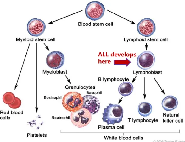
Leukemia is a type of cancer that affects the hematopoietic precursors of the lymphoid lineage. Acute Lymphoblastic Leukemia (ALL) is a fast progressing leukemic malignancy which results from an abnormal transformation and proliferation of lymphoid progenitor cells in the bone marrow and the blood.1-4 It is a result of the deregulated control of the blood stem cells which affects their ability to differentiate into healthy mature blood cells, thus affecting the number and functions of different blood components (i.e. red blood cells, white blood cells, and platelets) and consequently provoking a wide range of complications.2,4 Figure.1 provides a quick outlook on blood cells development showing the differentiation of diverse lineages of blood and immune cells from a common blood stem cell, including T and B lymphocytes.4
3
Figure 1. Differentiation of hematopoietic stem cells.
In the bone marrow, blood stem cells differentiate into either the myeloid or the lymphoid progenitor lines. Acute lymphoblastic leukemia is a result of aberrant differentiation of the lymphoid cells (B or T cell), leading to overproduction and accumulation in the blood, bone marrow, spleen and liver.
For the National Cancer Institute © 2008 Terese Winslow LLC, U.S. Govt. has certain rights. Reproduced with permission.
4
Generally, the presence of 20% lymphoblasts in the bone marrow or the blood is used as a cut-off to establish the ALL diagnosis.1,5 Almost 80% of ALL cases occur in pediatric population and is referred to as childhood acute lymphoblastic leukemia.3 The incidence can start as early as before birth, but there is a marked peak in between 1-5 years of age.2,6 However, another peak can also be observed at around the age of 50, giving rise to adult acute lymphoblastic leukemia, which is usually associated with less favourable outcomes.2,6 In fact, survival probability decreases with increasing patient’s age at diagnosis, and sadly, the long-term survival rate among patients over 60 years of age is only about 10-15%.1,7 Childhood ALL is the most common subtype of leukemia, accounting for approximately 25% of all childhood cancers and about 75-80% of leukemia cases in children.2,8,9 Furthermore, it is the most frequent cause of death from cancer before 20 years of age.6
Genetics can play an important role in the incidence of ALL as it was shown that ethnicity is significantly associated with the risk of developing ALL; with black race individuals being the least affected, followed by those of the white race and then Hispanics having the highest incidence.6 Moreover, in the same genetic context, male gender was found to be associated with a slightly higher, but significantly different, risk of childhood ALL than female gender (55% to 45%, respectively).6 This inherent vulnerability of male gender is not surprising since it has been previously pointed out that the variability in epigenetic signature between genders, and the differential ability of the Y vs. X chromosomes in repairing damage to their genes, can render boys at increased risk of
5
developing various types of health conditions including different cancers.6,8 For example, a recent study reported that the gene coding for the histone demethylase Ubiquitously Transcribed X-chromosome (UTX) tetratricopeptide repeat protein was found to be recurrently affected by somatic loss-of-function mutations in male T-cell acute lymphoblastic leukemia (T-ALL) patients and that UTX is capable of escaping X-inactivation in female T-ALL blasts as well as in normal T cells; thus adding to the growing body of evidence suggesting that UTX has a gender-specific tumor suppressor role in the context of T-ALL, among other cancers.10
1.2. Prognostic Factors
Classically, childhood ALL was majorly stratified into risk groups based on two important clinical factors, age and white blood cell counts at presentation.1 However, it is largely recognized nowadays that in addition to clinical features at diagnosis, immunophenotype, pathophysiology and cytogenetic changes of cancer cells, genetics of the host, as well as response to initial treatment (also known as early response), can all interact together to affect the risk and prognosis of childhood ALL and should be used collectively to guide treatment regimens.1
6
1.2.1. Age & WBC count
Briefly, older age and higher WBC count are associated with a worsening prognosis and two groups of risk can be defined based on these parameters according to the Consensus criteria of the Rome/National Cancer Institute Workshop:11 “standard risk” (1 > age < 10 years and initial WBC count of <50,000 per cubic millimeter) representing around two thirds of patients, and “high risk” (age ≥10 years, initial WBC count ≥50,000 per cubic millimeter, or both)12 which roughly makes one third of patients. It must be noted that ALL in children < 1 year of age at diagnosis is usually associated with a worse outcome and is considered a special subgroup.6,13,14
1.2.2. Immunophenotype
Immunophenotyping based on the expression of the surface markers of lineage can distinguish between two subtypes of childhood ALL known as precursor B-cell and T-cell, making reference to the otherwise healthy mature lymphocytes expressing these markers, and representing around 85% and 15% of childhood ALL cases, respectively. This is important to understand the distinction between immunophenotypes since it was shown that age and WBC count at diagnosis have limited prognostic importance in T-cell ALL.6,13
7
1.2.3. Cytogenetics & Molecular Genetics
While several factors have been reported to predispose to an increased risk of developing childhood ALL including exposure to ionizing radiation, chemicals such as pesticides & certain solvents, viral infections like Epstein-Barr virus or human immunodeficiency virus, these factors can only explain a minor percentage of cases.1,6,15,16
Differences in the genetic make-up between patients have recently driven considerable attention as genetic variability and chromosomal aberrations have been described as early, probably initiating events, in developing ALL, and were shown to play an important role in disease detection, prognosis and treatment response.1,6,17,18 Common genetic alterations include single nucleotide polymorphisms (SNPs), genomic insertions and deletions, as well as copy number variation.19 These variants can be divided into disease-causing variants -with high penetrance and a large pathogenic effect- which are usually rare and mostly seen in single-gene Mendelian disorders, or can have lower penetrance and smaller pathogenic effect -typically present in higher frequency in cases compared to controls in association studies.20 For instance, genes governing B-lymphoid development have been associated with ALL, most notably PAX5 gene, which was estimated to be mutated in 35% of childhood ALL patients 21 followed by IKZF1 gene reportedly mutated in 15% of cases.22 Several association studies identified polymorphic variants in various other genes to be linked to an increased risk of ALL or to specific subtypes of it such as variants in CEBPE, GATA3 and ARID5B genes.23-25 Likewise, copy number variation within
8
genes involved in B cell proliferation and differentiation is a very frequent event observed in B-cell ALL patients.18,21
Tumor-specific genetic alterations can include inter-chromosomal translocations, uniparental disomy, and loss of heterozygosity. For example, loss of heterozygosity in an allele of tumor suppressor gene can results in tumorigenesis and may also influence drug effects thus modulating the evolution of the disease and its progression.18,19,26 Several genetic translocations were extensively described in childhood ALL such as: t(12;21) [ETV6-RUNX1] gene fusion reported in around one quarter of cases; t(9;22) [BCR-ABL1] that results in the formation of an activated tyrosine-kinase and is also known as the Philadelphia chromosome (Ph-positive) ALL; and the translocation of t(1;19) [TCF3-PBX1] whose protein product alters cell differentiation arrest mechanisms among others. Additionally, multiple genomic rearrangement of the CRLF2 gene 6,27 as well as more than 70 different chromosomal rearrangements involving the chromosome 11q23 mixed-lineage leukemia (MLL) gene,1,21,28 have been described in ALL literature. Recently, a new subtype of ALL, characterized by exhibiting a gene expression profile similar to that of the Philadelphia chromosome but lacking the BCR-ABL1 rearrangement, has been identified, and is also known as Philadelphia (Ph)-like ALL (or previously as BCR-ABL-like ALL). Interestingly, 90% of Ph-like cases seem to harbor a plethora of genetic alterations lading to kinase-activation.1,6 The relative frequency of genetic alterations found in major B-ALLs and T-lineage subtypes of ALL as derived from front-line studies of childhood ALL are shown in Figure.2.
9
Figure 2. The relative frequency of major B-ALLs and T-lineage subtypes of ALL.
BCR-ABL1–like subtype and BCR-ABL1–positive ALL are shown in yellow to illustrate the high frequency of childhood B-ALL cases with genetic alterations activating tyrosine kinase and cytokine receptor signaling. Data are derived from front-line studies of childhood ALL. Reproduced with permission from (Mullighan CG. Molecular genetics of B-precursor acute lymphoblastic leukemia. J Clin Invest 122(10), 3407-3415 (2012)). American Society for Clinical Investigation.
10
Interestingly, it was shown that genetic background variability related to race can be associated with differential risk of developing particular subtypes of ALL such as
TCF3-PBX1 ALL in Blacks 12 and CRLF2-rearrangement ALL in Hispanics.27 Moreover, numerous genetic syndromes have also been associated with a higher risk of developing ALL in children, most notably being Down syndrome and Fanconi anemia, but ataxia telangiectasia Neurofibromatosis, Bloom syndrome, Li-Fraumeni syndrome and Nijmegen breakdown syndrome were also reported.1,4,6,29-32
It is highly important to have a detailed characterization of the patient’s ALL subtype as certain genetic alterations can have prognostic utility since they were shown to be associated with treatment outcome of childhood ALL.1,6,33 For example, high-risk of a poor outcome has been consistently reported for patient with intra-chromosomal amplification of chromosome 21,34 BCR-ABL1 gene fusion,35 Ph-like subtype of ALL,1,36 MLL rearrangement,37 and alterations of IKZF1;38,39 as well as for patients showing hypodiploidy with less than 44 chromosomes,40 and those with T-cell precursor ALL subtype.41,42 On the other hand, ETV6-RUNX1 translocation and high hyperdiploidy are associated with favourable outcome.6
Moreover, variability in epigenetic signature, such as an aberrant acetylation or methylation profile, can modulate genetic expression, thereby influencing drug effect, and is a common feature of cancer cells.19,43,44 Furthermore, it is increasingly recognized that even genomic regions that do not codify proteins such as micro inhibitory RNAs (miRNAs), which are RNA sequences that are around 22 nucleotides in size, can be strongly implicated in
11
regulatory functions as they can modulate the expression of over 60% of known genes, thus influencing sensitivity to drugs and treatment outcome.45 Indeed, some miRNA-related polymorphisms have been shown to affect miRNA levels and function, and the expression of some of those miRNAs has been associated with drug response in ALL treatment.45-49
1.2.4. Early Treatment Response:
Recently, early response to treatment (also referred to as response to the initial therapy) has received a lot of attention and emerged as an important and independent prognostic tool in ALL treatment. The determination of the phenotype (i.e. type of response) is based on the evaluation of the time required to bring down the initial leukemic-cell population to undetectable levels, known as minimum residual disease (MRD).1,6,13 This method uses molecular techniques such as the polymerase chain reaction or flow cytometry to monitor the disease at submicroscopic levels, which helps further refining the risk-stratification process at different stages of therapy, consequently improving the treatment outcome.50-54
12
1.3. ALL Treatment and Outcome
1.3.1. Improvement in Treatment Outcome
The first temporary remission of leukemia induced by chemotherapy was reported around 7 decades ago, in 1948.55 In the 1960s, the survival rate of childhood ALL was estimated to be less than 10%.6,56 Nowadays, the 5 years event-free survival (EFS) and overall survival (OS) rates are reported to surpass 85% and 90%, respectively, for most international treatment protocols;57-63 thus making childhood ALL an exemplary model for progressive improvement.1,6,8,56 Table.1 provides a short summary of outcomes derived from most recent front-line trials for children and adolescents newly diagnosed with ALL. Similar improvement was also reported for 10-year survival which has witnessed an increase of more than 20 percentage points in the last three decades in patients aged 0–14 years, which is being considered recently as a new cut-off value for age-based risk stratification in childhood ALL. 8
13
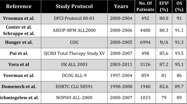
Table 1. Summary of outcomes derived from most recent front-line trials for children and adolescents newly diagnosed with acute lymphoblastic leukemia.
Reference
Study Protocol
Years
Patients No. Of EFS(%) § (%) OSVrooman et al. DFCI Protocol 00-01 2000-2004 492 80.0 91
Conter et al.
Schrappe et al. AIEOP-BFM ALL2000 2000-2006 4480 80.3 91.1
Hunger et al. COG 2000-2005 6994 N/A 91.3
Pui et al. SJCRH Total Therapy Study XV 2000-2007 498 85.6 93.5
Vora et al UK ALL 2003 2003-2011 3126 87.2 95.1
Veerman et al. DCOG ALL-9 1997-2004 859 81 86
Domenech et al. EORTC CLG 58591 1998-2008 1940 82.6 89.7
Schmiegelow et al. NOPHO ALL-2000 2000-2007 1023 79 89
AIEOP-BFM, denotes Italian Association of Pediatric Haematology and Oncology and Berlin-Frankfurt-Münster; ALL, Acute Lymphoblastic Leukemia; COG, Children’s Oncology Group; DCOG, Dutch Childhood Oncology Group; DFCI, Dana Farber Cancer Institute Consortium; EORTC CLG, European Organization for Research and Treatment of Cancer-Children's Leukemia Group; EFS, Event-free survival; OS, Overall Survival; NOPHO Nordic Society of Paediatric Haematology and Oncology; SJCRH, St Jude Children’s Research Hospital; UKALL, United Kingdom Acute Lymphoblastic Leukaemia.
§ Survival percentages shown are the rates at 5 years except for the rates for the AIEOP-BFM trial, which were reported at 7 years.
14
Reproduced with permission from (Hunger SP, Mullighan CG. Acute Lymphoblastic Leukemia in Children. N Engl J Med 373(16), 1541-1552 (2015)), Copyright Massachusetts Medical Society.6
This was achieved through the introduction and continuous refining of multi-agent chemotherapeutic regimens, paired with the progressive advancement in risk-stratification based on clinical features of the patients, a better understanding of the biological mechanisms underlying the disease, the ability to exploit genetic differences between cancer-cells and host-cells, as well as the incorporation of the initial treatment response as a dynamic parameter into the risk-calculation equation and the adoption of precision-medicine treatment strategies.6,13 Table 2 provides a brief comparison of ASNase exposure between four consequent treatment protocols of the Dana Farber Cancer Institute Consortium to highlight the evolution of the use of ASNase which is a main focus of this thesis and will be discussed in further details throughout different chapters.
15
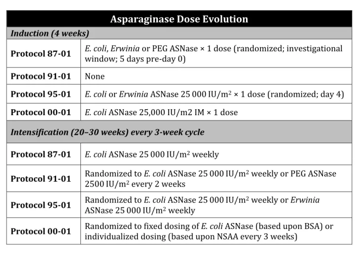
Table 2. Asparaginase exposure in Dana Farber Cancer Institute protocols.
Asparaginase Dose Evolution
Induction (4 weeks)
Protocol 87-01 E. coli, Erwinia or PEG ASNase × 1 dose (randomized; investigational window; 5 days pre-day 0)
Protocol 91-01 None
Protocol 95-01 E. coli or Erwinia ASNase 25 000 IU/m2 × 1 dose (randomized; day 4)
Protocol 00-01 E. coli ASNase 25,000 IU/m2 IM × 1 dose Intensification (20–30 weeks) every 3-week cycle
Protocol 87-01 E. coli ASNase 25 000 IU/m2 weekly
Protocol 91-01 Randomized to E. coli ASNase 25 000 IU/m2500 IU/m2 every 2 weeks 2 weekly or PEG ASNase
Protocol 95-01 Randomized to E. coli ASNase 25 000 IU/mASNase 25 000 IU/m2 weekly 2 weekly or Erwinia
Protocol 00-01 Randomized to fixed dosing of E. coli ASNase (based upon BSA) or individualized dosing (based upon NSAA every 3 weeks)
Abbreviations: ASNase, asparaginase; PEG, pegylated; BSA, body surface area; NSAA, nadir serum asparaginase activity.
Reproduced from author’s own article (Wolthers BO, Frandsen TL, Patel CJ et al. Trypsin encoding PRSS1-PRSS2 variation influence the risk of asparaginase-associated pancreatitis in children with acute lymphoblastic leukemia: a Ponte di Legno toxicity working group report. Haematologica doi:10.3324/haematol.2018.199356 (2018)), Copyright European Hematology Association.64
16
Nevertheless, while the landscape looks promising for childhood ALL, it is important to note that the prognosis for adulthood ALL is still dismal, with almost half of the patients failing to achieve long-term remission, up until recently.1,65,66 Encouragingly, it has been suggested lately that using pediatric-inspired protocols may be helpful in increasing survival of the adolescent and young adults population (i.e. 15-39 years), with some preliminary results showing a 5-year EFS of as high as 72%.67-69 Unfortunately, however, for infants that are less than 1 year of age, the survival remains low despite the ongoing efforts aiming at improving it. One possible contributing factor to this poor prognosis is the fact that infant ALL is usually associated with MLL gene rearrangement, which, on its own, is associated with unfavourable outcomes, and any further intensification of chemotherapy can cause significant long-term and short-term toxicities in this vulnerable population.8,70
1.3.2. Contemporary Therapy
A major milestone in anti-leukemia treatment was the introduction of an intensive regimen that employed sets of combinations of 8 drugs administered over two phases (induction and consolidation) for a period of 8 weeks. This treatment strategy was later referred to as protocol-I and became the backbone of most contemporary protocols for ALL.6 Indeed, modern treatment strategies for childhood ALL last 2–2.5 years in total and include distinct phases each of them having a specific objective. Table 3 summarizes the evolutionary history of the Dana Farber Cancer Institute Consortium (DFCI) protocols and Table 4 provides details on the Associazione Italiana Ematologia Oncologia Pediatrica and
17
the Berlin-Frankfurt-Munster study protocol AIEOP-BFM ALL 2000. Both of these protocols will be discussed extensively throughout this thesis.
The initial phase is essentially a remission induction therapy that usually lasts 4 to 6 weeks and includes an L-asparaginase (ASNase) formulation, a glucocorticoid (e.g. prednisone or dexamethasone; GCs) and vincristine (VCR), as wells as the optional use of an anthracycline. By the end of this phase, remission is successfully induced in most patients (85-95%), but relapse is still possible due to the submicroscopic residual disease. To reduce this risk and prevent the development of overt CNS leukemia, patients undergo a remission consolidation phase which includes 6 to 9 months of intensive combination chemotherapy. In general, high dose methotrexate (MTX) along with 6-mercaptopurine (6-MP) are commonly used in this phase, accompanied by frequent pulses of VCR, GCs and ASNase for 20–30 weeks. Basically, the drug combinations in this phase tend to include chemotherapeutic agents that have different mechanisms of action from those applied in the induction phase and might also include cytarabine, etoposide, and cyclophosphamide. This concept is important in order to minimize drug resistance and assure the elimination of submicroscopic residual disease by taking advantage of the synergistic effects obtained by combining the different molecules. The last phase is primarily a maintenance therapy and can last between 18 and 30 months depending on the protocol and the risk group. This is a low-intensity antimetabolite-based treatment comprising a daily oral 6-MP or thioguanine and a weekly oral MTX administered along with optional periodic pulses of glucocorticoids and vincristine every 5 to 7 days in certain protocols.6,13
18
Historically, cranial radiation was routinely employed in many protocols to further prevent CNS relapse, but its use was gradually abandoned (or reserved only for patients with the highest risk) due to its associated toxicities such as the risk of developing a second malignant neoplasms and the concerns about its long-term effects on cognitive skills leading to intellectual disability, especially in young adults. Instead, it was replaced by intrathecal therapy that was incorporated into the induction remission phase of most protocols and which includes the administration of intrathecal methotrexate, either alone, or in combination with cytarabine and hydrocortisone (referred to as triple intrathecal treatment). However, the administration of this therapy in other phases is variable across the different protocols, with some of them also administering it during the remission consolidation phase while others throughout the entire course of treatment.6,13,56
One of the hallmarks of childhood ALL treatment is the stratification of patients into risk groups. While the definition and treatment of high-risk childhood ALL remains controversial, the use of prognostic factors affecting the treatment outcome can allow the classification of patients into groups based on their risk of experiencing treatment failure. Protocols offer different blocks of chemotherapy with varying intensities and patients are then assigned to one of these blocks depending on their risk-stratified group. Accordingly, patients with favorable prognostic features can be treated with less toxic regimens while those at high-risk of failure or relapse can be assigned to receive more intense regimens to help eradicating the highly aggressive disease.6,13
19
Table 3. Therapy on DFCI ALL Consortium Protocols: 1981–2000.
Phase
Treatment
Induction
(4 weeks)
IT cytarabine* × 1 dose (day 0), IT chemotherapy day 14
Vincristine 1.5 mg/m2 q week (maximum=2 mg) (days 0, 7, 14, 21)
Prednisone 40 mg/m2/day (days 0–28)
Doxorubicin 30 mg/m2/dose (days 0 and 1) Protocol 81-01: 45 mg/m2/dose x 1 dose
Protocol 95-01: randomized +/− dexrazoxane 300 mg/m2 (HR only) Protocol 00-01: + dexrazoxane 300 mg/m2 (HR only)
Methotrexate × 1 dose (day 2): dose per protocol
Protocol 81-01: None Protocol 85-01: 40 mg/m2
Protocol 87-01: 40 mg/m2 or 4 g/m2 with leucovorin (randomized) Protocols 91-01 + 95-01 + 00-01: 4 g/m2 with leucovorin
Asparaginase
Protocol 81-01: None
Protocol 85-01: E.coli ASNase × 1 dose (investigational window; 5 days pre-day 0)
Protocol 87-01: E. coli, Erwinia or PEG ASNase × 1 dose (randomized; investigational window; 5days pre-day 0)
Protocol 91-01: None
Protocols 95-01: E.coli or Erwinia ASNase 25,000 IU/m2 × 1 dose (randomized; day 4)
Protocols 00-01: E.coli ASNase 25,000 IU/m2 × 1 dose (randomized; day 4)
CNS therapy
(3 weeks)
IT chemotherapy twice weekly × 2 weeks (4 doses) Vincristine 2.0 mg/m2 IV day 1 (maximum=2 mg)
6-MP 50 mg/m2/day orally (days 1–15)
HR only: doxorubicin 30 mg/m2 on day 1
Protocol 95-01: randomized +/− dexrazoxane 300 mg/m2
Cranial Radiation per protocol (beginning day 1)
Protocol 81-01: SR-18Gy; HR-28 Gy Protocol 85-01: SR-18Gy; HR-24 Gy Protocol 87-01: SR-No XRT; HR-18 Gy
Protocol 91-01: SR girls-No XRT; SR boys and HR-18 Gy.
Protocol 95-01: SR: randomized-No XRT versus 18 Gy; HR-18 Gy