Review
Molecular and Cellular Oncology 1, e1; July 2014; © 2014 Landes Bioscience
Review
The Lymphatic Network
The lymphatic vasculature consists of a network of lymph
ves-sels whose main function is to return protein-rich interstitial fluid
to the circulating blood. Fluid, macromolecules, and cells, such
as leukocytes and activated antigen-presenting cells, enter the
lymphatic system through the blind-ended lymphatic
capillar-ies. From here, lymph is transported toward collecting lymphatic
vessels and is returned to the blood circulation in the jugular
area through the lymphaticovenous junctions.
1On its way, lymph
is filtered through the lymph nodes, where foreign particles
taken up by antigen-presenting cells initiate specific immune
responses.
2In the small intestine, lacteal lymphatic vessels inside
the intestinal villi absorb the dietary fat released by enterocytes
in the form of lipid particles called chylomicron. In addition to
these physiologic functions, the lymphatic system contributes to
pathologic conditions such as lymphedema, inflammatory
dis-eases, and tumor metastasis. Many studies have demonstrated
the existence of proliferative peri- and intratumoral lymphatic
vessels.
3Additionally, tumoral lymphangiogenesis correlates with
an increase in metastases,
4,5and detection of lymphangiogenic
growth factors is associated with poor prognosis in many human
tumors.
6-8Similar to blood capillaries, lymphatic capillaries are
thin-walled, relatively large vessels composed of a single layer of
endo-thelial cells, but they are not covered by pericytes or smooth
muscle cells and have an absent or poorly developed basement
membrane.
9In addition, they lack tight junctions and adherens
junctions, which allows easy access for fluid, macromolecules,
and cells to enter the vessel lumen.
10Endothelial cells of
lym-phatic capillaries are oak leaf-shaped and are characterized by
discontinuous vascular endothelial (VE)-cadherin–positive
button-like junctions. Collecting lymphatic vessels downstream
have continuous zipper-like junctions previously described in
blood vessels.
9Initial lymphatics combine to form larger
ves-sels called precollectors and collectors, which in turn feed into
four major groups of lymph nodes in the axillary and inguinal
regions. Collecting lymphatic vessels have a smooth muscle cell
layer, basement membrane, and valves.
Lymphatic Markers
Lymphatic vessels were first described in the beginning of the
17th century; however, the first growth factors and molecular
markers specific for these vessels were discovered only 10 to 15 y ago.
These growth factors include Prox1, the main transcription
fac-tor implicated in lymphatic vasculature development;
11lymphatic
vascular endothelial-cell hyaluronan receptor-1 (LYVE-1),
12a new
homolog of CD44 glycoprotein that is a lymph-specific receptor
*Correspondence to: Barbara Garmy-Susini;
Email: barbara.garmy-susini@inserm.fr
Submitted: 05/31/2014; Revised: 06/30/2014; Accepted: 07/06/2014;
Published Online: 08/13/2014
Citation: Morfoisse F, Morfoisse F, Morfoisse F, Morfoisse F, Morfoisse F. Role
of hypoxia and vascular endothelial growth factors in lymphangiogenesis.
Molecular & Cellular Oncology 2014; 1:e29907; http://dx.doi.org/10.4161/
mco.29907
Role of hypoxia and vascular endothelial growth
factors in lymphangiogenesis
Florent Morfoisse
1,2, edith Renaud
2,3, Fransky Hantelys
2,3, Anne-Catherine Prats
2,3, and Barbara Garmy-Susini
1,2,*
1iNSeRM U1048; Toulouse, France; 2Université de Toulouse; UPS; Toulouse, France; 3UPS; TRADGeNe; eA4554; Toulouse, FranceKeywords: lymphangiogenesis, VEGF, hypoxia, transcription, translation
Abbreviations: ARE, AU-rich element; FGF2, fibroblast growth factor 2; HIF, inducible factor 1; HRE,
hypoxia-responsive element; IRES, internal ribosome entry site; LEC, lymphatic endothelial cell; PDGF, platelet-derived growth factor;
uORF, upstream open reading frame; UTR, untranslated region; VEGF, vascular endothelial growth factor; VEGFR, VEGF
receptor
Hypoxia is known to be a major factor in the induction of
angiogenesis during tumor development but its role in
lym-phangiogenesis remains unclear. Blood and lymphatic
vas-culatures are stimulated by the vascular endothelial family
of growth factors – the veGFs. in this review, we investigate
the role of hypoxia in the molecular regulation of
synthe-sis of the lymphangiogenic growth factors veGF-A, veGF-C,
and veGF-D. Gene expression can be regulated by hypoxia
at either transcriptional or translational levels. in contrast to
strong induction of DNA transcription by hypoxia-inducible
factors (HiFs), the majority of cellular stresses such as hypoxia
lead to inhibition of cap-dependent translation of mRNA and
downregulation of protein synthesis. Here, we describe how
initiation of translation of VEGF mRNA is induced by hypoxia
through an internal ribosome entry site (iReS)-dependent
mechanism. Considering the implications of the lymphatic
vasculature for metastatic dissemination, it is crucial to
under-stand the molecular regulation of lymphangiogenic growth
factors by hypoxia to obtain new insights into cancer therapy.
for
hyal-uronan;
13and podoplanin, a transmembrane glycoprotein
mol-ecule.
14Although the blood and lymphatic vascular systems are
structurally related and function in concert, these
lymphatic-spe-cific markers have allowed investigation of the spelymphatic-spe-cific features
of lymph vessels. Vascular endothelial growth factor receptor 3
(VEGFR-3, also known as FLT-4) has primarily been described
as an major marker of lymphatics
15,16because its expression in
adults becomes restricted to the lymphatic endothelium.
17-19However, recent studies have shown that VEGFR-3 is also
upreg-ulated on vascular endothelial cells in angiogenic sprouts and is
present on vessels in tumors and wounds.
20,21Lymphangiogenesis in Pathology
In adult organisms, lymphangiogenesis takes place only in
certain pathologic conditions. Abnormal function of the
lym-phatics is implicated in certain disease states, such as
lymph-edema, inflammation, immune diseases, and tumor metastasis.
Lymphedema is a disorder of the lymphatic vascular system
characterized by impaired lymphatic return and swelling of the
extremities. When the lymphatic system has been damaged
dur-ing surgery or radiation treatment, its capacity to absorb excess
water and cells from the interstitial space is reduced.
If the transport capacity of the lymphatic system is
reduced such so that it cannot handle this increase in
lymphatic load, an insufficiency of the lymphatic
sys-tem may occur. Lymphedema can be an unfortunate
side effect of cancer treatment and is a chronic
condi-tion that, if ignored, can lead to disfigurement,
immo-bilization, and severe infections. Without treatment,
the swelling may continue to increase.
Inflammation is thought to contribute to the
devel-opment and progression of various cancers, including
lung,
22breast,
23gastrointestinal,
24-26ovarian,
27pros-tate,
28skin,
29and liver cancers.
30Inflammatory breast
cancer exhibits increased angiogenesis and
lymphan-giogenesis and has a higher metastatic potential than
noninflammatory breast cancer.
31Blocking
lymphan-giogenesis in chronic inflammatory diseases may be an
important means of ameliorating the severity of some
of these pathologies.
The extent of lymph node metastasis is a major
determinant of staging and prognosis of most human
malignancies. Although the clinical significance of
lymph node involvement is well documented, the
molecular mechanisms that promote tumor spread
into the lymphatic or blood vascular systems and
wide-spread dissemination are not well understood. Recent
studies have provided a large body of evidence
indi-cating that newly visualized lymphatics facilitate the
formation of metastases. High tumor interstitial fluid
pressure is thought to promote tumor cell entry into
lymphatic vessels that have lower fluid pressure.
32,33Intratumoral lymphatic vessel growth often correlates
with metastasis of human melanoma, breast, or head
and neck cancers
34-36where tumor cells can be observed within
lymphatic vessels, demonstrating that lymphatic vessel growth is
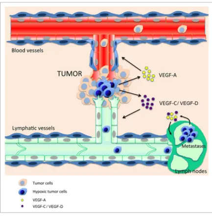
important for tumor spread (Fig. 1).
Tumor Growth, Hypoxia, and Lymphangiogenesis
As solid tumors grow, the cells within the expanding mass
frequently become hypoxic because of the increasing distance
from the nearest blood vessels. Without an adequate vascular
supply, solid tumors can grow only to a critical size of 1–2 mm
(approximately 10
6cells), primarily due to a lack of oxygen and
nutrients.
37A number of studies have been performed to
charac-terize and ultimately inhibit tumor angiogenesis. However, since
hypoxia also regulates the expression of lymphangiogenic factors,
it is crucial to consider tumor hypoxia and tumor
lymphangi-ogenesis as two tightly interlocked phenomena. In contrast to
blood vessels, the lymphatic vasculature does not promote tumor
growth by providing key elements for cell survival (i.e.,
oxy-gen and nutrients), but allows metastatic dissemination of solid
tumors through lymph nodes and finally to distant organs.
38,39The lymphatic network is not merely an alternative vehicle to
blood vessels for dissemination, but actually constitutes the main
Figure 1. Crosstalk between tumor hypoxia and the lymphatic and blood
vascu-latures. Hypoxic tumor cells (blue) near pre-existing blood and lymphatic vessels
secrete angiogenic and lymphangiogenic growth factors such as veGF-A, -C, and
–D. Blood vessels bring oxygen and nutriments to tumor cells, whereas
lymphat-ics drain debris and provide new routes for tumor metastasis. Lymphatic metastatic
tumor cells maintain lymphangiogenic growth factors synthesis in this poorly
oxy-genated environment to promote lymph node lymphangiogenesis and establish
the “metastatic niche.”
vascular system implicated in dissemination because lymphatic
vessels have an optimal structure for tumor cell invasion. Indeed,
the main difference between blood and lymphatic networks is the
structure and permeability of their capillaries: whereas lymphatic
capillaries are thin-walled, relatively large vessels, composed of
a single layer of endothelial cells, lymphatic capillaries are not
ensheathed by pericytes or smooth muscle cells and have little
or no basement membrane.
3As a result of this high permeabilty,
tumor cells can spread more easily in lymphatics than in blood
vessels. Moreover, this invasion is also not just a passive process
as tumors induce growth of new lymphatic vessels in draining
lymph nodes and enlargment of lymphatic endothelium before
metastasis. This remodelling of lymph nodes potentially
con-tributes to the migration, implantation, or survival of metastatic
tumor cells by inducing a specific tumor microenvironment.
Thus, a hypoxic tumor will not only ensure its survival through
activation of angiogenesis but will also become more
aggres-sive. This dual regulation of blood and lymphatic vasculature by
hypoxia during tumor growth impairs therapeutic efficacy. First,
there is real crosstalk between the tumor and blood and
lym-phatic endothelial cells. Blood vessel endothelial cells produce
lymphangiogenic factors such as vascular endothelial growth
fac-tor C (VEGFC), fibroblast growth facfac-tor 2 (FGF2), and
plate-let-derived growth factors (PDGFs) to facilitate tumor-induced
lymphangiogenesis.
40Both endothelial cell types produce matrix
metalloproteinases that promote tumor spreading. Lymphatic
endothelial cells (LECs) also express the CCL21 chemokine that
is physiologically implicated in dendritic cell mobilization
41and
interacts with the CCR7 receptor expressed by many tumors to
stimulate lymphatic dissemination.
42Several treatments have
been developed specifically to inhibit tumor angiogenesis (e.g.,
Avastin) and therefore suppress tumor oxygenation and destroy
tumor cells. However, these drugs also target lymphangiogenesis
and would generate severe tumor hypoxia, which induces
overex-pression of lymphangiogenic factors and increased tumor
dissem-ination. This cross talk between hypoxia and the two vascular
systems and the resultant spread of the tumor can in part explain
the failure of antiangiogenic drugs in cancer treatment (Fig. 1).
A key feature of the lymphatic system is its hypoxic
environ-ment as lymphatic vessels do not transport red blood cells. Lymph
vessels are often located in remote areas away from
oxygen-car-rying blood vessels and are therefore exposed to a milieu with
very low oxygen levels. Tumor cells have to adapt to this hostile
hypoxic environment in order to spread to the lymph nodes.
43-45Normoxia is defined as a milieu where the O
2concentration
is sufficient to ensure aerobic metabolism of cells, the basis of
eukaryotic physiology.
46In contrast, hypoxia is an environment
where the aerobic metabolism of cells is inhibited due to a lack of
oxygen. The major cellular response to hypoxia is stabilization of
hypoxia-inducible factor 1(HIF1). HIF1 is a transcription factor
that controls the expression of a battery of more than 40 target
genes.
47-49It is composed of an α subunit that is constitutively
expressed and a β subunit that is subject to rapid ubiquitination
and proteasomal degradation under normoxic conditions.
50The
molecular basis for this regulation is the O
2-dependent
hydrox-ylation of proline residues 402 and 564 in HIF-1α by any one
of three enzymes in mammals that have been designated prolyl
hydroxylase-domain proteins or HIF-1α prolyl hydroxylases.
51,52Prolyl hydroxylation of HIF-1α is required for binding of the von
Hippel–Lindau tumor suppressor protein (VHL), which is the
recognition component of an E3 ubiquitin-protein ligase that
tar-gets HIF-1α for proteasomal degradation.
53,54Hypoxia has been
shown to regulate not only angiogenesis, but also
lymphangio-genesis by promoting overexpression of specific lymphangiogenic
factors (e.g., VEGF-C) and growth factors that are shared by the
vascular and the lymphatic vasculature (e.g., VEGF-A, FGF2).
In this review we provide an overview of the link between
lym-phangiogenic factors and hypoxia and the consequences of this
relationship in a well-known hypoxic pathology: the
develop-ment and dissemination of solid tumors.
Hypoxia-Induced Molecular Regulation of VEGFs
The VEGF family is composed of growth factors involved in
vascular development. This family includes VEGF-A, -B, -C, -D,
and -E, and placental growth factor.
55All members of this
fam-ily stimulate proliferation and migration of endothelial cells in
vitro. These proteins bind and activate specific receptors on the
endothelial cell surface: VEGF recognizes VEGFR-1 (Flt-1) and
VEGFR-2 (KDR/Flk-1); placental growth factor and VEGF-B
recognize VEGFR-1; and VEGF-C and VEGF-D recognize
VEGFR-2 and VEGFR-3 (Flt-4). Lymphangiogenesis is induced
by VEGFs that promote angiogenesis (VEGF-A) or angiogenesis
and lymphangiogenesis (VEGF-C and VEGF-D).
Hypoxia-induced gene expression was first described as a
tran-scriptional mechanism mediated by hypoxia-responsive elements
(HREs) present in the promoter region of different genes that
are targets of the hypoxia-induced transcription factors (HIFs).
In particular, a functional HRE has been identified within the
5′ flanking region element of the human VEGFA gene
56,57that is
the target of both HIF1 and HIF2.
58This HRE allows
transcrip-tional induction of VEGFA by hypoxia in several physiologic (i.e.,
wound healing, inflammation) and pathologic (i.e., ischemia,
tumor development) states.
In addition to its transcriptional effects, hypoxia also regulates
gene expression post-transcriptionally at the levels of mRNA
sta-bility and translation. An important class of mRNAs is stabilized
by hypoxia: the so-called AU-rich mRNAs that bear AU-rich
elements (AREs) in their 3′ untranslated regions (3′UTR).
59,60AREs are found in mRNAs of most genes coding for cytokines,
growth factors, and proto-oncogenes (7–8% of the transcribed
genome), indicating that the stabilization of such mRNAs in
hypoxic conditions has important consequences regarding cell
pathophysiology. In particular, angiogenic cytokines including
VEGF-A are regulated by this mechanism.
61mRNA
stabiliza-tion is controlled by the binding of HuR protein to the ARE
in cooperation with polyA-binding protein interacting protein 2
(PAIP2).
62One proposed mechanism is that HuR acts in
com-petition with destabilizing proteins such as AUF1 or
tristetrap-rolin (TTP) to bind to the ARE.
61Another emerging concept
3′UTRs.
63In the case of Vegfa mRNA, the HuR binding site
overlaps with the binding site of miR-200b, thus HuR
antago-nizes the suppressive effect of this microRNA.
63Hypoxia also strongly regulates gene expression at the
trans-lational level. First, it silences global cell translation by
inhibit-ing mRNA cap-dependent translation through inactivation of
mTOR kinase. This results in hypophosphorylation of the 4E-BP
protein, which then sequesters the cap-binding factor eIF4E.
64,65In addition, hypoxia induces phosphorylation of the initiation
factor eIF2a by activation of PERK kinase, which also
gener-ates translational blockade.
66Two major alternative mechanisms
are able to overcome this global inhibition of translation that is
induced by hypoxia: upstream open reading frames (uORFs) and
internal ribosome entry sites (IRESs). uORFs are a key element of
translational control in response to stress. These elements precede
the initiation codon of the mRNA main coding regions and are
present in approximately 40–50% of mRNAs. They are
primar-ily translational inhibitors when eIF2a is dephosphorylated and
the complex of initiator tRNA with eIF2 and GTP is available
for translation initiation. In contrast, they allow the ribosome to
scan and reach the initiation codon of the main coding sequence
in conditions of stress when eIF2a is phosphorylated.
67IRESs are RNA structural elements present in the 5′
non-translated regions of a small number of mRNAs that allow
recruitment of the ribosome to a site that is a considerable
dis-tance from the cap structure, most frequently in the presence
of trans-acting factors.
64,68The majority of identified IRESs are
found in mRNAs of proteins associated with the control of cell
growth and death, including growth factors, proto-oncogenes,
and proteins required for apoptosis.
65,69IRES-dependent
trans-lation is cap-independent and, in the case of cellular mRNAs,
independent of eIF2a phosphorylation; this allows translation to
occur in stress conditions.
64,70Notably, HIF1α mRNA possesses
an IRES, suggesting that these structure are crucial for
trans-lational regulation occurring under hypoxia. IRESs have also
been identified in the mRNAs of three major lymphangiogenic
growth factors, FGF2, VEGF-A, and VEGF-C.
71,72Interestingly,
these IRESs are activated in hypoxic conditions, resulting in
translational induction of these factors.
45,73The regulation of
VEGF-A and -C expression and their relationship with hypoxia
is discussed below.
VEGF-A
VEGF-A, also called vascular permeability factor, is a
homodi-meric glycoprotein with a molecular weight of approximately 45
kDa. At least 9 VEGF isoforms exist as a result of alternative
pat-terns of splicing.
74Three of these, containing 121, 165, and 189
amino acids respectively, are preferentially expressed by VEGF-A
producing cells
75-77Each of these isoforms contributes to
forma-tion of a VEGF-A gradient essential for the proper migraforma-tion
of ECs/LECs during angiogenesis or lymphangiogenesis. The
larger species, VEGF-165, VEGF-189, and VEGF-206, are basic
and bind to isolated heparin and heparin proteoglycans
distrib-uted on cellular surfaces and extracellular matrices whereas the
smaller species, VEGF-121, is acidic and is freely diffusible.
78Although VEGF-A is mainly known as a growth factor that plays
an essential role in physiologic and pathologic angiogenesis
dur-ing both development and adulthood,
79it also has
prolymphan-giogenic properties.
80,81The proangiogenic activity of VEGF-A is
mediated by interaction with a high-affinity VEGFR2 receptor,
whereas the prolymphangiogenic activity is promoted by binding
to the VEGFR2/R3 heterodimeric receptor.
In addition to its transcriptional upregulation during hypoxia,
is probably the most highly post-transcriptionally regulated
factor.
74The gene structure of VEGF-A has been predicted in
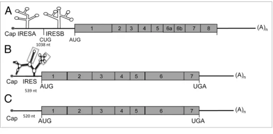
silico (Fig. 2A). VEGFA mRNA contains two IRESs
72located
upstream of the alternative initiation codons CUG and AUG that
are responsible for synthesis of alternative isoforms of VEGF-A.
74Figure 2. Schematic representation of VEGF-A, -C, and -D mRNAs. (A) VEGF-A mRNA is characterized by a long 5′UTR (1038 nt) containing two internal
ribosome entry sites (iReSs) (A and B). The VEGF-A gene encodes multiple isoforms generated by mRNA splicing of four constitutive and four alternative
exons. (B) VEGF-C mRNA possesses a GC-rich 5′UTR containing one iReS. The secondary structure of veGF-C iReS has been determined by shape analysis
and contains 2 motifs (indicated by boxes) showing a similar reactivity pattern between human and mouse mRNA. (C) Similar to VEGF-C, the VEGF-D
mRNA is generated from 7 exons.
Both IREs are activated by hypoxia.
73Vegfa IRESs are
differen-tially regulated by an upstream ORF and by binding of Mir16
to the 3′UTR.
82,83A study of VEGF-A IRES trans-acting
fac-tors revealed tight regulation by both positive regulafac-tors activated
by hypoxia (e.g., MAPK3 kinase) and negative regulators that
are inhibited during this stress (e.g., DEAD-box RNA helicase
6).
84Another mechanism implicated in translation regulation of
VEGF-A is riboswitch. Riboswitch refers to the ability of mRNAs
to alter their folding structure and hence rate of translation in
response to an environmental modification. During hypoxia,
intracellular accumulation of heterogeneous nuclear
ribonu-cleoprotein L (hnRNP L) promotes an active conformation and
increases the rate of translation of VEGF-A mRNA.
85VEGF-A
expression is strongly regulated at the level of mRNA stability,
a process primarily mediated by the AREs present in the Vegfa
mRNA. Indeed, the Vegfa mRNA is destabilized by several
pro-teins including AU-rich element RNA-binding protein 1 (AUF1,
also known as hnRNPD) and tristetraprolin (TTP), which target
the AREs.
61Destabilization of Vegfa mRNA by TTP is
respon-sible for its antiangiogenic activity.
86In contrast, Vegfa mRNA is
stabilized by hypoxia.
60This process is mediated by binding of
the RNA stabilizing protein HuR and its partner PAIP2 to the
AREs, which prevents binding of the destabilizing proteins.
60,61Interestingly, the MDM2 protein, which is translocated from the
nucleus to the cytoplasm under hypoxic conditions, increases
Vegfa mRNA stabilization.
87Vegfa mRNA stability is thus
con-trolled by interplay between stabilizing and destabilizing proteins
that compete for binding to the AREs. Moreover, it has been
proposed that export of VEGF-A mRNA from the nucleus and its
loading onto ribosomes can be increased during hypoxia by
extra-nuclear shuttling of mRNA-binding proteins such as hnRNP L
and A1, which also regulate VEGF-A mRNA stability.
88These
mechanisms, combined with transcriptional regulation induced
by HIFs, allow fast and massive overexpression of VEGF-A in
response to hypoxia.
VEGF-C
The VEGF-C/VEGFR3 signaling pathway is the major
pathway implicated in lymphangiogenesis. First identified in
1996,
89VEGF-C is produced as a precursor protein that is
acti-vated by intracellular proprotein convertases.
89,90The secreted
disulphide-linked VEGF-C subunits only bind VEGFR-3, but
the factor is further proteolyzed in the extracellular environment
by plasmin and other proteases to generate non–disulfide-linked
homodimeric proteins with high affinity for both VEGFR-2 and
VEGFR-3.
3,90VEGF-C is crucial for the induction of
prolifera-tion, migraprolifera-tion, and survival of endothelial cells.
91VEGF-C is
also an essential chemotactic and survival factor during
embry-onic lymphangiogenesis; homozygous deletion of VEGF-C leads
to complete absence of lymphatic vasculature in mouse embryos
whereas VEGF-C
+/−mice display severe lymphatic hypoplasia.
In VEGF-C null mice, lymphatic endothelial cells initially
dif-ferentiate in the cardinal veins but fail to migrate and form
pri-mary lymph sacs.
92Although several studies have shown positive
correlations between HIF-1α and VEGF-C in various
can-cers,
93-95for a long time the molecular mechanisms of
hypoxia-induced regulation of VEGF-C remained poorly understood.
The likelihood of direct transcriptional regulation of VEGF-C
by HIF1α is low because the VEGF-C promoter does not
con-tain a HRE sequence.
96Our recent work demonstrated the
exis-tence of a single IRES in the 5′UTR of both murine and human
VEGF-C mRNA (Fig. 2B). We have demonstrated that VEGF-C
IRES activity is upregulated in vivo during tumor growth in
three murine models of carcinoma, similar to the IREs of FGF2
and VEGF-A.
45Strikingly, we also observed that VEGF-C IRES
activity increases under hypoxia in vitro, but the presence of
HIF-1α is not required in cultured cells.
VEGF-D
Binding of VEGF-D, also called c-fos induced growth
fac-tor, to its receptor VEGFR-3 promotes lymphangiogenesis.
The VEGF-D gene encodes 7 exons (Fig. 2C). Maturation of
VEGF-D is similar to that of VEGF-C and occurs by protein
cleavage in N and C-terminal regions. VEGF-D has been poorly
studied because of the lack of a phenotype resulting from its
depletion in mice. Recent reports have shown that
overexpres-sion of VEGF-D induces tumor lymphangiogenesis and
pro-motes lymphatic metastasis in mouse tumor models.
97However,
few clinical studies have investigated the association between the
expression of VEGF-D and lymphatic metastasis. VEGF-D
over-expression correlates with an increase in lymphatic vessel growth
and lymphatic metastasis.
39Recent studies suggest that VEGF-D
is necessary for for the entry of tumor cells into the lymphatic
system that results in metastasis.
98VEGF-D promotes structural
changes in tumor-draining lymphatic vessels and induces
vaso-dilatation. VEGF-D also increases the endothelial response to
prostaglandin E2 (PGE2) by inhibiting the prostaglandin
dehy-drogenases (PGDH).
99,100The role of hypoxia in the promotion of VEGF-D
expres-sion has not been clearly established. Recent studies have
dem-onstrated correlations between VEGF-D and HIF-1α expression
in invasive breast ductal carcinoma
101and in resected esophageal
squamous cell carcinoma.
102These findings revealed that expression of lymphangiogenic
factors is tightly linked to hypoxia, which activates their
expres-sion at both transcriptional and translational levels. It is now well
known that, at least in solid tumors, hypoxia is a major
compo-nent of the tumor microenvironment and induces critical changes
in tumor cell metabolism, angiogenesis, and lymphangiogenesis.
Concluding Remarks and Perspectives
The lymphatic vasculature has long been considered the poor
relation of the blood vasculature. Compared with the vascular
network, which provides both oxygen and nutrients and is
there-fore obviously necessary for life, the lymphatic system appeared
to be a less important vascular network. In addition, until
recently it was challenging to differentiate lymph from blood
vessels due to lack of a specific marker. Recently, however, the
lymphatic system has emerged as a crucial player during
develop-ment and in adulthood. Although it is implicated specifically in
chronic inflammatory and vascular pathologies (such as psoriasis
and lymphedema), it is also able to interact with blood vessels in
cancer. Indeed, recent studies have highlighted hypoxia-induced
regulation of lymphangiogenic factors in the tumor
microenvi-ronent. Understanding the molecular regulation of
lymphangio-genesis in a wide range of organs and pathologies might lead to
new therapeutic solutions for diseases such as cancer.
Disclosure of Potential Conflicts of Interest
No potential conflicts of interest were disclosed.
References
1. Jeltsch M, Tammela T, Alitalo K, Wilting J. Genesis and pathogenesis of lymphatic vessels. Cell Tissue Res 2003; 314:69-84; PMID:12942362; http://dx.doi. org/10.1007/s00441-003-0777-2
2. Baluk P, Tammela T, Ator E, Lyubynska N, Achen MG, Hicklin DJ, Jeltsch M, Petrova TV, Pytowski B, Stacker SA, et al. Pathogenesis of persistent lymphatic vessel hyperplasia in chronic airway inflammation. J Clin Invest 2005; 115:247-57; PMID:15668734; http://dx.doi.org/10.1172/JCI200522037
3. Alitalo K, Tammela T, Petrova TV. Lymphangiogenesis in development and human dis-ease. Nature 2005; 438:946-53; PMID:16355212; http://dx.doi.org/10.1038/nature04480
4. Williams CS, Leek RD, Robson AM, Banerji S, Prevo R, Harris AL, Jackson DG. Absence of lymphangio-genesis and intratumoural lymph vessels in human metastatic breast cancer. J Pathol 2003; 200:195-206; PMID:12754740; http://dx.doi.org/10.1002/ path.1343
5. Mumprecht V, Detmar M. Lymphangiogenesis and cancer metastasis. J Cell Mol Med 2009; 13(8A):1405-16; PMID:19583813; http://dx.doi. org/10.1111/j.1582-4934.2009.00834.x
6. Renyi-Vamos F, Tovari J, Fillinger J, Timar J, Paku S, Kenessey I, Ostoros G, Agocs L, Soltesz I, Dome B. Lymphangiogenesis correlates with lymph node metastasis, prognosis, and angiogenic phenotype in human non-small cell lung cancer. Clin Cancer Res 2005; 11:7344-53; PMID:16243806; http://dx.doi. org/10.1158/1078-0432.CCR-05-1077
7. Zeng Y, Opeskin K, Horvath LG, Sutherland RL, Williams ED. Lymphatic vessel density and lymph node metastasis in prostate cancer. Prostate 2005; 65:222-30; PMID:15948136; http://dx.doi. org/10.1002/pros.20288
8. Stacker SA, Williams RA, Achen MG. Lymphangiogenic growth factors as mark-ers of tumor metastasis. APMIS 2004; 112:539-49; PMID:15563315; http://dx.doi. org/10.1111/j.1600-0463.2004.apm11207-0812.x 9. Baluk P, Fuxe J, Hashizume H, Romano T, Lashnits
E, Butz S, Vestweber D, Corada M, Molendini C, Dejana E, et al. Functionally specialized junctions between endothelial cells of lymphatic vessels. J Exp Med 2007; 204:2349-62; PMID:17846148; http:// dx.doi.org/10.1084/jem.20062596
10. Leak LV. The structure of lymphatic capillaries in lymph formation. Fed Proc 1976; 35:1863-71; PMID:1269772
11. Wigle JT, Oliver G. Prox1 function is required for the development of the murine lymphatic system. Cell 1999; 98:769-78; PMID:10499794; http://dx.doi. org/10.1016/S0092-8674(00)81511-1
12. Prevo R, Banerji S, Ferguson DJ, Clasper S, Jackson DG. Mouse LYVE-1 is an endocytic receptor for hyal-uronan in lymphatic endothelium. J Biol Chem 2001; 276:19420-30; PMID:11278811; http://dx.doi. org/10.1074/jbc.M011004200
13. Banerji S, Ni J, Wang SX, Clasper S, Su J, Tammi R, Jones M, Jackson DG. LYVE-1, a new homo-logue of the CD44 glycoprotein, is a lymph-specific receptor for hyaluronan. J Cell Biol 1999; 144:789-801; PMID:10037799; http://dx.doi.org/10.1083/ jcb.144.4.789
14. Breiteneder-Geleff S, Soleiman A, Horvat R, Amann G, Kowalski H, Kerjaschki D. [Podoplanin--a spe-cific marker for lymphatic endothelium expressed in angiosarcoma]. Verh Dtsch Ges Pathol 1999; 83:270-5; PMID:10714221
15. Karkkainen MJ, Ferrell RE, Lawrence EC, Kimak MA, Levinson KL, McTigue MA, Alitalo K, Finegold DN. Missense mutations interfere with VEGFR-3 signalling in primary lymphoedema. Nat Genet 2000; 25:153-9; PMID:10835628; http://dx.doi. org/10.1038/75997
16. Kilic N, Oliveira-Ferrer L, Neshat-Vahid S, Irmak S, Obst-Pernberg K, Wurmbach JH, Loges S, Kilic E, Weil J, Lauke H, et al. Lymphatic reprogram-ming of microvascular endothelial cells by CEA-related cell adhesion molecule-1 via interaction with VEGFR-3 and Prox1. Blood 2007; 110:4223-33; PMID:17761831; http://dx.doi.org/10.1182/ blood-2007-06-097592
17. Breslin JW, Gaudreault N, Watson KD, Reynoso R, Yuan SY, Wu MH. Vascular endothelial growth factor-C stimulates the lymphatic pump by a VEGF receptor-3-dependent mechanism. Am J Physiol Heart Circ Physiol 2007; 293:H709-18; PMID:17400713; http://dx.doi.org/10.1152/ajpheart.00102.2007 18. Bridenbaugh E. Literature watch. Complete and
spe-cific inhibition of adult lymphatic regeneration by a novel VEGFR-3 neutralizing antibody. Lymphat Res Biol 2005; 3:87-8; PMID:16000057; http://dx.doi. org/10.1089/lrb.2005.3.87
19. Mäkinen T, Veikkola T, Mustjoki S, Karpanen T, Catimel B, Nice EC, Wise L, Mercer A, Kowalski H, Kerjaschki D, et al. Isolated lymphatic endothelial cells transduce growth, survival and migratory sig-nals via the VEGF-C/D receptor VEGFR-3. EMBO J 2001; 20:4762-73; PMID:11532940; http://dx.doi. org/10.1093/emboj/20.17.4762
20. Tammela T, Zarkada G, Wallgard E, Murtomäki A, Suchting S, Wirzenius M, Waltari M, Hellström M, Schomber T, Peltonen R, et al. Blocking VEGFR-3 suppresses angiogenic sprouting and vas-cular network formation. Nature 2008; 454:656-60; PMID:18594512; http://dx.doi.org/10.1038/ nature07083
21. Petrova TV, Bono P, Holnthoner W, Chesnes J, Pytowski B, Sihto H, Laakkonen P, Heikkilä P, Joensuu H, Alitalo K. VEGFR-3 expres-sion is restricted to blood and lymphatic vessels in solid tumors. Cancer Cell 2008; 13:554-6; PMID:18538738; http://dx.doi.org/10.1016/j. ccr.2008.04.022
22. Ardies CM. Inflammation as cause for scar can-cers of the lung. Integr Cancer Ther 2003; 2:238-46; PMID:15035887; http://dx.doi. org/10.1177/1534735403256332
23. Van der Auwera I, Van Laere SJ, Van den Eynden GG, Benoy I, van Dam P, Colpaert CG, Fox SB, Turley H, Harris AL, Van Marck EA, et al. Increased angiogenesis and lymphangiogenesis in inflam-matory versus noninflaminflam-matory breast cancer by real-time reverse transcriptase-PCR gene expression quantification. Clin Cancer Res 2004; 10:7965-71; PMID:15585631; http://dx.doi.org/10.1158/1078-0432.CCR-04-0063
24. Jaiswal M, LaRusso NF, Gores GJ. Nitric oxide in gastrointestinal epithelial cell carcinogenesis: link-ing inflammation to oncogenesis. Am J Physiol Gastrointest Liver Physiol 2001; 281:G626-34; PMID:11518674
25. Brower V. Researchers attempting to define role of cytokines in cancer risk. J Natl Cancer Inst 2005; 97:1175-7; PMID:16106019; http://dx.doi. org/10.1093/jnci/dji269
26. Biarc J, Nguyen IS, Pini A, Gossé F, Richert S, Thiersé D, Van Dorsselaer A, Leize-Wagner E, Raul F, Klein JP, et al. Carcinogenic properties of proteins with pro-inflammatory activity from Streptococcus infantarius (formerly S.bovis). Carcinogenesis 2004; 25:1477-84; PMID:14742316; http://dx.doi. org/10.1093/carcin/bgh091
27. Altinoz MA, Korkmaz R. NF-kappaB, macrophage migration inhibitory factor and cyclooxygenase-inhi-bitions as likely mechanisms behind the acetamino-phen- and NSAID-prevention of the ovarian cancer. Neoplasma 2004; 51:239-47; PMID:15254653 28. Wang W, Bergh A, Damber JE. Chronic
inflamma-tion in benign prostate hyperplasia is associated with focal upregulation of cyclooxygenase-2, Bcl-2, and cell proliferation in the glandular epithelium. Prostate 2004; 61:60-72; PMID:15287094; http://dx.doi. org/10.1002/pros.20061
29. Hussein MR, Ahmed RA. Analysis of the mono-nuclear inflammatory cell infiltrate in the non-tumorigenic, pre-tumorigenic and tumorigenic keratinocytic hyperproliferative lesions of the skin. Cancer Biol Ther 2005; 4:819-21; PMID:16210913; http://dx.doi.org/10.4161/cbt.4.8.1864
30. Bartsch H, Nair J. Oxidative stress and lipid peroxi-dation-derived DNA-lesions in inflammation driven carcinogenesis. Cancer Detect Prev 2004; 28:385-91; PMID:15582261; http://dx.doi.org/10.1016/j. cdp.2004.07.004
31. Angelo LS, Kurzrock R. Vascular endothelial growth factor and its relationship to inflamma-tory mediators. Clin Cancer Res 2007; 13:2825-30; PMID:17504979; http://dx.doi.org/10.1158/1078-0432.CCR-06-2416
32. Jain RK. Barriers to drug delivery in solid tumors. Sci Am 1994; 271:58-65; PMID:8066425; http:// dx.doi.org/10.1038/scientificamerican0794-58 33. Padera TP, Kadambi A, di Tomaso E, Carreira CM,
Brown EB, Boucher Y, Choi NC, Mathisen D, Wain J, Mark EJ, et al. Lymphatic metastasis in the absence of functional intratumor lymphatics. Science 2002; 296:1883-6; PMID:11976409; http://dx.doi. org/10.1126/science.1071420
34. Dadras SS, Lange-Asschenfeldt B, Velasco P, Nguyen L, Vora A, Muzikansky A, Jahnke K, Hauschild A, Hirakawa S, Mihm MC, et al. Tumor lymphan-giogenesis predicts melanoma metastasis to senti-nel lymph nodes. Mod Pathol 2005; 18:1232-42; PMID:15803182; http://dx.doi.org/10.1038/ modpathol.3800410
35. Maula SM, Luukkaa M, Grénman R, Jackson D, Jalkanen S, Ristamäki R. Intratumoral lymphatics are essential for the metastatic spread and prognosis in squamous cell carcinomas of the head and neck region. Cancer Res 2003; 63:1920-6; PMID:12702584
36. Choi WW, Lewis MM, Lawson D, Yin-Goen Q, Birdsong GG, Cotsonis GA, Cohen C, Young AN. Angiogenic and lymphangiogenic microvessel density in breast carcinoma: correlation with clinicopatho-logic parameters and VEGF-family gene expression. Mod Pathol 2005; 18:143-52; PMID:15297858; http://dx.doi.org/10.1038/modpathol.3800253 37. Bergers G, Benjamin LE. Tumorigenesis and the
angiogenic switch. Nat Rev Cancer 2003; 3:401-10; PMID:12778130; http://dx.doi.org/10.1038/ nrc1093
38. Skobe M, Hawighorst T, Jackson DG, Prevo R, Janes L, Velasco P, Riccardi L, Alitalo K, Claffey K, Detmar M. Induction of tumor lymphangiogenesis by VEGF-C promotes breast cancer metastasis. Nat Med 2001; 7:192-8; PMID:11175850; http://dx.doi. org/10.1038/84643
39. Stacker SA, Caesar C, Baldwin ME, Thornton GE, Williams RA, Prevo R, Jackson DG, Nishikawa S, Kubo H, Achen MG. VEGF-D promotes the meta-static spread of tumor cells via the lymphatics. Nat Med 2001; 7:186-91; PMID:11175849; http:// dx.doi.org/10.1038/84635
40. Petrova TV, Mäkinen T, Mäkelä TP, Saarela J, Virtanen I, Ferrell RE, Finegold DN, Kerjaschki D, Ylä-Herttuala S, Alitalo K. Lymphatic endothe-lial reprogramming of vascular endotheendothe-lial cells by the Prox-1 homeobox transcription factor. EMBO J 2002; 21:4593-9; PMID:12198161; http://dx.doi. org/10.1093/emboj/cdf470
41. Tal O, Lim HY, Gurevich I, Milo I, Shipony Z, Ng LG, Angeli V, Shakhar G. DC mobilization from the skin requires docking to immobilized CCL21 on lymphatic endothelium and intralymphatic crawling. J Exp Med 2011; 208:2141-53; PMID:21930767; http://dx.doi.org/10.1084/jem.20102392
42. Issa A, Le TX, Shoushtari AN, Shields JD, Swartz MA. Vascular endothelial growth factor-C and C-C chemokine receptor 7 in tumor cell-lymphatic cross-talk promote invasive phenotype. Cancer Res 2009; 69:349-57; PMID:19118020; http://dx.doi. org/10.1158/0008-5472.CAN-08-1875
43. Ivanovic Z. Hypoxia or in situ normoxia: The stem cell paradigm. J Cell Physiol 2009; 219:271-5; PMID:19160417; http://dx.doi.org/10.1002/ jcp.21690
44. Guzy RD, Sharma B, Bell E, Chandel NS, Schumacker PT. Loss of the SdhB, but Not the SdhA, subunit of complex II triggers reactive oxygen species-dependent hypoxia-inducible factor activa-tion and tumorigenesis. Mol Cell Biol 2008; 28:718-31; PMID:17967865; http://dx.doi.org/10.1128/ MCB.01338-07
45. Morfoisse F, Kuchnio A, Frainay C, Gomez-Brouchet A, Delisle MB, Marzi S, Helfer AC, Hantelys F, Pujol F, Guillermet-Guibert J, et al. Hypoxia induces VEGF-C expression in metastatic tumor cells via a HIF-1α-independent translation-mediated mecha-nism. Cell Rep 2014; 6:155-67; PMID:24388748; http://dx.doi.org/10.1016/j.celrep.2013.12.011 46. Ivanovic Z. Physiological, ex vivo cell oxygenation is
necessary for a true insight into cytokine biology. Eur Cytokine Netw 2009; 20:7-9; PMID:19318314 47. Gerber HP, Condorelli F, Park J, Ferrara N.
Differential transcriptional regulation of the two vas-cular endothelial growth factor receptor genes. Flt-1, but not Flk-1/KDR, is up-regulated by hypoxia. J Biol Chem 1997; 272:23659-67; PMID:9295307; http:// dx.doi.org/10.1074/jbc.272.38.23659
48. Iyer NV, Kotch LE, Agani F, Leung SW, Laughner E, Wenger RH, Gassmann M, Gearhart JD, Lawler AM, Yu AY, et al. Cellular and developmental con-trol of O2 homeostasis by hypoxia-inducible factor 1 alpha. Genes Dev 1998; 12:149-62; PMID:9436976; http://dx.doi.org/10.1101/gad.12.2.149
49. Ryan HE, Lo J, Johnson RS. HIF-1 alpha is required for solid tumor formation and embryonic vasculariza-tion. EMBO J 1998; 17:3005-15; PMID:9606183; http://dx.doi.org/10.1093/emboj/17.11.3005
50. Huang LE, Gu J, Schau M, Bunn HF. Regulation of hypoxia-inducible factor 1alpha is mediated by an O2-dependent degradation domain via the ubiqui-tin-proteasome pathway. Proc Natl Acad Sci U S A 1998; 95:7987-92; PMID:9653127; http://dx.doi. org/10.1073/pnas.95.14.7987
51. Bruick RK, McKnight SL. A conserved family of prolyl-4-hydroxylases that modify HIF. Science 2001; 294:1337-40; PMID:11598268; http://dx.doi. org/10.1126/science.1066373
52. Mazzone M, Dettori D, Leite de Oliveira R, Loges S, Schmidt T, Jonckx B, Tian YM, Lanahan AA, Pollard P, Ruiz de Almodovar C, et al. Heterozygous deficiency of PHD2 restores tumor oxygenation and inhibits metastasis via endothelial normalization. Cell 2009; 136:839-51; PMID:19217150; http:// dx.doi.org/10.1016/j.cell.2009.01.020
53. Jaakkola P, Mole DR, Tian YM, Wilson MI, Gielbert J, Gaskell SJ, von Kriegsheim A, Hebestreit HF, Mukherji M, Schofield CJ, et al. Targeting of HIF-alpha to the von Hippel-Lindau ubiquitylation com-plex by O2-regulated prolyl hydroxylation. Science 2001; 292:468-72; PMID:11292861; http://dx.doi. org/10.1126/science.1059796
54. Ivan M, Kondo K, Yang H, Kim W, Valiando J, Ohh M, Salic A, Asara JM, Lane WS, Kaelin WG Jr. HIFalpha targeted for VHL-mediated destruction by proline hydroxylation: implications for O2 sensing. Science 2001; 292:464-8; PMID:11292862; http:// dx.doi.org/10.1126/science.1059817
55. Ferrara N. Vascular endothelial growth factor as a target for anticancer therapy. Oncologist 2004; 9(Suppl 1):2-10; PMID:15178810; http://dx.doi. org/10.1634/theoncologist.9-suppl_1-2
56. Pagès G, Pouysségur J. Transcriptional regulation of the Vascular Endothelial Growth Factor gene--a concert of activating factors. Cardiovasc Res 2005; 65:564-73; PMID:15664382; http://dx.doi. org/10.1016/j.cardiores.2004.09.032
57. Forsythe JA, Jiang BH, Iyer NV, Agani F, Leung SW, Koos RD, Semenza GL. Activation of vascular endo-thelial growth factor gene transcription by hypoxia-inducible factor 1. Mol Cell Biol 1996; 16:4604-13; PMID:8756616
58. Blancher C, Moore JW, Talks KL, Houlbrook S, Harris AL. Relationship of hypoxia-inducible factor (HIF)-1alpha and HIF-2alpha expression to vascular endothelial growth factor induction and hypoxia sur-vival in human breast cancer cell lines. Cancer Res 2000; 60:7106-13; PMID:11156418
59. Gruber AR, Fallmann J, Kratochvill F, Kovarik P, Hofacker IL. AREsite: a database for the compre-hensive investigation of AU-rich elements. Nucleic Acids Res 2011; 39:D66-9; PMID:21071424; http:// dx.doi.org/10.1093/nar/gkq990
60. Levy AP, Levy NS, Goldberg MA. Post-transcriptional regulation of vascular endothe-lial growth factor by hypoxia. J Biol Chem 1996; 271:2746-53; PMID:8576250; http://dx.doi. org/10.1074/jbc.271.5.2746
61. Griseri P, Pagès G. Control of pro-angiogenic cyto-kine mRNA half-life in cancer: the role of AU-rich elements and associated proteins. J Interferon Cytokine Res 2014; 34:242-54; PMID:24697202; http://dx.doi.org/10.1089/jir.2013.0140
62. Onesto C, Berra E, Grépin R, Pagès G. Poly(A)-binding protein-interacting protein 2, a strong regu-lator of vascular endothelial growth factor mRNA. J Biol Chem 2004; 279:34217-26; PMID:15175342; http://dx.doi.org/10.1074/jbc.M400219200 63. Chang SH, Hla T. Post-transcriptional gene regulation
by HuR and microRNAs in angiogenesis. Curr Opin Hematol 2014; 21:235-40; PMID:24714527; http:// dx.doi.org/10.1097/MOH.0000000000000040 64. Holcik M, Sonenberg N. Translational control
in stress and apoptosis. Nat Rev Mol Cell Biol 2005; 6:318-27; PMID:15803138; http://dx.doi. org/10.1038/nrm1618
65. Baird SD, Turcotte M, Korneluk RG, Holcik M. Searching for IRES. RNA 2006; 12:1755-85; PMID:16957278; http://dx.doi.org/10.1261/ rna.157806
66. Silvera D, Schneider RJ. Inflammatory breast cancer cells are constitutively adapted to hypoxia. Cell Cycle 2009; 8:3091-6; PMID:19755858; http://dx.doi. org/10.4161/cc.8.19.9637
67. Somers J, Pöyry T, Willis AE. A perspective on mammalian upstream open reading frame func-tion. Int J Biochem Cell Biol 2013; 45:1690-700; PMID:23624144; http://dx.doi.org/10.1016/j. biocel.2013.04.020
68. Vagner S, Galy B, Pyronnet S. Irresistible IRES. Attracting the translation machinery to internal ribosome entry sites. EMBO Rep 2001; 2:893-8; PMID:11600453; http://dx.doi.org/10.1093/ embo-reports/kve208
69. Bushell M, Stoneley M, Sarnow P, Willis AE. Translation inhibition during the induction of apop-tosis: RNA or protein degradation? Biochem Soc Trans 2004; 32:606-10; PMID:15270687; http:// dx.doi.org/10.1042/BST0320606
70. Thakor N, Holcik M. IRES-mediated translation of cellular messenger RNA operates in eIF2α- inde-pendent manner during stress. Nucleic Acids Res 2012; 40:541-52; PMID:21917851; http://dx.doi. org/10.1093/nar/gkr701
71. Vagner S, Gensac MC, Maret A, Bayard F, Amalric F, Prats H, Prats AC. Alternative translation of human fibroblast growth factor 2 mRNA occurs by internal entry of ribosomes. Mol Cell Biol 1995; 15:35-44; PMID:7799942
72. Huez I, Créancier L, Audigier S, Gensac MC, Prats AC, Prats H. Two independent internal ribosome entry sites are involved in translation initiation of vas-cular endothelial growth factor mRNA. Mol Cell Biol 1998; 18:6178-90; PMID:9774635
73. Bornes S, Prado-Lourenco L, Bastide A, Zanibellato C, Iacovoni JS, Lacazette E, Prats AC, Touriol C, Prats H. Translational induction of VEGF inter-nal ribosome entry site elements during the early response to ischemic stress. Circ Res 2007; 100:305-8; PMID:17255526; http://dx.doi.org/10.1161/01. RES.0000258873.08041.c9
74. Arcondéguy T, Lacazette E, Millevoi S, Prats H, Touriol C. VEGF-A mRNA processing, stability and translation: a paradigm for intricate regulation of gene expression at the post-transcriptional level. Nucleic Acids Res 2013; 41:7997-8010; PMID:23851566; http://dx.doi.org/10.1093/nar/gkt539
75. Keck PJ, Hauser SD, Krivi G, Sanzo K, Warren T, Feder J, Connolly DT. Vascular permeability factor, an endothelial cell mitogen related to PDGF. Science 1989; 246:1309-12; PMID:2479987; http://dx.doi. org/10.1126/science.2479987
76. Leung DW, Cachianes G, Kuang WJ, Goeddel DV, Ferrara N. Vascular endothelial growth fac-tor is a secreted angiogenic mitogen. Science 1989; 246:1306-9; PMID:2479986; http://dx.doi. org/10.1126/science.2479986
77. Tischer E, Mitchell R, Hartman T, Silva M, Gospodarowicz D, Fiddes JC, Abraham JA. The human gene for vascular endothelial growth factor. Multiple protein forms are encoded through alterna-tive exon splicing. J Biol Chem 1991; 266:11947-54; PMID:1711045
78. Houck KA, Leung DW, Rowland AM, Winer J, Ferrara N. Dual regulation of vascular endothelial growth factor bioavailability by genetic and proteo-lytic mechanisms. J Biol Chem 1992; 267:26031-7; PMID:1464614
79. Carmeliet P. Angiogenesis in life, disease and medi-cine. Nature 2005; 438:932-6; PMID:16355210; http://dx.doi.org/10.1038/nature04478
80. Dellinger MT, Brekken RA. Phosphorylation of Akt and ERK1/2 is required for VEGF-A/ VEGFR2-induced proliferation and migration of lymphatic endothelium. PLoS One 2011; 6:e28947; PMID:22174934; http://dx.doi.org/10.1371/journal. pone.0028947
81. Wuest TR, Carr DJ. VEGF-A expression by HSV-1-infected cells drives corneal lymphangiogenesis. J Exp Med 2010; 207:101-15; PMID:20026662; http:// dx.doi.org/10.1084/jem.20091385
82. Bastide A, Karaa Z, Bornes S, Hieblot C, Lacazette E, Prats H, Touriol C. An upstream open reading frame within an IRES controls expression of a specific VEGF-A isoform. Nucleic Acids Res 2008; 36:2434-45; PMID:18304943; http://dx.doi.org/10.1093/ nar/gkn093
83. Karaa ZS, Iacovoni JS, Bastide A, Lacazette E, Touriol C, Prats H. The VEGF IRESes are differen-tially susceptible to translation inhibition by miR-16. RNA 2009; 15:249-54; PMID:19144909; http:// dx.doi.org/10.1261/rna.1301109
84. Casanova CM, Sehr P, Putzker K, Hentze MW, Neumann B, Duncan KE, Thoma C. Automated high-throughput RNAi screening in human cells combined with reporter mRNA transfection to identify novel regulators of translation. PLoS One 2012; 7:e45943; PMID:23029333; http://dx.doi. org/10.1371/journal.pone.0045943
85. Ray PS, Jia J, Yao P, Majumder M, Hatzoglou M, Fox PL. A stress-responsive RNA switch regu-lates VEGFA expression. Nature 2009; 457:915-9; PMID:19098893; http://dx.doi.org/10.1038/ nature07598
86. Essafi-Benkhadir K, Onesto C, Stebe E, Moroni C, Pagès G. Tristetraprolin inhibits Ras-dependent tumor vascularization by inducing vascular endothe-lial growth factor mRNA degradation. Mol Biol Cell 2007; 18:4648-58; PMID:17855506; http://dx.doi. org/10.1091/mbc.E07-06-0570
87. Zhou S, Gu L, He J, Zhang H, Zhou M. MDM2 regulates vascular endothelial growth factor mRNA stabilization in hypoxia. Mol Cell Biol 2011; 31:4928-37; PMID:21986500; http://dx.doi.org/10.1128/ MCB.06085-11
88. Vumbaca F, Phoenix KN, Rodriguez-Pinto D, Han DK, Claffey KP. Double-stranded RNA-binding protein regulates vascular endothelial growth fac-tor mRNA stability, translation, and breast can-cer angiogenesis. Mol Cell Biol 2008; 28:772-83; PMID:18039850; http://dx.doi.org/10.1128/ MCB.02078-06
89. Joukov V, Pajusola K, Kaipainen A, Chilov D, Lahtinen I, Kukk E, Saksela O, Kalkkinen N, Alitalo K. A novel vascular endothelial growth fac-tor, VEGF-C, is a ligand for the Flt4 (VEGFR-3) and KDR (VEGFR-2) receptor tyrosine kinases. EMBO J 1996; 15:1751; PMID:8612600
90. Karpanen T, Alitalo K. Molecular biology and pathology of lymphangiogenesis. Annu Rev Pathol 2008; 3:367-97; PMID:18039141; http://dx.doi. org/10.1146/annurev.pathmechdis.3.121806.151515 91. Tammela T, Saaristo A, Lohela M, Morisada T,
Tornberg J, Norrmén C, Oike Y, Pajusola K, Thurston G, Suda T, et al. Angiopoietin-1 promotes lymphatic sprouting and hyperplasia. Blood 2005; 105:4642-8; PMID:15746084; http://dx.doi.org/10.1182/ blood-2004-08-3327
92. Karkkainen MJ, Haiko P, Sainio K, Partanen J, Taipale J, Petrova TV, Jeltsch M, Jackson DG, Talikka M, Rauvala H, et al. Vascular endothe-lial growth factor C is required for sprouting of the first lymphatic vessels from embryonic veins. Nat Immunol 2004; 5:74-80; PMID:14634646; http:// dx.doi.org/10.1038/ni1013
93. Liang X, Yang D, Hu J, Hao X, Gao J, Mao Z. Hypoxia inducible factor-alpha expression correlates with vascular endothelial growth factor-C expres-sion and lymphangiogenesis/angiogenesis in oral squamous cell carcinoma. Anticancer Res 2008; 28(3A):1659-66; PMID:18630523
94. Schoppmann SF, Fenzl A, Nagy K, Unger S, Bayer G, Geleff S, Gnant M, Horvat R, Jakesz R, Birner P. VEGF-C expressing tumor-associated macrophages in lymph node positive breast cancer: impact on lym-phangiogenesis and survival. Surgery 2006; 139:839-46; PMID:16782443; http://dx.doi.org/10.1016/j. surg.2005.12.008
95. Tao J, Li T, Li K, Xiong J, Yang Z, Wu H, Wang C. Effect of HIF-1alpha on VEGF-C induced lymphan-giogenesis and lymph nodes metastases of pancreatic cancer. J Huazhong Univ Sci Technolog Med Sci 2006; 26:562-4; PMID:17219968; http://dx.doi. org/10.1007/s11596-006-0520-9
96. Chilov D, Kukk E, Taira S, Jeltsch M, Kaukonen J, Palotie A, Joukov V, Alitalo K. Genomic organization of human and mouse genes for vascular endothelial growth factor C. J Biol Chem 1997; 272:25176-83; PMID:9312130; http://dx.doi.org/10.1074/ jbc.272.40.25176
97. Stacker SA, Achen MG. From anti-angiogenesis to anti-lymphangiogenesis: emerging trends in cancer therapy. Lymphat Res Biol 2008; 6:165-72; PMID:19093789; http://dx.doi.org/10.1089/ lrb.2008.1015
98. Karnezis T, Shayan R, Caesar C, Roufail S, Harris NC, Ardipradja K, Zhang YF, Williams SP, Farnsworth RH, Chai MG, et al. VEGF-D pro-motes tumor metastasis by regulating prostaglandins produced by the collecting lymphatic endothelium. Cancer Cell 2012; 21:181-95; PMID:22340592; http://dx.doi.org/10.1016/j.ccr.2011.12.026 99. Karnezis T, Shayan R, Fox S, Achen MG, Stacker SA.
The connection between lymphangiogenic signal-ling and prostaglandin biology: a missing link in the metastatic pathway. Oncotarget 2012; 3:893-906; PMID:23097685
100. Stacker SA, Williams SP, Karnezis T, Shayan R, Fox SB, Achen MG. Lymphangiogenesis and lym-phatic vessel remodelling in cancer. Nat Rev Cancer 2014; 14:159-72; PMID:24561443; http://dx.doi. org/10.1038/nrc3677
101. Okada K, Osaki M, Araki K, Ishiguro K, Ito H, Ohgi S. Expression of hypoxia-inducible factor (HIF-1alpha), VEGF-C and VEGF-D in non-invasive and invasive breast ductal carcinomas. Anticancer Res 2005; 25:3003-9; PMID:16080559
102. Tzao C, Lee SC, Tung HJ, Hsu HS, Hsu WH, Sun GH, Yu CP, Jin JS, Cheng YL. Expression of hypoxia-inducible factor (HIF)-1alpha and vascular endothe-lial growth factor (VEGF)-D as outcome predictors in resected esophageal squamous cell carcinoma. Dis Markers 2008; 25:141-8; PMID:19096126; http:// dx.doi.org/10.1155/2008/468323