Author’s Accepted Manuscript
Antihypertensive and vasorelaxant effects of
aqueous extract of Artemisia campestris L. from
Eastern Morocco
Ikram Dib, Monique Tits, Luc Angenot, Jean Noel
Wauters, Asmae Assaidi, Hassane Mekhfi,
Mohammed
Aziz,
Mohammed
Bnouham,
Abdelkhaleq
Legssyer,
Michel
Frederich,
Abderrahim Ziyyat
PII:
S0378-8741(17)30339-2
DOI:
http://dx.doi.org/10.1016/j.jep.2017.05.036
Reference:
JEP10877
To appear in:
Journal of Ethnopharmacology
Received date: 25 January 2017
Revised date:
5 May 2017
Accepted date: 30 May 2017
Cite this article as: Ikram Dib, Monique Tits, Luc Angenot, Jean Noel Wauters,
Asmae Assaidi, Hassane Mekhfi, Mohammed Aziz, Mohammed Bnouham,
Abdelkhaleq
Legssyer,
Michel
Frederich
and
Abderrahim
Ziyyat,
Antihypertensive and vasorelaxant effects of aqueous extract of Artemisia
campestris L. from Eastern Morocco, Journal of Ethnopharmacology,
http://dx.doi.org/10.1016/j.jep.2017.05.036
This is a PDF file of an unedited manuscript that has been accepted for
publication. As a service to our customers we are providing this early version of
the manuscript. The manuscript will undergo copyediting, typesetting, and
review of the resulting galley proof before it is published in its final citable form.
Please note that during the production process errors may be discovered which
could affect the content, and all legal disclaimers that apply to the journal pertain.
1
Antihypertensive and vasorelaxant effects of aqueous extract of Artemisia
campestris L. from Eastern Morocco
Ikram Diba1, Monique Titsb2, Luc Angenotb, Jean Noel Wautersb3, Asmae Assaidia4,
Hassane Mekhfia5, Mohammed Aziza, Mohammed Bnouhama, Abdelkhaleq Legssyera,
Michel Frederichb6, Abderrahim Ziyyata*
a
Laboratoire de Physiologie, Génétique et Ethnopharmacologie URAC-40, Département de Biologie, Faculté des Sciences, Université Mohammed Premier, Oujda-Maroc.
b
Laboratoire de Pharmacognosie, Centre Interfacultaire de Recherche sur les Médicaments (CIRM), Université de Liège, Belgique
[email protected] [email protected] [email protected] [email protected] [email protected] [email protected] [email protected] [email protected] [email protected] [email protected] 1+212 (0)6 78 19 07 50 2+32 (0)43 66 43 34 3+32 (0)43 66 43 35 4+212 (0)6 16 19 74 54 5+212 (0)5 36 50 90 77 (N° tel de laboratoire) 6+32 (0)43 66 43 31
2
*Correspondence to : Pr Abderrahim ZIYYAT Laboratoire de Physiologie, Génétique et Ethnopharmacologie, Département de Biologie, Faculté des Sciences, Université Mohamed Premier, Oujda – Maroc. Tel.: +(212) 6 67 08 61 22.
Abstract
Ethnopharmacological relevance:
Artemisia campestris L. (Asteraceae) has many traditional uses, among which
treatment of diabetes and hypertension.
Aim of the study
This study was conducted in order to confirm the antihypertensive and hypotensive effects of A. campestris L. aqueous extract (AcAE) and to explore the underlying mechanism of action of its vasorelaxant effect, besides the acute toxicity. Also, the chemical composition of AcAE was investigated.
Material and methods
the chemical content of AcAE was determined by using HPLC and NMR techniques. The antihypertensive effect was assessed indirectly by tail-cuff method on L-NAME induced hypertensive rats, while the hypotensive action was monitored intravenously by invasive method on normotensive rats. The vasorelaxant effect and vascular mechanism of action were studied in the presence of antagonists and blockers on aorta isolated from normotensive rats. On the other side, the acute toxicity was studied by oral feeding of extract to the mice.
3
Results
The global phytochemical profile of AcAE reveals the presence of several polyphenols as main components. A. campestris L. infusion was characterized by mono- and di-cinnamoyl compounds, with 3,5-dicaffeoylquinic (isochlorogenic A) acid being the main compound, followed by 5-caffeoylquinic (chlorogenic) acid. Vicenin-2 (apigenin 6,8-di-C-glucoside) appeared to be the most abundant compound among flavonoids.The daily treatment with AcAE at 150 mg/Kg/day prevented the installation of hypertension on L-NAME hypertensive rats, and reduced SBP from 172 mmHg up to 144 mmHg. At the dose 40 mg/Kg, AcAE provoked reduction of systolic (SBP), diastolic (DBP) and mean arterial pressure (MAP), without affecting the heart rate. Also, AcAE (10-2-2 mg/ml)
relaxed the precontracted aorta by 95.8 ± 1.3%. The denudation and preincubation of aorta with atropine, calmidazolium, L-NAME, hydroxycobalamin, ODQ, 8-RP-Br-PET-cGMP, thapsigargin and verapamil attenuated the vasorelaxant response, while the pre-treatment with 4-AP, TEA, glibenclamide and BaCl2 did not alter this effect. The
oral administration of AcAE (0-6 g/Kg) reveals no mortality or toxicity. Conclusions
our study proved that AcAE possess an important antihypertensive, hypotensive and vasorelaxant effect, which is mediated via calmodulin-NO-cGC-PKG pathway, and via inhibition of calcium influx through voltage-operated calcium channels and activation of intracellular calcium mobilization into sarcoplasmic reticulum. Therefore, our findings give first evidence about the traditional use of A. campestris L. as antihypertensive plant.
4 fx1
Key words
Artemisia campestris L., aqueous extract, phytochemical analysis, antihypertensive,
hypotensive, vasorelaxant.
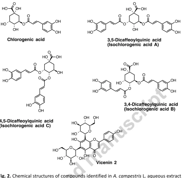
Chemical compounds studied in this article
Chlorogenic acid (PubChem CID: 1794427); 3,4-Dicaffeoylquinic acid (PubChem CID: 5281780); 3,5- Dicaffeoylquinic acid (PubChem CID: 6474310); 4,5- Dicaffeoylquinic acid (PubChem CID: 6436237); Vicenin-2 (PubChem CID: 442664).
5
1. Introduction
Hypertension is considered as the main current concern of public health due to its high prevalence and its related risk factors. In Africa, the global prevalence attained it maximum by the year 2000 and was estimated by 30.8%, with 33.3% as prevalence average in Northern Africa (Adeloye and Basquill, 2014). In Morocco, the prevalence of this disease was about 33.6% (Tazi et al., 2003), while the frequency of hypertension in Eastern Morocco was accounted for 31.7% (Ziyyat et al., 2014). Even though, the antihypertensive drugs consumption among the Moroccan hypertensive population remains very low for their needs (Berrada El Azizi et al., 2012), which lead to the utilization of traditional herbal medicine as an alternative to treat high blood pressure. According to ethnomedicinal surveys conducted in four regions of Morocco, 67.5% to 80% of patients use medicinal plants to treat hypertension (Eddouks et al., 2002; Jouad et al., 2001; Tahraoui et al., 2007; Ziyyat et al., 1997). Artemisia campestris L. (Asteraceae) is an Eastern Morocco medicinal plant (Fakchich and Elachouri, 2014), that has been botanically identified and characterized based on its essential oil chemo-typing (Dib et al., 2017a). This plant is commonly used as antihypertensive (Boudjelal et al., 2013) besides to other common uses like antidiabetic (Bnouham et al., 2002; Boudjelal et al., 2013) diuretic (Benchelah et al., 2004; Ferchichi et al., 2006), emmenagogue (Benchelah et al., 2004; Hammiche and Maiza, 2006; Popović et al., 2012), against digestive (Djidel et al., 2009; Fakchich and Elachouri, 2014; Hammiche and Maiza, 2006; Leporatti and Ghedira, 2009) and cutaneous problems (Benítez et al., 2010; El Hassani et al., 2013), as febrifuge (Guarino et al., 2008), anthelmintic (Hammiche and Maiza, 2006; Popović et al., 2012). The overall of traditional uses and
6
pharmacological studies of this plant were detailed in a review published by our team (Dib et al., 2016).
According to Ben-Nasr et al. (2014a), it has been evidenced that the water extract of A.
campestris decreased significantly the diastolic pressure and heart rate in both smoker
and non-smoker men, but the systolic pressure did not significantly change. The same author performed experiments on envenomed pregnant and non-pregnant rats with hypertensive phase induced by the venom, and proved that the pre-treatment with the aqueous decoction of A. campestris leaves exhibited a preventive antihypertensive effect, and abolished the venom induced hypertensive shock (Ben Nasr et al., 2014b). Recently, we have showed that the essential oil of A. campestris L. exhibited a powerful vasorelaxant effect, which was endothelium-independent, and mediated via calcium channels (Dib et al 2017b). In this perspective, this study was carried out with the objective to confirm the antihypertensive action of A. campestris L. aqueous extract (AcAE), on L-NAME-induced hypertensive rats. The effect of AcAE on normal blood pressure rats was also explored, and its vasorelaxant effect was studied, with an emphasis to the vascular mechanism of action. Finally, the phytochemical composition of AcAE was characterized with the aim to give a chemical fingerprinting of this plant according to its occurrence in Oriental Morocco.
2. Material and methods
2.1. Plant material
Aerial part of A. campestris L. was collected in 2014 at flowering stage in a desert area situated between Tendrara and Figuig (South-Eastern Morocco), called chott Tiggri (32°49’48’’ N, 1°39’36’’ w). The plant material was identified by Pr. Atika Mihamou
7
botanist from biology department, and a voucher specimen was deposited in the Herbarium of Faculty of Sciences, University Mohamed First (Oujda, Morocco) under the reference number (HUMPOM-151). The name of the plant was checked and confirmed according the official website www.theplantlist.org.
2.2. Preparation of aqueous extract
A quantity of 250g of A. campestris L. aerial part was infused in boiled distilled water during 4 hours. Then, the extract (AcAE) was filtered and the pooled extract was evaporated to dryness in a rotary evaporator. The yield extract was about 8.37%. The extract was stored at -20°C until use.
2.3. Chemicals and drugs
The following drugs and solvents were used in this study: (±)-verapamil hydrochloride (Sigma Aldrich, China), (R)-(-)-phenylephrine hydrochloride [Phe] (Sigma Aldrich, Germany), 1H-[1,2,4]Oxadiazolo[4,3-a]quinoxalin-1-one [ODQ] (Cayman Chemical, USA) 3,5-Dicaffeoylquinic acid (Chromadex, USA), 3,4-Dicaffeoylquinic acid (Chromadex, USA), 4,5-Dicaffeoylquinic acid (Chromadex, USA)4-aminopyridine [4-AP] (Alfa Aesar, Germany), atropine (Sigma Aldrich, China), barium chloride dehydrate [BaCl2] (AnalaR Normapur - VWR International, Belgium), calcium chloride dehydrate [CaCl2, 2H2O] (Scharlau chemie, Spain), calmidazolium chloride (Sigma Aldrich, USA), carbamylcholine chloride [carbachol, CCH] (Sigma Aldrich, USA), chlorogenic acid (Sigma Aldrich, China), D(+)-glucose anhydrous (Sigma Aldrich_Riedel-de Haen, Germany), Enalapril maleate [Renitec®20mg] (Afric-Phar, Morocco), glybenclamide ( Sigma Aldrich, USA), hydroxocobalamin hydrochloride (Fluka, USA), heparin sodium salt (Sigma Aldrich, united kingdom), indomethacin (Sigma Aldrich-Fluka, Italy),
8
magnesium sulfate [MgSO4] (Sigma Aldrich, Germany), Nω-Nitro-L-arginine methyl ester hydrochloride [L-NAME] (Sigma Aldrich, Switzerland), potassium di-hydrogen phosphate [KH2PO4] (Panreac, Spain), Rp-8-bromo-β-phenyl-1,N2-ethenoguanosine3′,5′-cyclicmonophosphorothioate sodium salt [Rp-8-Br-PET-cGMP] (Sigma Aldrich, Germany), sodium chloride [NaCl] (Sigma Aldrich_Riedel-de Haen, Denmark), sodium hydrogen carbonate [NaHCO3] (Farco chemical, Puerto Rico], sodium nitroprusside [SNP] (Farco chemical, Puerto Rico), sodium pentobarbital (Ceva Santé Animale, France), potassium chloride [KCl] (Sigma Aldrich_Riedel-de Haen, Germany), tetraethyl ammonium chloride hydrate [TEA] (Sigma Aldrich, USA), thapsigargin (Sigma Aldrich, Israel). The solvents utilized were: acetonitrile (Merck, Germany),diethyl ether (Sigma Aldrich, Germany), dimethyl sulfoxide [DMSO] (Sigma Aldrich_Riedel-de Haen, Germany), trifluoroacetic acid [TFA] (Merck, Germany). All chemicals and solvents used were analytical grade. The stock solutions of ODQ, thapsigargin and Rp-8-Br-PET-cGMP were prepared in DMSO whereas indomethacin was prepared in 5% (w/v) sodium bicarbonate solution. All other drugs were dissolved in distilled water.
2.4. High performance Liquid Chromatography analysis (HPLC/UV-VIS-DAD)
A sample of 10 mg/ml of AcAE was prepared in methanol and filtered through Chromafil® Pet -45/25 Macherey-Nagel (Germany) 0.45 µm filters. The analysis was carried out on an Agilent 1100 HPLC coupled with a DAD (Diode-Array Detector). A volume of 10 µl of the sample was injected on hypersil ODS column (5 µm, 250 × 4.6 mm) and the analysis was operated at 25 ºC. The mobile phase was constituted of: TFA 0.05 % in water (A), (pH=2.2) and acetonitrile (B). the sample was eluted at flow rate of
9
1 ml/min and with a linear gradient as follows: 100% (A) and 0% (B) at 0 min, 97% A and 3% (B) at 1 min, 60% A and 40% (B) at 45 min, 60% A and 40% (B) at 55 min, 40% A and 60% (B) at 56 min, 40% A and 60% (B) at 66 min and 100% (A) and 0% (B) at 67 min. The obtained peaks were recorded using UV absorbance at 340 nm, and identified by comparing their retention time and UV spectra to those of standards from an internal database. The identified peaks were numbered 1 to 5 on the chromatogram (Fig. 1)
Identification of peak 2
The peak 2 was identified after carrying out a preparative HPLC (Varian). A concentration of 1g/10ml of AcAE was prepared in methanol, then, filtered through Acrodisc PSF GXF/GHP 0.45 nm filter. This solution was injected and eluted through Lichrospher 100 RP 18 column Merck® (250 x 25 mm, 12 µm), by using a mobile phase consisting of TFA 0.05% (A) and acetonitrile (B). A gradient elution was applied: 100% (A) at 0 min, 60% (A) at 30 min, 40% (A) at 40 min, 20% (A) at 45 min and 0% A at 55 min. Flow rate was 30 ml/min. Detection was performed at 340 nm and time of collect is 0.20 min. The isolated peak (2) was re-analysed by LC/DAD, and identified by Mass Spectrometry and NMR.
Mass spectra were set to negative ion mode. Experiments were performed with a 9.4 tesla Apex-Qe FTICR mass spectrometer (Bruker Daltonics, Billerica, MA). NMR spectra were recorded in DMSO-d6 on a Bruker 500MHz NMR AVI spectrometer equipped with a cryoprobe operating at 500MHz for 1h and 125.7 MHz for 13C, with TMS as an internal reference
10
Wistar rats and albino mice were provided from the local colonies of department of Biology (Faculty of Sciences-Oujda, Morocco), they were maintained in standard conditions, with a photoperiod of 12 hours light and dark, at 24±2°C, and they were allowed to free access of water and food. All animals were cared for in compliance with the internationally accepted Guide for the care and use of laboratory animals, published by the US National Institutes of Health (NIH Publication No. 85-23, Revised in 1985).
2.6. Acute toxicity test
The acute toxicity of AcAE was carried out on mice of either sex weighing between 23 and 43 g. Animals were divided into five groups of six mice each. The doses 1, 2, 4 and 6 g/kg of AcAE were orally administered to the mice, while the control group was fed with tap water. The signs of toxicity or mortality were observed for the next 4 hours, then for the 48 hours and after the two weeks succeeding the administration of AcAE.
2.7. Hypotensive effect on anesthetized rats
Rats were anesthetized by an intraperitoneal injection of sodium pentobarbital (50 mg/kg body weight) and kept at 37°C during the experiment. Catheters filled with heparin-saline solution (200 IU/ml) were inserted in the femoral vein for drug administration and in the femoral artery for arterial blood pressure recording by using a pressure transducer BP-T (EMKA Technologies, France). The systolic (SBP), diastolic (DBP), mean arterial pressure (MAP) and heart rate (HR) were visualized and analysed by using an acquisition card “National Instrument” and software Labview 6.1. The control values of MBP and HR were recorded just after 30 minutes of stabilisation. then, the baroreflex response of the animal was tested by assessing changes in MBP
11
and HR after an intravenous (i.v.) injection of bolus (25 µl/100 g of BW) of phenylephrine (PHE, 1.5 µg/kg) and sodium nitroprusside (SNP, 10 µg/kg). After 10 minutes, the baseline values of MPB and HR were recorded, again, before (control) and after injection of AcAE (1, 2.5, 5, 10, 20, 30, 40 mg/kg). The injections were spaced by a time interval of 10 minutes, after stabilisation of the signal.
2.8. Antihypertensive effect on L-NAME hypertensive rats.
Male Wistar rats (N=39) weighing 180-320g were randomly divided into five groups; the control group received tap water (n=5), L-NAME group (n=6) received L-NAME (32 mg/Kg/day), L-NAME + Enalapril (n=7) was co-treated with L-NAME 32 mg/Kg/day and Enalapril 15 mg/Kg/day, while, L-NAME + AcAE 50 (n=7) and L-NAME + AcAE 150 (n=7) received concomitantly L-NAME (32 mg/Kg/day) and respectively the doses 50 mg/Kg/day and 150 mg/Kg/day of AcAE. All treatments were administered daily and orally at volume of 1ml/100g during 4 weeks. The SBP was indirectly measured at the beginning of the treatment and weekly throughout the treatment period, using the non-invasive tail cuff method. The rats were maintained on a holder restrained to get immobilised and placed at a heating table, kept approximately at 37°C for 15 minutes until they got stabilised and to ensure the blood flow on the caudal artery. The transducer placed around the tail and related to an inflation-deflation system permitted the detection of systolic blood pressure signal on plethysmograph apparatus (Innovators in Instrumentation, Landings, USA).
2.9. Vasorelaxant effect on aorta isolated from normotensive rats
Wistar rats weighing 200-300g were used. Rats were anesthetized with sodium pentobarbital (0.1ml/100g body weight). The thoracic aorta was quickly and gently
12
removed, cleaned of adherent connective tissue and cut into rings (3-4 mm in length). Rings were gently introduced between two stainless-steel hooks and placed in organ chamber (Emka technologies, France) containing 11 ml of Krebs solution gassed with 95% O2 and 5% CO2 and maintained at 37 °C and pH 7.4. One hook was connected to
an isometric force transducer (Emka technologies, France) and a tension of 1g was applied to the vessels then they were allowed to stabilize for 30 minutes. The composition of Krebs solution was as follow (mmol/L): NaCl 119, KCl 4.7, CaCl2 2.6,
MgSO4 1.2, KH2PO4 1.2, NaHCO3 25, and Glucose 11. Endothelial integrity was
monitored by the percentage of relaxation evoked by carbachol (10-4 M) after a steady
contraction was reached with phenylephrine (10-6 M). Rings with carbachol-induced
relaxation less than 50% were discarded.
2.9.1. Vasorelaxant effect of AcAE on intact and denuded precontracted aorta, and in the presence of Atropine and Calmidazolium
In endothelium-intact aorta (n = 6), steady tension was evoked by phenylephrine (1 µM), then, AcAE (0.1–100 μg/ml) was cumulatively added to the Krebs solution. To verify if the relaxant effect of AcAE was mediated via the endothelium, denuded rings (n=6) were obtained by gentile rubbing of the lumen of aorta with a plier curved end, and the denudation was verified by the absence of any degree of relaxation caused by carbachol (10-4 M), then, AcAE (0.1–100 μg/ml) was cumulatively added. In another set
of experiments, endothelium-intact rings were pre-incubated with the muscarinic receptor antagonist atropine (1 μM; n=6) and Ca2+-Calmodulin binding to NOS blocker
calmidazolium chloride (10-3µM; n=6) for 20 minutes prior the contraction with
13
were constructed and compared with those obtained with untreated rings.
2.9.2. Vasorelaxant effect of AcAE in the presence of L-NAME, Hydroxycobalamin, ODQ and 8-RP-Br-PET-cGMP
Endothelium-intact rings were pre-incubated with the NO synthase inhibitor, Nω-Nitro-L-arginine methyl ester (L-NAME; 10-4 M; n = 6), the NO scavenger,
hydroxocobalamin (3.10-5 M; n = 6), the guanylyl cyclase inhibitor,
1H-[1,2,4]oxadiazolo[4,3-a]quinoxalin-1-one (ODQ; 10-5M; n = 6), and the competitive
cGMP-dependent protein kinase G (PKG) inhibitor, Rp-8-Br-PET-cGMP (3.10-6 M; n = 6)
for 20 minutes prior the contraction with phenylephrine (10-6 M), then, the cumulative
concentration–response curves of AcAE were constructed and compared with those obtained with untreated rings.
2.9.3. Vasorelaxant effect of AcAE in the presence of potassium channels blockers, either TEA, or 4-AP, or BaCl2,or Glibenclamide
Endothelium-intact rings were incubated with the Ca2+-activated potassium channels,
tetraethyl ammonium (TEA; 10-2M; n=6), the selective voltage-activated potassium
channel (Kv) blocker, 4-aminopyridine (4-AP; 10-4M; n=6), the selective
inwardly-rectifying potassium channel blocker, barium chloride (BaCl2; 10-4M; n=6), and the
selective ATP-sensitive potassium channel blocker, glibenclamide (10-5M; n=6) for 20
minutes prior to contraction with phenylephrine (10-6M); then, the cumulative
concentration–response curves of AcAE were constructed and compared with those obtained with untreated rings.
2.9.4. Vasorelaxant effect of AcAE in the presence of either Indomethacin,or Thapsigargin, or Verapamil
14
To determine if prostanoid (PGI2) was responsible of the relaxant effect of AcAE,
endothelium-intact rings were pre-incubated with the non-selective cyclooxygenase inhibitor, indomethacin (10-5M; n=6), and to explore the role of calcium channels in the
vasorelaxant effect, endothelium-intact rings were incubated with the Ca2+-channel
type VOC, verapamil (10-5 M; n=6) and the endoplasmic reticulum Ca2+-ATPase (SERCA)
inhibitor, thapsigargin (10-7 M; n=6) for 20 minutes prior to contraction with
phenylephrine (10-6 M), then, the cumulative concentration–response curves of AcAE
were constructed and compared to those obtained with untreated rings.
2.10. Statistical analysis
The data were expressed as the mean ± standard error of mean (SEM). The results were analysed using one-way and two-way analysis of variance (ANOVA), followed by Bonferroni’s as a post-test. A value of p < 0.05 was considered significant. The chemical structures have been drawn by using the freeware version of the software ACD/ChemSketch (Freeware) 14.01.
3. Results
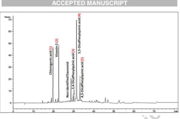
3.1. High performance liquid chromatography analysis (HPLC/UV-VIS-DAD)
The HPLC analysis of AcAE reveals a quite simple chromatographic profile (see Fig. 1), with three major peaks: chlorogenic acid (peak 1), vicenin-2 (peak 2) and 3,5-Dicaffeoylquinic acid (peak4). The peaks 1 to 5 were identified by means of their UV/vis spectra and their relative retention times, and literature data (Scifinder, 2016) corroborated these assignments. Co-injection with reference standards confirmed our hypothesis for the caffeoylquinic acids (chlorogenic and the three isochlorogenic acids).
15
Fig. 1. . LC chromatogram of A. campestris L. aqueous extract ( AcAE) visualized at the
wave lenght of 340 nm. Peaks were detected using DAD detector. Peak (1): chlorogenic acid, peak (2): vicenin-2, peak (3): 3,4-dicaffeoylquinic acid (isochlogenic acid C), peak (4): 3,5-dicaffeoylquinic acid (isochlogenic acid A), peak (5): 4,5-dicaffeoylquinic acid (isochlogenic acid B).
Vicenin-2 (apigenin 6, 8-di-C-glucoside) was identified by spectroscopic analysis (UV,MS and NMR) and comparison with literature data (Sawabe et al., 1989; Scifinder, 2016). Chemical structures of identified compounds are represented in Fig. 2.
3.2. Acute toxicity test
The single oral dose of 1, 2, 4 and 6 g/Kg of AcAE administered to the mice did not produce any symptoms of toxicity or mortality during the 2 weeks observations.
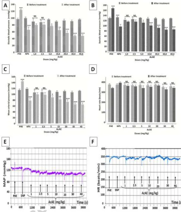
3.3. Hypotensive effect on anesthetized rats
AcAE at doses 1, 2.5, 5, 10, 20, 30 and 40 mg/Kg reduced the systolic blood pressure (SBP) respectively, by 8.9±0.7, 8.1±2.4, 14.4±3.4, 21.3±2, 28.4±5.2, 32.5±2.7, 33.2±4 %, and the diastolic blood pressure (DBP) respectively, by 15.6±1.4, 8.4±2.5, 19.6±3.1, 28.1±3.2, 38.0±6.7 and 46.9±3.6.
16
Fig. 2. Chemical structures of compounds identified in A. campestris L. aqueous extract
(AcAE).
Similarly, the mean arterial pressure (MAP) was lowered by treatment with AcAE; the reduction of MAP was: 7.6±1.3, 7.7±2.4, 20.2±4.6, 23.3±2.8, 32.1±6.3, 37.0 ±3.2, 41.1±4.7 %, respectively at doses 1, 2.5, 5, 10, 20, 30 and 40 mg/Kg. Heart rate were not significantly affected by treatment with AcAE (Fig.3).
17
Fig. 3. Effect of intravenous injection of A. campestris L. (AcAE) on (A) systolic blood
pressure (SBP) , (B) diastolic blood pressure (DBP), (C) mean arterial pressure (MAP) and (D) heart rat (HR) in anesthetized rats. Values are mean ± SEM, n=5. Two way ANOVA followed by Bonferroni’s post-test; * p < 0.05 and *** p <0.001 vs control (before treatment), NS: not significant. Original tracings showing the effect of intravenous injection of cumulative doses of A.campestris L. aqueous extract (AcAE) on
18
3.4 Antihypertensive effect
As shown in Fig. 4, at the beginning of the experiment, the baseline SBP values did not differ between the experimental groups. Daily administration of L-NAME (32 mg/Kg/j) induced a progressive increase in systolic blood pressure of L-NAME group from 123.3±1.05 mmHg and reached 172.5±2.8 mmHg at the end of treatment.
Fig. 4. Effect of four-weeks treatment on systolic bood pressure (SBP) of rats fed with
L-NAME (L-NAME group), Enalapril (L-NAME+Enalapril group), and A.campestris L. aqueous extract at 50 mg/Kg/day NAME+AcAE 50 group) and at 150 mg/Kg/day (L-NAME+AcAE150 group). Data are mean ± S.E.M (n= 5-7 rats per group). Two way ANOVA followed by Bonferroni’s post-test; b: p < 0.01; c: p < 0.001 vs. control group; β: p < 0.01; γ: p < 0.001 vs. L-NAME group.
This increase was significant from the first week until the end of experiment compared to control group (122.6 ± 1.9 mmHg; p < 0.001). At the end of the fourth week, the SBP of the rats in the L-NAME+Enalapril group (139.3 ± 2.5 mmHg; p < 0.001), L-NAME +
19
AcAE 50 group (146.4 ± 1.6 mmHg; p < 0.001) and L-NAME + AcAE 150 group (144.2 ± 1.5 mmHg; p < 0.001) was significantly lower than that of L-NAME group.
3.5. Vasorelaxant effect on aorta isolated from normotensive rats
3.5.1. Vasorelaxant effect of AcAE on intact and denuded precontracted aorta, and in the presence of Atropine and Calmidazolium
AcAE (10-2 - 2 mg/ml) induced 95.8 ±1.8% (n=6) of relaxation of aorta contracted by
phenylephrine (10-6 M). The vasorelaxation produced by AcAE was markedly
attenuated after denudation of aorta (26.9% ±2.1; p<0.001). Also, the vasodilator effect was significantly reduced when aorta was pretreated with atropine, the muscarinic receptor inhibitor (69.9% ±7.1; p<0.001) and with calmidazolium, the calcium-calmodulin binding to NO synthase blocker (54.2% ±14.8; p<0.001) (Fig. 5).
3.5.2. Vasorelaxant effect of AcAE in the presence of L-NAME, Hydroxycobalamin, ODQ and 8-RP-Br-PET-cGMP
Vasorelaxant effect produced by AcAE (10-2 - 2 mg/ml) was decreased after the
pre-incubation of aorta with L-NAME, NO synthase inhibitor (35.1 ±5.1%; p<0.001), hydroxocobalamin, NO scavenger (38.9 ± 7.1%; p<0.001), ODQ, the guanylyl cyclase inhibitor (67.4 ±3.4%; p<0.001) and Rp-8-Br-PET-cGMP, the competitive cGMP-dependent protein kinase G (PKG) inhibitor (69.1 ±8.2%; p<0.001) (Fig. 6).
20
Fig.5. (I) Concentration–response curves of the vasorelaxant effect of A.campestris L.
aqueous extract (AcAE) on aorta isolated from normotensive rats and pre-contracted with phenylephrine 10-6 M (●), on denuded aorta (■), and in the presence of Atropine (▲) and Calmidazolium (▼).Values are mean ± SEM, n=5-6.Two way ANOVA followed by Bonferroni’s post-test; ** p<0.01 and ***p < 0.001 vs control. (II) Original tracing showing the effect of charbachol 10-4 M on precontracte aorta with phenylephrine 10-6 M (endothelium integrity control) (A), and the vasorelaxant effect of A. campestris L. aqueous extract (AcAE) on isolated aorta pre-contracted with phenylephrine 10-6 M (B), on denuded aorta (C), and in the presence of atropine (D) and calmodizalium (E).
21
Fig.6. (I) Concentration–response curves of the vasorelaxant effect of A.campestris L.
aqueous extract (AcAE) on aorta isolated from normotensive rats and pre-contracted with phenylephrine 10-6 M (●), and in the presence of L-NAME (■), Hydroxocobalamin (▲), ODQ (▼) and 8-RP-Br-PET-cGMP (◆).Values are mean ± SEM, n=5-6.Two way ANOVA followed by Bonferroni’s post-test; *p < 0.05 and *** p < 0.001 vs control. (II) Original tracing showing the effect of charbachol 10-4 M on precontracte aorta with phenylephrine 10-6 M (endothelium integrity control) (A), and the vasorelaxant effect of A. campestris L. aqueous extract (AcAE) on isolated aorta pre-contracted with phenylephrine 10-6 M (B), in the presence of L-NAME (C), hydoroxocobalamine (D), ODQ (E), and 8-RP-Br-PET-cGMP (F).
22
3.5.3. Vasorelaxant effect of AcAE in the presence of Indomethacin, Thapsigargin, Verapamil
The vasorelaxant effect of AcAE (10-2 - 2 mg/ml) was inhibited after exposing aorta to
the blocker of VOC-calcium channels, verapamil (36.2 ±3.3%; p<0.001) and to the inhibitor of endoplasmic reticulum calcium-ATPase, thapsigargin (30.7 ±3.8%; p<0.001). However, the inhibition of cyclooxygenase, by pretreatment with indomethacin did not alter the vasorelaxant effect evoked by AcAE (88.3 ±4.3%; p>0.05) (Fig. 7).
3.5.4. Vasorelaxant effect of AcAE in the presence of potassium channels blockers, TEA, 4-AP, BaCl2 and Glibenclamide
The relaxation triggered by AcAE (10-2-2 mg/ml) was not influenced after pretreatment
of aorta with various potassium channels blockers such as TEA (93.1 ±5.6%; p> 0.05), 4-AP (90.8 ± 2.4%; p> 0.05), BaCl2 (87.7 ± 4.4%; p> 0.05) and glibenclamide (81.7 ±
7.4%; p> 0.05) (Fig.8).
4. Discussion
The present work investigated the phytochemical composition of the aqueous extract of A. campestris L. (AcAE) from Oriental Morocco, and showed no acute toxicity when tested on mice. Moreover, the hypotensive effect of AcAE intravenous injection to normotensive rats and antihypertensive effect of oral administration of the extract to L-NAME induced hypertensive rats during four weeks have been highlighted; also, the vasorelaxant effect mechanism of action was explored on isolated rat aorta and pointed out that AcAE act through both endothelium dependent and independent vasorelaxant pathways.
23
Fig.7. (I) Concentration–response curves of the vasorelaxant effect of A.campestris L.
aqueous extract (AcAE) on aorta isolated from normtensive rats andpre-contracted with phenylephrine 10-6 M (●) and in the presence of Thapsigargin (■), Verapamil (▲) and Indomethacin (▼). Values are mean ± SEM, n=6.Two way ANOVA followed by Bonferroni’s post-test; *** p < 0.001 vs control.(II) Original tracing showing the effect of charbachol 10-4 M on precontracte aorta with phenylephrine 10-6 M (endothelium integrity control) (A), and the vasorelaxant effect of A. campestris L. aqueous extract (AcAE) on isolated aorta pre-contracted with phenylephrine 10-6 M (B), in the presence of thapsigargin (C), verapamil(D), and indomethacin (E).
24
The HPLC analysis confirmed the presence of chlorogenic acid and three isochlorogenic acids: 3,4-dicaffeoylquinic acid (isochlorogenic acid B), 3,5-dicaffeoylquinic acid (isochlorogenic acid A) and 4,5-dicaffeoylquinic acid (isochlorogenic acid C). These findings are in congruence with a previous work carried out by Riedel et al. (2010), that found the same HPLC profile of cell culture of A. campestris L. occurring in Germany. Additional studies are available, showing the presence of chlorogenic acid in the phenolic-rich extract (Djeridane et al., 2007) and in the aqueous extract (Sebai et al., 2014) of A. campestris L.
The isochlorogenic acids were also detected in the water fraction and aqueous extract of A. campestris L. from Tunisia (Megdiche-Ksouri et al., 2015; Sebai et al., 2014). Data of other authors reported the occurrence of flavonoids such as chrysin, apigenin, luteolin, kaempferol, quercetin, myricetin and their derivatives (Akkari et al., 2014; De Pascual Teresa et al., 1986; De Pascual Teresa et al., 1984; Ferchichi et al., 2006; Hurabielle et al., 1982; Karabegović et al., 2011; Megdiche-Ksouri et al., 2015; Rauter et al., 1989; Sebai et al., 2014; Valant-Vetschera et al., 2003; Vasconcelos et al., 1998). Nevertheless, our study represents the first evidence about the existence of the glycoside flavonoid vicenin 2 as the main flavonoid present in the aqueous extract of A.
campestris L. (AcAE) from Eastern Morocco.
In a preliminary step to ensure the safety of AcAE, the extract was tested for it oral acute toxicity. Consequently, the LD50 was greater than 6 g/Kg, which seems to be
much higher than that of the aqueous extract of Tunisian A.campestris L. orally administered to the mice (LD50 > 3.2 g/Kg) (Sebai et al., 2014), and injected
25
Fig.8. (I) Concentration–response curves of the vasorelaxant effect of A.campestris L.
aqueous extract (AcAE) on aorta isolated from normotensive rats andpre-contracted with phenylephrine 10-6 M (●), and in the presence of TEA (■), 4-AP (▲), BaCl2 (▼) and Glibenclamide (◆).Values are mean ± SEM, n=6. (II) Original tracing showing the effect of charbachol 10-4 M on precontracte aorta with phenylephrine 10-6 M (endothelium integrity control) (A), and the vasorelaxant effect of A. campestris L. aqueous extract (AcAE) on isolated aorta pre-contracted with phenylephrine 10-6 M (B), in the presence of TEA (C), 4-AP (D), BaCl2 (E), and glibenclamide (F).
26
The extract also revealed a positive safety profile explained by the absence of any behavioural perturbations during the 2 weeks following the administration.
In our experiment, we proved the potential activity of A. campestris L. at lowering the blood pressure levels by testing the effect of AcAE on normotensive and hypertensive rats. Accordingly, the extract induced 41.1% drop of MAP of normal rats (with, 33.2% for SBP and 46.9% for DBP), at the dose of 40 mg/Kg, while the heart rate was not affected. The absence of any effect on the heart rate may lead to speculate that the hypotensive effect of AcAE is mainly mediated via a vascular pathway.
Besides, an antihypertensive action on L-NAME induced hypertensive rats was evidenced, and the results showed that AcAE at doses 50 and 150 mg/Kg/day given concomitantly to the NO synthase inhibitor (L-NAME) exerted about the same degree of preventive action against the rise of SBP and which was comparable to the effect of reference antihypertensive drug (enalapril; angiotensin-converting enzyme (ACE) inhibitor). These data may suggest that the antihypertensive effect of AcAE is probably attributed to its ability to inhibit the impairment of NO signalling associated to L-NAME treatment (Paulis et al., 2008). In the light of these findings, it is noteworthy to point out that it has been shown in a previous study that aqueous extract of A. campestris leaves, prevents the induced hypertensive phase induced by the scorpion venom in rats, probably through adrenergic pathway (Ben Nasr et al., 2014b).
The antihypertensive mechanism of action of AcAE was explored on vascular signalling pathways. Indeed, AcAE provoked a total relaxation of contracted aorta isolated from normotensive rats, which seems to be endothelium dependent, since the relaxant effect disappeared after the denudation process. Given the major importance of
27
endothelium in the regulation of vascular tone by releasing vasodilator factors such as nitric oxide (NO), prostacyclin (PGI2) and endothelium-derived hyperpolarizing factor
(EDHF) (Sandoo et al., 2010), the participation of endothelium mediators in the obtained vasorelaxant response was investigated. It is well known that the muscarinic vasorelaxation is mainly mediated via NO production in endothelial cells, which is triggered by the activation of endothelial nitric oxide synthase (eNOS), induced by Ca2+-calmodulin complex (Furchgott and Zawadzki, 1980; Harvey, 2012). Our results
showed that the pre-treatment of aorta with atropine, a selective muscarinic receptor antagonist, and with calmidazolium, a Ca2+-calmodulin binding to NOS blocker,
significantly reduced AcAE-induced vasorelaxation, which may suggest that the aqueous extract act via muscarinic receptor and interfere with the site of action of Ca2+-calmodulin complex in the vascular endothelium. More interestingly, NO was
identified as the pilot modulator of vascular tone; once synthetized within the cytosol of endothelial cell by NOS, NO diffuses into adjacent vascular smooth muscle cells (VSMC) and bind to soluble guanylyl cyclase (sGC) which mediate the vasorelaxation action (Walford and Loscalzo, 2003). In this concern, we performed separate experiments, in which we preincubated aorta with L-NAME (NOS inhibitor) and with hydroxycobalamin (NO scavenger). In the presence of both blockers, AcAE induced-vasorelaxation seems to be totally blunted, indicating the involvement of NO in the observed effect.
The cyclic GMP-PKG signalling within the VSMC represents the final cascade of vasorelaxation initiated by NO production (Furchgott, 1983). Hence, it was reported that NO activates sGC that enhances the conversion of GTP to cGMP. Subsequently,
28
cGMP activates PKG that decreases the cytosolic free calcium level through several mechanisms, which finally resulted in the vessel relaxation (Hofmann et al., 2000). This asseveration was proved when vascular relaxation was lost on aorta submitted to ODQ, the inhibitor of sGC, and to Rp-8-Br-PET-cGMPs, the inhibitor of PKG; thing that may lead to postulate about the contribution of sGC and PKG in the vasodilatory effect of AcAE.
The cyclooxygenase (COX) product, prostacyclin (PGI2), is an endogenous mediator of
endothelium derived relaxation (Félétou et al., 2011), via activation of cAMP (Parkington et al., 2004). Nevertheless, the participation of prostacyclin in the vasorelaxant effect of AcAE was ruled out, because the relaxation was not affected by the treatment with the COX inhibitor, indomethacin.
The intracellular calcium concentration [Ca2+]i is an ubiquitous parameter that
regulates vascular smooth muscle contractile state. The major [Ca2+]i, is principally
linked to the opening of voltage operated calcium (VOC) channels, which are finely controlled by the membrane potential (Ledoux et al., 2006). One the other hand, the re-uptake of intracellular calcium is considered as regulating mechanism of the vascular contractility, and the sarcoplasmic reticulum represents the major storage site of cytosolic calcium; in fact, the sequestration of excessive intracellular calcium occurs via an energy-dependent sarco-endoplasmic reticulum Ca2+-ATPase pump (SERCA)
(Laporte et al., 2004). Our data reported significant decrease of vasoactivity of AcAE after the incubation with verapamil, the L-type calcium inhibitor, and thapsigargin (inhibitor of the SERCA pump), which indicates that SERCA activation and VOCC channels inhibition account for the vascular dilation produced by the extract.
29
Otherwise, the opening of potassium channels (K+) triggers an increase in K+ efflux,
provoking membrane potential (Em) hyperpolarization and subsequent closure of VOC
calcium channels, causing a decrease of intracellular Ca2+ mobilisation followed by a
vasodilation (Ko et al., 2008; Sobey, 2001). Our findings reveal that pretreatment of aorta with potassium channels blockers did not modify the vascular effect of AcAE, which discarded the possible intervention of potassium channels in the vasorelaxant effect.
As mentioned above, A.campestris L. extract (AcAE) exhibited a relevant antihypertensive effect on L-NAME-induced hypertensive rats, where the NO synthesis is assumed to be inhibited. So, one may wonder how AcAE is acting on a model where NOS is completely inhibited by L-NAME whereas its action is mediated mainly through the NO pathway. This effect, controversial and confusing at the first glimpse, can be well justified taking into account that the present treatment is aimed at exploring the preventive antihypertensive effect of AcAE rather than its curative effect, because the extract was administered concomitantly with L-NAME (the selective inhibitor of NOS), and so, the supplementation of AcAE for four weeks tended likely to prevent the presumable action of L-NAME to inhibit NO release by blockade of NOS and to avoid the development of hypertension. It is probable that the co-administration of L-NAME and the extract triggered a competitive action between the two products on NOS activity. Hence, the activator effect of AcAE on NOS seems to be more pronounced and succeeded to reduce the inhibitory effect of L-NAME, and so neutralizing the blockade of NOS. This hypothesis may lead to postulate that the treatment with AcAE is most efficient to prevent, with an earlier way, the NO deficiency, by acting on the vascular
30
compartment to increase the NO bioavailability leading to the vasorelaxant effect on the favor of the antihypertensive action.With respect to the human projected dose or human equivalent dose (HED), it was calculated according to the method proposed by Reagan-Shaw et al. (2008) and based on comparison of the body surface area of the different animal species. The HED for the aqueous extract of A. campestris L. (AcAE) are 8.1 mg/kg (486.5 mg for a 60 kg person) for the first dose of 50 mg/kg in the rat and 24.3 mg/kg (1.46 g of AcAE for a 60 kg person) for the second dose of 150 mg/kg in rats. Since the antihypertensive effect of the aqueous extract of A. campestris L. (AcAE) at the dose of 50 mg/kg was comparable to that of the standard drug (Enalapril) and taking into account the extraction yield (8.7%), the effective dose to be consumed by humans corresponds to 93.1 mg of the plant /kg of weight /day. For an adult person of 60 kg, the daily necessary quantity is 5.59 g. Compared to infusion bags (which contain about 2 g of plant for one cup of water), this amount corresponds to 3 cups of herbal tea per day. Based on information from three herbalists interviewed in Oujda market, the recommended dose for the traditional use of A. campestris was estimated in a handful of the aerial part (approximately 20 g) in a cup (about 100 ml) of boiled water. The preparation can be consumed three times a day. Using the yield of the extraction found in our study (8.7%), the dose taken would be about 1.74 g of plant / 100 ml for each cup, which is the equivalent of 5.22 g of the net weight of the plant distributed three times a day.
On the basis of these data, it can be concluded that the human dose projected (8.1 mg/kg in humans, equivalent to 50 mg/kg in rats) is quite similar to the dose commonly used in traditional medicine.
31
Taking into account the antihypertensive effects of chlorogenic acids and mechanisms of improving endothelial function in hypertensive rats (Suzuki et al., 2006; Zhao et al., 2012), and considering the abundance of AcAE on chlorogenic and isochlorogenic acids it can be hypothesized that such compounds are at least partially or even mainly responsible of the antihypertensive and vasorelaxant observed effects. Nevertheless, the effect of vicenin-2 (6,8-di-C-glucosylapigenin) could also play an important role in the effect of AcAE. Indeed, this flavonoid exhibited the strongest hypotensive activity for hypertensive rats among seventy-three glycosides investigated. This activity was not only found with intravenous administration (Sawabe et al., 1989), but also after oral administration. In the latter case, C-glucosyl flavonoids are considered to be not hydrolyzed by the digestive system and hydrochloric acid (Sawabe and Matsubara, 1999). This hypothesis was recently confirmed by the investigations carried on vicenin-2, the active principle of an anti-inflammatory Brazilian medicinal plant (Buqui et al., 2015). Furthermore, vicenin-2 is a constituent of Crataegus monogyna, an old hypotensive medicinal plant used in Europe (Nikolov et al., 1981).
5. Conclusion
The present study gives evidence that A. campestris L. aqueous extract (AcAE) possess potent hypotensive, antihypertensive and vasorelaxant effects. The vasorelaxant effect of AcAE seems to be due to activation of muscarinic receptor and stimulating calmodulin-NO-sGC-PK pathway. An additional mechanism of action of AcAE is mediated via activation of SERCA pump and inhibition of VOCC channels. In relation with the chemical composition of AcAE it appears that the extract is predominantly composed of phenolic compounds (chlorogenic and isochlorogenic acids as well as
32
vicenin-2) which may be principally involved in the vasodilator effect.
Author’s contributions
I. Dib conducted the animal studies, performed the statistical analysis and wrote the
manuscript.
A Ziyyat is the supervisor of the student Ikram Dib who oversaw the work in its
entirety; It obviously discussed and approved all the protocols of pharmacological studies and reviewed and corrected the article several times
A. Assaidi contributed to the achievement of the hypotensive and antihypertensive
experiments.
A. Legssyer gives technical directives to perform the tail cuff method for caudal arterial
pressure measuring.
M. Tits, L. Angenot, M. Frédérich, and J.-N. Wauters approved the chromatographic
technics applied to the analyzed samples, and assisted the analyses of the aqueous extract of Artemisia campestris L., and participated in the correction of the manuscript.
H.Mekhfi, M. Bnouham, and M. Aziz helped in the correction of the manuscript.
All the authors read the manuscript and approved the final version.
Acknowledgments
This work was supported by «la Commission Universitaire pour le Développement du CIUF (CIUF-CUD)». We are grateful to Professor Mihamou Atika (Laboratoire de Biologie des Plantes et des Microorganismes, Département de Biologie, Faculté des
33
Sciences, Université Mohammed Premier, Oujda, Maroc) for botanical identification. We thank Miss. Delphine Etienne (Laboratoire de pharmacognosie, Liège-Belgique) for technical assistance and Mr. Mostafa Bedraoui from our laboratory (Laboratoire de Physiologie, Génétique et Ethnopharmacologie-Faculté des Sciences, Université Mohamed Premier, Oujda) for the reliable care of animals breeding.
References
Adeloye, D., Basquill, C., 2014. Estimating the prevalence and awareness rates of hypertension in Africa: a systematic analysis. PLoS ONE 9, e104300- e104316.
Akkari, H., Rtibi, K., B’chir, F., Rekik, M., Darghouth, M.A., Gharbi, M., 2014. In vitro evidence that the pastoral Artemisia campestris species exerts an anthelmintic effect on Haemonchus contortus from sheep. Veterinary Research Communication 38, 249-255.
Ben-Nasr, H., Salama, M., Ksouda, K., Zeghal, K.M., Hammami, S., 2014a. Hemodynamic effects of aqueous extract of Artemisia campestris in adult men. Journal of Applied Pharmaceutical Science 4, 38-42.
Ben Nasr, H., Hammami, T.S., Mahmoudi, L., Zeghal, K., 2014b. Aqueous leaves extract of Artemisia campestris inhibition of the scorpion venom induced hypertension. Journal of Medicinal Plant Research 8, 538-542.
Benchelah, A.-C., Bouziane, H., Maka, M., 2004. Fleurs du Sahara, arbres et arbustes, voyage au coeur de leurs usages avec les Touaregs du Tassili. Phytothérapie 2, 191-197.
34
Benítez, G., González-Tejero, M., Molero-Mesa, J., 2010. Pharmaceutical ethnobotany in the western part of Granada province (southern Spain): Ethnopharmacological synthesis. Journal of Ethnopharmacology 129, 87-105.
Berrada El Azizi, G., Ahid, S., Ghanname, I., Belaiche, A., Hassar, M., Cherrah, Y., 2012. Trends in antihypertensives use among Moroccan patients. Pharmacoepidemiology and drug safety 21, 1067-1073.
Bnouham, M., Mekhfi, H., Legssyer, A., Ziyyat, A., 2002. Ethnopharmacology forum medicinal plants used in the treatment of diabetes in Morocco. International Journal of Diabetes & Metabolism 10, 33-50.
Boudjelal, A., Henchiri, C., Sari, M., Sarri, D., Hendel, N., Benkhaled, A., Ruberto, G., 2013. Herbalists and wild medicinal plants in M'Sila (North Algeria): An ethnopharmacology survey. Journal of Ethnopharmacology 148, 395-402.
Buqui, G.A., Sy, S.K., Merino-Sanjuán, M., Gouvea, D.R., Nixdorf, S.L., Kimura, E., Derendorf, H., Lopes, N.P., Diniz, A., 2015. Characterization of intestinal absorption of C-glycoside flavonoid vicenin-2 from Lychnophora ericoides leafs in rats by nonlinear mixed effects modeling. Revista Brasileira de Farmacognosia 25, 212-218.
De Pascual Teresa, J., Gonzalez, M., Muriel, M., Arcocha, A., Bellido, I., 1986. Flavonoids from Artemisia campestris ssp. glutinosa. Journal of Natural Products 49, 177-177.
De Pascual Teresa, J., González, M.S., Muriel, M.R., Bellido, I.S., 1984. Phenolic derivatives from Artemisia campestris subsp. Glutinosa. Phytochemistry 23, 1819-1821.
35
Dib, I., Angenot, L., Mihamou, A., Ziyyat, A., Tits, M. ,2016. Artemisia campestris L.: Ethnomedicinal, phytochemical and pharmacological review. Journal of Herbal Medicine, http://dx.doi.org/10.1016/j.hermed.2016.10.005.
Dib, I., Mihamou, A., Berrabah, M., Mekhf,i H., Aziz, M., Legssyer, A., Bnouham, M., Ziyyat, A, 2017a. Identification of Artemisia campestris L. subsp. glutinosa (Besser) Batt. from Oriental Morocco based on its morphological traits and essential oil profile. Journal of Material and Environmental Sciences. 8, 180-187
Dib, I., Fauconnier, M.-L., Sindic, M., Belmekki, F., Assaidi, A., Berrabah, M., Mekhfi, H., Aziz, M., Legssyer, A., Bnouham,M., Ziyyat, A.,2017b. Chemical composition, vasorelaxant, antioxidant and antiplatelet effects of essential oil of Artemisia campestris L. from Oriental Morocco. BMC Complementary and Alternative Medicine 17, DOI: 10.1186/s12906-017-1598-2.
Djeridane, A., Yousfi, M., Nadjemi, B., Vidal, N., Lesgards, J., Stocker, P., 2007. Screening of some Algerian medicinal plants for the phenolic compounds and their antioxidant activity. European Food Research and Technology 224, 801-809.
Djidel, S., Khennouf, S., Baghiani, A., Harzallah, D., Arrar, L., 2009. Medicinal plants used traditionally in the Algerian folk medicine for gastrointestinal disorders and hypertension: total polyphenols, flavonoids and antioxidant activity. Acta Horticulturae 854, 59-65.
Eddouks, M., Maghrani, M., Lemhadri, A., Ouahidi, M.-L., Jouad, H., 2002. Ethnopharmacological survey of medicinal plants used for the treatment of diabetes mellitus, hypertension and cardiac diseases in the south-east region of Morocco (Tafilalet). Journal of Ethnopharmacology 82, 97-103.
36
El Hassani, M., Douiri, E., Bammi, J., Zidane, L., Badoc, A., Douira, A., 2013. Plantes médicinales de la Moyenne Moulouya (Nord-Est du Maroc). Ethnopharmacologia 50, 39-53.
Fakchich, J., Elachouri, M., 2014. Ethnobotanical survey of medicinal plants used by people in Oriental Morocco to manage various ailments. Journal of Ethnopharmacology 154, 76-87.
Félétou, M., Huang, Y., Vanhoutte, P.M., 2011. Endothelium‐mediated control of vascular tone: COX‐1 and COX‐2 products. British journal of pharmacology 164, 894-912.
Ferchichi, L., Merza, J., Landreau, A., Le Ray, A.M., Legseir, B., Seraphin, D., Richomme, P., 2006. Occurrence of isocoumarinic and phenolic derivatives in Artemisia campestris L. subsp. campestris. Biochemical Systematics and Ecology 34, 829-832.
Furchgott, R.F., 1983. Role of endothelium in responses of vascular smooth muscle. Circulation research 53, 557-573.
Furchgott, R.F., Zawadzki, J.V., 1980. The obligatory role of endothelial cells in the relaxation of arterial smooth muscle by acetylcholine. Nature 288, 373-376.
Guarino, C., De Simone, L., Santoro, S., 2008. Ethnobotanical study of the Sannio area, Campania, Southern Italy. Ethnobotany Research & Applications 6, 255-317.
Hammiche, V., Maiza, K., 2006. Traditional medicine in Central Sahara: pharmacopoeia of Tassili N’ajjer. Journal of Ethnopharmacology 105, 358-367.
Harvey, R.D., 2012. Muscarinic receptor agonists and antagonists: effects on cardiovascular function, in: Fryerl, A.D., Christopoulos, A., Nathanson, N.M. (Eds.), Muscarinic Receptors. Springer, New York, pp. 299-316.
37
Hofmann, F., Ammendola, A., Schlossmann, J., 2000. Rising behind NO: cGMP-dependent protein kinases. Journal of cell science 113, 1671-1676.
Hurabielle, M., Eberle, J., Paris, M., 1982. Etude des flavonoïdes d'Artemisia campestris sous-espèce Glutinosa [Flavonoids of Artemisia campestris, ssp. glutinosa]. Planta Medica 46, 124-125.
Jouad, H., Haloui, M., Rhiouani, H., El Hilaly, J., Eddouks, M., 2001. Ethnobotanical survey of medicinal plants used for the treatment of diabetes, cardiac and renal diseases in the North centre region of Morocco (Fez–Boulemane). Journal of Ethnopharmacology 77, 175-182.
Karabegović, I., Nikolova, M., Veličković, D., Stojičević, S., Veljković, V., Lazić, M., 2011. Comparison of antioxidant and antimicrobial activities of methanolic extracts of the Artemisia sp. recovered by different extraction techniques. Chinese Journal of Chemical Engineering 19, 504-511.
Ko, E.A., Han, J., Jung, I.D., Park, W.S., 2008. Physiological roles of K+ channels in vascular smooth muscle cells. Journal of Smooth Muscle Research 44, 65-81.
Laporte, R., Hui, A., Laher, I., 2004. Pharmacological modulation of sarcoplasmic reticulum function in smooth muscle. Pharmacological reviews 56, 439-513.
Ledoux, J., Werner, M.E., Brayden, J.E., Nelson, M.T., 2006. Calcium-activated potassium channels and the regulation of vascular tone. Physiology 21, 69-78.
Leporatti, M.L., Ghedira, K., 2009. Comparative analysis of medicinal plants used in traditional medicine in Italy and Tunisia. Journal of Ethnobiology and Ethnomedicine 5, 1-8.
38
Megdiche-Ksouri, W., Trabelsi, N., Mkadmini, K., Bourgou, S., Noumi, A., Snoussi, M., Barbria, R., Tebourbi, O., Ksouri, R., 2015. Artemisia campestris phenolic compounds have antioxidant and antimicrobial activity. Industrial Crops and Products 63, 104-113. Nikolov, N., DellaMonic, G., Chopin, J., 1981. Di-C-glycosylflavones from Crataegus monogyna. Phytochemistry 20, 2780-2781.
Parkington, H.C., Coleman, H.A., Tare, M., 2004. Prostacyclin and endothelium-dependent hyperpolarization. Pharmacological Research 49, 509-514.
Paulis, L., Zicha, J., Kunes, J., Hojna, S., Behuliak, M., Celec, P., Kojsova, S., Pechanova, O., Simko, F., 2008. Regression of L-NAME-induced hypertension: the role of nitric oxide and endothelium-derived constricting factor. Hypertension Research 31, 793. Popović, Z., Smiljanić, M., Matić, R., Kostić, M., Nikić, P., Bojović, S., 2012. Phytotherapeutical plants from the Deliblato Sands (Serbia): Traditional pharmacopoeia and implications for conservation. Indian Journal of Traditional Knowledge 11, 385-400.
Rauter, A.P., Branco, I., Tosrão, Z., Pais, M.S., Gonzalez, A.G., Bermejo, J.B., 1989. Flavonoids from Artemisia campestris subsp. marítima. Phytochemistry 28, 2173-2175. Reagan-Shaw, S., Nihal, M., Ahmad, N., 2008. Dose translation from animal to human studies revisited. The FASEB Journal 22, 659-661.
Riedel, H., Cai, Z., Smetanska, I., 2010. Obtaining phenolic acids from cell cultures of various Artemisia species. African Journal of Biotechnology 9, 8805-8809.
Sandoo, A., van Zanten, J.J.V., Metsios, G.S., Carroll, D., Kitas, G.D., 2010. The endothelium and its role in regulating vascular tone. The open cardiovascular medicine journal 4, 302-312.
39
Sawabe, A., Matsubara, Y., 1999. Bioactive glycosides in citrus fruit peels, in: Chong-Ren, Y., Osamu, T. (Eds.), Studies in Plant Science. Elsevier, pp. 261-274.
Sawabe, A., Matsubara, Y., Iizuka, Y., Okamoto, K., 1989. Structures and hypotensive effect of flavonoid glycosides in young Citrus unshiu peelings. Journal of Japan Oil Chemists' Society 38, 53-59.
Scifinder, 2016. American Chemical Society www.cas.org/products/scifinder.
Sebai, H., Jabri, M.-A., Souli, A., Hosni, K., Selmi, S., Tounsi, H., Tebourbi, O., Boubaker, S., El-Benna, J., Sakly, M., 2014. Protective effect of Artemisia campestris extract against aspirin-induced gastric lesions and oxidative stress in rat. RSC Advances 4, 49831-49841.
Sefi, M., Fetoui, H., Makni, M., Zeghal, N., 2010. Mitigating effects of antioxidant properties of Artemisia campestris leaf extract on hyperlipidemia, advanced glycation end products and oxidative stress in alloxan-induced diabetic rats. Food and Chemical Toxicology 48, 1986-1993.
Sobey, C.G., 2001. Potassium channel function in vascular disease. Arteriosclerosis, Thrombosis, and Vascular Biology 21, 28-38.
Suzuki, A., Yamamoto, N., Jokura, H., Yamamoto, M., Fujii, A., Tokimitsu, I., Saito, I., 2006. Chlorogenic acid attenuates hypertension and improves endothelial function in spontaneously hypertensive rats. Journal of hypertension 24, 1065-1073.
Tahraoui, A., El-Hilaly, J., Israili, Z., Lyoussi, B., 2007. Ethnopharmacological survey of plants used in the traditional treatment of hypertension and diabetes in south-eastern Morocco (Errachidia province). Journal of Ethnopharmacology 110, 105-117.
40
Tazi, M.A., Abir-Khalil, S., Chaouki, N., Cherqaoui, S., Lahmouz, F., Sraïri, J.E., Mahjour, J., 2003. Prevalence of the main cardiovascular risk factors in Morocco: results of a National Survey, 2000. Journal of hypertension 21, 897-903.
Valant-Vetschera, K.M., Fischer, R., Wollenweber, E., 2003. Exudate flavonoids in species of Artemisia (Asteraceae—Anthemideae): new results and chemosystematic interpretation. Biochemical Systematics and Ecology 31, 487-498.
Vasconcelos, J.M.J., Silva, A.M.S., Cavaleiro, J.A.S., 1998. Chromones and flavanones from Artemisia campestris subsp. maritima. Phytochemistry 49, 1421-1424.
Walford, G., Loscalzo, J., 2003. Nitric oxide in vascular biology. Journal of Thrombosis and Haemostasis 1, 2112-2118.
Zhao, Y., Wang, J., Ballevre, O., Luo, H., Zhang, W., 2012. Antihypertensive effects and mechanisms of chlorogenic acids. Hypertension Research 35, 370-374.
Ziyyat, A., Legssyer, A., Mekhfi, H., Dassouli, A., Serhrouchni, M., Benjelloun, W., 1997. Phytotherapy of hypertension and diabetes in oriental Morocco. Journal of Ethnopharmacology 58, 45-54.
Ziyyat, A., Ramdani, N., Bouanani, N.E.H., Vanderpas, J., Hassani, B., Boutayeb, A., Aziz, M., Mekhfi, H., Bnouham, M., Legssyer, A., 2014. Epidemiology of hypertension and its relationship with type 2 diabetes and obesity in eastern Morocco. SpringerPlus 3, 644-650.
Glossary
41
campestris L. aqueous extract; BaCl2: barium chloride;BP-T: blood pressure transducer;
cAMP: cyclic adenosine monophosphate; CCH: carbachol; cGMP: cyclic guanosine monophosphate; COX: cyclooxygenase; DAD: diode array detector; DBP; diastolic blood pressure; DMSO: diméthylsulfoxyde; Em: membrane potentiel; EDHF:
endothelium derived hyperpoliryzing factor; eNOS: endothelial nitrous oxide synthase; GTP: guanosine triphosphate; HR: heart rate; L-NAME: Nω-nitro-L-arginine methyl ester hydrochloride; LD50 : median lethal dose; MAP: mean arterial pressure; NMR:
nuclear resonance magnetic; NO: nitrous oxide; NOS: nitrous oxide synthase; ODQ: 1H-[1,2,4]oxadiazolo[4,3-a]quinoxalin-1-one; PGI2: prostacyclin ; Phe/PHE: phenylephrine;
PKG: protein kinase G; Rp-8-Br-PET-cGMPS: Rp-8-bromo-β-phenyl-1,N2-ethenoguanosine 3′,5′-cyclic monophosphorothioate sodium salt; SBP: systolic blood pressure; SERCA: sarco/endoplasmic reticulum Ca2+-ATPase; sGC: soluble guanylyl
cyclase; SNP: sodium nitroprusside; TEA: tetraethylammonium; TFA: trifluoroacetic acid; UV: ultarvaiolet; VOC: voltage-operated channels; VOCC: voltage-operated calcium channels; VSMC: vascular smooth muscle cell.