HAL Id: hal-03004639
https://hal-cnrs.archives-ouvertes.fr/hal-03004639
Submitted on 20 Nov 2020
HAL is a multi-disciplinary open access
archive for the deposit and dissemination of
sci-entific research documents, whether they are
pub-lished or not. The documents may come from
teaching and research institutions in France or
abroad, or from public or private research centers.
L’archive ouverte pluridisciplinaire HAL, est
destinée au dépôt et à la diffusion de documents
scientifiques de niveau recherche, publiés ou non,
émanant des établissements d’enseignement et de
recherche français ou étrangers, des laboratoires
publics ou privés.
Small-Angle x-ray scattering and crystallographic
studies of arcelin-1: An insecticidal lectin-like
glycoprotein fromPhaseolus vulgaris L
Lionel Mourey, Jean-Denis Pedelacq, Christine Fabre, Henri Causse, Pierre
Rougé, Jean-Pierre Samama
To cite this version:
Lionel Mourey, Jean-Denis Pedelacq, Christine Fabre, Henri Causse, Pierre Rougé, et al..
Small-Angle x-ray scattering and crystallographic studies of arcelin-1: An insecticidal lectin-like glycoprotein
fromPhaseolus vulgaris L. Proteins: Structure, Function, and Genetics, Wiley, 1997, 29 (4),
pp.433-442. �10.1002/(sici)1097-0134(199712)29:43.0.co;2-9�. �hal-03004639�
Small-Angle X-Ray Scattering and Crystallographic
Studies of Arcelin-1: An Insecticidal Lectin-Like
Glycoprotein From Phaseolus vulgaris L
Lionel Mourey,1Jean-Denis Pe´delacq,1Christine Fabre,2Henri Causse,2Pierre Rouge´,2 and Jean-Pierre Samama1*
1Groupe de Cristallographie Biologique, Institut de Pharmacologie et de Biologie Structurale,
UPR 9062 CNRS, F-31077 Toulouse Cedex, France
2Groupe Lectines et Reconnaissance, Institut de Pharmacologie et de Biologie Structurale,
UPR 9062 CNRS, F-31077 Toulouse Cedex, France
ABSTRACT Arcelin-1 and a-amylase
in-hibitor are two lectin-like glycoproteins ex-pressed in the seeds of the kidney bean (Phaseolus vulgaris). They display insecticidal activities and protect the seeds from predation by larvae of various bruchids through differ-ent biological actions. Solution-state investiga-tions by small-angle X-ray scattering (SAXS) show the dimeric structure of arcelin-1, a re-quirement for its hemagglutinating ties. Anions were found to have specific proper-ties in their effectiveness to disrupt protein aggregates, affect solubility, and improve crys-tallizability. The SAXS results were used to improve crystallization conditions, and single crystals diffracting beyond 1.9 Å resolution were obtained. X-ray diffraction data analysis shows that noncrystallographic symmetry-related arcelin-1 molecules form a lectin-like dimer and reveals the presence of a solvent-exposed anion binding site on the protein, at a crystal-packing interface. The solution state properties of arcelin-1 and crystal twinning may be explained by the anion specificity of this binding site. Proteins 29:433–442, 1997.
r1997 Wiley-Liss, Inc.
Key words: glycoprotein; insecticide; bru-chids; crystallization; X-ray struc-ture
INTRODUCTION
Lectins form a large superfamily of proteins in-volved in many interactions at the cell surface through specific carbohydrate recognition. They are widely spread in plants, animals, viruses, and bacte-ria, and many of them have been isolated and characterized.1Precise understanding of their
biologi-cal function remains unclear, despite several X-ray structure determinations that provided the struc-tural basis for sugar recognition and binding.2
Five different structural classes of plant lectins, differing in primary sequences and overall folds, can
be distinguished: legume lectins, hevein-related (ce-real) lectins, monocot mannose-binding (bulb) lectins, jacalin, and ribosome-inactivating proteins (RIPs). Lectins of the Leguminosae share a common b-sandwich tertiary structure that can be described as an ellipsoidal dome. However, these closely re-lated proteins differ in their sugar specificities and quaternary organizations.3 Dimers (pea lectin,4
Lathyrus ochrus isolectins,5,6Erythrina
coralloden-dron lectin,7Griffonia simplicifolia lectin IV,8lentil
lectin9) and tetramers (concanavalin A,10 peanut
agglutinin,11soybean agglutinin,12
phytohemaggluti-nin-L13) have been described from the X-ray
struc-tures. Within this class, four glycoproteins are found in the seeds of kidney bean (Phaseolus vulgaris): two lectins, phytohemagglutinin-L (PHA-L) and phytohe-magglutinin-E, and the related lectin-like proteins, arcelins anda-amylase inhibitors (a-AI). These pro-teins have different biochemical properties, are en-coded by a cluster of physically linked genes,14and
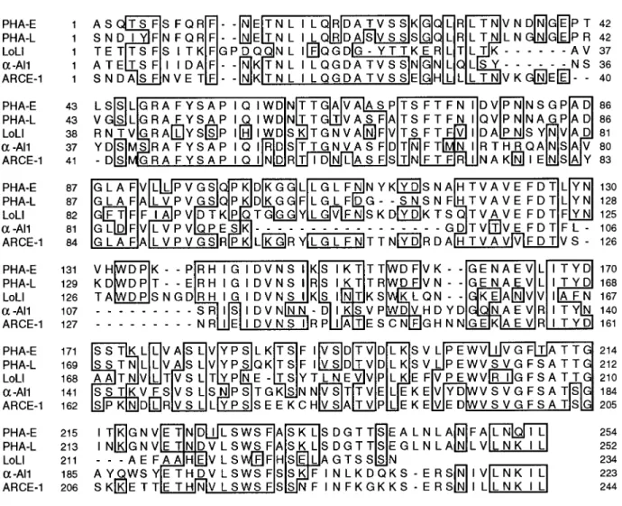
share 55–60% sequence identity. Sequence align-ment suggests that the canonical monosaccharide-binding site of legume lectins, delineated by four loop regions, is altered in arcelin and a-AI due to the deletion of one and two of these loops, respectively (Fig. 1).15
PHA-L binds complex-type oligosaccharides, and its three-dimensional structure pointed out a pos-sible binding site for adenine-derived plant hor-mones.13a-AI protects bean seeds against predation
by some insect pests through the inactivation of a-amylase. The molecular basis of the enzyme inhibi-tion was recently revealed by the crystal structure of the tight 1:2 complex formed between the inhibitor and pig pancreatic a-amylase.16Arcelin-1 also
pro-tects kidney bean seeds against the larvae of various
Abbreviations: MES, 2-Morpholinoethanesulfonic acid; PEG,
polyethylene glycol; SAXS, small angle X-ray scattering. *Correspondence to: Jean-Pierre Samama, IPBS-CNRS, 205 route de Narbonne, 31077 Toulouse Cedex, France.
E-mail: [email protected]
Received 27 March 1997; Accepted 26 June 1997
PROTEINS: Structure, Function, and Genetics 29:433–442 (1997)
bruchids, such as the Mexican bean weevil (Zabrotes
subfasciatus).17 However, this protection occurs
through a different mechanism than that ofa-AI and is still not well understood. A step toward the understanding of the structure-function relation-ships of arcelin-1 should be provided by solution-state investigations and high resolution X-ray struc-ture determination.
In this article, we describe the first results of the crystallographic study of arcelin-1 and emphasize the use of small-angle X-ray scattering as a diagnos-tic tool for producing crystals of this glycoprotein, diffracting beyond 1.9 Å resolution.
MATERIALS AND METHODS Solution X-Ray Scattering
Arcelin-1 was purified from seed flour by using a two-step procedure based on ion exchange chromatog-raphy and gel chromatofocusing (P. Rouge´, unpub-lished data). All SAXS and crystallization experi-ments were performed by using the same purification
batch. Stock protein solutions (10 mg/ml) were pre-pared by dissolving lyophilized arcelin-1 in water. They were then centrifuged for 1 h at 4°C and 15,000 rpm to discard any insoluble material. The protein solutions used for SAXS measurements were pre-pared by mixing equal volumes of stock arcelin-1 solution and twice-concentrated solutions of the dif-ferent media (Table I). Ionic strength values were calculated by using I 5 1⁄2Sc
iZi
2 with c
i and Zi the
concentration and valency of all ionic species present in the solution. In the case of the polyacid sodium citrate, the concentrations of each anion were calcu-lated by using the program EQS4WIN (Mathtrek Systems) and the different pK values18at the given
pH. All samples contained 1.3 mM DTT to eliminate the free radicals formed in solution under X-ray irradiation.
X-ray scattering curves were recorded on the small-angle-scattering instrument D2419 by using
synchrotron radiation at LURE-DCI (Orsay, France). The instrument and the data acquisition system
Fig. 1. Sequence alignement ofLathyrus ochrus isolectin I (LoLI), erythroagglutinating (PHA-E), and leucoagglutinating (PHA-L) subunits of phytohemagglutinin,a-amylase inhibitor 1 (a-AI1), and arcelin-1 (ARCE-1). The sequence identities between
arcelin-1 and LoLI, PHA-E, PHA-L, anda-AI1 are 45, 55, 58, and 60%, respectively. The four loop regions implicated in monosaccha-ride binding in LoLI are: 73–81, 95–102, 123–135, and 210–215.
were described previously.20The wavelength of the
X-rays was 1.488 Å (K-edge of Ni), and the sample to detector distance was set to 1827 mm. Scattered intensities were recorded in the angular range 0.00132, s (Å21), 0.02976, where s is the
scatter-ing vector (2 sinu)/l, and 2u and l are the scattering angle and wavelength of the X-rays, respectively. All experiments were performed at 4°C by using a temperature-controlled cell.21 Eight successive
frames of 100 s each were collected for each sample. The scattering intensity of a reference sample of carbon black, recorded immediately before and after each sample, was used to normalize all data to the transmitted intensity. The scattering contribution of the buffer was substracted before further analysis.
The radius of gyration (Rg) was deduced from
Guinier’s law:
I(s)5 I(0) exp (24p2R
g
2s2/3)
where I(s) is the SAXS intensity, I(0) is the value of the extrapolated scattered intensity at s5 0.22For
monodisperse solutions, the Guinier plot of ln I(s) versus s2approximates a straight line with a slope
proportional to Rg in the small-angle region. After
normalization to protein concentration (c), the value of I(0)/c is proportional to the molecular weight of the scattering entity. To calibrate this value, we used a monodisperse solution of 3-phosphoglycerate kinase (PGK, MW 44,500 Da). Measurements with PGK were performed in 20 mM Tris buffer pH 7.5 by using identical experimental settings.
Crystallization
All crystallization trials were performed at 4°C by vapor diffusion using the hanging drop method and freshly prepared aqueous protein solutions (10 mg/ ml). Crystals diffracting to 1.9 Å resolution were obtained by mixing 2 µl of arcelin-1 solution and 1 µl of reservoir solution containing 25% PEG400 and 300 mM ammonium sulfate in 100 mM MES buffer at pH 4.8 or 4.9.
Data Collection and Processing: Molecular Replacement
Crystals were characterized in the laboratory with a Rigaku RU300 rotating anode producing Cu-Ka X-rays collimated with the Yale double-mirror focus-ing (Molecular Structure Corporation) and an R-Axis II imaging plate detector (Rigaku).
A 1.9 Å native data set was collected on the W32 wiggler beam line at LURE-DCI (Orsay, France), tuned at a wavelength of 0.938 Å, and equipped with a large MarResearch scanner. The crystal to detector distance was set to 270 mm, and the crystal tempera-ture was maintained at 4°C. A total of 58 frames (1.5° oscillation per frame and 3-min time exposure) was collected from a single arcelin-1 crystal (6603 400 3 180 µm3). Data were processed with MOSFLM23and
the CCP4 suite of programs.24
Molecular replacement calculations were per-formed with the AMoRe program25as implemented
in the CCP4 package. The Lathyrus ochrus isolectin I crystallographic structure5 (PDB26 access code
1LOE) was used as a search model for the rotation and the translation functions. It was truncated as a polyalanine with B factors set to 20.0 Å2. In addition,
residues 125–134, which are absent in arcelin-1, according to sequence alignment, were removed.
RESULTS SAXS Experiments
The curvature of the Guinier plot at small s values indicated the presence of protein aggregates when experiments were conducted in MES buffer pH 4.9,
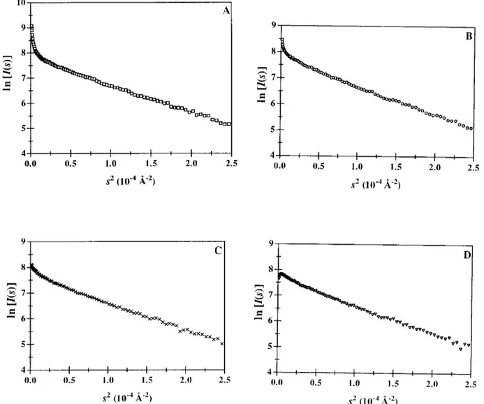
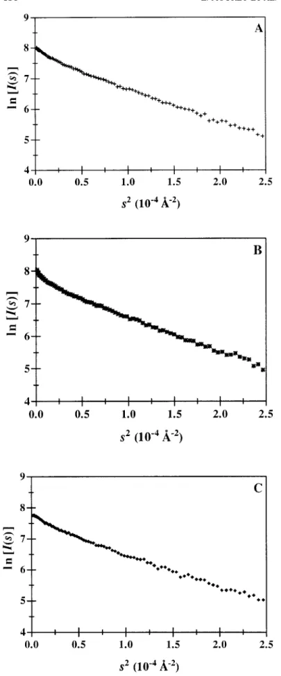
I 5 0.1 M (Fig. 2A). The effect of anions on the
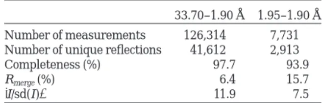
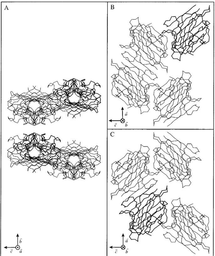
monodispersity of arcelin-1 solutions was investi-gated with SAXS measurements performed in the presence of thiocyanate, chloride, and citrate ions (Fig. 2B–D), in the same buffer and at low ionic strength (I5 0.4–0.5 M). We found that the improve-ment of monodispersity followed the lyotropic charac-ter of the anions (citrate. chloride . thiocyanate).* The most chaotropic thiocyanate ions, used at higher concentration (I5 1.1 M), further decreased aggrega-tion (Fig. 3B versus 2B), suggesting that the ionic strength was a second parameter that improved monodispersity. Accordingly, the Guinier plot indi-cated that aggregation was nearly abolished when the most lyotropic sulfate ions were used at I5 1.0 M (Fig. 3C).
With regard to protein crystallization, the relative contributions of the ionic strength and of the lyo-tropic properties of anions were approached by com-paring experiments performed in 100 mM sodium
*The lyotropic to chaotropic classification of anions has been related to several properties of biological macromolecules, such as folding, stability, and solubility.27,28
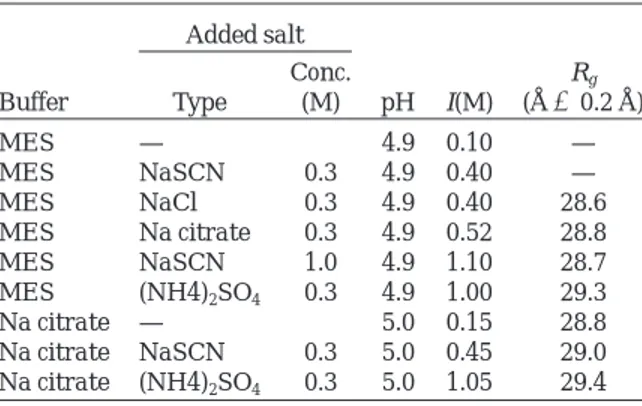
TABLE I. Conditions Used for Arcelin-1 SAXS Experiments* Buffer Added salt pH I(M) Rg (Å6 0.2 Å) Type Conc. (M) MES — 4.9 0.10 — MES NaSCN 0.3 4.9 0.40 — MES NaCl 0.3 4.9 0.40 28.6 MES Na citrate 0.3 4.9 0.52 28.8 MES NaSCN 1.0 4.9 1.10 28.7 MES (NH4)2SO4 0.3 4.9 1.00 29.3 Na citrate — 5.0 0.15 28.8 Na citrate NaSCN 0.3 5.0 0.45 29.0 Na citrate (NH4)2SO4 0.3 5.0 1.05 29.4 *All protein and buffers concentrations were 5 mg/ml and 0.1 M, respectively.
435
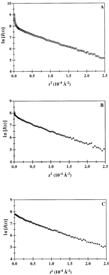
citrate buffer alone (I5 0.15 M) and in this buffer supplemented with 300 mM sodium thiocyanate (I5 0.45 M) or 300 mM ammonium sulfate (I 5 1.05 M). The nearly monodispersed character of the pro-tein solution in citrate buffer (Fig. 4A) was slightly altered in the presence of the chaotropic thiocyanate ions (Fig. 4B), even though the ionic strength was increased. On the contrary, sulfate ions had no significant effect (Fig. 4C).
The Guinier plots for arcelin-1, computed for all monodispersed conditions, gave a similar radius of gyration. The average Rgvalue is 29.06 0.3 Å. The
extrapolated intensities I(0) were brought to abso-lute scale by measuring the small-angle X-ray scat-tering of a 3-phosphoglycerate kinase solution of known concentration. The calculated average molecu-lar weight of the arcelin-1 scattering species was 60.1 6 3.1 kDa, which according to the molecular
weight of the monomer from SDS-PAGE electropho-resis, indicated that arcelin-1 exists as dimeric spe-cies in solution.
Crystallization
Thin plates were obtained from a large screening of the crystallization parameters, among which acidic pH and PEG400 appeared to be favorable for nucle-ation. The crystallization media were then chosen to also take into account the requirements for monodis-persed protein solutions. They were conducted in the presence of 300 mM ammonium sulfate, added to three different buffers: 100 mM MES, 100 mM sodium acetate, or 100 mM sodium citrate. The first and the last conditions correspond to the SAXS experiments shown in Figures 3C and 4C. In all three conditions, the ionic strength and the pH were similar (I< 1.0 M, pH < 4.9). Sulfate ions
contrib-Fig. 2. Guinier plots of 5 mg/ml arcelin-1 solutions in (A) 0.1 M MES pH 4.9 (I50.1 M), and in the same buffer supplemented with (B) 0.3 M sodium thiocyanate (I50.4 M). (C) 0.3 M sodium chloride (I50.4 M). (D) 0.3 M sodium citrate (I50.5 M).
Fig. 3. Guinier plots of 5 mg/ml arcelin-1 solutions in (A) 0.1 M MES pH54.9 and in the same buffer supplemented with (B) 1.0 M so-dium thiocyanate (I51.1 M). (C) 0.3 M ammo-nium sulfate (I51.0 M).
437
Fig. 4. Guinier plots of 5 mg/ml arcelin-1 solutions in (A) 0.1 M sodium citrate pH 5.0 (I50.15 M) and in the same buffer supple-mented with (B) 0.3 M sodium thiocyanate (I50.45 M). (C) 0.3 M ammonium sulfate (I51.05 M).
uted as both ionic strength enhancer and as the most lyotropic agent. In the presence of citrate buffer, the protein solution remained clear during the whole crystallization process, and crystals appeared within 1 week. Skin formation was observed in the case of acetate buffer, and crystals were obtained after 1 month. However, in these two conditions, polynucle-ation was substantial, and the crystals were often twinned. By using MES buffer, single crystals ap-peared after some precipitation of the protein, and 2 months were needed to reach equilibrium. It thus appeared that acetate and citrate ions increasingly counteract the effectiveness of sulfate ions in lower-ing protein solubility.
Crystallographic Data and Molecular Replacement
Crystals belong to the orthorhombic space group
P21212 with cell parameters a5 85.6 Å, b 5 92.6 Å,
c 5 67.3 Å. The asymmetric unit contains two
molecules of arcelin-1, which gave a calculated crys-tal volume per unit of molecular weight VM of
2.2 Å3/Da and a solvent content of 44%. These values
are in the range generally found for protein crys-tals.29Diffraction data were collected to 1.9 Å
resolu-tion from a single crystal of arcelin-1 (Table II). Molecular replacement was performed by using one promoter of isolectin I (LoLI) as a model. The sequence identity between arcelin-1 and LoLI is 45% (Fig. 1). The cross-rotation function, with an outer radius of integration of 25 Å, and the translation function gave the best results between 8.0 and 3.0 Å resolution. The highest peak of the rotation function had a value of 16.5 (the r.m.s. deviation from mean density is 2.2), which, when compared with the height of the second peak (10.0), indicated a possible solution. It indeed gave, after a translation search, the best correlation coefficient (C5 0.173) and crys-tallographic R-factor (Rf5 0.685), whereas other
solutions of the cross-rotation function had correla-tion coefficients not higher than 0.132. The posicorrela-tion of this molecule was fixed, and the search was pursued to locate a second molecule in the asymmet-ric unit. One solution was apparent according to the crystallographic criteria (C5 0.537 and Rf5 0.529).
It should be mentioned that the unambiguous assign-ment of the crystal space group, among the eight possible ones in the orthorhombic system, was pro-vided by the translation function. Final C and Rf
with other space groups than P21212 were not better
than 0.499 and 0.552, respectively. The two mol-ecules in the asymmetric unit were found in the same orientation and related by the translation vector (0.5976, 0.4999, 0.5005). These results were in agreement with the Patterson map computed from the native data set, which displayed a strong peak (0.52 of the origin peak) at (0.5972, 0.5, 0.5), indicat-ing pseudocenterindicat-ing of the primitive lattice.
A rigid body fitting dropped the Rfvalue to 0.494 in
the 8.0–3.0 Å resolution range and increased C to 0.624. Crystal packing showed no bad contact be-tween crystallographically and noncrystallographi-cally related molecules (Fig. 5).
DISCUSSION
Crystallization of glycoproteins is sometimes diffi-cult to achieve due to the heterogeneity and the conformational flexibility of their carbohydrate moi-eties. Despite a total carbohydrate content of 10% (w/w), single crystals of arcelin-1 diffracting to 1.9 Å resolution were obtained when taking into account the results of the physicochemical investigations on the protein solution.
SAXS is well suited to investigate the macromo-lecular organization and the properties of proteins in solution,30which may, in turn, provide insights into
their function.31The concentrations of arcelin-1 used
in these experiments (5 mg/ml, 0.1 mM) are close to the initial protein concentration in the crystalliza-tion trials and may not be unrealistic with respect to the situation in vivo, because arcelin-1 is expressed at a high level in the seeds.17 SAXS experiments
showed that arcelin-1, at low ionic strength in zwit-terionic buffer, and in undersaturating conditions, was prone to aggregation. This behavior might be surprising in view of the accepted assumption that electrostatic interactions dominate, at low ionic strength, for protein bearing a net charge different from zero. Indeed, according to the calculation made from the number and the type of charged residues,32
arcelin-1 (pIexp5 6.8, pIcalc5 6.3) bears nine positive
charges at pH 4.9, and attractive protein-protein interactions should not be expected. At the protein concentration used in these experiments, the form factor dominates the scattered intensities, and the curvature near the origin seems best explained by the presence of oligomers. Their formation could be mediated by the glycan chains, because arcelin-1 also displays complex-glycan binding properties, or due to nonspecific electrostatic interactions between clusters of positive and negative regions at the protein surface, if the charged residues are not randomly distributed. Monodispersity of the arce-lin-1 solution, which seems to be a prerequisite to crystallization,33,34 was dependent on two
param-TABLE II. Data Processing Statistics for Native Arcelin-1 Crystal*
33.70–1.90 Å 1.95–1.90 Å Number of measurements 126,314 7,731 Number of unique reflections 41,612 2,913 Completeness (%) 97.7 93.9
Rmerge(%) 6.4 15.7
7I/sd(I)8 11.9 7.5
*Values are given for the entire resolution range and for the highest resolution shell.
439
eters. The first one was the ionic strength, which likely contributes through the nonspecific screening effect of anions on the excess of positive charges.35
The second parameter was the type of anions. Their effectiveness against aggregation followed the Hofmeister series (sulfate< citrate . chloride .
Fig. 5. Crystal packing of arcelin-1. A: The eight molecules in the unit cell are shown with the crystallographicb and c axes running vertically and horizontally in the plane of the figure, respectively. Each motif corresponds to four arcelin-1 molecules.
B,C: Orthogonal view of each of these motifs, looking down theb axis. The lectin-type dimers are now apparent. The two molecules that were found by molecular replacement to be related by a translation vector are displayed with thicker lines.
thiocyanate). Interestingly, the effectiveness of an-ions to lower arcelin-1 solubility (sulfate. acetate . citrate) also followed this classification.
The aggregation tendency of arcelin-1 and the effectiveness of anions with respect to protein solubil-ity, are opposite to what has been described for other positively charged proteins.32With lysozyme, it was
shown that the presence of 13 positive charges induced repulsive protein-protein electrostatic inter-actions in undersaturating and low ionic strength conditions and that the effectiveness of anions to lower protein solubility followed the reverse order of the Hofmeister series.30However, the
physicochemi-cal properties of arcelin-1 might be explained by the presence of a surface-exposed anion binding site and preferential protein-sulfate interactions. Anion bind-ing would disrupt aggregation by neutralizbind-ing this specific positively charged region, but the binding efficiency and thus the effect on aggregation, will depend on the geometry and the number of charges on the anion. Citrate and sulfate ions seem most efficient in that respect. By decreasing the positive charge of the protein, anion binding would decrease its solubility. In that respect, sulfate seems much more efficient than citrate. Surface plasmon reso-nance experiments could not provide firm conclu-sions with regard to protein-sulfate specific interac-tions. However, a (3Fobs-2Fcalc) exp(iacalc) map,
computed in the course of structure refinement, shows the typical tetrahedral electron density of a sulfate ion, in the immediate vicinity of an arginine and a histidine residue at the protein surface. This may explain why acidic pH is required for crystalliza-tion. This sulfate ion also defines a packing interface between two arcelin-1 molecules. Citrate, acetate (and thiocyanate) ions binding to this preferential sulfate binding site would influence crystal packing and may explain the formation of twinned crystals observed in these conditions.
Two molecules of arcelin-1, related by a pure translation vector, were found from molecular re-placement. However, examination of the crystal pack-ing allowed a second description of noncrystallo-graphically related molecules leading to a compact dimer. In this case, the two molecules in the asymmet-ric unit are related by a noncrystallographic twofold axis lying parallel to the b crystallographic axis (Fig. 5B,C). This dimer closely resembles the canonical dimeric lectins organization in which the two mono-mers are associated to form a dimerwide b-sheet. This dimer likely corresponds to the molecular spe-cies observed in SAXS experiments. The experimen-tal Rgvalue for arcelin-1 (29.0 Å) was higher than
that calculated from the crystallographic coordi-nates of LoLI (24.9 Å) using the program CRYSOL.36
This may arise from the contribution of the two or three glycan chains linked to each arcelin-1 mono-mer (P. Rouge´, unpublished data), which have no
equivalent in the legume lectin used for the compari-son.
The dimeric structure of arcelin-1 has implications for its biological function. The presence of oligomeric forms is a prerequisite for the bridging properties associated with the hemagglutinating activity of the protein.37Surprisingly, arcelin-5, which displays 62%
sequence identity to arcelin-1, crystallized as a mono-mer, and crystal packing did not reveal dimeric species.38 Such observations are puzzling with
re-spect to the previously reported hemagglutination properties of this protein.39 The 1.9 Å resolution
three-dimensional structure of arcelin-1 should pro-vide a better understanding of the insecticidal prop-erties of this protein and will help to relate the molecular features of arcelin-1,a-amylase inhibitor, and PHA-L to their different biochemical properties.
ACKNOWLEDGMENTS
We thank Roger Fourme and Patrice Vachette (LURE, Orsay, France) for excellent data collection facilities. We are grateful to Joe¨l Janin for providing us with pure 3-phosphoglycerate kinase. We also thank Martin Welch for critical reading of the manu-script.
REFERENCES
1. Goldstein, I.J., Poretz, R.D. Isolation, physicochemical characterization and carbohydrate-binding specificity of lectins. In: ‘‘The Lectins: Properties, Functions and Applica-tions in Biology and Medicine.’’ Liener, I.E., Sharon, N., Goldstein, I.J. (eds.). New York: Academic Press Inc., 1986:33–247.
2. Weis, W.I., Drickamer, K. Structural basis of lectin-carbohydrate recognition. Annu. Rev. Biochem. 65:441– 473, 1996.
3. Rouge´, P., Cambillau, C., Bourne, Y. The three-dimensional structure of legume lectins. In: ‘‘Lectin Reviews.’’ Kilpat-rick, D.C., Van Driessche, E., Bog-Hansen, T.C. (eds.). Vol. 1. St. Louis, MO: Sigma Chemical Company, 1991:143– 159.
4. Einspahr, H., Parks, E.H., Suguna, K., Subramanian, E., Suddath, F.L. The crystal structure of pea lectin at 3.0 Å resolution. J. Biol. Chem. 261:16518–16527, 1986. 5. Bourne, Y., Abergel, C., Cambillau, C., Frey, M., Rouge´, P.,
Fontecilla-Camps, J.C. X-Ray crystal structure determina-tion and refinement at 1.9 angstroms resoludetermina-tion of isolectin I from the seeds of Lathyrus ochrus. J. Mol. Biol. 214:571– 584, 1990.
6. Bourne, Y., Mazurier, J., Legrand, D., Rouge´, P., Montreuil, J., Spik, G., Cambillau, C. Structures of a legume lectin complexed with the human lactotransferrin N2 fragment, and with an isolated biantennary glycopeptide: Role of the fucose moiety. Structure 2:209–219, 1994.
7. Shaanan, B., Lis, H., Sharon, N. Structure of a legume lectin with an ordered N-linked carbohydrate in complex with lactose. Science 254:862–866, 1991.
8. Delbaere, L.T.J., Vandonselaar, M., Prasad, L., Quail, J.W., Wilson, K.S., Dauter, Z. Structures of the lectin IV of
Griffonia simplicifolia and its complex with the Lewis B
human blood group determinant at 2.0 angstroms resolu-tion. J. Mol. Biol. 230:950–965, 1993.
9. Loris, R., Steyaert, J., Maes, D., Lisgarten, J., Pickersgill, R., Wyns, L. Crystal structure determination and refine-ment at 2.3 angstroms resolution of the lentil lectin. Biochemistry 32:8772–8781, 1993.
10. Reeke, G.N., Jr., Becker, J.W., Edelman, G.M. The covalent and three-dimensional structure of concanavalin A. IV.
441
Atomic coordinates, hydrogen bonding, and quaternary structure. J. Biol. Chem. 250:1525–1547, 1975.
11. Banerjee, R., Mande, S.C., Ganesh, V., Das, K., Dhanaraj, V., Manhanta, S.K., Suguna, K., Surolia, A., Vijayan, M. Crystal structure of peanut lectin, a protein with an unusual quaternary structure. Proc. Natl. Acad. Sci. USA 91:227–231, 1994.
12. Dessen, A., Gupta, D., Sabesan, S., Brewer, C.F., Sac-chetini, J.C. X-ray crystal structure of the soybean aggluti-nin cross-linked with a biantennary analog of the blood group I carbohydrate antigen. Biochemistry 34:4933–4942, 1995.
13. Hamelryck, T.W., Dao-Thi, M.H., Poortmans, F., Chris-peels, M.J., Wyns, L., Loris, R. The crystallographic struc-ture of phytohemagglutinin-L. J. Biol. Chem. 271:20479– 20485, 1996.
14. Chrispeels, M.J., Raikhel, N.V. Lectins, lectin genes, and their role in plant defense. Plant Cell 3:1–9, 1991. 15. Rouge´, P., Barre, A., Causse, H., Chatelain, C., Porte´, G.
Arcelin anda-amylase inhibitor from the seeds of common bean (Phaseolus vulgaris L.) are truncated lectins. Bio-chem. Syst. Ecol. 21:695–703, 1993.
16. Bompard-Gilles, C., Rousseau, P., Rouge´, P., Payan, F. Substrate mimicry in the active center of a mammalian a-amylase: Structural analysis of an enzyme-inhibitor complex. Structure 4:1441–1452, 1996.
17. Osborn, T.C., Alexander, D.C., Sun, S.S.M., Cardona, C., Bliss, F.A. Insecticidal activity and lectin homology of arcelin seed protein. Science 240:207–210, 1988.
18. Dawson, R.M.C., Elliott, D.C., Elliott, W.H., Jones, K.M. In: ‘‘Data for Biochemical Research.’’ Oxford: Oxford Univer-sity Press, 1986.
19. Depautex, C., Desvignes, C., Leboucher, P., Lemonnier, M., Dagneaux, D., Benoit, J.P., Vachette, P. LURE Annual report 1985–1987, Doc. CEN Saclay, 1987.
20. Bordas, J., Koch, M.H.J., Clout, P.N., Dorrington, E., Boulin, C., Gabriel, A. A synchrotron radiation camera and data acquisition system for time resolved x-ray scattering studies. J. Phys., E: Sci. Instrum. 13:938–944, 1980. 21. Dubuisson, J.M., Decamps, T., Vachette, P. Improved
signal-to-background ratio in small-angle X-ray scattering experi-ments with synchrotron radiation using an evacuated cell for solutions. J. Appl. Crystallogr. 30:49–54, 1997. 22. Guinier, A., Fournet, G. In: ‘‘Small Angle X-ray Scattering.’’
New York: Wiley, 1955.
23. Leslie, A.G.W. Recent changes to the MOSFLM package for processing film and image plate data. CCP4 and ESF-EACMB Newsletter on Protein Crystallography, Number 26, 1992.
24. Collaborative Computational Project 4. The CCP4 suite: Programs for protein crystallography. Acta Crystallogr. D 50:760–763, 1994.
25. Navaza, J. AMoRe: An automated package for molecular replacement. Acta Crystallogr. A 50:157–163, 1994.
26. Bernstein, F.C., Koetzle, T.F., Williams, G.J.B., Meyer, E.F., Jr, Brice, M.D., Rodgers, J.R., Kennard, O., Shimanouchi, T., Tasumi, M. The protein data bank: A computer-based archival file for macromolecular structures. J. Mol. Biol. 112:535–542, 1977.
27. von Hippel, H., Schleich, T. The effects of neutral salts on the structure and conformational stability of macromol-ecules in solution. In: ‘‘Structure and Stability of Biological Macromolecules.’’ Timasheff, S.N., Fashman, G.D. (eds.). New York: Dekker, 1969:417–574.
28. Collins, K.D., Washabaugh, M.W. The Hofmeister effect and the behaviour of water at interfaces. Q. Rev. Biophys. 18:323–422, 1985.
29. Matthews, B.W. Solvent content of protein crystals. J. Mol. Biol. 33:491–497, 1968.
30. Ducruix, A., Guilloteau, J.P., Rie`s-Kautt, M., Tardieu, A. Protein interactions as seen by solution X-ray scattering prior to cristallogenesis. J. Crystal Growth 168:28–39, 1996.
31. Birck, C., Vachette, P., Welch, M., Sware´n, P., Samama, J.P. Is the function of the cdc2 kinase subunit proteins tuned by their propensities to oligomerize? Conformational states in solution of the cdc2 kinase partners p13suc1and p9cksphy. Biochemistry 35:5577–5585, 1996.
32. Rie`s-Kautt, M., Ducruix, A. Inferences drawn from physico-chemical studies of cristallogenesis and precrystalline state. Methods Enzymol. 276:23–59, 1997.
33. Zulauf, M., D’Arcy, A. Light scattering of proteins as a criterion for crystallization. J. Crystal Growth 122:102– 106, 1992.
34. Veesler, S., Marcq, S., Lafont, S., Astier, J.P., Boistelle, R. Influence of polydispersity on protein crystallization: A quasi-elastic light scattering study applied toa-amylase. Acta Crystallogr. D50:355–360, 1994.
35. Melander, W., Horva`th, C. Salt effects on hydrophobic interactions in the precipitation and chromatography of proteins: An interpretation of the lyotropic series. Arch. Biochem. Biophys. 183:200–215, 1977.
36. Svergun, D., Barberato, C., Koch, M.H.J. CRYSOL—a program to evaluate X-ray solution scattering of biological macromolecules from atomic coordinates. J. Appl. Crystal-logr. 28:768–773, 1995.
37. Hartweck, L.M., Vogelzang, R.D., Osborn, T.C. Character-ization and comparison of arcelin seed protein variants from common bean. Plant Physiol. 97:204–211, 1991. 38. Hamelryck, T.W., Poortmans, F., Goossens, A., Angenon,
G., Van Montagu, M., Wyns, L., Loris, R. Crystal structure of arcelin-5, a lectin-like defense protein from Phaseolus
vulgaris. J. Biol. Chem. 271:32796–32802, 1997.
39. Goossens, A., Geremia, R., Bauw, G., Van Montagu, M., Angenon, G. Isolation and characterisation of arcelin-5 proteins and cDNAs. Eur. J. Biochem. 225:787–795, 1994.