Université de Montréal
Pharmacokinetic/pharmacodynamic relationship
of pulmonary administration methods for
milrinone:
A translational approach
par Paul Gavra
Faculté de Pharmacie
Thèse présentée à la Faculté des Études Supérieures en vue de l’obtention du grade de
Philosophiae Doctor en Sciences Pharmaceutiques Option pharmacologie
Août 2016
Résumé
Suite à l'interruption de la circulation extracorporelle (CEC) chez les patients qui subissent une chirurgie cardiaque, le rétablissement de la circulation sanguine au niveau du poumon cause souvent une hypertension pulmonaire qui, à son tour, peut engendrer des complications au cœur droit. Pour traiter cette hypertension, la milrinone, un inhibiteur de la phosphodiestérase 3 (PDEIII) est souvent donnée par voie intraveineuse. Par contre, cette administration cause également une hypotension systémique qui peut nécessiter l’administration d'agents vasoactifs. Une administration de la milrinone par nébulisation a donc été adoptée, ciblant ainsi directement les poumons [1]. En cas d'urgence, la milrinone peut être injectée ou atomisée directement au niveau de la trachée.
Lors de chirurgies cardiaques nécessitant une CEC, un protocole expérimental s'avère souvent difficile à respecter chez le patient. En effet, les contraintes temporelles, les nombreux médicaments co-administrés et le stress physique causé par la chirurgie sont autant de facteurs qui peuvent biaiser une étude dont le but est caractériser l’effet pharmacologique de la milrinone. Il s'est donc avéré important, en premier lieu, de quantifier les effets des artéfacts reliés aux manipulations chirurgicales sur notre biomarqueur, soit le ratio de la pression systémique moyenne sur la pression pulmonaire moyenne. Chez le porc, nous avons observé des changements de 30-50% au niveau des pressions systémiques moyennes, soit le double des changements au niveau des pressions pulmonaires pour le même artéfact. La durée moyenne de tous les effets, des artéfacts, (au niveau systémique et pulmonaire conjointement) fut de 4.5±2.5 min (n=152). Dû au biais potentiel apporté par ces artéfacts, l’utilisation d’un modèle animal dépourvu de manipulations chirurgicales s’est avérée être pleinement justifiée.
En second lieu, la voie pulmonaire étant une voie d'administration non homologuée pour la milrinone, le site optimal de déposition pulmonaire permettant un effet local maximal n’a pas été identifié. La milrinone cible la PDEIII au niveau des muscles lisses vasculaires. Ainsi, l’effet anti-hypertensif local de la milrinone se situe au niveau des artères musculaires côtoyant l’arbre trachéobronchique. Nos études se concentrent sur les nébulisations par simple jet (SJ), par mèche vibrante (VM) ainsi que sur les administrations d’urgence, soit l’instillation (ITI) et l’atomisation (ITA) intratrachéale. De ce fait, nous avons conduit des études in vitro pour caractériser la taille des particules générée par les deux formes de nébulisation, qui est un facteur influençant la déposition pulmonaire. Nous avons aussi tenté, grâce à des études in et ex vivo, de démontrer une différence d’efficacité entre les deux méthodes de nébulisation. Les études in vitro ont permis, de quantifier la dose se rendant au poumon, soit la dose inhalée, et ont démontré une différence au niveau de la distribution de la taille des particules des gouttelettes générées par les deux nébulisations. Les résultats indiquent une taille de particules plus fine, visant préférentiellement la zone alvéolaire pour la nébulisation par mèche vibrante. Ils indiquent aussi une plus grande dose inhalée pour la nébulisation par mèche vibrante mais de façon surprenante, en tenant compte de la dose inhalée par le patient, une meilleure relaxation pulmonaire par simple jet. En plus d’une meilleure oxygénation suite à l’administration de la milrinone, nos études démontrent une sensibilité de 20 fois plus grande, tel que démontrée par le IC50 de la relaxation à l’acétylcholine, et un effet de relaxation près de 25% supérieur chez la nébulisation par simple jet
En troisième lieu, la relation pharmacocinétique (PK) et pharmacodynamie (PD) de la milrinone par nébulisation n'a pas été caractérisée en absence d’artéfacts chirurgicaux et encore moins pour les voies d’urgence. Suite au développement d’un modèle d’hypertension pulmonaire sans manipulations chirurgicales, nous avons déterminé une relation concentration/effet (PK/PD) pour les méthodes d’administration pulmonaire et comparé leurs efficacités après correction pour la dose réellement inhalée, déterminée par collecte urinaire. Cette dernière étude démontre un EC50 de
l’impact de la milrinone sur le ratio des pressions artérielles systémiques moyennes sur les pressions artérielles pulmonaires plus faible suite à l’administration de la milrinone par simple jet ainsi qu’une augmentation de ce ratio de près 40%.
Les résultats obtenus au cours de ces études suggèrent l’administration par simple jet semble plus efficace. Par conséquent, un meilleur jugement clinique pourra être appliqué lors de la méthode d’administration ainsi que de la dose utilisée de la milrinone.
Mots-clés : milrinone; inhalation; intratrachéale; nébuliseur; pharmacocinétique;
pharmacodynamie; hypertension pulmonaire; circulation extracorporelle; chirurgie cardiaque; in vivo; in vitro.
Abstract
Following weaning from cardiopulmonary bypass (CPB), the cardiac surgery patients undergoing a procedure are often faced with the pulmonary reperfusion syndrome which may lead to pulmonary hypertension condition which, in turn, is likely to precipitate right ventricular dysfunction. Milrinone, a phosphodiesterase type 3 inhibitor (PDEIII) is used, among others, to facilitate CPB weaning in patients by reducing the severity of pulmonary hypertension through vasodilation. Milrinone is typically given intravenously. However, this mode of administration is frequently associated with systemic hypotension which may require administration of vasoactive agents. Administration of milrinone throughnebulization has therefore been developed, targeting the lungs directly and avoiding systemic hypotension. In case of emergency, milrinone may even be injected directly into the trachea or even possibly atomized intratrachealy. In this thesis we will describe an experimental protocol where the administration of milrinone through inhalation was studied.
Unfortunately, experimental protocols during surgeries requiring CPB are often challenging due to time and procedural constraints. Indeed, the signal-to-noise ratio makes it difficult to isolate the effect of a specific intervention. The effect of the other administered medications and the physical stress of surgery may result in potentially incomplete or biased pharmacological studies. This aspect is the basis of the first portion of the thesis. The identification and and the quantification of the surgical manipulation artifacts on our main biomarker, the mean systemic pressure over the mean pulmonary pressure, was our first objective. In our first study, following our experimentation, we witnessed a 30-50% changes on systemic pressures and these changes were twice those observed on pulmonary pressures for the same artefact. Pooled durations of all effects on both mAP and mPAP averaged 4.5±2.5 min (n=152). These changes fully justified the creation of a new model due to the potential biais of the artefacts.
Secondly, as nebulization is a novel way to administer milrinone, the pharmacokinetic-pharmacodynamics (PK-PD) relationship has not been completely characterized. Even less information is available for the emergency routes. Milrinone targets PDEIII in vascular smooth muscle, thus the local antihypertensive effect of milrinone is most likely to occur at the level of muscular arteries alongside the tracheobronchial tree. We studied four methods of administration: simple jet (SJ), vibrating mesh (VM) nebulization, intratracheal instillation (ITI) and atomization (ITA). In addition, the optimal pulmonary deposition and absorption site for maximum local effect remains as of yet unknown. We have strived to identify the factors influencing the lung deposition of milrinone through nebulization and part of the work discussed in this thesis is aimed at characterizing the amount of milrinone deposited on the various sections of the tracheobronchial tree by the mentioned methods. Through in and ex vivo studies we have investigated the differences in efficacy of the two nebulization methods. We have found each method to generate a significantly different deposition pattern and as such, influencing lung deposition. Our results show that VM targets the lower airways and alveoli, whereas SJ targets preferably the higher and middle airways. Our study also shows a higher inhaled dose for VM but surprisingly a better dose-related efficacy for SJ. Indeed, in addition to a better oxygenation, our study shows, as demonstrated by the acetylcholine-induced relaxation IC50, a 20 times higher sensitivity and a 25% higher maximum relaxation following simple jet nebulization.
Subsequently, following the development of a pulmonary hypertension model devoid of surgical manipulations and, by association, artefacts, we have determined a urinary-dose-corrected PK/PD relationship for the nebulization and emergency methods of milrinone administration. We explored the methods of intratracheal bolus and atomization and compared their efficacy to those of nebulization. This study shows a lower EC50 of milrinone impact and an almost 40% increase of the mean systemic arterial pressure over mean pulmonary arterial pressures ratio following simple jet adminstrationThe information
obtained by these studies offers the potential to aid clinicians in making a more informed judgment in the use of milrinone.
Keywords : milrinone; inhalation; intratracheal; nebulizer; pharmacokinetics;
pharmacodynamic; pulmonary hypertension; cardiopulmonary bypass; cardiac surgery; in vivo; in vitro.
Table of contents
Résumé ... ii
Abstract ... v
Table of contents ... viii
Table List ... xiv
Figures List ... xv
Equation list ... xviii
Abbreviations ... xix
Acknowledgements ... xxv
Foreword ... xxvii
SECTION I: INTRODUCTION ... 1
CHAPTER 1. Cardiovascular physiology ... 2
1.1. Introduction ... 2
1.2. Heart and circulation ... 2
1.2.1. Heart structure ... 2 1.2.2. Systemic vasculature ... 7 1.2.3. Hemodynamics ... 8 1.3. Lungs ... 10 1.3.1. Airways structure ... 10 1.3.2. Upper airways ... 11
1.3.3. Middle and lower airways ... 12
1.3.4. Cells ... 14
1.3.5. Airways vasculature ... 16
1.3.6. Vasculature composition ... 19
1.3.7. Neural and humoral control of pulmonary vasculature ... 21
CHAPTER 2. Cardiopulmonary Bypass and Pulmonary Hypertension ... 23
2.1. Methodology, functioning and pathophysiological consequences ... 24
2.2.1. Vascular pathways involved in pulmonary hypertension ... 28
2.3. Clinical View of Pulmonary Hypertension ... 34
2.3.1. Diagnosis and mechanism ... 35
2.3.2. Pulmonary hypertension treatment ... 36
2.4. Pulmonary hypertension animal models ... 39
2.4.1. Prevention of Pulmonary Hypertension in Cardiac Surgery ... 41
CHAPTER 3. Milrinone ... 42
3.1. Physicochemistry and Mechanism of Action ... 42
3.1. Milrinone pharmacokinetics ... 44
3.2. Clinical use ... 44
3.2.1. Intravenous milrinone ... 44
3.2.2. Inhaled milrinone ... 44
CHAPTER 4. Pulmonary administration and deposition ... 48
4.1. Nebulization ... 48
4.1.1. Nebulizer types ... 48
4.2. Particle size distribution ... 50
4.1. Deposition mechanisms... 51 4.1.1. Inertial impaction ... 52 4.1.2. Sedimentation ... 53 4.1.3. Brownian diffusion ... 54 4.1.4. Airway geometry ... 54 4.1.5. Aerodynamic diameter ... 55 4.1.6. Other factors ... 56 4.1.7. Cascade impaction ... 57
CHAPTER 5. High performance liquid chromatography and tandem mass spectrometry ... 63
5.1. High performance liquid chromatography ... 63
5.1.1. Regular or reverse phase chromatography and mobile phase ... 64
5.1.2. Detection ... 64
5.2. Mass spectrometry ... 65
6.1. Transmembranary movement... 69
6.2. Absorption ... 72
6.2.1. Pulmonary absorption ... 73
6.2.2 Inhalation versus Instillation ... 76
6.3. Distribution ... 78
6.3.1. Plasma protein binding ... 79
6.3.2. Lung distribution... 80
6.4. Metabolism and hepatic clearance ... 80
6.4.1. Phase I metabolism ... 82
6.4.2. Phase II metabolism ... 83
6.4.3. Hepatic clearance ... 83
6.4.4. First pass effect ... 85
6.4.5. Lung metabolism ... 85 6.5. Elimination ... 87 6.5.1. Glomerular filtration ... 87 6.5.2. Tubular secretion ... 88 6.5.3. Tubular reabsorption ... 88 6.6. Drug effect ... 89 CHAPTER 7. Modeling ... 93 7.1. Non-compartmental analysis ... 94 7.2. Compartmental analysis ... 97
7.2.1. Objective Function and Minimization ... 99
7.2.2. Model quality appreciation ... 101
CHAPTER 8. Research hypotheses and objectives ... 102
8.1. Working hypotheses ... 102
8.2. Specific objectives ... 103
SECTION II: BODY OF WORKS ... 106
CHAPTER 9. Manuscript #1: Impact of Surgical Procedure Artefacts on the Hemodynamic Parameters of an Isoflurane-Anesthetized Swine Cardiopulmonary Bypass Model. ... 107
Introduction ... 109
Material and methods... 110
Results ... 115 Discussion ... 117 Conclusion ... 120 Tables ... 121 Figures ... 122 References ... 124
CHAPTER 10. Manuscript #2: A specific and sensitive HPLC-MS/MS micromethod for milrinone plasma levels determination after inhalation in cardiac patients. ... 126
Abstract ... 127
Introduction ... 128
Material and Methods... 130
Results and Discussion ... 135
Conclusion ... 138
Tables ... 139
Figures ... 141
References ... 145
CHAPTER 11. Manuscript #3: Use of nebulized milrinone in cardiac surgery; Comparison of vibrating mesh and simple jet nebulizers. ... 148
Abstract ... 149
Introduction ... 150
Material and methods... 152
Results ... 160 Discussion ... 163 Conclusion ... 170 Tables ... 171 Figures ... 173 References ... 178
CHAPTER 12. Manuscript #4:Pharmacokinetics and pharmacodynamics of nebulized and intratracheal milrinone in a hypercapnic swine
pulmonary hypertension model. ... 183
Abstract ... 184
Introduction ... 186
Material and methods... 188
Results ... 192 Discussion ... 195 Conclusions ... 201 Tables ... 202 Figures ... 204 References ... 209
SECTION III: CONCLUSION ... 214
CHAPTER 13: General Discussion ... 215
13.1 Quantification of surgical artefacts ... 215
13.2 Bioanalytical method ... 217
13.3 In vitro, in vivo and ex vivo nebulizer studies ... 217
13.3.1 In vitro studies ... 218
13.3.2 In vivo and ex vivo studies ... 219
13.4 PK/PD model ... 221
13.4.1 Animal model... 222
13.4.2 Pharmacokinetic Modeling ... 222
13.4.3 Pharmacodynamic modeling. ... 224
13.5 Conclusions and Perspectives ... 226
13.6 References ... 227 Annexe I. Manuscript #5 (co-first author): High-performance liquid
chromatography assay using ultraviolet detection for urinary quantification of milrinone concentrations in cardiac surgery patients undergoing cardiopulmonary bypass... I
Abstract ... III Introduction ... IV
Methods ... VI Results and Discussion ... XI Conclusion ... XV Tables ... XVI Figures ... XVII References ... XX Annexe II. Excerpt of manuscript #6: Multicentered Randomized
Controlled Trial of Inhaled Milrinone in High-Risk Cardiac Surgical Patients ... XXII
Methods excerpt ... XXIII Results excerpt ... XXIV
Table List
Table I: Current clinical classification of pulmonary hypertension. ... 27
Table II: Mediators of pulmonary vascular responses in pulmonary arterial hypertension. ... 34
Table III: Randomized controlled trial in the treatment of pulmonary
hypertension in adult cardiac surgery. ... 37
Table IV: Clinical use of inhaled milrinone. ... 46 Table V: Deposition of monodisperse merosols in the aerodynamic
size range 1 to 8 µm. ... 56
Table VI: Summary of drug transporter expression in human lungs. ... 75 Table VII: Overview of the expression of metabolic enzymes detected
Figures List
Figure 1: Frontal section of the heart, in anterior view, showing internal
structures. ... 3
Figure 2: Heart valves in superior view. ... 5
Figure 3: Autonomic innervation of the heart. ... 7
Figure 4: Hormones that affect blood pressure. ... 9
Figure 5: Schematic representation of airway branching in the human lung ... 11
Figure 6: Schematic representation of gas exchange between the tissues of the body and the environment. ... 14
Figure 7: Comparison of the lung epithelium at different sites within the lungs. ... 15
Figure 8: Illustration of the main anatomic features of the bronchial circulation. ... 17
Figure 9: Structure of an artery, arteriole, capillary network, venule, and vein. ... 18
Figure 10: The arterial adventitia. ... 19
Figure 11: Regulation of smooth muscle contraction. ... 20
Figure 12: Components of cardiopulmonary bypass. ... 24
Figure 13: Systemic and pulmonary artery pressure during cardiopulmonary bypass ... 26
Figure 14: The three mechanistic pathways known to be disturbed in patients with pulmonary arterial hypertension. ... 29
Figure 15: Endothelin-1 (ET-1) signaling pathway in the regulation of pulmonary vascular tone. ... 32
Figure 16: The most common mechanisms that could induce pulmonary hypertension in cardiac surgery. ... 35
Figure 17: Rat pulmonary arterioles. ... 39
Figure 19: Signal transduction pathway ... 43
Figure 20: Typical structure of air jet nebulizers. ... 49
Figure 21: Typical structure of ultrasonic mesh nebulizers. ... 49
Figure 22: Typical structure of ultrasonic mesh nebulizers. ... 50
Figure 23: Relationship between particle size and lung deposition. ... 51
Figure 24: Factors that determine the deposition of inhaled particles. ... 52
Figure 25: The calibration curves of the ACI ... 58
Figure 26: Impactor stage deposition. ... 59
Figure 27: Andersen 8-stage cascade impactor cutoff sizes at 28.3 L/min. ... 60
Figure 28: Andersen 8-stage impactor. ... 61
Figure 29: New generation cascade impactor. ... 61
Figure 30: Typical chromatogram for sildenafil ... 65
Figure 31: A schematic representation of the electrospray ionisation ion source. ... 66
Figure 32: A typical representation of the electrospray ionisation mass spectrum in positive ion mode. ... 67
Figure 33: The basic components of the electrospray ionisation mass spectrometer. ... 67
Figure 34: Inhaled morphine (dose = 8.8 mg) compared with intravenous injection (dose = 4 mg) in human volunteers. ... 73
Figure 35: Differences in particle distribution following an instillation or inhalation exposure. ... 76
Figure 36: Macroscopic features of lungs after high speed bolus administration of an india ink suspension. ... 77
Figure 37: Schematic view of the cut surface of the liver ... 82
Figure 38: Example of hepatic extraction and clearance.. ... 84
Figure 39: Illustration of EC50 and Emax... 90
Figure 40: Types of hysteresis. ... 92
Figure 41: Semilog plot demonstrating the estimation of λz. ... 94
Figure 43: A schematical representation of a bi-compartmental model ... 97 Figure 44: Modeling example. ... 99 Figure 45: Illustration of the concept of least squares linear regression. ... 100
Equation list
Equation 1: Poiseuille's Law.. ... 8
Equation 2: Inertial impaction. ... 53
Equation 3: Sedimentation... 53
Equation 4: Brownian diffusion.. ... 54
Equation 5: Fick's law. ... 69
Equation 6: Michaelis Menten. ... 71
Equation 7: Hepatic clearance. ... 83
Equation 8: Well-stirred clearance model. ... 84
Equation 9: Renal clearance.. ... 89
Equation 10: Hill equation. ... 90
Equation 11: Hill equation with baseline.. ... 91
Equation 12: Area under the concentration-time curve until time of last sample. ... 95
Equation 13: Relationship between area under the concentration-time curve and clearance.. ... 95
Equation 14: Extrapolated area under the concentration-time curve. ... 95
Equation 15: Overall area under the concentration-time curve. ... 96
Equation 16: Differential equations generated from a bi-compartmental model. ... 98
Equation 17: Bi-compartmental integrated equation. ... 98
Abbreviations
∆t min Time inerval
A, B, α et β --- Pharmacokinetic macroconstants
ACI --- Andersen cascade impactor
Ach --- Acetylcholine
ADH --- Antidiuretic hormone
ADME --- Absorption, distribution, metabolism,
elimination
AMP --- Adenosin monophosphate
AMPc --- Cyclical adenosin monophosphate
ANP --- Atrial natriuretic petide
ATP --- Adenosine triphosphate
AUC ng ml-1 min-1 Area under the curve
AV --- Atrioventricular
BK Bradykinin
C ng/ml Plasma concentration
Cl L/h Clairance
Clast ng/ml Last sample concentration
ClR L/h Renal clairance
Cmax ng/ml Concentrations plasmatiques
maximales
CO --- Cardiac output
CPB --- Cardiopulmonary bypass
D --- Dose
E0 --- Baseline effect
EC50 ng/ml Concentration producing 50% of effect
ECD μm Effective cutoff diameter
ELS --- Extended least squares
Emax --- Maximum effect
ET-1 --- Endothelin
e-αt --- Distribution phase
e-βt --- Elimination phase
F --- Bioavailability
GLS --- Generalized least squares
GTP --- Guanosine triphosphate
HPLC --- High performance liquid chromatography
I0 --- Baseline inhibition
I0 --- Baseline inhibitory effect
IC50 ng/mL Half maximal inhibiting concentration
IL-6 --- Interleukine-6
iMil --- Inhaled milrinone;
iNO --- Inhaled nitric oxide;
iPGI2 --- Inhaled prostacyclin
ITA --- Intratracheal atomization
ITI --- Intratracheal instillation
IV --- Intravenous
k10 min-1 Elimination constant
ke, min-1 Elimination constant
LLOQ ng/ml Lower limit of quantification
Log P --- Octanol/water partition coefficient
LV --- Ventricule gauche/Left ventricule
MAD μm Mean aerodynamic diameter
mAP mmHg Mean arterial pressure
mPAP mmHg Mean pulmonary artery pressure
MS/MS: --- Tandem mass spectrometry
MS --- Mass spectrometry
NO --- Nitric oxide
OAT --- Organic anion transporter
OATP --- Organic anion transporting polypetide
OCT --- Organic cation transporter
OF --- Objective function
OLS --- Ordinary least squares
PA mmHg Pulmonary arterial hypertension
PAP mmHg Pulmonary arterial pressures
PD --- Pharmacodynamic
PDEIII --- Posphodiesterase III
PG --- Prostaglandin
PGI2 --- Prostacyclin
P-gp --- Acid p-glycoprotein
PH --- Pulmonary hypertension
pKa --- Acid dissociation constant
PM g/mol Molecular weight
PPM --- Patient-prosthesis mismatch
PVR dyn·sec·cm-5 Pulmonary vascular resistance
Q mL/min Blood flow
RV --- Right ventricule
SA --- Sinoatrial
SJ --- Simple jet nebulizer
SMC --- Smooth muscle cell
t min Time
t0 min Time zero
t1/2 α min Distribution half-life
t1/2 β min Elimination half-life
t1/2 min Apparent half-life
tlast min Time of last sample
tmax min Time of maximum effect
TNF-α --- Tumor necrosis factor
TXA2 --- Thromboxane A2
UV --- Ultraviolet
V1 L Central compartment volume
V2 L Peripheral compartment volume
Vd L Apparent volume of distribution
VEGF --- Vascular endothelial growth factor
VIP --- Vasoactive intestinal peptide
WLS --- Weighted least squares
γ: --- Hill coefficient
Dragi mei parinţii, Aceste sunt aripile ce mi-aţi dat pentru a zbura. Vă mulţumesc.
Acknowledgements
To my directors
First and foremost, I would like to thank my director, Dr. France Varin. You welcomed me into your laboratory and, amongst all the hypotheses, theories, discussions, vials and life lessons, you made a space for me to call a second home. From day one, you’ve guided every step of my way and helped mould the critical thinking that allowed me to become the scientist and human being that I am today. Words will never be able to express how thankful I am for your supervision and tutelage.
I would also like to thank my co-director, Dr. André Y. Denault for his tremendous help during the span of my doctorate. Dr. Denault, your innovation, determination and implication are truly inspiring. I have never left your office without the feeling that I could accomplish everything. Our meetings have always pushed me to delve deeper into the subject and your optimism has been a remarkable source of encouragement.
To my colleagues.
We have shared much over the span of our work together and the memories and bonds that we’ve made are a testimony to how much we’ve grown since we first met. Francois Gaudreault, you were my colleague, my teacher and the lessons that I’ve learned from you, I will carry for years to come. Anne Nguyen, thank you for all the discussions, thank you for all the nights working together, thank you for so many other reasons. Fady Thomas, my first supervisor, thank you for our work together and thank you for your friendship. Thank you Véronique for all the lovely moments. Thank you Natasha and Hélène for your hard work. Johanne Couture, thank you for everything you’ve taught me throughout the years. Your advice helped shaped the work ethic that I have today. Thank you Dr. Théoret for your supervision and help at the CHU Sainte Justine. These projects would not have succeeded without your contribution.
Thank you Christiane and Audrey from the CHU Sainte-Justine as well for your availabilities and contributions to these projects. Thank you Marie-Pierre Mathieu for your wonderful technical help at the Montreal Heart Institute.
To the Université de Montréal
I would like to express my thanks to the members of the Faculté de Pharmacie of the Université de Montréal. The administrative and technical support provided created the beneficial work environement required for the success of doctoral endeavours.
Funding agencies
I am thankful also for the grants which made this research possible The funding provided by the Groupe de Recherche Universitaire sur le Médicament was key in securing the material need for the experiments.
To my family
I would like to thank my parents, whose support made my graduate studies possible. Their sacrifices allowed me to pursue a higher education and their unwavering care throughout the years gave me the will to move forward.
To my friends
Thank you for your patience and for believing in me. Richy, palabras no pueden expresar cuanto te agradezco tu apoyo. Gracias por todo.
To the members of the jury,
Thank you for your time and for being part of the jury. I am thankful for your input and for the scientific dialogue that we about to share.
Foreword
This thesis was performed through funds from the Groupe de recherche universitaire sur le médicament (Fonds de recherche en santé du Quebec). The studies were conducted under the supervison of Dr. France Varin of the Université de Montréal Faculty of Pharmacy. This project was part of an ongoing cooperation between Dr. Varin’s laboratory and Dr. André Y. Denault (co-director) and Dr Perrault’s animal surgery laboratory at the Montreal Heart Institute (MHI). The long-term objective of the MHI team is the optimization of vasoactive drug use in cardiac surgery requiring cardiopulmonary bypass.
The thesis is divided into the following three main sections: a general introduction (Section I), followed by the main body of research works (Section II), and a general conclusion (Section III). The first section covers an introduction to the principles of heart and lung anatomy and cardiopulmonary bypass as well as basic concepts related to pulmonary hypertension, pulmonary drug administration, inhaled particle deposition theories, chromatography basics and a reminder of the basic concepts of pharmacokinetics and pharmacodynamics as well as modeling principles. The second section consists of four scientific articles resulting from research. Finally, the last section is a general discussion of the results and research perspectives.
CHAPTER 1. Cardiovascular physiology
1.1. Introduction
In order to perform cardiac surgery, the heart has to be still. In order to maintain adequate circulation in the rest of the body during cardiac surgery, cardiopulmonary bypass (CPB) is currently used. During CPB, all the organs will be perfused except the heart and the lung. Consequently at the end of cardiac surgery, during the reperfusion period, significant vascular consequences can occur and in some patients, reperfusion injury can be lethal [2, 3]. The treatment of these conditions is however based on the anatomical and physiology concepts of the cardiovascular system, the respiratory system and, of course, their interaction. In the following chapter, we will both define and address the normal physiology of the cardiorespiratory system so as to set up a better understanding of the factors playing into the treatment of the pulmonary hypertension generated by the reperfusion syndrome and, by extension, cardiopulmonary bypass.
1.2. Heart and circulation
The most devastating consequence of pulmonary hypertension is, through the small, low-pressure pulmonary circulation, its effect on the right heart [4]. In turn, this effect impacts in many ways the higher-pressure systemic circulation. As such, it is impossible to approach only the pulmonary paradigm without an overview of the “heart of the matter”.
1.2.1. Heart structure
The heart, biomechanical motor that keeps us going, is divided into two separate pumping chambers, ushering blood into two distinct circulations at different pressures. These movements require specific timing, coordination of a neural network, heart valves, vascular supply, connective tissue and muscle cells. The different heart structures are lined with a very smooth and simple squamous
epithelium layer called endocardium, the smoothness of which acts as prevention of abnormal blood clotting. The heart anatomy (Figure 1) may be separated in the four cardiac chambers (two atria and two ventricles) and the valves separating them from each other as well as from the two circulations to which the chambers are connected.[5] When blood passes into one of the atria or from atrium to ventricle, it flows through four cardiac valves; the tricuspid, mitral, aortic and pulmonary valves. The annulus is the fibrous ring which holds together these four cardiac valves. By the means of interdigitated connective tissue, the bases of these valves mesh into the annulus and are thus interconnected.
Figure 1: Frontal section of the heart, in anterior view, showing
The cardiac atria are mainly the filling chambers of the heart. They are two thin-walled low pressure reservoirs. The right atrium receives the whole venous return from the body by means of the superior and inferior venae cavae that respectively drain the upper and lower bodies. The left atrium receives blood from the small pulmonary circulation through the four pulmonary veins. The left atrium also produces a hormone called the atrial natriuretic peptide which is highly involved in blood pressure maintenance. It decreases the reabsorption of sodium ions by the kidneys, therefore boosting fluid excretion. This elimination of sodium also increases water elimination, which, in turns, lowers blood volume and consequently, blood pressure.
The ventricles are the actual pumps which keep the blood flowing. By contracting, each of the ventricles pushes blood into either the small, pulmonary circulation for the right ventricle or into the larger systemic circulation from the left ventricle. In this thesis, as previously mentioned in the hypertension section, the main clinical outcome is directly related to the right ventricle and the thickening of its wall due to pulmonary-hypertension-induced cellular hypertrophy. When blood passes into, or from one chamber to another, it passes almost inevitably through one of the four cardiac valves. The foremost purpose of these valves is stopping potential blood backflow. The cardiac valves are shown in Figure 2:
Figure 2: Heart valves in superior view. With permission from
Scanlon [5].
As blood drains from the venae cavae, it reaches the right atrium and subsequently makes its way into the right ventricle via the right atrioventricular valve, also known as the tricuspid valve. It is so named for the three connective-tissue-reinforced endothelium flaps which make up the valve. Once the ventricle begins its contraction, the pressure generated closes the tricuspid and the blood is diverted towards the small circulation after passing through the pulmonary semilunar valve. After re-oxygenation of the blood by passage through the lungs, it returns to the left atrium and into the left ventricle through the left atrioventricular valve, most commonly known as the mitral valve that is made of two endothelium cups. Again, as the left ventricle contracts, the pressure generated closes the mitral valve and the blood is diverted into the systemic circulation.
The heart wall is composed of three layers. Firstly, the epicardium, also named visceral pericardium, is a thin serous membrane that covers the heart. The
second layer is the myocardium which is the thick muscular middle layer and third one is, as mentioned previously, the endocardium which is the squamous inner-layer of endothelial cells. The cardiac muscular cells are quite unique as they show features of both smooth and skeletal muscle. Unlike skeletal muscle, they are mononucleated while still exhibiting skeletal muscle striations. The cellular membranes at the end of the cells fold and fit into the folds of adjacent cells. These interlocking folds, named intercalated disks, are distinctive of cardiac muscle histology. These discs contain so-called gap junctions which have the property of low resistance, thus allowing freer diffusion of ions and, consequently, electrical impulse. The properties of these muscular cells are what keep the electrical activity and spontaneous contraction through a lifetime of heartbeats. Cardiomyocytes exhibit excitability, contractility, rhythmicity and conductivity.
The heart is intricately innervated by both the sympathetic and parasympathetic systems (Figure 3) to respectively either increase heart rate and contractility or conversely, decrease the heart rate through the vagus nerve. The action potential enters the heart through the sinoatrial (SA) node which then transmits to the atrioventricular (AV) node, followed by depolarization of the Purkinje fibers. Chamber contraction order is based on the electrical impulse transfer speed of the different parts of the cardiac autonomous nervous system.
Figure 3: Autonomic innervation of the heart. With permission
from Marieb [6].
1.2.2. Systemic vasculature
Systemic vascular anatomy is very variable, depending on the part of the body. The ranges are very wide in terms permeability (e.g. hepatic sinusoids versus brain-blood barrier) size (e.g. capillaries versus aorta). This range translates into varying thicknesses of the three sheaths which compose all vessels: adventitia, media and intima, which will be discussed in detail in section 2.3.5.
1.2.3. Hemodynamics
Blood flow is an extremely complex system regulated by many parameters, both mechanical and/or biochemical. These parameters are aortic and venous pressures and vessel resistance, viscosity, length and radius of vessel. These parameters are regrouped by Poiseuille’s law which characterizes vascular flow.
∝ ×
×
Equation 1: Poiseuille's Law. F: blood flow; : pressure gradient; r:
vessel radius; : viscosity; L: length of the vessel.
Blood pressure is the force which blood flow exerts on the walls of the vessels through which it flows. When a measure is taken, two numbers are determined: the systolic and diastolic pressure, the former always being the highest. To keep these numbers within normal ranges, several factors are involved, including of course the strength of the pump itself. Firstly, venous return, which is the amount of blood returning to the heart via the two venae cavae regulates through Starling’s Law (which states that the more cardiac fibers are distended, the more they will subsequently contract) the force at the end of a systole. Another factor to be accounted for is the skeletal muscle pumps. This factor involves in particular deep-leg venous return and could be best described as the pumping of venous blood content towards the heart through the contraction of surrounding skeletal muscles.
Blood flow is inversely proportional to vessel resistance, at the capillary level. Two major factors in vascular hemodynamics are both peripheral resistance and large artery elasticity. Peripheral resistance can be seen as a container holding the full volume of blood. Indeed, the smaller the container, the greater the pressure exerted by the body of liquid held within. Large artery elasticity may be
seen as the recoil from the left ventricular contraction which helps lower systolic pressure and raise diastolic.
Viscosity of blood also factors into blood flow. This viscosity depends highly on the presence of blood proteins, mainly albumin as well as the hematocrit.
Lastly, hormonal control is summarized in Figure 4. The adrenal medulla secretes epinephrine and norepinephrine, both of which have an effect through direct stimulation of vasoconstriction, thus raising blood pressure. On the other hand, hormonal control through aldosterone, antidiuretic hormone and atrial natriuretic peptide has an indirect effect on blood pressure as it generates change mainly through changes in blood water content and volume.
Figure 4: Hormones that affect blood pressure. Where ADH:
anti-diuretic hormone, ANP: atrial natriuretic peptide and B.P.: blood pressure. With permission from Scanlon [5].
1.3. Lungs
When discussing a highly complex organ, such as the respiratory tract, it might be thought convenient to firstly compartmentalize it into different sections. The anatomical units making up the luminal part of the lungs can be divided into the large airways, such as the nose, mouth pharynx, larynx trachea and main bronchi, the smaller airways such as the smaller bronchi and bronchioles and lastly, the respiratory acini which are made up by the terminal bronchioles, alveoli ducts and alveoli. Running parallel to the luminal respiratory part and interweaving with the airways structure’s mucosa are the vascular networks making up the pulmonary and bronchial circulations.
1.3.1. Airways structure
The respiratory tract can be roughly divided into two major parts. Firstly, the upper respiratory tract may be identified, which is made up of the portions outside the thoracic cavity: the nose, pharynx, larynx and the upper part the trachea. Secondly, the lower airways are made up of the parts inside the chest cavity, the tracheobronchial tree which begins with the lower trachea, the bronchi, bronchioles and alveoli. The tracheobronchial tree is made up of 23 generations (Figure 5) beginning at the trachea and ending with the alveolar sacs.
Figure 5: Schematic representation of airway branching in the
human lung with approximate dimensions. With permission from Levitsky [7].
1.3.2. Upper airways
The nose is the entry and exit point of air in the respiratory tract. Made of cartilage, the nose’s nostrils are equipped with hairs whose role is to halt the progression of dust. The nasal mucosa is made up of ciliated epithelium and mucus-producing goblet cells. The ciliated cells continuously displace the mucus towards the nasopahrynx, oropharynx and pharynx in view of it later being swallowed, leading to the destruction of the bacteria it may contain by means of the hydrochloric acid present in the stomach. By passing through the nostrils, air is not only filtered of major particulates, but also warmed by the rich vasculature and humidified [7].
Stemming from the larynx is the flexible cartilaginous tube called the trachea. In humans, about 20 C-shaped cartilages prevent the trachea from collapsing upon itself [8]. The cartilages are surrounded by muscular tissue and the inner walls are covered with mucus produced by goblet cells and mucous glands. This mucus is constantly moved towards the larynx in view of deglutition [8].
1.3.3. Middle and lower airways
The trachea branches at the carina into the first generation bronchi which further divide into the three right-lobar and two left-lobar bronchi. The lobar bronchi further arborize 15 to 20 times in a dichotomous fashion into an extensive network of segmental and subsegmental bronchi up until the terminal bronchi which is the final generation of the tracheobronchial tree before the beginning of the respiratory region. The daughter/mother branch ratio for branches with a diameter greater than 1 mm is 0.76 whereas, in 1 mm and lower branches, the ratio increases to 0.87 [9].
Alveoli are the functional units of the lung where gaseous exchange occurs. Disposed in the form of clusters, these units are held together by connective tissue whose elasticity is necessary for exhalation. The area of all these functional units combined is around 75 square meters. Alveolar walls are made up of type I cells which are simple squamous epithelium. The air-side of alveoli is coated with a thin layer of a fluid. This serous liquid, which approaches a total intrapulmonary volume of 10-30 mL is absolutely necessary for gaseous exchange as gases need dissolution for the exchange to occur.[6] Mixed in the tissue fluid can be found a lipoprotein named pulmonary surfactant which is secreted by alveolar type II cells. Its role is to reduce the liquid’s surface tension and prevent alveolar collapse [8].
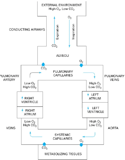
Normal gas exchange (Figure 6) involves the absorption of an adequate quantity of O2 and an externalization of a suitable amount of CO2. For an average individual, this translates into a ventilation rhythm of 12-15 breaths per minute with a ventilation volume of 500 ml of air.[8] Typical pulmonary blood flow is
equivalent to cardiac output which, in an average individual is around 5 L/min. Contrary to the high hydrostatic pressure generated in high resistance systemic arteries (85 mmHg), the thin and elastic pulmonary arteries generate pressures of 15 mmHg. Due to the change in total-vessel lumen area between the higher lungs and the respiratory section, the transit time of blood in the alveolar-adjacent capillaries is slow and averages 0.75 sec. This transition time allows time for the gaseous exchange where the blood O2 pressure increases from 40 to 100 mmHg and CO2 pressure decreases from 45 to 40mmHg [7].
Figure 6: Schematic representation of gas exchange between the
tissues of the body and the environment. With permission from Levitsky [7].
1.3.4. Cells
The trachea and main bronchi epithelium is a pseudostratified epithelium made up of many types of cells as described below (Figure 7).
Figure 7: Comparison of the lung epithelium at different sites
within the lungs. With permission from Patton [10].
Ciliated cells are columnar epithelial cells, which is a cell type characterised by its height being greater than its width [11]. The apical region of these cells is covered with cilia, whose role is the displacement of mucus. Goblet cells, like ciliated ones, are also part of the columnar epithelial cell type. Their main purpose is the production and secretion of mucins, which are major component of pulmonary mucus. These cells’ cytoplasm is in great part occupied by mucin-containing vesicles [11]. Mucins are released through both merocrine and apocrine secretion. In addition to the baseline secretions of these cells, production may be increased in the presence of external factors such as particulates or microorganisms [10].
Basal cells, part of the basal lamina, basal cells are yet undifferentiated cells. Though little is known about the mechanisms of differentiation of these cells, their role is to replenish the surface layers of ciliated and mucous cells of the pulmonary epithelium [8].
Mast cells are cells of which the main function is the release of cytokines and chemokines which play a critical role in allergic responses. These chemical mediators include, but are not limited to histamine, serotonin, heparin and eosinophil chemotactic factor [8].
Clara cells located mostly in bronchioles, their function is a secretory one. In addition, they contain cytochrome P450 isoenzymes which play a role in drug metabolism.
Pneumocytes are cells make up the lining of the alveoli, thus participating in gas exchange control. Type I cells cover up over 95% of the alveolar surface of the lungs. They have a 25 nm thickness, which is critical in gaseous exchanges. Their role is to provide a barrier as thin as possible while still remaining permeable to O2 and CO2. Type II cells cover the remaining 5% of alveolar surface. However, conversely to surface, in terms of cell number, type-II cells represent 60% of the cells lining the alveoli. Their main purpose is to produce and secrete surfactant phospholipids [8].
Finally, alveolar machophages are relatively large cells which possess the capacity for engulfing local or foreign through endocytosis (phagocytosis for solid matter or pinocytosis for liquids). Freely moving between vessels and tissues through extravasation, they also participate actively in cellular communication through the use of cytokines, most notably the pro-inflammatory ones: tumor necrosis factor alpha, interleukin 1, 6, 8 and 12. They also play a role in pulmonary drug clearance through their endocytotic capabilities [9].
1.3.5. Airways vasculature
Certainly, the main focus of airway vasculature is to reach the tracheobronchial respiratory region and allow gaseous exchanges between the blood and the luminal side of the lungs. However, that is not its only function. Indeed, two circulations are thus distinguished: firstly, the pulmonary circulation whose aim is the aforementioned exchange and, secondly, the bronchial circulation which
nourishes the tracheobronchial tree. The interactions and anastomoses between the two can be seen in Figure 8.
Figure 8: Illustration of the main anatomic features of the
bronchial circulation. With permission from Levitsky [7].
In normal subjects, most of the venous return will circulate into the pulmonary circulation. Venous blood will pass through the pulmonary artery, branching out through the pulmonary parenchyma and returning to the main systemic circulation by passing through the left ventricle and atrium, with the scope of oxygenation. Many exceptions occur however as some subjects exhibit intracardiac and/or intrapulmonary shunting and some are afflicted with congenital anomalies. The bronchial circulation’s aim is to supply the lung parenchyma with nutrients through an extensive capillary bed circulating through the bronchial walls. All intrapulmonary structures related to the bronchial
circulation drain into the pulmonary veins. As such, these two circulations have extensive anastomoses that mix together oxygen-rich and less-rich blood [12]. However, the perihilar region (region around main bronchi and pulmonary arteries and veins) drains to the azygos system [13]. The bronchial circulation accounts to roughly one percent of the cardiac output.
Both these circulations are, to varying degrees, made up of the standard vascular tunics which can be seen in Figure 9.
Figure 9: Structure of an artery, arteriole, capillary network,
1.3.6. Vasculature composition
The adventitia is commonly known to be the collagen-rich elastic sheath surrounding blood vessels. It is considered a scaffold through which intermesh fibroblasts, perivascular nerves, nourishing vessels and a wide array of cellular populations such as, but not limited to macrophages, T-cells, B-Cells and mast cells while aiding in the regulation of lumen size through smooth muscle tone (Figure 10). It has also been shown to participate in signaling related to remodelling purposes [14].
Figure 10: The arterial adventitia. Tissue represents the
conjunctive tissue scaffold. Where Lu: vascular lumen; In: intima; Me: media; Ad: adventia; Tissue: conjunctive tissue scaffold. With permission from Majesky [14].
Whether in the pulmonary or systemic circulation, the tunica media is seen as the muscular layer of blood vessels. It is made up almost entirely of smooth muscle cells, positioned concentrically in lamellae around the intimal layer. It is
within this layer that contraction occurs through the action of several receptors and mediators (Figure 11).
Figure 11: Regulation of smooth muscle contraction. Where G:
G-protein; α, β and γ; G-protein subunits; DG: diacylglycerol; IP3: inositol 1,4,5-trisphosphate; Ca2+: calcium; PKC: phosphokinase C; MLC: myosin light chain. Following stimulation, either by an influx of calcium or the presence of an agonist which acts through phosphatidylinositol 4,5-bisphosphate, there is a release of calcium from the sarcoplasmic reticulum which phosphorylates myosin light chain kinase which initiates the contraction of the smooth muscle cell. Adapted with permission from Webb [15].
The intima is the part of the vessel that is actually in contact with the blood [5]. It is composed of squamous epithelium called endothelium from which many different vasoconstrictors and vasodilators which mediate the modulation of
vascular resistance in the pulmonary system are derived [14]. Impairment in the hemodynamic regulation of the airway vasculature can lead to a multitude of pathologies, including pulmonary hypertension.
As the tracheobronchial tree’s anatomy evolves with each passing branch generation, so does the vascularisation around it. Indeed, the composition and thickness of the different sections making up the pulmonary arteries changes with tracheobronchial depth. Furthermore, arterial matches tracheobronchial caliber as both jointly decrease in diameter. The pulmonary circulation is made up, initially, of elastic arteries, transitioning into muscular ones and ending with the capillary network. The walls of the larger arteries (≥ 1000 µm in diameter) are made up mostly of elastic lamellae, few smooth muscle cell (SMC) and collagen fibrils [14]. As the caliber of arteries decreases, the elasticity decreases and the percentage of smooth muscle mass increases, up until arteries with a 40 to 50 µm diameter.
Indeed, larger arteries with a diameter greater than 3200 µm are considered elastic, between 3200-2000 µm, they are transitional between elastic and muscular and, when having an internal diameter between 1000-50 µm, they are muscular [12]. Subsequently, SMC in arteries progressively diminishes in quantity and, when approaching the capillary bed structure, a single smooth muscle cell may remain in the artery wall [12].
1.3.7. Neural and humoral control of pulmonary vasculature
Pulmonary vessels are innervated by both sympathetic and parasympathetic fibers of the autonomic nervous system. However, when compared to systemic vasculature, the innervation is sparser. Larger elastic vessels tend to show more innervation than small muscular ones and no innervation is witnessed for vessels lower than 30 µm in diameter. The stimulation of the sympathetic nerves of the airways causes vasoconstriction by the release of certain neurotransmitters, including norepinephrine, neuropeptide Y, and substance P. The catecholamines epinephrine and norepinephrine both increase pulmonary
vascular resistance when injected into the pulmonary circulation. Histamine, found in the lung in mast cells, is a pulmonary vasoconstrictor. Certain prostaglandins and related substances, such as F2α, E2 and thromboxane, are also pulmonary vasoconstrictors, as is endothelin (ET-1), a 21 amino acid peptide synthesized by the vascular endothelium. In addition, alveolar hypoxia and hypercapnia also cause pulmonary vasoconstriction. At the opposite, parasympathetic stimulation results in acetylcholine and vasoactive intestinal peptide release, leading to vasodilatation and increased mucosal blood flow. The β-adrenergic agonist isoproterenol, nitric oxide, and certain prostaglandins (PG), such as PGE1 and PGI2 (prostacyclin), are additional pulmonary vasodilators. In the following section, we will explore how CPB and the reperfusion injury can alter the normal pulmonary vasculature physiology.
CHAPTER 2. Cardiopulmonary Bypass and
Pulmonary Hypertension
Cardiopulmonary bypass in action at the Montreal Heart Institute
Cardiopulmonary bypass (CPB), the heart lung machine has been viewed as one of the foremost medical advances of the 20th century [16]. Indeed, thanks to this procedure, cardiac surgical corrections are now common. Roughly 2000 surgeries involving CPB are performed worldwide per hour [16]. In Canada, specifically, there are 36 000 heart surgeries performed yearly, of which 8000 are performed in Quebec [4]. This medical advance having permitted scalpel entry within the cardiac walls is, however, not without risks. The CPB procedure has been shown to have a profound effect on pulmonary vasculature.
2.1. Methodology, functioning and pathophysiological
consequences
CPB management has, through the ages, evolved into a highly complex labyrinth of tubing as can be seen in Figure 12.
Figure 12: Components of cardiopulmonary bypass. Taken from
Aslati [17].
In rough terms, this method implies the transport of blood from the vena cavae and the right atrium, into a venous reservoir through an oxygenation chamber where gaseous exchange is allowed via a membrane interface. Inside this membrane interface chamber is also added the anesthesia mixture. Subsequently, blood is warmed or cooled at will and then redirected to the aorta.
There are several types of consequences associated with this procedure.
Physiological. Following the extensive interaction with foreign materials and substances, CPB exerts an important toll on the outcome of surgery. Indeed, CPB duration was shown to be a significant factor in predicting the clinical outcome of cardiac [18].
Structural. CPB is associated with an increase in pulmonary vascular permeability, resulting in enhanced extravascular water and edema, which causes endothelial and epithelial damages [19].Other changes related to CPB are: thickening of the alveolar septa, vascular congestion, mitochondrial swelling of the alveolar epithelial cells, vacuolation of endothelial cells, blood vessel congestion, and diffuse alveolar damage [20].
Serological. The diversion of blood results in the activation of an inflammatory response through the activation of neutrophils and the release of a multitude of mediators. The activation of neutrophils leads to free radical production, degradation of the interstitium by the proteinases contained in their granules and increased levels of circulating catecholamines [21]. As will be covered in a later section, some mediators have been claimed to play an important role in pulmonary hypertension (PH) following CPB, including ET-1, lysosomal enzymes, interleukin-6 and anaphylatoxins [20, 21]. Most of the effects related to the physiological damages caused by the CPB procedure can be traced back to the blood/biomaterial interactions. Protein adsorption onto the inner surface of materials joint with platelets reaction, intrinsic coagulation activation, fibrinolytic activity and complement activation are at the core of the damaging CPB effect on vasculature [22]. Everything starts with an initial adsorption of a protein layer on the surface of the biomaterials, particularly fibrinogen, albumin and gamma-globulin. This layer of proteins induces platelet adhesion and aggregation which, as erythrocytes and leukocytes are gradually trapped in fibrinogen, is followed
by the formation of a thrombus. This same protein adsorption also leads to activation of the intrinsic coagulation pathway involving the participation of the contact proteins factor XII, factor XI, HMWK (definer) and prekallikrein [22]. In addition an important part of the blood/biomaterial interactions includes the activation of the complement system, notably anaphylatoxins C3a, C4a and C5a which mediate the chemotactic, adhesive and phagocytic responses of leucocytes in the inflammatory process.
These physiological changes and activations within the blood result in a possibly disastrous effect on the lungs called the reperfusion syndrome (Figure 13) where the pulmonary pressures surge, leading to pulmonary hypertension, an increase in cardiac post-charge and, potentially, to right-heart failure [23, 24].
Figure 13: Systemic and pulmonary artery pressure during
cardiopulmonary bypass (CPB). At the end of CPB shortly following reperfusion significant increase in pulmonary artery pressure is observed in relation to the systemic pressure. With permission from Denault et al [4].
2.2. Pulmonary hypertension
Depending on the basis of mechanisms, the World Health Organization classifies pulmonary hypertension into five specific groups, as can be seen in Table I.
Table I: Current clinical classification of pulmonary hypertension.
1. Pulmonary arterial hypertension (PH) 1.1. Idiopathic
1.2. Familial
1.3. Associated with
1.3.1. Collagen vascular disease
1.3.2. Congenital systemic-to-pulmonary shunts 1.3.3. Portal hypertension
1.3.4. Human immunodeficiency virus infection 1.3.5. Drugs and toxins
1.3.6. Other (thyroid disorders, glycogen storage disease, Gaucher disease, hereditary hemorrhagic telangiectasia, hemoglobinopathies, chronic myeloproliferative disorders, splenectomy)
1.4. Associated with substantial venous or capillary involvement 1.4.1. Pulmonary veno-occlusive disease
1.4.2. Pulmonary capillary hemangiomatosis
1.5. Persistent pulmonary hypertension of the newborn 2. Pulmonary hypertension with left-sided heart disease
2.1. Left-sided atrial or ventricular heart disease 2.2. Left-sided valvular heart disease
3. Pulmonary hypertension associated with lung diseases and/or hypoxemia 3.1. Chronic obstructive pulmonary disease
3.2. Interstitial lung disease 3.3. Sleep-disordered breathing 3.4. Alveolar hypoventilation disorders 3.5. Long-term exposure to high altitude 3.6. Developmental abnormalities
4. Pulmonary hypertension due to chronic thrombotic and/or embolic disease 4.1. Thromboembolic obstruction of proximal pulmonary arteries 4.2. Thromboembolic obstruction of distal pulmonary arteries
4.3. Nonthrombotic pulmonary embolism (tumor, parasites, foreign material) 5. Miscellaneous
Sarcoidosis, histiocytosis X, lymphangiomatosis, compression of pulmonary vessels (adenopathy, tumor, fibrosing mediastinitis)
Pulmonary arterial hypertension (PH) is defined as a mean pulmonary artery pressure superior to 25 mmHg at rest, or to 30 mmHg during exercise [26]. This type of pulmonary hypertension also comprises idiopathic PH, which is a disease of unknown cause, and can just as much lead to right ventricular failure because of the elevation in pulmonary artery pressure as the other groups [27]. The other conditions making up Group I of the World Health Organization table have very similar histological appearances.
2.2.1. Vascular pathways involved in pulmonary hypertension
The histology of pulmonary arteries in patients with PH shows a higher vessel wall thickness, and an unusual layering characterized by thickening of the intimal layer and wall architecture irregularities such as intimal fibrosis [28]. Indeed, though stemming from several underlying causes, PH comprises a group of clinical and pathophysiological entities with comparable features. The chief vascular alterations in PH result in hemodynamic alterations from thrombosis, increase of endothelial and smooth-muscle cells and vasoconstriction. Regardless of the underlying cause of HP, there are many cellular mechanisms (Figure 14) acting together in the maintenance of the disease.
Figure 14: The three mechanistic pathways known to be disturbed
in patients with pulmonary arterial hypertension. The points at which drug treatment affects these mechanistic processes are shown in gray circles.
AA: arachidonic acid; CCB: calcium channel blocker; ETRA: endothelin receptor antagonist (eg, bosentan [dual], ambrisentan, and sitaxsentan [receptor A selective]); PDE5i: phosphodiesterase 5 inhibitor (eg, sildenafil). Left, The nitric oxide (NO) pathway. Nitric oxide is created in endothelial cells by type III (ie, endothelial) NO synthase (eNOS), which in pulmonary arterial smooth muscle cells (PASMCs) induces guanylate cyclase (GC) to convert guanylate triphosphate (GTP) to cyclic guanylate monophosphate (cGMP). Cyclic GMP is a second messenger that constitutively maintains PASMC relaxation and inhibition of PASMC proliferation by ultimately reducing inward flux of calcium
ions (Ca++). Cyclic GMP is removed by the PDE5 enzyme to yield the inactive product 5′GMP. Patients with PH have reduced expression and activity of eNOS. Middle, The prostacyclin pathway. The production of prostaglandin I2 (PGI2 [ie, prostacyclin]) is catalyzed by prostacyclin synthase (PS) in endothelial cells. In PASMCs, PGI2 stimulates adenylate cyclase (AC), thus increasing production of cyclic adenosine monophosphate (cAMP) from adenosine triphosphate (ATP). Cyclic AMP is a second messenger that constitutively maintains PASMC relaxation and inhibition of PASMC proliferation. Patients with PH have reduced expression and activity of PS. Right, The endothelin (ET) pathway. Big- (ie, pro-) ET is converted in endothelial cells to ET1 (a 21–amino acid peptide) by endothelin-converting enzyme (ECE). ET1 binds to PASMC ETA and ETB receptors, ultimately leading to PASMC contraction, proliferation, and hypertrophy. Endothelin 1 also binds to endothelial cell ETB receptors (not illustrated). Patients with PH have increased expression and activity of ECE. With permission from McGoon [25].
Nitric Oxide (NO) is a highly potent vasodilator and inhibitor of platelet activation and smooth-cell proliferation. The impairment of the NO pathway, contributing to pulmonary vasodilatation, can also be a cause of PH. This mediator activates a signaling cascade in the endothelial cell that results in smooth muscle cell relaxation [29]. It has also been shown that inhalation of NO by PH patients produces selective pulmonary vasodilatation, and thus enables a diminution of pulmonary vascular resistance. No change in systemic vascular resistance is observed because NO is rapidly sequestrated and inactivated by hemoglobin [30].
Prostacyclin (PGI2) and Thromboxane A2 (TxA2) are derivatives of arachidonic acid produced by endothelial cell. PGI2 shows a three-way activity as vasodilator, as platelet activator and proliferation inhibitor [31, 32]. On the contrary, TxA2 is a platelet aggregator and a vasoconstrictor. In PH, it was found that the balance between these two vascular effectors was shifted towards TxA2 [32] as shown by metabolite dosing in urine; thus indicating that this imbalance is part of the underlying causes of PH [32]. As will be discussed later, shifting the balance towards prostacyclin is one of the current treatment options being developed. Indeed, when administered by inhalation, prostacyclin was shown to be efficient in decreasing pulmonary artery pressure by inducing vasodilatation in patients with PH [33].
Endothelin-1 is a potent arterial and venous vasoconstrictor involved in the development of PH as can be seen in Figure 15. It can affect vascular resistance by binding to ETA receptors located on vascular smooth muscle cells and inducing vasoconstriction. ETB receptors, expressed on vascular endothelial and smooth muscle cells, reduce pulmonary ET-1 clearance through downregulation of ET-1 cleaving enzyme, also increasing vasoconstriction. However, in contrast, ETB receptor stimulation increases the release of NO and prostacyclin [34-36]. In addition to its vasoconstrictor effects, ET-1 possesses a stimulating effect on vascular smooth muscle cells, leading to architectural changes in the vascular walls and increased muscularisation.

![Figure 2: Heart valves in superior view. With permission from Scanlon [5].](https://thumb-eu.123doks.com/thumbv2/123doknet/11644340.307584/32.918.313.706.123.495/figure-heart-valves-superior-view-permission-scanlon.webp)
![Figure 3: Autonomic innervation of the heart. With permission from Marieb [6]](https://thumb-eu.123doks.com/thumbv2/123doknet/11644340.307584/34.918.338.630.125.621/figure-autonomic-innervation-heart-permission-marieb.webp)