HAL Id: hal-02661775
https://hal.inrae.fr/hal-02661775
Submitted on 30 May 2020
HAL is a multi-disciplinary open access
archive for the deposit and dissemination of
sci-entific research documents, whether they are
pub-lished or not. The documents may come from
teaching and research institutions in France or
abroad, or from public or private research centers.
L’archive ouverte pluridisciplinaire HAL, est
destinée au dépôt et à la diffusion de documents
scientifiques de niveau recherche, publiés ou non,
émanant des établissements d’enseignement et de
recherche français ou étrangers, des laboratoires
publics ou privés.
Proprotein convertase subtilisin kexin type 9 null mice
are protected from postprandial triglyceridemia
Cedric Le May, Sanae Kourimate, Cédric Langhi, Maud Chétiveaux, Anne
Jarry, Christine Coméra, Xavier Collet, Folkert Kuipers, Michel Krempf,
Bertrand Cariou, et al.
To cite this version:
Cedric Le May, Sanae Kourimate, Cédric Langhi, Maud Chétiveaux, Anne Jarry, et al.. Proprotein
convertase subtilisin kexin type 9 null mice are protected from postprandial triglyceridemia.
Arte-riosclerosis, Thrombosis, and Vascular Biology, American Heart Association, 2009, 29 (5), pp.684-690.
�10.1161/ATVBAHA.108.181586�. �hal-02661775�
Version preprint
Comment citer ce document :
Are Protected From Postprandial Triglyceridemia
Ce´dric Le May, Sanae Kourimate, Ce´dric Langhi, Maud Che´tiveaux, Anne Jarry, Christine Comera,
Xavier Collet, Folkert Kuipers, Michel Krempf, Bertrand Cariou, Philippe Costet
Objectives—Proprotein convertase subtilisin kexin type 9 (PCSK9) is a natural inhibitor of the low-density lipoprotein
receptor, and its deficiency in humans results in low plasma LDL-cholesterol and protection against cardiovascular disease. We explored whether PCSK9 expression impacts postprandial triglyceridemia, another important cardiovas-cular risk factor.
Methods and Results—Real-time PCR and confocal microscopy were used to show that PCSK9 is expressed throughout
the entire small intestine and in human enterocytes. On olive oil gavage, PCSK9-deficient mice showed a dramatically decreased postprandial triglyceridemia compared with their wild-type littermates. Lymph analysis revealed that intestinal TG output is not quantitatively modified by PCSK9 deletion. However, PCSK9⫺/⫺ mice present with a significant reduction of lymphatic apoB secretion compared to PCSK9⫹/⫹ mice. Modulating PCSK9 expression in polarized CaCo-2 cells confirmed the relationship between PCSK9 and apoB secretion; PCSK9⫺/⫺mice consistently secrete larger TG-rich lipoprotein than wild-type littermates. Finally, kinetic studies showed that PCSK9-deficient mice have an increased ability to clear chylomicrons compared to wild-type littermates.
Conclusion—These findings indicate that in addition to its effect on LDL-cholesterol, PCSK9 deficiency might protect against
cardiovascular disease by reducing postprandial triglyceridemia. (Arterioscler Thromb Vasc Biol. 2009;29:684-690.)
Key Words: PCSK9 䡲 chylomicron 䡲 postprandial 䡲 intestine 䡲 CaCo-2
G
ain-of-function mutations affecting proprotein conver-tase subtilisin/kexin type 9 (PCSK9) are associated with autosomal dominant hypercholesterolemia and premature atherosclerosis.1It is now established that PCSK9 is a naturalinhibitor of the low-density lipoprotein receptor (LDLr), acting posttranscriptionally.2Circulating PCSK9 binds to the
EGF-A extracellular domain of the hepatic LDLr and pre-vents its recycling to the cell surface.3
A breakthrough study reported that blacks harboring PCSK9 loss-of-function mutations had an 88% risk reduction for coronary heart disease.4 Although the lower
concentra-tions of LDL cholesterol over one’s lifetime is suggested to be the main reason for this very high level of protection, we hypothesized that PCSK9 deficiency might also affect other risk factors. Low levels of HDL cholesterol (HDL-c) and elevated nonfasted triglycerides (TG) levels are associated with increased risk for cardiovascular disease. No association between PCSK9 loss-of-function mutations and plasma HDL-c levels has been noticed.4,5 However, to our
knowl-edge, nonfasting plasma TG were not measured in PCSK9-deficient individuals. They are an important contributor to the risk for cardiovascular disease.6,7
Rashid et al reported the phenotype of PCSK9-deficient mice (PCSK9⫺/⫺) mice and observed that they exhibit lower plasma cholesterol levels (⫺50%) and are hypersensitive to statins.8The potential role of PCSK9 in the intestine in mice
and humans remains unexplored.
During the postprandial period, dietary lipids are ac-tively absorbed into the enterocyte. After intracellular reesterification, long chain fatty acids and cholesterol esters are associated with phospholipids and apoteins, particularly apoB48, to produce TG-rich lipopro-teins, mainly chylomicrons.9
Chylomicrons are secreted into the mesenteric lymph to eventually deliver lipids to the peripheral tissues and organs.9
It is believed that the number of chylomicrons does not change during the postprandial phase, but their size increases because of their TG enrichment.10Because a single copy of
apoB48 is present per chylomicron,11the intestinal output of
apoB remains constant during the absorptive process. ApoB48 is the preferred protein for the intestine to coat chylomicrons in mice.12 Chylomicron remnants are cleared
from the blood by the LDLr and the LDLr-related protein
Received November 25, 2008; revision accepted February 18, 2009.
From the INSERM U915 (C.L.M., S.K., C.L., M.C., M.K., B.C., P.C.), CHU de Nantes, France; Universite´ de Nantes, EA Biometadys (A.J.), Nantes, France; INSERM U563 (C.C., X.C.), Toulouse, France; the Center for Liver, Digestive, and Metabolic Diseases (F.K.), University of Groningen, The Netherlands; Universite´ de Nantes, l’institut du thorax (M.K., B.C.), Clinique d’Endocrinologie et Nutrition, Nantes, France; the Centre de Recherche en Nutrition Humaine de Nantes (M.K., P.C.), Nantes, France; and INRA UR66 (C.C.), France.
Correspondence to Philippe Costet, INSERM U915, CHU Hoˆtel Dieu, 3ENORD, 9, Quai Moncousu, 44000, Nantes, France. E-mail philippe.costet@univ-nantes.fr
© 2009 American Heart Association, Inc.
Arterioscler Thromb Vasc Biol is available at http://atvb.ahajournals.org DOI: 10.1161/ATVBAHA.108.181586
684
Version preprint
Comment citer ce document :
(LRP).13PCSK9-deficient mice display an increase in hepatic
LDLr protein, but not in LRP.8
Our aim was to determine PCSK9 localization in murine and human intestines and to determine its role in postprandial lipemia. The main outcome is that PCSK9 deficiency results in dramatically less plasma TG accumulation in 14 hour– fasted mice, challenged with an olive oil gavage, because of increased hepatic clearance of larger chylomicrons.
Methods
An expanded Methods section can be found in the supplemental materials (available online at http://atvb.ahajournals.org).
Human Tissues
The human tissue fragments were processed according to the French
Guidelines for Research on Human Tissues.14 Informed patient
consent was obtained, according to the French bioethics law.15 A
detailed protocol and information concerning patients are available in the supplemental materials.
Animals
PCSK9⫹/⫺ mice were purchased from Jackson Laboratories (Bar
Harbor, Me), interbred to produce PCSK9⫺/⫺ and PCSK9⫹/⫹
(wild-type [wt]) littermates, and genotyped as described elsewhere.8
Mice had free access to food and water under a 12-hour light/12-hour dark cycle in a temperature-controlled environment. All animal studies were conducted with age-matched male mice (2-month-old), and approved by the Unite´ de The´rapeutique Expe´rimentale (Agree-ment No. BP44015). Before surgery, mice were anesthetized with a
single intraperitoneal injection of 100 L of ketamin/ivermectine
(Imalge`ne 1000, Merial).
Statistics
Each experiment was representative of at least 2 independent experiments with a minimum of triplicates per condition. Values are
reported as means⫾SEM. Statistical significance was analyzed using
a Student unpaired t test. Values of P⬍0.05 were considered
as significant.
Results
PCSK9 Is Expressed in the Small Intestine of Mice and Humans
Using real-time quantitative PCR, we first examined PCSK9 and the LDLr mRNA quantities along the gastrointestinal tract in mice (Figure 1A). PCSK9 expression in the stomach was virtually undetectable. However, PCSK9 was expressed throughout the small intestine and colon, at a level which did not vary significantly along the intestinal cephalo-caudal axis, and which was similar to that in the liver (Figure 1A). As previously published,17the LDLr was abundantly expressed
along the digestive tract.
Using confocal microscopy, we determined for the first time the localization of PCSK9 protein in frozen sections of normal, human, small intestines. PCSK9 was expressed almost exclusively in the epithelial barrier of the human duodenum and ileum, both in enterocytes and goblet cells (Figure 1B). PCSK9 immunostaining was observed in the cytoplasm of epithelial cells and accumulated at the apical and basolateral sides of the cells (Figure 1B). Blocking of the PCSK9 antibody with an excess of antigen peptide abolished the staining (Figure 1B, upper panel, right), confirming the specificity of the antibody. We also examined the localization of PCSK9 in the human colonic, enterocyte-like cell line,
CaCo-2, maintained as polarized by culturing on filters. As shown in Figure 1B (lower panel), the xy image (left) shows a punctuated staining within CaCo-2 cells, and the xz image (right) shows a strong PCSK9 immunostaining mainly in the apical compartment of the cells.
PCSK9 Is Upregulated by Pravastatin in CaCo-2 Cells and the LDLr Expression Is Increased in Intestines of PCSK9ⴚ/ⴚ Mice
We next examined whether PCSK9 is upregulated by statins in CaCo-2 cells as it has been reported in hepatocytes18,19
(supplemental Figure IA). We exposed the cells for 48 hours to 10mol/L pravastatin and observed a 160% increase in PCSK9 mRNA, and a 110% increase in LDLr mRNA (supplemental Figure IA). We next confirmed that the LDLr content is increased in the liver of PCSK9⫺/⫺mice compared to wt littermates, and observed a similar phenotype in the medial intestine (⫹621%, P⬍0.05; supplemental Figure IB).
PCSK9 Deficiency Protects Mice Against Postprandial Lipemia
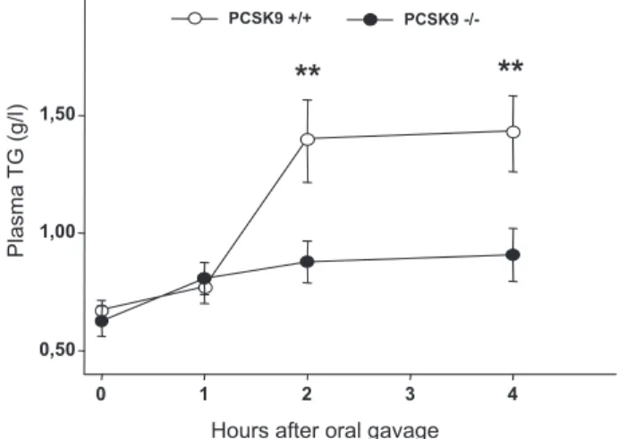
Random-fed PCSK9⫺/⫺mice had lower total cholesterol than wt littermates (⫺35% in 3-month-old mice, P⬍0.001), and similar plasma TG concentrations (supplemental Figure IIA). Plasma TG levels were measured over a 4-hour period in 14 hour–fasted mice after they received an intragastric bolus of olive oil (200L; Figure 2). At T0, plasma levels of TG were comparable between PCSK9⫹/⫹ and PCSK9⫺/⫺ mice. Two hours after gavage, PCSK9⫹/⫹mice showed a 209% increase in plasma TG, whereas PCSK9⫺/⫺ littermates displayed a
A
B
Caco2 Duodenum Ileum * LP LP*
P C SK9 / cycl ophilin LDLr/ cycl o philinLiver Stomach Proximal Medial Distal Colon
0 0,4 0,8 1,2 1,6 0 1 2 Small Intestine xy xz
Figure 1. PCSK9 is highly expressed in the small intestine and
enterocytes. A, Real-time PCR measurement of PCSK9, LDLr
gene expression in PCSK9⫹/⫹mice. B, Immunofluorescence
staining in human duodenum and ileum, and CaCo-2. See also supplement Methods.
Le May et al PCSK9 Function in Postprandial Lipemia 685
Version preprint
Comment citer ce document :
strongly attenuated TG postprandial response. Mean TG appearance rates were, respectively, 0.19⫾0.032 and 0.082⫾0.022 g/L/h for PCSK9⫹/⫹ and PCSK9⫺/⫺ mice, ie, 62% less (n⫽12 PCSK9⫹/⫹and n⫽14 PCSK9⫺/⫺mice from to 2 independent experiments).
Fractionation by fast protein liquid chomatography (FPLC) showed that the difference in triglyceridemia observed 2 hours after gavage was attributable to a decrease in the TG content of chylomicrons/VLDL lipoproteins (supplemental Figure IIB and supplemental Methods and Results).
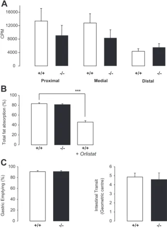
Gastric Emptying, Intestinal Transit, and Fat Absorption After Oral Oil Challenge
The defect in postprandial lipemia observed in PCSK9⫺/⫺ mice could be attributable to (1) decreased intestinal lipid absorption, or chylomicron production, or (2) accelerated lipolysis or clearance of TG-rich lipoproteins. We first measured the radioactivity content of intestinal segments of PCSK9⫹/⫹and PCSK9⫺/⫺littermates 2 hours after a gavage with olive oil containing3H-triolein (Figure 3A). Triolein is a triglyceride and unsaturated fat formed from oleic acid, the main constituent of olive oil. This method reflects (1) the enterocytic uptake of 3H-oleate, (2) the intracellular non
esterified or reesterified3H-oleate, (3) the reuptake of
radio-labeled nascent particles. PCSK9⫺/⫺mice had a tendency to accumulate less3H in the proximal and medial part of their
intestine compared to wt mice, suggesting that lipid uptake could be defective in these segments. To determine intestinal fat absorption, we measured food intake and collected feces every 24 hours for 3 consecutive days. Food intake, weight of excreted feces, and fecal fat excretion were not significantly different between mice (supplemental Table II). PCSK9⫹/⫹ and PCSK9⫺/⫺ mice absorb fat with the same efficiency (Figure 3B) demonstrating that reduced lipemia in PCSK9⫺/⫺ mice is not attributable to fat malabsorption. As a control, PCSK9⫹/⫹mice fed with orlistat, an inhibitor of pancre-atic lipases, showed a 45% decrease in total fat absorption compared to PCSK9⫹/⫹mice under normal chow (Figure 3B).
Altered postprandial lipemia could also be attributable to gastric emptying or intestinal motility defect. We performed an oral gavage with a nonabsorbable 70-KDa FITC conjugated dextran marker diluted in olive oil. After 30 minutes, about 10% of the marker was still present in the stomach of PCSK9⫹/⫹ and PCSK9⫺/⫺ mice (Figure 3C), demonstrating that PCSK9 deficiency does not alter gastric emptying. Intestinal transit was also similar in both genotypes (Figure 3C).
Thus, although there was a trend toward less accumulation of 3H-triolein in the proximal and medial intestine of
PCSK9⫺/⫺mice after an olive oil gavage, they do not show a defect in fat absorption, gastric emptying or intestinal transit.
Chylomicron Secretion in PCSK9ⴚ/ⴚMice
To directly explore the impact of PCSK9 deficiency on chylomicron output, we cannulated the lymph ducts from 2 hour–fasted PCSK9⫹/⫹ and PCSK9⫺/⫺ littermates. After establishing a 30-minute baseline collection, we injected a bolus of olive oil in their duodenums, and collected the lymph at timed intervals for 210 minutes (Figure 4A). Lymph flow was reduced by 20% in knockout mice (0.59L/min versus 0.47 L/min, P⬍0.05). No difference in cumulative TG contents was observed in the first 1.5 hour. In a second phase, the accumulation tended to be reduced in PCSK9⫺/⫺mice, although it did not reach significance (Figure 4A).
Lymph samples collected at various times after olive oil injection were pooled, and equal amounts were loaded on a gel for separation of the proteins by electrophoresis (Figure 4B). As expected, the quantity of apoB was relatively constant during the absorption process within each genotype, and apoB48 was more abundant than apoB100.10However,
apoB100 and apoB48 contents were significantly lower in lymph from PCSK9⫺/⫺ mice (⫺39%, P⬍0.05 and ⫺40%,
P⬍0.01), suggesting that PCSK9 deficiency results in less
chylomicrons being secreted. The positive relation between PCSK9 expression and the apoB output was confirmed in CaCo-2 cells (supplemental Figure III and supplemental Methods and Results). We also determined the content of the other apolipoprotein by electrophoresis. No striking differ-ence between genotypes was observed. There was a tendency toward an accumulation of apoE in lymph from PCSK9⫺/⫺ mice (Figure 4C).
As lymphatic TG output was unchanged and lymphatic apoB secretion was decreased, it suggests that PCSK9⫺/⫺ mice might secrete larger chylomicrons than wt littermates. Using dynamic light scattering techniques, we showed that chylomicron diameters from PCSK9⫺/⫺mice were increased by 10% compared to wt littermates (Figure 4D). Assuming that they are spherical particles, PCSK9⫺/⫺ mice secrete chylomicrons with a volume at least 25% larger than PCSK9⫹/⫹mice (Figure 4D).
Chylomicron Catabolism in PCSK9ⴙ/ⴙand PCSK9ⴚ/ⴚMice
We next measured the hepatic TG content after olive oil gavage. There was no change in hepatic TG contents in fed PCSK9⫹/⫹ and PCSK9⫺/⫺ mice (Figure 5A). Fasting
in-Hours after oral gavage
0 1 2 3 4 Plasma TG (g/l) 0,50 1,00 1,50 PCSK9 -/-PCSK9 +/+
**
**
Figure 2. Postprandial lipemia is reduced in PCSK9⫺/⫺mice. 14
hour–fasted PCSK9⫹/⫹(white circles, n⫽12 mice) and
PCSK9⫺/⫺littermates (black circles, n⫽14 mice) received an
intragastric olive oil load. Blood samples were collected before gavage (time 0) and at 1, 2, 3, and 4 hours after gavage and
plasma triglycerides concentrations determined. **P⬍0.01.
Version preprint
Comment citer ce document :
duced an accumulation of TG by 100% in both genotypes (P⬍0.01). Two hours after olive oil gavage, hepatic TG contents were similar to those of PCSK9⫹/⫹mice in the fed state, but remained elevated in PCSK9⫺/⫺ mice. Conse-quently, there was 54% more TG in livers of knockout mice after gavage (P⬍0.05), suggesting that TG originated from olive oil fatty acids. To confirm this,3H-triolein diluted in
olive oil was orally administered to fasted mice, and 2 hours later the amount of hepatic radioactivity was measured. PCSK9⫺/⫺ mice exhibited almost 200% more radioactivity than wt littermates showing that the hepatic uptake of chylomicrons was increased in these mice (supplemental Figure IV).
We verified whether the clearance of chylomicrons is enhanced in knockout mice (Figure 5B). Chylomicrons ob-tained by mesenteric lymph duct cannulation of wt and knockout mice (chylo⫹/⫹ and chylo⫺/⫺) were labeled with
125
I and injected into the circulatory systems of mice from both genotypes. To mimic the postprandial status, we injected chylo⫹/⫹ in PCSK9⫹/⫹ mice and chylo⫺/⫺ in PCSK9⫺/⫺ mice. Consistent with the phenotype observed in Figure 2, PCSK9⫺/⫺ mice cleared chylo⫺/⫺ 37% faster than PCSK9⫹/⫹ cleared chylo⫹/⫹ (P⬍0.05). We also performed the experiment crossing the donors and receivers. PCSK9⫺/⫺ mice cleared chylo⫺/⫺21% faster than PCSK9⫹/⫹mice did
(P⬍0.01). These results reflect probably the higher hepatic LDLr content observed in PCSK9⫺/⫺ mice (supplemental Figure IB). Surprisingly, we observed (1) no difference in chylo⫹/⫹ clearance rates between the genotypes; and (2) PCSK9⫺/⫺mice cleared chylo⫺/⫺56% more efficiently than chylo⫹/⫹ (P⬍0.001).
Taken together, these data showed that the reduced post-prandial lipemia in PCSK9⫺/⫺ mice is caused in part by increased hepatic uptake.
Discussion
This study shows that PCSK9 is highly expressed in entero-cytes and plays a critical role in postprandial lipemia in mice. We first determined the expression of PCSK9 in the gastro-intestinal tract. We showed that in mice PCSK9 is expressed throughout the digestive tract and colon at levels equivalent to those found in the liver. In the human intestine, PCSK9 is localized in the cytoplasm probably in the endoplasmic reticulum and the golgi, and it accumulates at the subapical and basolateral compartments of the enterocyte. The hetero-geneity of this cellular distribution depending on the intesti-nal segment considered is puzzling. PCSK9 is expressed at both the apical and basolateral pole in the duodenum, and mainly at the apical pole in the ileum. It is possible that PCSK9 is directed toward both poles of the enterocyte in the upper part of the small intestine, in relation with the absorp-tive process and the lipoprotein secretion, and only to the apical side in the ileum that secretes less lipoproteins. It appears that CaCo-2 cells would be more representative of an ileal enterocyte, in respect to PCSK9 cellular expression. Indeed, in differentiated CaCo-2 cells, most of the signal was concentrated at the apical side. We observed high levels of expression in the ileum where the bile acid reuptake takes place. Since we recently showed that bile acids repress PCSK9,20it is tempting to speculate that PCSK9 might play
a role in their metabolism. We also observed some PCSK9 protein in goblet cells, although its function in this cell type remains unknown.
PCSK9-deficient mice clearly exhibited reduced postpran-dial lipemia after olive oil gavage. A balance study showed that intestinal fat absorption was not significantly different between genotypes, and that PCSK9 deficiency does not alter the gastric emptying and intestinal transit. We have no clear explanation concerning the trend toward a decrease of accu-mulation of3H-triolein in the proximal and medial segments
of PCSK9⫺/⫺mice. It could explain the weak trend observed during lymph cannulation, with PCSK9⫺/⫺ mice secreting less TG. Thus, it might reflect some aspects of PCSK9 function in the intestine, in relation or not with the strong phenotype observed concerning the intestinal apoB secretion and postprandial lipemia in PCSK9⫺/⫺mice. We investigated whether this phenotype could be attribtuable to chylomicron production or catabolism. We directly collected the mesen-teric lymph after olive oil gavage. TG outputs in lymph were virtually similar in both genotypes. We observed a 20% reduction of lymph flow in PCSK9⫺/⫺mice. There is a large number of plausible causes for lymph flow reduction,21and
more work is needed to determine which one is responsible for this phenotype. Direct measurement on lymph samples
B
Gastric Emptyin g (%) 0 20 40 60 80 100 +/+ -/-Intestin al Transit (Geo metric centre) 0 1 2 3 4 5 6 +/+ -/-Total fat ab sorpti o n (%) 0 20 40 60 80 100 +/+ -/-*** +/+ + OrlistatC
Proximal Medial Distal
A
+/+ -/-+/+ -/-+/+ -/-CPM 0 4000 8000 12000 16000Figure 3. PCSK9⫺/⫺mice do not present with altered intestinal fat absorption. A, Radioactivity in intestinal segments of
PCSK9⫹/⫹and PCSK9⫺/⫺mice (n⫽8 mice) 2 hours after
gavage. B, Total fat absorption in mice on chow diet (n⫽18
mice). ***P⬍0.001. C, Gastric emptying and intestinal transit
(n⫽4 mice).
Le May et al PCSK9 Function in Postprandial Lipemia 687
Version preprint
Comment citer ce document :
collected in mice after oil gavage revealed that PCSK9⫺/⫺ mice secreted less apoB than wt littermates did. The reduction in lymph flow could not account for this, as it was estimated with equal amounts of lymph from both genotypes. Experi-ments in differentiated CaCo-2 cells confirmed that PCSK9 deficiency results in a decrease in apoB secretion. Interest-ingly, Rashid et al showed that primary hepatocytes from PCSK9⫺/⫺ mice exhibit reduced apoB48 secretion.8
How-ever, on a high-fat diet, transient PCSK9 knockdown in mice led to a decrease in serum apoB100 content and an increase in apoB48 content, probably because of the upregulation of apobec1.22 Apobec 1 is the enzyme responsible for the
production of apoB48 in human intestine and is also ex-pressed in rodent liver.23We did not observe a change in the
ratio of apoB48 to apoB100 in lymph samples of PCSK9⫺/⫺ and PCSK9⫹/⫹mice, suggesting that apobec1 is not involved in the differences we observed. The lower lymphatic apoB output in PCSK9⫺/⫺ mice indicates that they secrete less chylomicrons. A direct measurement of their size showed an
average diameter 10% larger than that found in wild-type littermates. The corresponding 25% increase in volume could compensate for the lower number of particles and results in the absence of a significant difference concerning the result-ing TG output. Another plausible explanation is the higher expression of LDLr we observed in the intestine of PCSK9⫺/⫺mice. In humans and rodents, LDLr is present at the basolateral side of enterocytes, and is able to efficiently capture LDL particles.17However, the functional importance
of the LDLr in the intestine is unclear. In the liver, Twisk et al showed that the LDLr controls apoB secretion by promot-ing the reuptake of newly synthetized apoB containpromot-ing lipoprotein particles.24We can speculate that intestinal
TG-rich lipoproteins are more avidly recaptured in PCSK9⫺/⫺ mice than in wt mice leading to the secretion of larger particles. Consistent with our results, LDLr-deficient mice present with increased postprandial lipemia after oil gavage compared to wt mice.25 Furthermore, kinetic studies
sug-gested that patients with familial hypercholesterolemia are
0 1000 2000 3000 4000 30 60 90 120 150 180 210 Cumul a tive TG Conte n t in µ g min
A
30 90 150 210 30 90 150 210 min. PCSK9+/+ PCSK9 -/-ApoB100 ApoB48 ApoB100 ApoB48 1 ±0,022 3.06 ± 0.16 0.61 ± 0,06 ***B
5.1± 0,49 PCSK9+/+mice PCSK9-/-miceD
Lymphatic li poprotein size (nm) 120 140 160 180 30 90 150Time after lipid bolus (min)
*
PCSK9+/+micePCSK9-/-mice
Time after lipid bolus (min)
PCSK9+/+mice PCSK9-/-mice
C
PCSK9+/+ PCSK9 -/-98kd 64kd 50kd 36kd 16kd 6kd ApoE ApoAII ApoCs ApoAI ApoE 1 ±0,29 1.43 ± 0,09 PCSK9+/+ PCSK9 -/-NSFigure 4. Mesenteric lymph output, composition, and size. A, Plasma cumulative TG (n⫽10 mice each genotype). B, ApoB100, apoB48
in pooled lymph samples. C, ApoE, AI, AII, apoCs in lymph samples (n⫽4 mice), collected after olive oil gavage (t⫽30 minutes). D,
Lipoproteins size (nm) in lymph samples. *P⬍0.05, **P⬍0.01.
Version preprint
Comment citer ce document :
characterized by an increased intestinal chylomicron produc-tion.26Our experiments also indicated a contribution from the
liver to the reduced postprandial lipemia observed in PCSK9⫺/⫺ mice. It is believed that chylomicron remnant clearance in mice is accountable for 75% of the LDLr, mainly in an apoE-dependent fashion.25 Thus, it makes sense that
PCSK9⫺/⫺ mice that exhibit more LDLr clear their own remnants faster than wt mice. However, a hypothesis entirely based on an LDLr-dependent pathway is unsatisfactory given the fact that PCSK9⫺/⫺mice do not clear chylomicrons from wt mice significantly faster than control mice. It seems that there is a combination of specificities attributable to both the lack of hepatic PCSK9 and the nature of PCSK9⫺/⫺ chylo-microns. We were unable to detect any difference between genotypes in chylomicron lipoprotein contents. They differed only by their larger size, together with a trend to a higher TG to PL ratio, suggesting that other factors may be at play. Future work will determine whether wt chylomicrons actually contain some PCSK9 protein that could interact with their clearance.
To summarize, we believe that the dramatic reduction of postprandial lipemia we observed in PCSK9⫺/⫺mice results from the combination of various effects, including the intes-tinal secretion of larger chylomicrons, and their higher hepatic clearance rate. We can only speculate that the intestinal LDLr plays a role in the reuptake of nascent particles. It is tempting to postulate that a specific intestinal
target of PCSK9 that is still to be discovered is involved. All together these results make postprandial kinetic investiga-tions in human with partial or complete loss of PCSK9 highly desirable. We propose that PCSK9 inhibitors would help manage LDL cholesterol and also postprandial triglycer-idemia, 2 important risk factors of cardiovascular disease.
Acknowledgments
We thank Lucie Arnaud and Rick Havinga for their excellent technical help. The authors are grateful to Athina Kalopissis and Raphael Moriez for helpful discussions and to Joshua Musgrave for the correction of the manuscript.
Sources of Funding
Funding for this research was obtained from the Agence Nationale de la Recherche (PNRA 2006 “ABSINTE”). C. Le May was supported by the Fondation pour la Recherche Me´dicale, Sanae Kourimate by Re´gion Pays de la Loire and Centre de Recherche en Nutrition Humaine de Nantes, and Ce´dric Langhi by Nouvelle Socie´te´ Fran-c¸aise d’Athe´roscle´rose. Philippe Costet is the titular of a “contrat d’interface CHU de Nantes-INSERM.”
Disclosures
None.
References
1. Abifadel M, Varret M, Rabes JP, Allard D, Ouguerram K, Devillers M, Cruaud C, Benjannet S, Wickham L, Erlich D, Derre A, Villeger L, Farnier M, Beucler I, Bruckert E, Chambaz J, Chanu B, Lecerf JM, Luc G, Moulin P, Weissenbach J, Prat A, Krempf M, Junien C, Seidah NG, Boileau C. Mutations in PCSK9 cause autosomal dominant hypercholes-terolemia. Nat Genet 2003;34:154 –156.
2. Horton JD, Cohen JC, Hobbs HH. Molecular biology of PCSK9: its role in LDL metabolism. Trends Biochem Sci. 2007;32:71–77.
3. Zhang DW, Lagace TA, Garuti R, Zhao Z, McDonald M, Horton JD, Cohen JC, Hobbs HH. Binding of PCSK9 to EGF-A repeat of LDL receptor decreases receptor recycling and increases degradation. J Biol
Chem. 2007;282:18602–18612.
4. Cohen JC, Boerwinkle E, Mosley TH Jr, Hobbs HH. Sequence variations in PCSK9, low LDL, and protection against coronary heart disease.
N Engl J Med. 2006;354:1264 –1272.
5. Cohen J, Pertsemlidis A, Kotowski IK, Graham R, Garcia CK, Hobbs HH. Low LDL cholesterol in individuals of African descent resulting from frequent nonsense mutations in PCSK9. Nat Genet. 2005;37: 161–165.
6. Bansal S, Buring JE, Rifai N, Mora S, Sacks FM, Ridker PM. Fasting compared with nonfasting triglycerides and risk of cardiovascular events in women. JAMA. 2007;298:309 –316.
7. Nordestgaard BG, Benn M, Schnoh P, Tybjaerg-Hansen A. Nonfasting triglycerides and risk of myocardial infarction, ischemic heart disease, and death in men and women. JAMA. 2007;298:299 –308.
8. Rashid S, Curtis DE, Garuti R, Anderson NN, Bashmakov Y, Ho YK, Hammer RE, Moon YA, Horton JD. Decreased plasma cholesterol and hypersensitivity to statins in mice lacking Pcsk9. Proc Natl Acad Sci
U S A. 2005;102:5374 –5379.
9. Mansbach CM, Gorelick F. Development and physiological regulation of intestinal lipid absorption. II. Dietary lipid absorption, complex lipid synthesis, and the intracellular packaging and secretion of chylomicrons.
Am J Physiol Gastrointest Liver Physiol. 2007;293:G645–G650.
10. Hayashi H, Fujimoto K, Cardelli JA, Nutting DF, Bergstedt S, Tso P. Fat feeding increases size, but not number, of chylomicrons produced by small intestine. Am J Physiol. 1990;259:G709 –G719.
11. Phillips ML, Pullinger C, Kroes I, Kroes J, Hardman DA, Chen G, Curtiss LK, Gutierrez MM, Kane JP, Schumaker VN. A single copy of apoli-poprotein B-48 is present on the human chylomicron remnant. J Lipid
Res. 1997;38:1170 –1177.
12. Lo CM, Nordskog BK, Nauli AM, Zheng S, Vonlehmden SB, Yang Q, Lee D, Swift LL, Davidson NO, Tso P. Why does the gut choose apolipoprotein B48 but not B100 for chylomicron formation? Am J
Physiol Gastrointest Liver Physiol. 2008;294:G344 –G352.
A
B
0 0,5 1 1,5 2 2,5 PCSK9-/-chylo. 125 I clearance rate % clearance / minute) PCSK9+/+chylo. +/+ -/-+/+ -/-** *** TG mg/g 0 10 20 30 Liver TG contentFed Fasted Oral
oil gavage +/+ * -/-+/+ -/-+/+ -/-*
Figure 5. Lipoprotein kinetics in PCSK9⫺/⫺mice. A, Hepatic TG
contents 2 hours after the gavage (n⫽10 mice of each
geno-type). B,125I-labeled chylomicrons clearance. Data represent
mean percentages of total125I clearance rate for each
group⫾SEM *P⬍0.05, **P⬍0.01, ***P⬍0.001.
Le May et al PCSK9 Function in Postprandial Lipemia 689
Version preprint
Comment citer ce document :
13. Martins IJ, Hone E, Chi C, Seydel U, Martins RN, Redgrave TG. Relative roles of LDLr and LRP in the metabolism of chylomicron remnants in genetically manipulated mice. J Lipid Res. 2000;41:205–213. 14. [Recommendations for cryopreservation of cells tumor tissues to be used
for molecular analyses]. Ann Pathol. 2001;21:184 –201. 15. French Bioethic Law No. 2004-800. 2008.
16. Deleted in proof.
17. Fong LG, Bonney E, Kosek JC, Cooper AD. Immunohistochemical localization of low density lipoprotein receptors in adrenal gland, liver, and intestine. J Clin Invest. 1989;84:847– 856.
18. Dubuc G, Chamberland A, Wassef H, Davignon J, Seidah NG, Bernier L, Prat A. Statins upregulate PCSK9, the gene encoding the proprotein convertase neural apoptosis-regulated convertase-1 implicated in familial hypercholesterolemia. Arterioscler Thomb Vasc Biol. 2004;24: 1454 –1459.
19. Kourimate S, Le May C, Langhi C, Jarnoux AL, Ouguerram K, Zair Y, Nguyen P, Krempf M, Cariou B, Costet P. Dual mechanisms for the fibrate-mediated repression of proprotein convertase subtilisin/kexin type 9. J Biol Chem. 2008;283:9666 –9673.
20. Langhi C, Le May C, Kourimate S, Caron S, Staels B, Krempf M, Costet P, Cariou B. Activation of the farnesoid X receptor represses pcsk9 expression in human hepatocytes. FEBS Lett. 2008;19:949 –955.
21. Fanous MY, Phillips AJ, Windsor JA. Mesenteric lymph: the bridge to future management of critical illness. JOP. 2007;8:374 –399.
22. Graham MJ, Lemonidis KM, Whipple CP, Subramaniam A, Monia BP, Crooke ST, Crooke RM. Antisense inhibition of proprotein convertase subtilisin/kexin type 9 reduces serum LDL in hyperlipidemic mice.
J Lipid Res. 2007;48:763–767.
23. Greeve J, Altkemper I, Dieterich JH, Greten H, Windler E. Apoli-poprotein B mRNA editing in 12 different mammalian species: hepatic expression is reflected in low concentrations of apoB-containing plasma lipoproteins. J Lipid Res. 1993;34:1367–1383.
24. Twisk J, Gillian-Daniel DL, Tebon A, Wang L, Barrett PH, Attie AD. The role of the LDL receptor in apolipoprotein B secretion. J Clin Invest. 2000;105:521–532.
25. Ishibashi S, Perrey S, Chen Z, Osuga J, Shimada M, Ohashi K, Harada K, Yazaki Y, Yamada N. Role of the low density lipoprotein (LDL) receptor pathway in the metabolism of chylomicron remnants. A quantitative study in knockout mice lacking the LDL receptor, apolipoprotein E, or both.
J Biol Chem. 1996;271:22422–22427.
26. Tremblay AJ, Lamarche B, Ruel I, Hogue JC, Bergeron J, Gagne C, Couture P. Lack of evidence for reduced plasma apo B48 catabolism in patients with heterozygous familial hypercholesterolemia carrying the same null LDL receptor gene mutation. Atherosclerosis. 2004;172: 367–373.