Science Arts & Métiers (SAM)
is an open access repository that collects the work of Arts et Métiers Institute of Technology researchers and makes it freely available over the web where possible.
This is an author-deposited version published in: https://sam.ensam.eu Handle ID: .http://hdl.handle.net/10985/19918
To cite this version :
Alexandre HARDY, C RODAIX, Claudio VERGARI, Raphaël VIALLE - Normal Range of Patellar Tendon Elasticity Using the Sharewave Elastography Technique: An In Vivo Study in Normal Volunteers - Surgical Technology International - Vol. 31, p.1-7 - 2017
Any correspondence concerning this service should be sent to the repository Administrator : archiveouverte@ensam.eu
Normal range of patellar tendon elasticity using the shear wave
elastography technique: an in vivo study in healthy volunteers
Alexandre Hardy 1 , Camille Rodaix 1, Claudio Vergari 2, Raphael Vialle 3
1 Department of Pediatric Orthopaedics, Universite Pierre et Marie Curie Paris, Armand
Trousseau Hospital, Paris, France.
2 Arts et Métiers, Institut de Biomécanique Humaine Georges Charpak, Paris, France. 3 Hospital-University Department for Innovative Therapies in Musculoskeletal Diseases,
The MAMUTH-DHU, Armand Trousseau Hospital, Pierre and Marie Curie University, Paris, France.
Abstract
In-vivo investigation of tendon mechanical properties in healthy subjects using Shear Wave Elastography (SWE) techniques is a relatively new field of study. This work aims to evaluate the elastic properties of patellar tendon in various knee range of flexion. Twenty healthy adult subjects were enrolled in the study. Shear wave speed (SWS) in the patellar tendon was measured in three different positions: Knee extended, Knee semi-flexed (30°) and Knee flexed (90°). Mean shear modulus was 50.9 ± 33.1 kPa in knee extension position, 137.5 ± 50.7 kPa in 30° flexion position and 226.5 ± 60.3 kPa in 90° flexion position. The lowest shear modulus was obtained at rest with the knee in a fully extended position. These results are in agreement with those previously reported on Achilles tendon and triceps muscle. Shear modulus values obtained in our study could be considered as baseline values for further investigations in adult.
Introduction
Shear Wave Elastography (SWE) was recently developed(1,2) allowing fast, non-invasive and quantitative measurement of soft tissue stiffness using ultrasound. It is based on the measurement of speed of remotely induced shear waves, which is determined by the tissue’s mechanical properties. While SWE is well described for applications on adults with liver or breast diseases(3,4), several clinical applications for the musculoskeletal apparatus are still at research stage.
Gastrocnemius muscle elasticity was investigated using SWE in several studies(5–8) with relevant data regarding the inter-operator reliability and baseline values on adult volunteers(6,8). Upper limbs are currently less studied, although the biceps brachii muscle has been measured(8,9). Recently, relevant data in a pediatric population were reported using a SWE protocol on biceps brachii and gastrocnemius medialis muscles(10). However, in-vivo investigation of tendon mechanical properties in healthy subjects is a relatively new field of study(11–13). Patellar tendinopathy is a common cause of chronic pain among athletes especially in jumping sports with up to 45% incidence (14,15). Tendinopathy can influence the elasticity of the patellar tendon, and although it is generally accepted that injured tendon has lower elastic modulus, recent results are conflicting (16–18). Still, measurement of tendon mechanical properties has a strong potential clinical interest, as shown by a recent study reporting a correlation between SWE measurements and pain and VISA-P score in patellar tendinopathy (19). Measurement protocol, however, is not yet standardized; in particular, knee angle can widely vary between studies, making comparisons difficult.
The purpose of this study was to evaluate the elastic properties of patellar tendon in various knee range of flexion in a cohort of healthy adult volunteers, in order to establish baseline values under the hypothesis that knee angle has large impact on tendon mechanical properties.
Materials and Methods
Twenty healthy subjects (12 males, 8 females), aged 20-40 years old, were enrolled in the study. A questionnaire was administered to ensure the absence of any musculoskeletal pathology. Before participation, all the volunteers were informed of the purpose of the study and signed consents were collected. The study was approved by the local Ethical Committee.
Measurements
An Aixplorer ultrasound scanner (Supersonic Imagine, Aix-en-Provence, France), with a 50 mm 8 MHz linear probe was used to measure shear wave speed (SWS) in the patellar tendon. The probe was applied perpendicularly to the skin and parallel to patellar tendon fibers. The transducer’s pressure applied was as light as possible during the images acquisition, in order to avoid tissue deformation (21). T
he operator defined a rectangular region of interest (ROI) in the elastographic image and the mean SWS was then calculated by the machine. The ROI was defined to evaluate the largest area of the mid third (mid distance between the patella and the tibial tubercle) of
the patellar tendon. This part of the tendon was chosen as Hsia et al (22) showed better reproducibility of the measures in this area.
Procedure
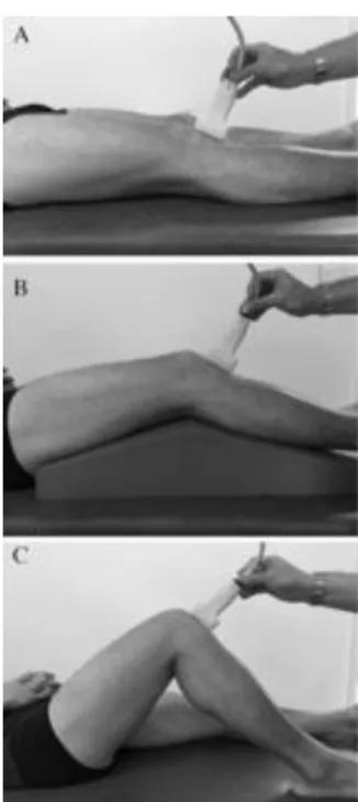
The subjects were at rest, and they were asked to be as relaxed as possible during the following protocol (Figure 1):
• Knee extended: subject was supine with a fully-extended knee.
• Knee semi-flexed: subject was supine with the knee on a 30° positioning splint. • Knee flexed: subject was supine with the knee flexed at 90° (controlled with a
goniometer)
Measurements were performed on both lower limbs for each subject. Three experimented operators were responsible for data acquisition. This protocol lasted about 20 minutes per subject.
Statistical analysis
Previous studies showed excellent intra- and inter-operator concordance of measurement in patellar tendon (22). Non-parametric Mann-Whitney test was used to assess difference in SWS according to age and sex, while Wilcoxon test was used to assess side differences (paired data). Kruskal-Wallis was used to determine differences between the 3 knee positions; normality was assessed with Lilliefors’ test. Calculations were done in Matlab 2016a (The MathWorks, Inc., Natick MA) P < 0.05 was considered significant. Results are reported as mean ± standard deviation.
Results
Mean SWS was 3.9 ± 1.2 m/s in knee extension position (0°), 6.7 ± 1.3 m/s in 30° flexion position and 8.6 ± 1.3 m/s in 90° flexion position; all positions presented a SWS significantly different from the others (p = 1.6e-18). Mean ROI area was 0.8 ± 0.2cm2. No difference was
Figure 1: The probe was placed parallel to patellar tendon fibers on the middle of the tendon in three different positions: A - Knee extended: subject was supine with a fully-extended knee, B - Knee semi-flexed: subject was supine with the knee on a 30° positioning splint. C - Knee flexed: subject was supine with the knee flexed at 90°.
observed between sides at any knee position (p > 0.19). The relevant data concerning side analysis is presented in table 1 and figure 2. According to gender, the statistical analysis showed no differences of SWS in the three positions (Table 2).
SWS variations were analyzed between the extension to 30° flexion, the 30° to 90° flexion and the 0° to 90° flexion. The increase of SWS in the left side was slightly higher than the right side when knee angle changed from 30° to 90° (2.4 against 1.5 m/s, p = 0.03) and from 0° to
90° (5.2 against 4.1 m/s, p = 0.04). SWS variations between knee angles was not affected by gender.
Discussion
The aim of this study was to provide normal range of values regarding patellar tendon elasticity in healthy adult subjects. The reported values could lead to an underestimation of the actual tendon mechanical properties; patellar tendon has a small cross-sectional area, compared to the wavelength of the propagating shear waves, and therefore it behaves as a wave-guide, resulting in a slower apparent wave velocity. However, it was shown that the apparent shear wave speed, as measured in this study, retains its clinical value because it is strongly correlated with the results of a full shear wave dispersion analysis. Besides, automatic wave dispersion analysis is still not available in clinic.
Patellar tendon is also transversely isotropic, therefore calculating tendon mechanical properties is not straightforward. At first approximation, however, tendon shear modulus (μ) can be estimated as μ = ρ·SWS2. This allows comparing our results with the previous
literature that applied this simplification. The calculation yields and average patellar tendon shear modulus with knee at 30° of 45.9 kPa. This value is higher than that reported by Zhang et al (19) at 30° knee flexion (27.5 ± 11.3 kPa), which was relative to the proximal region of the tendon (while in this study measurements were performed in the middle region). The overall average SWS obtained in this study with knee flexion of 90° (8.6 ± 1.3 m/s) is similar to that reported by Hsiao et al. (22) at 90° knee flexion (8.9 ± 1.1 m/s), who performed the measurements in the tendon middle region.
The lowest SWS was obtained at rest with the knee in a fully extended position (i.e., with the patellar tendon at full rest). This is in agreement with the result reported on Achilles tendon and muscles by Dubois(6), Lacourpaille(8). In healthy volunteers, lowest SWS were obtained in a fully non-contracted muscle-tendon couple.
SWS values obtained in our study could be now considered as baseline values for further pathological cohorts investigations in young adults, and they show the importance of proper control of knee angle. SWS in patellar tendon more than doubled between 0° and 90° knee angles.
Figure 2: Mean shear modulus values of patellar tendon according to the side and the knee flexion amplitude.
Conclusion
The SWE is a new, fast and non-invasive technique to assess mechanical properties of musculoskeletal structures and is promising for further investigations on adults or children with various musculoskeletal pathologies. Shear modulus values obtained in our study could be considered as baseline values for further investigations in young adults.
References
1. Gennisson J-L, Catheline S, Chaffaï S, Fink M. Transient elastography in anisotropic medium: application to the measurement of slow and fast shear wave speeds in muscles. J Acoust Soc Am. 2003 Jul;114(1):536–41.
2. Bercoff J, Tanter M, Muller M, Fink M. The role of viscosity in the impulse diffraction field of elastic waves induced by the acoustic radiation force. IEEE Trans Ultrason Ferroelectr Freq Control. 2004 Nov;51(11):1523–36.
3. Friedrich-Rust M, Romen D, Vermehren J, Kriener S, Sadet D, Herrmann E, et al. Acoustic radiation force impulse-imaging and transient elastography for non-invasive assessment of liver fibrosis and steatosis in NAFLD. Eur J Radiol. 2012 Mar;81(3):e325– 331.
4. Cosgrove DO, Berg WA, Doré CJ, Skyba DM, Henry J-P, Gay J, et al. Shear wave elastography for breast masses is highly reproducible. Eur Radiol. 2012 May;22(5):1023– 32.
5. Arda K, Ciledag N, Aktas E, Aribas BK, Köse K. Quantitative assessment of normal soft-tissue elasticity using shear-wave ultrasound elastography. AJR Am J Roentgenol. 2011 Sep;197(3):532–6.
6. Dubois G, Kheireddine W, Vergari C, Bonneau D, Thoreux P, Rouch P, et al. Reliable Protocol for Shear Wave Elastography of Lower Limb Muscles at Rest and During Passive Stretching. Ultrasound Med Biol. 2015 Sep;41(9):2284–91.
7. Hug F, Lacourpaille L, Maïsetti O, Nordez A. Slack length of gastrocnemius medialis and Achilles tendon occurs at different ankle angles. J Biomech. 2013 Sep 27;46(14):2534–8.
8. Lacourpaille L, Hug F, Bouillard K, Hogrel J-Y, Nordez A. Supersonic shear imaging provides a reliable measurement of resting muscle shear elastic modulus. Physiol Meas. 2012 Mar;33(3):N19–28.
9. Berko NS, Fitzgerald EF, Amaral TD, Payares M, Levin TL. Ultrasound elastography in children: establishing the normal range of muscle elasticity. Pediatr Radiol. 2014 Feb;44(2):158–63.
10. Lallemant-Dudek P, Dubois G, Vergari C, Forin V, Vialle R, Skalli W. Reproducibility of shearwave elastography on children, a reliability study.
11. DeWall RJ, Slane LC, Lee KS, Thelen DG. Spatial variations in Achilles tendon shear wave speed. J Biomech. 2014 Aug 22;47(11):2685–92.
12. Slane LC, DeWall R, Martin J, Lee K, Thelen DG. Middle-aged adults exhibit altered spatial variations in Achilles tendon wave speed. Physiol Meas. 2015 Jul;36(7):1485–96. 13. Brum J, Bernal M, Gennisson JL, Tanter M. In vivo evaluation of the elastic anisotropy of the human Achilles tendon using shear wave dispersion analysis. Phys Med Biol. 2014 Feb 7;59(3):505–23.
14. Witvrouw E, Bellemans J, Lysens R, Danneels L, Cambier D. Intrinsic risk factors for the development of patellar tendinitis in an athletic population. A two-year prospective study. Am J Sports Med. 2001 Apr;29(2):190–5.
15. Lian OB, Engebretsen L, Bahr R. Prevalence of jumper’s knee among elite athletes from different sports: a cross-sectional study. Am J Sports Med. 2005 Apr;33(4):561–7. 16. Helland C, Bojsen-Møller J, Raastad T, Seynnes OR, Moltubakk MM, Jakobsen V, et al. Mechanical properties of the patellar tendon in elite volleyball players with and without patellar tendinopathy. Br J Sports Med. 2013 Sep;47(13):862–8.
17. Kongsgaard M, Qvortrup K, Larsen J, Aagaard P, Doessing S, Hansen P, et al. Fibril morphology and tendon mechanical properties in patellar tendinopathy: effects of heavy slow resistance training. Am J Sports Med. 2010 Apr;38(4):749–56.
18. Couppé C, Kongsgaard M, Aagaard P, Vinther A, Boesen M, Kjaer M, et al. Differences in tendon properties in elite badminton players with or without patellar tendinopathy. Scand J Med Sci Sports. 2013 Mar;23(2):e89–95.
19. Zhang ZJ, Ng GY, Lee WC, Fu SN. Changes in morphological and elastic properties of patellar tendon in athletes with unilateral patellar tendinopathy and their relationships with pain and functional disability. PloS One. 2014;9(10):e108337.
20. Royer D, Gennisson J-L, Deffieux T, Tanter M. On the elasticity of transverse isotropic soft tissues (L). J Acoust Soc Am. 2011 May;129(5):2757–60.
21. Kot BCW, Zhang ZJ, Lee AWC, Leung VYF, Fu SN. Elastic modulus of muscle and tendon with shear wave ultrasound elastography: variations with different technical settings. PloS One. 2012;7(8):e44348.
22. Hsiao M-Y, Chen Y-C, Lin C-Y, Chen W-S, Wang T-G. Reduced Patellar Tendon Elasticity with Aging: In Vivo Assessment by Shear Wave Elastography. Ultrasound Med Biol. 2015 Nov;41(11):2899–905.
Tables:
Table 1: Statistical data according to the side
SWS [m/s] N p
Full extension Right side 4.2 ± 1.3 20 0.285
Left side 3.7 ± 1.0 20
30° flexion Right side 6.8 ± 1.3 20 0.465
Left side 6.5 ± 1.2 20
90° flexion Right side 8.3 ± 1.6 20 0.185
Left side 8.9 ± 1.0 20
Table 2: Statistical data according to the gender
SWS [m/s] N p
Full extension Male 3.9 ± 1.2 12 0.901
Female 4.0 ± 1.2 8
30° flexion Male 6.8 ± 1.1 12 0.355
Female 6.5 ± 1.5 8
90° flexion Male 8.8 ± 1.0 12 0.341