HAL Id: hal-00553416
https://hal.archives-ouvertes.fr/hal-00553416
Submitted on 10 Mar 2011
HAL is a multi-disciplinary open access
archive for the deposit and dissemination of
sci-entific research documents, whether they are
pub-lished or not. The documents may come from
teaching and research institutions in France or
abroad, or from public or private research centers.
L’archive ouverte pluridisciplinaire HAL, est
destinée au dépôt et à la diffusion de documents
scientifiques de niveau recherche, publiés ou non,
émanant des établissements d’enseignement et de
recherche français ou étrangers, des laboratoires
publics ou privés.
Performance analysis of an off-line BMI for the control
of a robotic digit
Sofiane Ouanezar, Selim Eskiizmirliler, Marc Maier
To cite this version:
Sofiane Ouanezar, Selim Eskiizmirliler, Marc Maier. Performance analysis of an off-line BMI for the
control of a robotic digit. Cinquième conférence plénière française de Neurosciences
Computation-nelles, ”Neurocomp’10”, Aug 2010, Lyon, France. �hal-00553416�
Performance analysis of an off-line BMI for the control of a robotic digit
(1,3)
Ouanezar S
.,
(1,2)Eskiizmirliler S. and(1,2)Maier M.A.1
Université Paris Descartes / CESEM – UMR 8194 CNRS, 2Université Paris Diderot
3
TELECOM ParisTech / Département TSI Signal-Images [email protected]
ABSTRACT
Various recent brain-machine interface (BMI) applications have emphasized the potential of using cortical signals for the kinematic control of artificial devices such as robotic arms or legs [1], [2]. However, it is less clear whether hand and digit movements can be controlled in the same way in order to potentially restore manual dexterity. Recently, we have provided a proof-of-concept that an anthropomorphic robot finger with 4 degrees of freedom (DoF) can be controlled off-line by intracortical signals recorded in the behaving monkey [3]. Here we report the performance analysis based on the same data and robot, the statistical results including aspects of learning/training, prediction, as well as control.
KEY WORDS
BMI, neurobotics, motor cortex, EMG, neural networks, computational neuroscience
1. Introduction
Recent developments in BMI technologies have shown how cortical activities can be used to control the position and/or the state of different types of external devices in real time. These efforts have focused mainly on controlling a computer cursor [1], [2], the state of on/off switches or the decoding and control of reach trajectories of a robotic arm, sometimes equipped with a simple 1 DoF gripper [3-6].
However, studies dedicated to the decoding of cortical signals for the eventual control of a dexterous hand with multiple degrees of freedom are rare. The goal would be to control individuated and/or combined finger movements. First attempts of decoding finger movements have only been undertaken very recently [7-11]. None of these previous results have so far been applied to robot artifacts.
Many studies on the cortical activity of the motor cortex in behaving non-human primates (e.g. [12]) have shown that neurons in the primary motor cortex (M1) code for some aspects of finger movements, such as force. In the aim of providing a proof-of-concept that a robot finger of multiple DoF can be controlled by intra-cortical signals, we first set up an experimental site (robotic digit) and implemented a kinematic control of its movement (i.e. positional control). To mimic the
monkey finger, we opted for an anthropomorphic robot finger of 4 DoF (designed and manufactured by Shadow Robot Company, UK) actuated by McKibben-like pneumatic artificial muscles in an antagonist actuation scheme. This robot digit is equipped with hall effect position sensors and with fingertip quantum tunnel tactile sensors. The robot digit was then mounted on an experimental set-up, which contained additional mechanical parts and sensors to imitate the monkey task (precision grip). Finally, a hybrid motor control scheme composed of an artificial neural network and PID modules has been implemented [3]. We then performed a statistical analysis on the decoding of previously recorded cortical data for controlling finger position. This is reported in the following.
2. Method
The sensory-motor control of an anthropomorphic robot finger using intra-cortical signals is composed of three main steps:
• Recording the data.
• Decoding the neural data to predict grip motion (BMI part).
• Controlling the robot. 2.1 Data recording
The biological signal consists of the activity of 33 corticomotoneuronal (CM) cells recorded in M1 of a macaque monkey performing a precision grip task. Recordings were obtained in 7 separate behavioral sessions. In addition, EMG signals were recorded from up to eight intrinsic hand and forearm muscles. These recordings were obtained from the Institute of Neurology (UCL, London, UK, courtesy of RN Lemon).
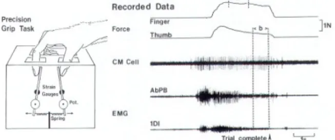
Fig. 1. Left: Schematic diagram of the manipulandum used for the precision grip task. Right: example of the recorded signal during the precision grip task. From top to bottom: force of the index finger and
of the thumb, CM cell activity, EMG of two muscles (AbPB and IDI). Figure taken from [12].
2.2 Decoding spiking data
We attempted to decode two different signals from the CM cell spike trains: (i) the time-varying endpoint position of the index finger during precision grip. The position was recorded via a spring-loaded lever in the experimental setup. (ii) the EMG of extrinsic or intrinsic hand muscles recorded during this same task. We used a neural network approach, a Time Delayed Multi-Layer Perceptron (TDMLP) to decode and thus to predict either the finger position or the EMG from the CM cell spike trains. The input vector consisted of a binary sequence corresponding to the presence/absence of spikes of the CM cell (down-sampled to 250 Hz) over a given period, i.e. the duration of the sliding input window, which varied between 25 ms and 400 ms. The TDMLP, a three-layer feed-forward neural network was implemented with the Matlab neural network tool box.
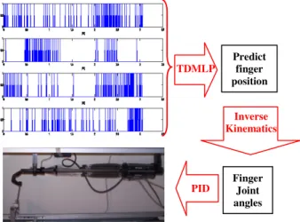
Fig. 2. The control of a robotic digit of 4 DoF using activity of multiple CM cells.
The input layer consists of D × NC units (D is the number of bins in the sliding window, and NC the number of CM cells used) which represent the spiking response recorded between time t-D and t. Sigmoid activation functions (range 0-1) were used in the hidden layer, which consisted of 10 units. The one output unit, also using a sigmoid activation function, provides a positive scalar that corresponds to the predicted position (or EMG) at time t. This is illustrated in Fig. 3.
Hidden layer units and the output unit also received bias inputs. Bias and connection weights are adjusted during the learning process, based on the gradient descent error to minimize the sum of the squared errors between the desired output and the network output. The number of units in the hidden layer and the particular learning algorithm have been chosen by the technique of cross-validation.
Learning of the input-output transformation occurred through the use of multiple trials (min: 15, max: 92 trials) depending on the recording session. The number of learning epochs was fixed to 1000, each epoch containing all trials. The Nguyen-Widrow algorithm was used for weight initialization.
Fig. 3. Schematic of the TDMLP and its input-output transformation.
2.3 Controlling the robot digit
The third and final part of the BMI based motor control chain consists in the reproduction of estimated index finger movement pushing on one of the manipulandum's lever, shown in Fig. 2, using the 4 DoF robot finger. On the finger there are four joints and three human-sized phalanges. Both middle and proximal (MCP) joints are actuated in the flexion/extension plane by a pair of antagonist artificial muscles. The MCP joint, also responsible for the abduction/adduction motion of the proximal phalange, is in addition driven by one muscle with an opposing return spring. Similar to the human finger the distal joint is coupled to the middle joint such that the angle of the middle joint is always greater than or equal to the angle of the distal joint. The joint angles relative to the estimated fingertip trajectory were calculated by an inverse kinematics model of the robot finger taking into account the coupled motion of the distal phalange. Finally, we used a classical PID controller, the last link of the control chain, to ensure the tracking of the desired trajectory as shown in Figure 4.
3. Results
The performance of the TDMLP-based prediction was systematically analysed by varying the size D of the sliding window and the number of cortical cells NC used as inputs. This was done with a TDMLP with 10 units in the hidden layer, a number of hidden units found to be optimal across different sizes of input windows, irrespective of the type of the output signal, i.e. position or EMG. Performance (Per) was calculated as follows:
∑
∑
∑
∑
∑
∑
∑
∑
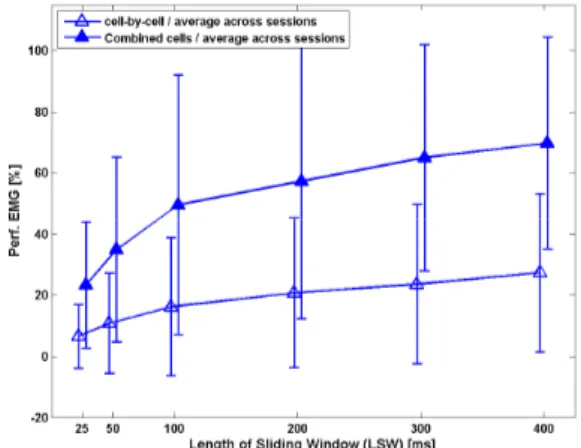
= = = = − −− − = = = = = == = 2 2 max where ) max( 100 100 Per )) -mean(Y (Y (error) ) Y (Y error error error * -des des est des 001000110100100100001000010000100000 Predict finger position TDMLP Inverse Kinematics Finger Joint angles PIDFig. 4 shows the performance of the TDMLP in estimating the recorded position for different sizes of sliding input windows, as well as for two modes of training: (i) a prediction based on the spike train of a single CM cell, and then averaged across cells in a given recording session. This was called "cell-by-cell" prediction. (ii) a prediction based on spike trains of multiple, simultaneously recorded CM cells. This was called prediction of "combined cells". The number of combined cells varied between 2 and 6 depending on the session. Fig. 4 shows the grand average across three recording sessions.
Fig. 4. Performance of prediction for fingertip position as a function of window size and number of cortical cells. Open triangles: “cell-by-cell” estimation. Filled triangles: “combined “cell-by-cell” estimation. Grand average across 3 sessions. Similar results were found for the remaining 4 sessions.
Fig. 5 shows the performance of a similar network for predicting the EMG signal of Abductor Pollicis Longus (AbPL) muscle.
Fig. 5. Performance of prediction for EMG activity of AbPL muscle as function of window size and number of cortical cells. Same conventions as in Fig. 4.
The network behaved similarly for predicting position or EMG. Three results were found: (i) In both cases the performance increased continuously as a function of increasing input window size (from 25 to 400 ms). Saturation was observed at a window size of about 400 ms. (ii) Performance was significantly better when using multiple simultaneously recorded spike trains compared to prediction based on single cells. (iii)
Independent of input size and number of cells, the prediction of position was better than that of EMG. How the observed performance translates to trial-by-trial prediction is shown in Fig. 6. Recorded (blue) and estimated (red) position signals for three representative trials, based on a TDMLP with a 400 ms input window of 4 concurrent CM cells. Also shown is the prediction of two corresponding EMG signals for Flexor Digitorum Superficialis (FDS) and Extensor Digitorum Communis (EDC). 0 1 2 3 4 5 6 7 8 0 0.2 0.4 0.6 0.8 1
Recorded and estimated position
time[s] Norm. Ampl. 0 1 2 3 4 5 6 7 8 0 0.2 0.4 0.6 0.8 1
Recorded and estimated EMG of FDS
time[s] Norm. Ampl. 0 1 2 3 4 5 6 7 8 0 0.2 0.4 0.6 0.8 1
Recorded and estimated EMG of EDC
time[s]
Norm. Ampl.
Fig. 6. The recorded (blue) and estimated (red) finger trajectories (top) and EMG activity of FDS and EDC for three successive trials.
Since each CM cells facilitates a given set of target muscles, it was of interest to investigate whether prediction of EMG varied as a function of the muscle field of the input cells. We hypothesised that the prediction of a target EMG would be better when based on CM cells that facilitated that particular muscle. Presence of facilitation was estimated by post-spike-facilitation (PSF) of spike-triggered averaging of the EMG [12].
Fig. 7. Prediction of FDS EMG with CM cells having FDS as target muscle compared to a prediction based on CM cells without facilitation of FDS (same 3 sessions as in Fig. 5).
Fig. 7 shows the performance of predicting (over 3 sessions) the FDS EMG as a function of the window size. Prediction based on CM cells that facilitated the FDS EMG (green triangles) tends to be better than those based on CM cells that facilitated other muscles than the FDS (yellow triangles).
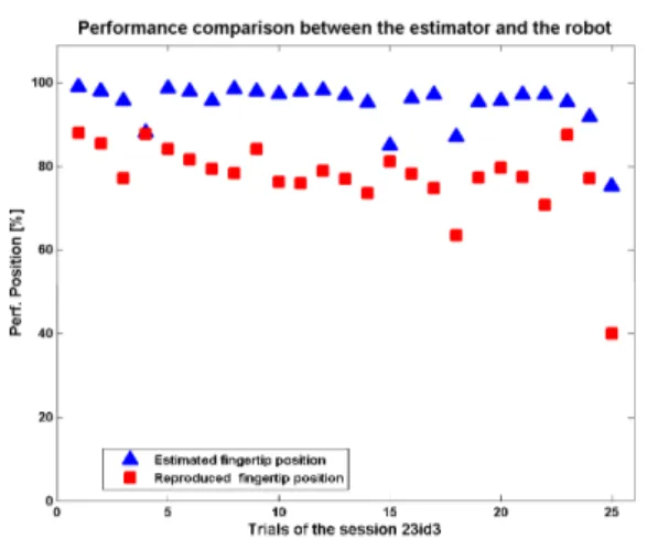
Finally, we used the predicted position in order to control the robot digit. A comparison between the performance achieved by the TDMLP and that realized by the robot digit shows a systematic lower performance for the robot. The decrease in performance, measured over 25 trails, is in the order of 10% to 20% (Fig. 8). However, this still allows for a functional reproduction of the monkey’s digit movement.
Fig. 8. The performance comparison between the TDMLP-estimated and reproduced fingertip endpoint positions over 25 trials recorded during one session.
4. Discussion
With few exceptions [7-11], most studies on the control of the upper limb through data from a cortical invasive BMI have focused on reaching [e.g. 1,2,5,6], not on hand or finger movements. Our results on the control of a single finger demonstrate that neural activity from cells recorded in the M1 hand area contain sufficient information to enable decoding of an asynchronous finger movement.
Several differences distinguish our approach from that of Georgopoulos et al. [7] or from those of Schieber and Thakor [8-11]. Both of these groups also worked on decoding of digit movements.
First, we used a neural network approach to obtain a transfer function between spike trains and finger trajectory, whereas they used a coding based on the population vector [7], on non-linear filters [8], or on a maximum-likelihood scheme [11].
Second, they decoded a binary movement direction (flexion or extension) together with a decoding of one of the five digits to be moved [7]. Essentially, their decoder worked as a movement classifier [9,11]. In our
case, decoding concerned a single finger whose endpoint trajectory was a time-varying scalar. We showed that the accuracy of the prediction increased as a function of the size (duration) of the input window. This was shown previously for hand trajectories [13]. Third, they used the activity of up to 50 simultaneously recorded but unidentified neurons in the M1 hand area as inputs, whereas we used few (up to 5) but identified CM cells. We showed that the accuracy of the prediction increased as a function of the number of simultaneously recorded CM cells acting as inputs. Such an increase in performance has been shown previously for unidentified cells in the case of classifying digit movements [8,11], as well as in decoding of the arm trajectory [5, 14].
We also predicted EMG activity. Although EMG is an inherently more noisy signal than position, prediction accuracy reached up to 60%, for windows of ≥ 200 ms and multiple CM cells. Slightly higher levels of accuracy (70-75%), but based on many more and unidentified cells, have been obtained previously for EMGs during arm movements [7]. Furthermore, we show a trend for improved accuracy if the prediction is based on CM cells that facilitate the target muscle EMG.
Finally, the TDMLP-based prediction of finger position was used to replicate the recorded movement with a robotic digit. Our robot differs from those of other BMI applications. We used a robot finger actuated by artificial muscles in an antagonist actuation scheme. Three properties make this robot biologically more plausible than conventional robots: i) one can establish a correspondence between a particular anatomical muscle and its artificial counterpart in the robot. ii) The dynamic control -future work in this project - needs a model of the actuators. It has been demonstrated that actuators of this type are biologically highly plausible [15]. iii) Finally, an analogy between the EMG signal and the pressurized air used to contract the artificial muscle looks promising. The observed performance difference between estimated and realized robot movement are most likely due to non-resolved compatibility issues in the inverse kinematics model as well as in the PID controller.
In conclusion, decoding of spike trains from simultaneously recorded CM cells in M1 allows for an off-line reproduction of a digit movement during precision grip.
Acknowledgements
This study was in part funded through a grant by the ‘Fondation pour la Recherche Médicale’ (FRM), France. DBC20080713368.
References
[1] W. Wu, M. J. Black, Y. Gao, E. Bienenstock, M. Serruya, A. Shaikhouni, and J. P. Donoghue. Neural decoding of cursor motion using a Kalman filter.
Advances in Neural Information Processing Systems, 2003, 15:133–140.
[2] Mijail D. Serruya, Nicholas G. Hatsopoulos, Liam Paninski, Matthew R. Fellows, and John P. Donoghue. Brain-machine interface: Instant neural control of a movement signal
.
Nature,
March 2002, 416(6877):141–142.[3] S. Ouanezar, S. Eskiizmirliler, and M. A. Maier, A BMI application for the control of a robotic digit.
Proceedings of the Neurocomp 2009, Bordeaux, France.
[4] M. Velliste, S. Perel, M. C. Spalding, A. S. Whitford & A. B. Schwartz. Cortical control of a prosthetic arm for self-feeding, Nature, 2008, 453, 1098-1101.
[5] J. M. Carmena, M. A. Lebedev, R. E. Crist, J. E. O'Doherty, D. M. Santucci, D. F. Dimitrov, P. G. Patil, C. S. Henriquez, M. A. Nicolelis. Learning to control a brain-machine interface for reaching and grasping by primates. PLoS Biol. 2005 Dec;3(12):e426.
[6] D. M. Taylor, S. H. I. Tillery, and A. B. Schwartz. Direct cortical control of 3d neuroprosthetic devices
.
Science, June 2002, 296(5574):1829–1832.
[7] A. P. Georgopoulos, G. Pellizzer, A. V. Poliakov, M. H. Schieber. Neural coding of finger and wrist movements
.
J Comput Neurosc, 1999i6: 279–288. [8] V. Aggarwal, S. Acharya, F. Tenore, H. C. Shin, R. Etienne-Cummings, M. H. Schieber, N. V Thakor. Asynchronous decoding of dexterous finger movements using M1 neurons. IEEE Trans Neural Syst Rehabil Eng.
2008 Aug;16(4):421.[
9] S. Acharya, F. Tenore, V. Aggarwal, R. Etienne-Cummings, M.H. Schieber, N.V. Thakor.
Decoding individuated finger movements using volume-constrained neuronal ensembles in the M1 hand area.IEEE Trans Neural Syst Rehabil Eng. 2008
Feb;16(1):15-23.
[10] V. Aggarwal, F. Tenore, S. Acharya, M.H. Schieber, N.V. Thakor. Cortical decoding of individual finger and wrist kinematics for an upper-limb neuroprosthesis. Conf Proc IEEE Eng Med Biol Soc. 2009;2009:4535-8.
[11] H.C. Shin, V. Aggarwal, S. Acharya, M.H. Schieber, N.V. Thakor. Neural decoding of finger movements using Skellam-based maximum-likelihood decoding. IEEE Trans Biomed Eng. 2010 Mar;57(3):754-60.
[12] M. A. Maier, M. C. Hepp-Reymond and R. N. Lemon. Contribution of the monkey corticomotoneuronal system to the control of force in precision grip
.
J Neurophysiol, 1993 Mar;69(3):772-85. [13] L. Paninski, S. Shoham,M. R. Fellows, N. G. Hatsopoulos, J. P. Donoghue. Superlinear populationencoding of dynamic hand trajectory in primary motor cortex. J Neurosci, 2004 Sep 29;24(39):8551-61. [14] J. C. Sanchez, D. Erdogmus, Y. N. Rao, S. P. Kim, M. A. Nicolelis. Interpreting neural activity through linear and nonlinear models for brain machine interfaces
.
Proc. 25th IEEE of Eng. Med. Biol. Soc., 2003, vol3 2160–2163[15] B. Tondu and P. Lopez. The McKibben muscle and its use in actuating robot-arms showing similarities with human arm behavior, Industrial Robot, 1997, Vol:24, No:6, pp:432-439.