UNIVERSITÉ DU QUÉBEC À MONTRÉAL
L'APNÉE OBSTRUCTIVE DU SOMMEIL. IDENTIFICATION DES PERSONNES ÂGÉES SUSCEPTIDLES DE DÉCLIN COGNTIF ANORMAL.
THÈSE PRÉSENTÉ
COMME EXIGENCE P ARTITIELLE DU DOCTORAT EN PSYCHOLOGIE
PAR
KA TIA GAGNON
Avertissement
La diffusion de cette thèse se fait dans le respect des droits de son auteur, qui a signé le formulaire Autorisation de reproduire et de diffuser un travail de recherche de cycles supérieurs (SDU-522 - Rév.1 0-2015). Cette autorisation stipule que «conformément à l'article 11 du Règlement no 8 des études de cycles supérieurs, [l'auteur] concède à l'Université du Québec à Montréal une licence non exclusive d'utilisation et de publication de la totalité ou d'une partie importante de [son] travail de recherche pour des fins pédagogiques et non commerciales. Plus précisément, [l'auteur] autorise l'Université du Québec à Montréal à reproduire, diffuser, prêter, distribuer ou vendre des copies de [son] travail de recherche à des fins non commerciales sur quelque support que ce soit, y compris l'Internet. Cette licence et cette autorisation n'entraînent pas une renonciation de [la] part [de l'auteur] à [ses] droits moraux ni à [ses] droits de propriété intellectuelle. Sauf entente contraire, [l'auteur] conserve la liberté de diffuser et de commercialiser ou non ce travail dont [il] possède un exemplaire.»
REMERCIMENTS
Je tiens tout d'abord à remercier mes directeurs de recherche. Jean-François Gagnon qui m'a transmis son intérêt pour la recherche en sommeil, et Nadia Gosselin qui m'a soutenue dans ce long processus et m'a permis d'atteindre mes objectifs dans un contexte de recherche stimulant et agréable. Nadia est une personne exceptionnelle, elle m'a permis de grandir sur le plan professionnel et personnel, elle demeure pour moi un modèle et une mentore exemplaire.
Un remerciement spécial à Andée-Ann Baril, ma coéquipière dans ce projet, pour son implication, son soutien, sa collaboration dans l'élaboration des articles, et nos nombreuses discussions concernant le projet de recherche. Andrée-Ann a contribué à rendre mon parcours doctoral agréable avec son sens de l'humour et sa folie, mais par-dessus tout je la remercie pour son amitié sans faille. Mes remerciements à Hélène Blais pour son inestimable soutien et son immense générosité, tant sur le plan pers01mel que pour la coordination du protocole de recherche et son implication dans l'analyse des tracés EEG et des données respiratoires. Merci à Caroline D'Aragon pour son soutien et son travail impeccable dans l'acquisition de données.
Un grand merci à l'équipe de techniciens du laboratoire, Benoît Adam, Nancy Poirier, Mélina-Crécia Babala et Jean-Claude Aubertin, sans lesquels les emegistrements des tracés EEG n'auraient été possibles. Merci à Marie-Josée Quinn pour l'acquisition des données génétiques. Merci également à Jean Paquet pour son soutien au plan statistique, ainsi qu'à Gaétan Poirier et à Sébastien Saucier pour leur soutien technique. Merci aux étudiants en neuropsychologie qui ont pris le relais lors de mon départ en stage et internats, Catherine Chapados, Matjolaine Lafortune, Frédérique Escudier, Jonathan Deslauriers. Je tiens à remercier Jessica Cole pour son aide et sa
rigueur. Merci beaucoup à tous les étudiants qui ont contribué à rendre ma formation doctorale agréable et stimulante : Caroline AJ·bour, Catherine Duclos, Solène Van der Maren, Héjar El-Khatib, Sit·in Chami, Erlan Sanchez, Marc-André Gareau, Maxime Fortin.
Je remercie toutes les personnes qui ont participé aux études et qui ont généreusement accepté de passer tous les tests nécessaires pour ma thèse. Plusieurs participants m'ont touché par leur enthousiasme, leur générosité et leur vulnérabilité. J'espère que ma contribution leur ont permis d'entreprendre des démarches afin d'améliorer leur état de santé.
Merci à mes superviseurs de stage clinique Isabelle Soulières et Bruno Gauthier pour leur patience, leur générosité, mais aussi pour m'avait· transmis leur intérêt pour la clinique en pédiatrie. Merci à mes collègues de stage, Emmanuelle, Claudia et Miriam d'avait· contribué à rendre mon expérience clinique agréable. Merci à Anne Décary, pour les nombreuses discussions stimulantes que nous avons eues autant sur la recherche que sur la clinique. Ces conversations m'ont aidé à orienter mes choix de carrière. Merci à mes superviseurs d'internat, Geneviève Gagnon et David Fontaine, pour leur rigueur, leur expertise et leurs généreux conseils. Merci à mon superviseur Stéphane Dubé, pour avoir généreusement partagé sa riche expérience en pédopsychiatrie et m'avait· donné confiance en mes capacités de clinicienne. Son implication envers ses clients dépasse largement ce qui est attendu d'un neuro psychologue.
Merci à Atmie Ménard, qut supervise mon travail clinique depuis 3 ans, pour sa générosité, sa douceur et sa flexibilité. Son approche avec les clients, son jugement et son raisonnement clinique font d'elle une clinicienne et une supervtseure exceptionnelle. Un merci très spécial à Julie Brault, ainsi qu'à mes collègues de travail du Centre d'Apprentissage aux 1001 Astuces, Valérie Choquette, Pénéloppe
IV
Guillard, Sonia Prince, Esther Ptovin, Annie-Claude Granger et Annick Cotnoir qui rendent mes vendredis et samedis incroyablement plaisants et qui contribuent à améliorer ma compréhension clinique par leurs différentes expertises.
Merci à mes amis, Josée Verreault, Roberts Melançon, Luc Boudrias, Jessica Peter, Marco Sauvé, Caroline Dubuc, Stéfani Desrochers, Manon Duchesneau et Marie Lacroix pour leur compréhension et leur soutien moral constant. Merci à mes cousines Maggie Gagnon, Véronique Gagnon et Cynthia Gagnon pour leurs encouragements. Merci à ma belle-famille Danielle Bénard, Ronald Vézina, Cynthia Jeanneau, Émile et James pour lem compréhension et leur soutien. Un remerciement tout spécial à Kevin Jeanneau pour son soutien inébranlable, sa patience et son amour. Je tiens à exprimer ma plus profonde gratitude envers ma mère et mon beau -père pour toutes les petites et grandes attentions, ainsi que leurs encouragements constants au cours de ces dernières années. Merci à tous d'avoir cru en moi.
TABLE DES MATIÈRES
REMERCIMENTS ... ii
TABLE DES MATIÈRES ... vi LISTE DES FIGURES ... viii
LISTE DES TABLEAUX ... ix
LISTE DES ABRÉVIATIONS, DES SIGLES ET DES ACRONYMES ... x
RÉSUMÉ ... xii ABSTRACT ... xiv CHAPITRE 1 ARTICLE 1 ET CONTEXTE THÉORIQUE ... 4
ARTICLE 1: COGNITIVE IMPAIRMENT IN OBSTRUCTIVE SLEEP APNEA. ... 4
1.2. Le déclin cognitif anormal chez les personnes âgées ... 36
1.2.1. Définitions ... 36
1.2.2. Épidémiologie du trouble cognitif léger dans l' AOS et facteurs de risque ··· 37
1.2.3. Risque de progression vers une démence ... 38
1.2.4. Les mécanismes associés à la démence et aux dysfonctions cognitives dans l'apnée obstructive du sommeil. ... 39
1.3. La plainte cognitive subjective ... 43 1.3.1. Définition de la plainte cognitive subjective ... 43
1.3.2. Évaluation de la plainte cognitive ... 44
1.3.3. Épidémiologie de la plainte cognitive et facteurs de risques ... 46
1.3.4. Évolution de la plainte cognitive à la démence ... 46
1.3.5. La plainte cognitive et l' AOS ... 48 1.4. Les tests de dépistages des troubles cognitifs ... 50
1.4.1. Mini-Mental State Examination ... 50
1.4.2. Montreal Cognitive Assessment.. ... 51 1.5. Objectifs et hypothèses de recherche ... 52
1.5.1. Article 2 : Disconnection of subjective and objective cognitive impairment in obstructive sleep apnea ... 53
1.5.2. Article 3 : Detection of mild cognitive impairment in older individuals with obstructive sleep apnea: is the Montreal Cognitive Assessment or the Mini -Mental State Examination the most useful? ... 54
CHAPITRE
rr
ARTICLE 2 ... 562.1 Approche méthodologique ... 56
DISCONNECTION OF SUBJECTIVE AND OBJECTIVE COGNITIVE IMPAIRMENT IN OBSTRUCTIVE SLEEP APNEA. ... 57
CHAPITRE III ARTICLE 3 ... 81
DETECTION OF MILO COGNITIVE IMPAJRMENT IN OLDER INDIVIDUALS WITH OBSTRUCTIVE SLEEP APNEA: IS THE MONTREAL COGNITIVE ASSESSMENT OR THE MINI-MENTAL STATE EXAMINATION THE MOST USEFUL? ... 81
CHAPITRE IV DISCUSSION GÉNÉRALE ... 120
4.1. Rappel de l'objectif de la thèse ... 120
4.2. Caractéristiques de la plainte cognitive chez les personnes apnéiques âgées de 55 ans et plus ... 121
4.2.2. La nature de la plainte cognitive est-elle différente dans l' AOS? ... 124
4.2.3 La plainte cognitive chez les apnéiques, une atteinte précoce des processus n1étacognitifs ... 125
4.2.4. L 'anosognosie et ses corrélats neuronaux ... 126
4.3. Efficacité des tests de dépistage à détecter le déclin cognitif chez les apnéiques ... 127
4.4. Forces de la thèse ... 131
4.5. Limites de la thèse ... 132
4.6. Implications cliniques ... 134
4.7. Perspectives futures ... 137
CONCLUSION ... 139
LISTE DES FiGURES
2.1 Scores on cognitive complaint questionnaires according to group and cognitive
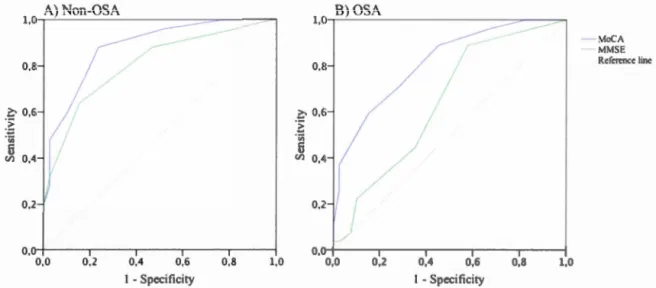
status ... 80 3.1 ROC curve comparisons between the MoCA and the MMSE in detecting MCI in
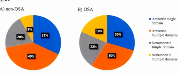
(A) non-OSA and (B) OSA participants ... 109 J.El Proportions ofMCI subtypes in (A) non-OSA and (B) OSA participants ... 115
2.3 Hierarchical regression of risk factors predicting each SCC questiotmaires scores ... 79 3.1. Demographie and clinicat characteristics of non-OSA and OSA participants
according to the ir cognitive statu s . ... 1 02 3.2 Validity ofthe MoCA and the MMSE in detecting MCI in OSA participants ... 104 3.3 Validity ofthe MoCA and the MMSE in detecting MCI in non-OSA participants ... 105 3.4. Neuropsychological results . ... 106 3.E 1 Nemopsychological tests and variables used to identify MCI. ... 113
LISTE DES ABRÉVIATIONS, DES SIGLES ET DES ACRONYMES Français ANOVA AOS ApoE4 Et al. P. Ex. Anglais AHI ANOVA ApoE4 AUC BAI BD I-II BLOJ BMI BNT BVMT-R CI CPAP CPT-II CWlT DNA EEG E.g.
E
RP
ESS I.e. MCI MMSE MoCA MRI NIA NPV PPV PSG PSQI RAVLT REMAnalyse de la variance
Apnée obstructive du sommeil Apolipoprotéine Epsilon 4 Et alii (et autre)
Par exemple
Apnea-Hypopnea Index Analyse ofvariance Apolipoproteine Epsilon 4 Area under the curve Beek Anxiety Inventory Beek Depression Inventory-II Benton Line Orientation Judgment Body mass index
Boston Naming Test
BriefVisuospatial Memory Test-revised Confidence interval
Continuous positive airway pressure Continuous Performance Test-II Co lor-Word Interference Test Deoxyribonucleic acid
Electroencephalography For example
Event-Related Potential Epworth Sleepiness Scale lsto es (That is)
Mild ognitive impairment Mini-Mental State Examination Montreal Cognitive Assessment Magnetic Resonnance Imaging Not Applicable
Negative predictive value Positive predictive value Ploysomnography
Pittsburgh Sleep Quality Index Rey Auditory Verbal Learning Test Rapid Eye Movement
ROC ROCF
sec
SENS SPEC Sp02 TOL TST VBI Yrs Reciever Operating Characteristic Rey-Osterreith Complex Figure Subjective cognitive complaint SensitivitySpecificity
Oxygen saturation Tower of London Total sleep time Vascular Burden Index Years
RÉSUMÉ
L'apnée obstructive du sommeil (AOS) augmente le risque de trouble cognitif léger et de démence chez les individus âgés. Dans ce contexte, l'identification des patients apnéiques susceptibles de déci in cognitif pourrait améliorer les pratiques cliniques. Les travaux de cette thèse ont pour objectif d'évaluer l'efficacité d'outils cliniques permettant d'identifier les individus risquant de présenter un déclin cognitif anormal dans un échantillon d'apnéiques âgé de 55 ans et plus.
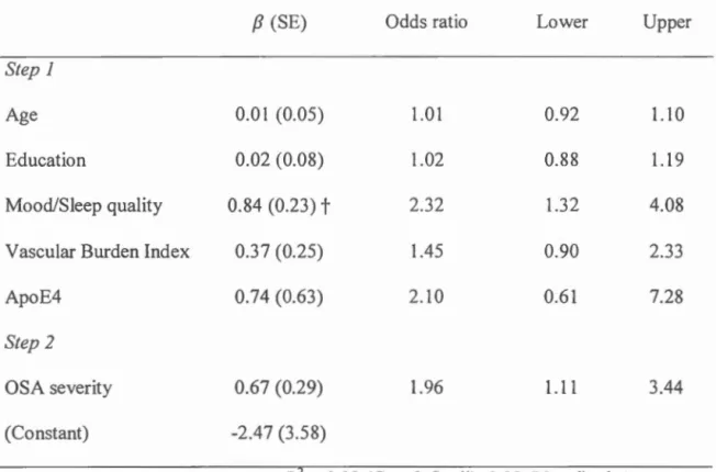
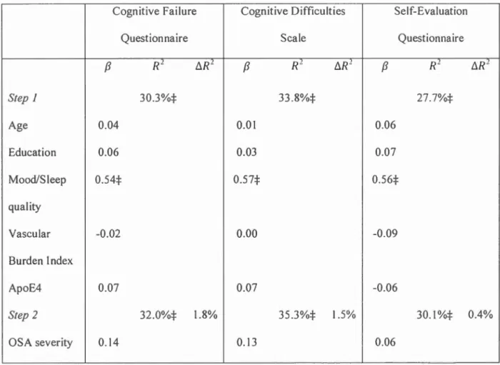
Le premier article (Chapitre I) est une revue de littérature sur la pathophysiologie de l' AOS, les dysfonctions cognitives qui lui sont associées, de même que les mécanismes et marqueurs (électroencéphalographie et neuroimagerie) liés à ces dysfonctions. L'efficacité du traitement par pression positive continue afin d'améliorer les atteintes cognitives est discutée. Les récents résultats concernant la relation entre l' AOS et le risque de déclin cognitif anormal sont finalement abordés. Le second article (Chapitre II) a pour objectif de vérifier si la plainte cognitive subjective est un outil utile pour identifier les apnéiques risquant de présenter un déclin cognitif anormal. Elle vise à déterminer si la sévérité de l' AOS contribue à la plainte cognitive, mais aussi à explorer le lien entre la plainte cognitive subjective et l'atteinte cognitive objective. Cinquante-sept participants apnéiques et 54 non-apnéiques âgés de 55 à 85 ans ont effectué une nuit d'emegistrement polysomnographique suivie d'une évaluation neuropsychologique. Ils ont rempli des questioru1aires évaluant l'humeur, la qualité du sommeil et la perception de leur foncti01mement cognitif. Nos analyses ont révélé que la sévérité de l' AOS (p. ex. index respiratoire, saturation en oxygène et fragmentation du sommeil) ne contribuait pas significativement à la sévérité de la plainte cognitive subjective. En fait, des scores plus élevés aux questionnaires portant sur l'humeur et la qualité du sommeil, contribuait significativement à augmenter la plainte cognitive. Les analyses ont aussi montré que les participants apnéiques ne sont pas en mesure de bien évaluer leur foncti01mement cognitif objectif En effet, nous avons trouvé que, parmi les participants sans déficit cognitif objectif, les apnéiques rapportaient plus de plaintes cognitives comparativement aux non-apnéiques. Inversement, parmi les participants avec des déficits cognitifs objectifs, les apnéiques rapportaient moins de plaintes cognitives comparativement au non-apnéiques.
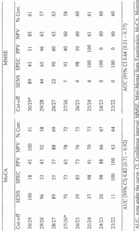
Le troisième article (Chapitre III) a pour but de comparer les propriétés psychométriques du Montreal Cognitive Assessment (Nasreddine et al., 2005) et du Mini-Mental State Examina/ion (Folstein, Folstein, & McHugh, 1975) pour détecter
le trouble cognitif léger dans un échantillon de parttctpants apnéiques et no n-apnéiques âgés de 55 ans et plus. L'étude cherchait aussi à déterminer si l'écart de validité entre les tests de dépistages pouvait être attribuable à une différence à propos du sous-type de trouble cognitif léger (amnésique ou non-amnésique ; domaine simple ou multiple). Soixante-sept participants apnéiques et 64 non-apnéiques ont effectué une évaluation neuropsycho logique, ainsi qu'une nuit d'enregistrement polysomnographique. Les résultats ont révélé que, chez les non-apnéiques, les deux tests avaient une validité discriminante similaire. Cependant, chez les participants apnéiques, le Montreal Cognitive Assessment s'est montré supérieur au Mini-Mental State Examination pour identifier les apnéiques ayant un trouble cognitif léger. En fait, le Mini-Mental State Examination ne devrait pas être utilisé pour dépister les troubles cognitifs chez les apnéiques. Nos analyses des différences de sous-types de trouble cognitif léger entre les apnéiques et les non-apnéiques se sont révélées non significatives. Des analyses supplémentaires sur chaque test neuropsychologique ont révélé que les non-apnéiques ayant un trouble cognitif léger avaient un score significativement plus bas au Test de Dénomination de Boston comparativement aux non-apnéiques sans trouble cognitif léger. Cette différence n'a pas été observée dans le groupe d'apnéiques. Ce résultat pourrait expliquer la validité plus élevée du Mini -Mental State Examination chez les non-apnéiques. Les implications théoriques et cliniques de ces résultats seront discutées dans le dernier chapitre de la thèse (Chapitre IV).
Mots-clés : Apnée obstructive du sommeil, cognition, neuropsychologie, plainte cognitive subjective, trouble cognitif léger, Montreal Cognitive Assessment, Mini-Mental State Examination, vieillissement.
XIV
ABSTRACT
Obstructive sleep apnea (OSA) increases the risk of mild cognitive impairment and dementia in the elderly. In this context, being able to identify patients at risk of cognitive decline could improve clinical decisions. The work presented in this thesis aimed at evaluate the effeteness of clinical tools to identify individuals at risk of abnormal cognitive decline in a sample ofapneic aged 55 years and older.
The first article (Chapter I) is a literature review on OSA pathophysiology, cognitive dysfunctions associated with OSA and also mechanism linked to these dysfunctions. This paper review markers of cognitive dysfunctions measured by electroencephalography and neuroimaging. ft also reviews the effect of continuous positive airway pressure to improve cognitive functioning. Recent findings regarding the relationship between OSA and the risk of abnormal cognitive decline are final! y addressed.
The second article (Chapter II) aim at verifying whether subjective cognrt1ve complaint could be a useful tool for identifying apneic individuals aged 55 and older, at risk of abnormal cognitive decline. The objective was to determine if the severity of OSA contributed to cognitive complaint, but also to explore the link: between subjective cognitive complaint and objective cognitive impairment. Fifty-seven apneic and 54 non-apneic participants aged between 55 and 85 years underwent a polysomnographic recording followed by a neuropsychological assessment. They completed questionnaires assessing mood, sleep quality and their perceptions of their cognitive functioning. Our analyzes revealed that the severity of OSA (i.e. Respiratory index, oxygen saturation, and sleep fragmentation) did not significantly contribute to the severity of subjective cognitive complaint. In fact, higher scores on mood and sleep quality questionnaires significantly contribute to increasing cognitive complaint. The results also showed that apneic participants are not able to accurately evaluate their objective cognitive functioning. We found, among participants without objective cognitive impairment, that apneic participants bad more cognitive complaint compared to non-apneic. Conversely, among participants with objective cognitive impairment, apneic participants bad fewer cognitive complaint compared to non-apne1c.
The third article (Chapter III) was designed to compare the psychometrie properties of the Montreal Cognitive Assessmenl (Nasreddine et al., 2005) and the Mini-Mental State Examina/ion (Folstein et al., 1975) to detect mild cognitive impairment in a sample of apneic and non-apneic participants aged 55 years and older. The study a Iso investigated whether the difference in validity between screening tests could be the consequence of a mild cognitive impairment subtype difference (amnesie or no
n-anmesic, single or multiple domain). Sixty-seven apneic and 64 non-apneic participants had a neuropsychological assessment and a polysomnographic recording. The results showed that, in non-apneic participants, both screening tests had similar discriminant validity. However, among apneic participants, the Montreal Cognitive Assessment was superior to the Mini-Mental Stale Examination. lndeed, the Mini-Mental State Examination should not be used to screen for cognitive impairment in apneic patients. We found no significant mild cognitive impairment subtype differences between apneic and non-apneic. Additional analyses on neuropsychological tests revealed that non-apneic participants with mild cognitive impairment had a significant lower score at the Boston Naming Test compared to non-apneic without mild cognitive impairment. This difference was not observed in the apneic group. This result could explain the higher validity of the Mini-Mental State Examination in non-apneic parttctpants. The theoretical and clinical implications of these results will be discussed in the last chapter of the thesis (Chapter IV)
Keywords: Obstructive sleep apnea, cognition, neuropsychology, subjective cognitive complaint, mild cognitive impairment, Montreal Cognitive Assessment, Mini-Mental State Examination, aging.
INTRODUCTION
L'apnée obstructive du sommeil (AOS) est un problème de santé majeur, qui touche environ 20 % de la population âgée (Ancoli-Israel, Kripke, et al., 1991; Duran, Esnaola, Ru bio, & Iztueta, 2001; Johansson, Alehagen, Svanborg, Dahlstrom, & Brostrom, 2009; Sforza, Chouchou, et al., 201 0; Young, Peppard, & Gottlieb, 2002). L' AOS cause des perturbations du sommeil et des hypoxémies intermittentes, qui peuvent entraîner de la somnolence diurne, des déficits cognitifs et des dysfonctions cérébrales (Daulatzai, 2015). L'AOS est aussi recormue comme un facteur de risque pour l'hypertension, le diabète, les maladies cardiovasculaires, les accidents vasculaires cérébraux et la démence (Ancoli-Israel, Kripke, et al., 1991; Kato, Adachi, Koshino, & Somers, 2009; Silverberg, Oksenberg, & laina, 1998; Somers et al., 2008; Yaffe et al., 2011; Young et al., 2009). Plusieurs études récentes de larges cohottes ont identifié l' AOS comme un facteur de risque du trouble cognitif léger et de la démence (p. ex. maladie d'Alzheimer) (Chang et al., 2013; Leng, McEvoy, Allen, & Yaffe, 2017; Osorio et al., 2015; Saint Martin et al., 2016; Yaffe et al., 2011). Une étude a d'ailleurs montré que la présence d' AOS était associée à un diagnostic plus précoce de trouble cognitif léger et de maladie d'Alzheimer chez des sujets âgés de 55 à 90 ans (Osorio et al., 20 15). D'autres études suggèrent que les problèmes cognitifs associés à l' AOS pourraient être, du moins partiellement, réversibles à l'aide d'un traitement par pression positive continue (Bardwell, Anco li-Israel, Berry, & Dimsdale, 2001; Bedard, Montplaisir, Malo, Richer, & Rouleau,
1993; Ferini-Strambi et al., 2003; Lau, Eskes, Morrison, Rajda, & Spurr, 201 0; Montserrat et al., 2001; Munoz, Mayoralas, Barbe, Pericas, & Agusti, 2000; Naegele et al., 1998; Pan & Kastin, 2014). Dans ce contexte, il est donc crucial d'identifier rapidement les individus apnéiques à risque de développer une démence. Malgré une augmentation de la sensibilisation des intervenants de la santé concernant le rôle de l' AOS dans le déclin cognitif anormal, le transfert des connaissances aux pratiques
cliniques ne semble pas s'effectuer facilement. En fait, les cliniciens ne disposent pas d'outils valides pour identifier les patients apnéiques âgés qui ont des problèmes cognitifs ou qui sont susceptibles de développer de démence.
Cette thèse vise à valider des outils cliniques dans un échantillon d'individus d'âge moyen et plus âgés présentant de l' AOS afin que les professionnels de la santé puissent identifier les individus risquant de présenter un déclin cognitif anormal. Parmi les outils disponibles, la plainte cognitive subjective est une information facilement accessible pour les cliniciens. Bien que la plainte cognitive ait longtemps été considérée comme faisant partie du vieillissement normal, plusieurs études ont démontré que cette dernière pourrait augmenter le risque de démence (Giffard et al., 2014; Jonker, Geerlings, & Sclm1and, 2000; Mendonca, Alves, & Bugalho, 2016; Reisberg, Shulman, T01·ossian, Leng, & Zhu, 2010). D'ailleurs, la plainte cognitive subjective fait partie des critères diagnostiques du désordre neurocognitif léger (American Psychiatrie Association. Diagnostic and statistical manual of mental disorders (5th ed.), 2013). Dans la mesure où une grande proportion de personnes présentant de l' AOS ont des plaintes cognitives, il serait utile pour les cliniciens du sommeil d'en saisir la nature, mais aussi de comprendre le lien entre la plainte cognitive et les atteintes cognitives objectives régulièrement observées dans l' AOS. À ce jour, plusieurs questionnaires de plainte cognitive ont été développés pour la population âgée, il serait donc intéressant d'en connaître la valeur clinique chez les apnéiques d'âge moyen et plus âgés.
Les outils de dépistage permettant d'objectiver des atteintes cognitives, tel que le Montreal Cognitive Assessment (Nasreddine et al., 2005) et le Mini-Mental State Examina/ion (Folstein et al., 1975) sont régulièrement utilisés par les cliniciens.
.., .)
Toutefois, ces tests n'ont pas été validés pour dépister le trouble cognitif léger 1 dans une population apnéique. De plus, ces deux tests présentent des différences dans la nature des fonctions cognitives évaluées et leur niveau de difficulté ne semble pas équivalent. Il se pourrait donc qu'un des deux outils soit plus sensible pour détecter le trouble cognitif léger chez les apnéiques. Puisque le trouble cognitif léger est lui aussi un facteur de risque de démence (Petersen & Jack, 2009), il serait important que les cliniciens soient en mesure d'identifier les patients apnéiques ayant des atteintes cognitives afin de prendre des décisions cliniques plus éclairées et d'assurer un suivi adéquat.
Cette thèse pourrait avoir des retombées importantes au plan clinique. En effet, en identifiant correctement les patients apnéiques de plus de 55 ans susceptibles de présenter un déclin cognitif anormal, les cliniciens pourront améliorer la prise en charge et le suivi de ces patients. Ce travail pourrait éventuellement contribuer à améliorer la santé cognitive de plusieurs patients apnéiques âgés à l'aide d'un traitement (p. ex. pression positive continue ou appareil d'avancement mandibulaire) et peut-être même retarder les manifestations du processus neurodégénératif.
La première partie de l'introduction débutera avec un premier article qui présente une revue de littérature de l' AOS et des troubles cognitifs qui lui sont associés. La deuxième partie présentera le trouble cognitif léger et les mécanismes neurodégénératifs possiblement associés à l' AOS. La troisième partie traitera de la plainte cognitive, de son évolution vers un trouble cognitif, et de son lien avec l' AOS. La quatrième partie traitera des outils de dépistage. Cette introduction nous amènera par la suite à la présentation des objectifs et des hypothèses de cette thèse.
1
Les différents concepts associés au fonctionnement cognitif anormal chez les personnes âgées seront
ARTICLE 1: COGNITIVE IMPAIRMENT IN OBSTRUCTIVE SLEEP APNEA.
AUTEURS: Katia Gagnon, Andrée-Ann Baril, Jean-François Gagnon, Maxime Fmiin, Anne Décary, Chantal Lafond, Alex Desautels, Jacques Montplaisir, Nadia Gosse lin.
Pathol Biol (Paris). 2014 Oct;62(5):233-40.
TITRE EN FRANÇAIS :Troubles cognitifs dans l'apnée obstructive du sonuneil
ABSTRACT
Obstructive sleep apnea (OSA) is characterised by repetitive cessation or reduction of airflow due to upper airway obstructions. These respiratory events lead to chronic sleep fragmentation and intermittent hypoxemia. Severa! studies have shawn that OSA is associated with daytime sleepiness as weil as cognitive dysfunctions, including impairments of attention, episodic memory, working memory, and executive functions. This paper reviews the cognitive profile of adults presenting OSA and discusses the relative role of altered sleep quality/quantity and hypoxemia in the aetiology ofthese cognitive deficits. Markers of cognitive dysf1.mctions such as those measured with waking electroencephalography and neuroimaging are also presented. The effects of continuous positive airway pressure (CP AP) on cognitive
5
functioning and the possibility of permanent brain damage associated with OSA are also discussed. Finally, this paper reviews the evidence suggesting that OSA represents an increased risk of developing mild cognitive impairment and dementia in the aging population and stresses the importance of its earl y diagnosis and treatment.
Key words: Obstructive sleep apnea; cognition; neuropsychology; continuous positive airway pressure; electroencephalography; neuroimaging; aging.
RÉSUMÉ
L'apnée obstructive du sommeil est caractérisée par des pauses respiratoires ou des
réductions du débit aérien répétées qui sont dues à une obstruction des voies aériennes supérieures. Ces évènements respiratoires entraînent une fragmentation
clu·onique du sornn1eil et une hypoxémie intermittente. Plusieurs études ont montré
que l'apnée obstructive du sommeil est associée à la somnolence diurne ainsi qu'aux dysfonctions cognitives incluant des déficits au niveau de l'attention, de la mémoire épisodique, de la mémoire de travail et des fonctions exécutives. Cet article examine le profil cognitif observé chez les adultes présentant l'apnée obstructive du sommeil.
Les rôles spécifiques de l'altération de la qualité/quantité du so1ru11eil et de
l'hypoxémie dans l'étiologie des dysfonctions cognitive seront également abordés. Les marqueurs de dysfonctions cérébrales tels que ceux mesurés en électroencéphalographie et en neuroimagerie seront aussi présentés. Les études examinant l'efficacité du traitement par pression positive continue afin d'améliorer les déficits cognitifs dans cette population seront discutées ainsi que la possibilité que l'apnée obstructive du sommeil puisse causer des donm1ages permanents au cerveau. Enfin, cet aLiicle présente les récents résultats concernant la relation entre l'apnée
obstructive du so11U11eil et le risque de développer un trouble cognitif léger et une
démence dans la population âgée et souligne l'importance d'un diagnostic précoce en vue d'initier un traitement de l'apnée obstructive du sonuneil.
Mots clés: Apnée obstructive du sornn1eil; cognition; neuropsychologie; pression
7
1. Introduction
Obstructive sleep apnea (OSA) is a sleep disorder characterised by frequent breathing cessation and/or reduction of ait·flow due to partial or complete obstruction of the. upper respit·atory airways. These respiratory events occurring during sleep lead to intermittent hypoxemia and micro-arousals or awakenings. Sleep fragmentation is one of the leading causes of excessive daytime sleepiness, a predominant clinical manifestation of OSA. Daytime sleepiness in OSA is known to increase from two to se ven times the risk of road accidents. [ 1] However, des pite an increasing awareness of this health problem, 80% of individuals remain undiagnosed and untreated. [2]
1.1. Nighttime and daytime symptoms
Spouses or relatives sleepit1g in or near the bedroom of individuals presenting OSA usually report loud snoring that sometimes disturbs theit· own sleep. OSA patients report awaking breathless and choking during the night. In addition, they evaluate theit· sleep as non-restorative and report fi·equent awakenings overnight. [3,4] When waking up in the morning, subjects feel tired and may also have headaches and dry throat. [3] Most OSA patients complain of daytime sleepiness that usually occurs when performing tasks that require Jess attention (i.e. reading or watching television). [3,5] With increasing OSA severity, sleepiness may occur while they drive, eat or work. [3,5] Most frequent cognitive complaints are a decrease in alertness, short-tenn memory problems, and Jack of concentration. [3,5] However, our preliminary study recently showed a Jack of insight in individuals with OSA with significantly more cognitive impairment. [6] Relatives and patients observe changes in theit· mood and personality, which includes irritability, depressed mood and anxiety. They often have sexual dysfunctions including Joss of libido and impotence. [3]
OSA is diagnosed through clinicat history and polysomnography (PSG). Clinicat history should include a complaint of excessive daytime sleepiness or two of the following symptoms: choking at night, recurrent awakenings, sensation of non-restorative sleep, and fatigue or inability to focus. [5] The PSG recording enables the confirmation of the OSA diagnosis. According to the recent update of the American Academy of Sleep Medicine criteria, [7] obstructive apnea is a decrease grea ter th an 90% of base! ine airflow for at !east 10 seconds, which is accompanied by a sustained or an increased respiratory effort. Obstructive hypopnea is a decrease greater than 30% of the aidlow amplitude for at !east 10 seconds accompanied with either a desaturation higher than 3% or an arousal. Frequency of respiratory events is measured by the apnea-hypopnea index (AHI) obtained by summing the respiratory events and dividing by the number of hours of sleep. Recommended criteria for the diagnosis of OSA are an AHI higher than 5 for young and middle-aged adults while criteria of AHI higher than 20 was suggested for the elderly. [8,9] OSA severity depends on the presence and intensity of daytime sleepiness and the fi·equency of respiratory events. The assessment of the severity of sleepiness relies on the type of task during which the episode occurs (i.e. reading vs. driving) and the impact of sleepiness on the social and functional aspects of life. The assessment of severity of respiratory events includes tlu·ee levels in young and middle-aged adults: mild OSA (AHI between 5 and 15), moderate OSA (AHI between 15 and 30) and severe OSA (AHI higher than 30). [5]
1.3. Epidemiology
Epidemiological findings vary according to age, sex and apnea severity. When a criterion of an AHI ~ 15 is used, the prevalence is estimated at 2 to 14% in middle -aged adults (39-59 years), but shows an increase to 20% in those aged 60 to 95 years. Some authors suppose that the increase in prevalence with age is due to anatomical
9
changes in the upper respi.ratory airways or the coexistence of a medical disorder.
[! 0] Others explain that OSA is more prevalent in the elderly than in middle-aged individuals because OSA cases accumulate from a constant incident rate. [2] Moreover, the prevalence is twice as high among men (25%) than women (Il%). Finally, in a population aged between 39 and 99 years old, mild OSA is more prevalent (29%) than moderate to severe OSA (18%). [2]
1.4. Pathophysiology
Partial or complete obstructions of the upper airway are due to the relaxation of the
throat muscles and tongue, which collapse the ai.rways during sleep. Airway
obstructions create intermittent hypoxemia and hypercapnia. Peripheral (a011ic) and
central (medulla oblongata) chemoreceptors both stimulate respi.ratory centers to
increase ventilation since they are sensitive hypoxemia and hypercapnia. The end of
respi.ratory events usually occurs through the occurrence of awakening or arousal and
is associated with sympathetic activation. [ l 1] OSA also creates inflammation and
endothelial dysfunction, which reduce vascular elasticity and increase coagulation. Altogether, these factors predispose to atherosclerosis. [ 11] The combination of reduced oxygenation reaching tissues and vascular damages can lead to cellular
dysfunctions in severa! organs such as the heart and the brain.
1.5. Risk factors and comorbidities
Overweight, obesity, central body fat distribution and large neck gu·th have been
identified as important risk factors for OSA. [ 12] Morphological factors such as
dimorphisms related to the size or position of the mandible or maxilla, narrow nasal
cavity, a low soft palate, and enlarged tonsils also play an important role in the development ofOSA. [12] Other factors may contribute to the risk ofOSA, including hormonal changes, alcohol consumption, smoking and nasal congestion, but thei.r
causal role is not weil established. Hypertension, diabetes, coronary heart disease, myocardial infarction, congestive heart failure and stroke were found to be associated with OSA. The emergence of these comorbid conditions may be due, in part, to common risk factors (i.e. obesity and hypertension), but OSA may also have a causal rote in these diseases since hypoxemia and hypercapnia can lead to vascular dysfunctions. [13] Genetically, it has been reported that the isoform E4 from the ApoE gene, identified as a risk factor for Alzheimer disease, is more frequently found in OSA patients than in healthy control individuals, [ 14, 15] but a meta-analysis based on published literature failed to support this association between ApoE4 and OSA. [16]
1.6. Continuous positive ai.rway pressure treatment description
Continuo us positive ai.rway pressure (CP AP) improves overall OSA symptoms by avoiding obstructions and keeping the upper au-ways open using air pressure, which consequently decreases hypoxemia and sleep fragmentation. [17] CP AP treatment globally improves subjective daytime functioning and performance in cognitive tests (see details in the following sections). [18 -24] In addition, CPAP treatment normalizes electroencephalographic (EEG) recordings performed during the waking state among OSA subjects and this normalization in EEG is associated with decreased daytime sleepiness. [25,26] However, CP AP should be used for at !east four hours per night for a subjective effect on sleepiness, and a minimum of six hours per night for an effect on objectively measured sleepiness. [27]
2. Cognitive impau·ment in OSA
Severa! studies have investigated the cognitive pro file of OSA patients and recently, a meta-review analysed neurocognitive functions in OSA based on systematic reviews and meta-analyses. [28] Published studies have identified attention, episodic memory,
Il
working memory, and executive functions as the most affected cognitive domains in
OSA, [22,29-36] whereas most aspects of language are preserved in this population [22,29,38-45] and heterogeneous results are found for psychomotor speed. [46] ln the
following section, cognitive deficits observed in OSA for each cognitive domain are
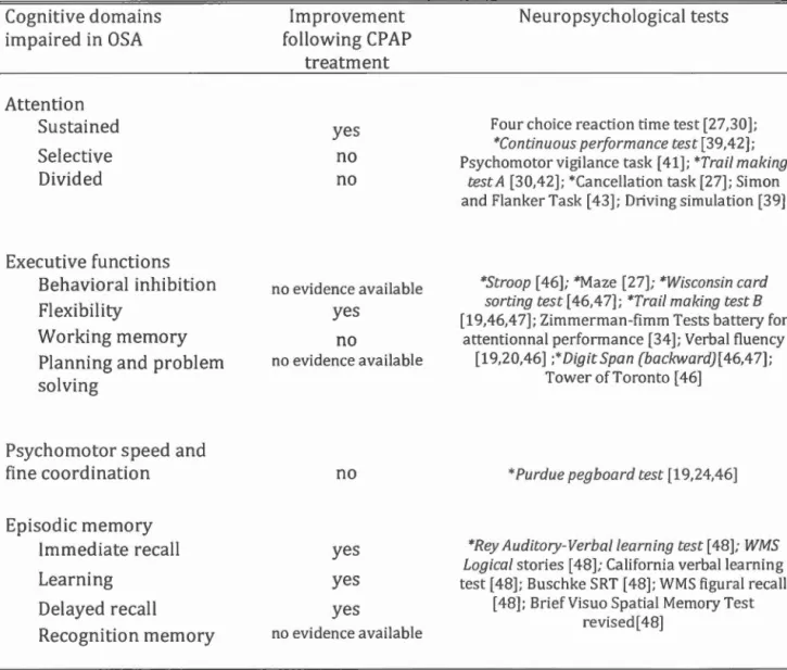
described. Table 1 presents an overview of cognitive domains affected in OSA, neuropsychological tests the most sensitive and the efficiency of CPAP to improve each cognitive function.
2.1. Attention
Attention IS generally divided into sustained, selective and divided attention.
Sustained attention, which also refers to vigilance in the present paper, is a mechanism that involves alertness and receptivity to stimuli for a continuing time period; selective attention enables to treat or ignore stimuli according to their relevance; and divided attention allows multiple tasks to be executed simultaneously. [ 40] Severa! studies have demonstrated that OSA subjects show impairments for ali tlu·ee attention components [29,39,41] and these observations were confi.rmed by a meta-review. [28] When compared to healthy controls, OSA subjects have more
lapses and/or longer reaction times in tasks requiring sustained attention, selective
attention, or vigilance. [30,32,41,43-45] In conditions requiring divided attention,
such as driving in a simulator while performing another cognitive task, individuals
with OSA showed an increase in reaction times and "off-road" events compared to controls. [ 41] Thus, the efficient allocation of attentional resources to multiple relevant stimuli bas proven difficult in individuals with OSA. Given the severity and the extent of attention deficits, it bas been suggested that vigilance and attention deficits could influence other aspects of cognitive deficits attributed to OSA.
[36,37,41] According to this hypothesis, attention deficits probably worsen other
cognitive deficits that are secondary to OSA, such as executive functions and episodic memory impairments.
A review of cognitive changes associated with CPAP treatment reported that li of the 17 studies showed a significant improvement in vigilance and attention with CPAP. (47] It was also observed that as few as 15 days of CPAP treatment was sufficient to obtain significant improvement in sustained attention tests. [22] However, despite significantly improving attention, CPAP is often unable to fully normalize attention processes in OSA. In fact, according to a study by Lau et al., selective and divided attention deficits were still observed after three months of CPAP treatment when compared to control subjects. [23] These results suggest that attention deficits in OSA are partially caused by sleep fragmentation and hypoxemia, but that OSA probably causes permanent damage to regions of the brain involved in attention processes. (22,23,29]
2.2. Executive functions
Executive functions are a complex concept that compnses many cognitive skills including behavioural inhibition, mental flexibility, working memory, fluid reasoning, and problem solving. They allow individuals to adaptively use their basic skills (e.g. language, visuo-perceptual, memory) to perform adequately in a changing environment. [38] A recent meta-analysis reported that executive ft.mctions are impaired in OSA for ali five sub-domains studied, namely: inhibition, shifting, updating/monitoring information in working memory, generating new information and fluid reasoning or problem solving. (42] Inhibition, which refers to the capacity to stop an automatic or ongoing response to an event, [49] is required in cognitive tests such as Stroop (inhibition condition) or Go No-Go tasks. In these tasks, OSA subjects make significantly more mistakes or have increased reaction times compared to contrais. (50] Moderate to severe OSA subjects also made more impulsive errors on maze completion. [30]
13
Shifting, or mental flexibility, is the ability to move from one cognitive or behavioural strategy to another. In the Wisconsin Card Sorting Tests, subjects with OSA showed an increased number of perseverative responses when compared to control subjects. [50,51] Reduced mental flexibility was also documented in studies using the Trail making test B and the computerized Zimmerma1m-Fimm Tests battery for Attentional Performance, where OSA subjects required more time to complete the tasks than controls. [30,50,51]
Working memory refers to the ability to retain, manipulate, update and monitor information for the duration of a task. The concept of working me mory encompasses the central executive, which enables information manipulation by subsystems. [52] For example, the ability to mentally reverse a digit string or visual sequences to repeat it backward reflects a working memory capacity. Studies using the Digit backward test showed that OSA subjects perform poorly on this working memory task compared to contra ls. [ 50,51] Saunamaki and Jehkonen (2007) a Iso fou nd th at working memory was among the most fi·equently impaired components of the executive functions in this population. [ 48] However, more studies investigating working memory among OSA patients are needed to confirm these observations.
Problem solving, which involve the evaluation and selection of a sequence of actions in order to achieve a goal [40], was found to be impaired in individuals with OSA. In fact, in tasks that are typically used to assess this component of executive functions, namely the Tower Tasks, one study showed that OSA subjects need a higher number of steps before solving problems. [50] Some executive aspects of verbal behaviour, such as mental processing speed, flexibility and synthetic skills are also diminished in adults with OSA, despite otherwise normal language skills. [22,30,50]
In sum, studies have found deficits in most aspects of executive functioning in OSA, characterised by decreased processing speed, increased perseverative responses or
behaviours, impulsivity, and difficulty with problems solving. However, it is important to mention the high leve! of heterogeneity in the results found among studies that could be partly due to population heterogeneity (including premorbid intellectual functioning and education), various disease severities and the large number of executive function tasks. [ 42]
Both a recent meta-analysis and a review showed that CP AP treatments induce a small to moderate improvement in executive functions. [42,53] For example, a study found an improvement of mental flexibility (Trail B) and verbal fluency (semantic) in OSA after a short CPAP treatment (15 days). Long treatments (four months) did not result in any further improvement on executive cognitive tests. [22] Behavioural inhibition and working memory were not irnproved following a short ( 15 da ys) and a long (four months) CP AP treatments when compared to pre-treatment condition [22]. Moreover, it was reported that mental flexibility of OSA subjects did not reach the level of control subjects after a three-month treatment. [23] Taken together, these studies suggest that only certain aspects of executive functions improve after a short CP AP treatment wh ile not systematically reaching the leve! of controls performance. [21-23] The fact that other cognitive functions, such as attention and vigilance, are necessary to perform most executive function tasks, and that OSA may cause permanent damage to the prefiontal cortex [39] could explain part of the variability in studies investigating the effects of CP AP on this cognitive domain.
2.3. Psychomotor speed and fine coordination
Tests involving psychomotor speed and fine coordination are generally used to investigate motor dysfunctions that occLu· in the context of intact capacity for normal movement. In individuals with OSA, reduction of manual dexterity was reported on Purdue Pegboard Test [21 ,30,50] and fine mo tor coordination was found to be more sensitive to chronic hypoxemia than sleep fragmentation. [29] A recent literature
15
review showed that ha if of the published studies reported that patients with OSA have a reduced information processing speed compared to control subjects. [46] This reduced information processing probably impacts performances in severa! cognitive tasks such as those evaluating executive f1.mctions (i.e. Tower tasks or Trait B) and visual attention. It is not surprising that OSA patients performed worse than healthy control subjects on ali timed tests that included a visuomotor coordination component. [47]
Contrary to other cognitive domains, psychomotor speed and fine coordination are not significantly improved with CP AP treatments, which suggests that OSA may cause permanent damages to cotiical and subcortical areas involved in motor skills. [17, 26,46]
2.4. Episodic memory
Episodic memory was extensively studied among OSA subjects and refers to the memorization of verbal or visual information in a spatio-temporal context. [54] Episodic memory tasks generally include immediate recall, total recall over multiple trials or learnings, delayed recall, and recognition memory. Learning and recall of list of words, such as in the Rey Auditory-Verbal Learning Tests and the California Verbal Learning Tests, are examples of episodic memory tests. A recent met a-analysis showed that OSA patients have different patterns of deficits for verbal and visual episodic memory. Indeed, for verbal material, each memory component, namely immediate and delayed recall, learning and recognition, was impaired in OSA patients when compared to controls. [55] However, in visuospatial episodic memory tasks, OSA patients showed impairment only for immediate and delayed recalls, and had normal results for learning slope and recognition. [55,56] It was suggested that this impairment was not entirely accounted for by a reduction of attention or by OSA severity. [57]
A review on the effect of CP AP treatments found an improvement in me mory in about half of the studies. [47] Although all components of verbal episodic memory evaluation are affected in OSA [55], a three-month CPAP treatment resulted in normalisation of performances for immediate and delayed memory, and for both verbal and visuospatiallearning. [22,23]
2.5. Role of sleep characteristics and hypoxemia in cognitive deficits observed m OSA
OSA causes sleep disruption, which leads to changes in sleep architecture and excessive daytime sleepiness. Intermittent hypoxemia is also a major consequence of OSA. Severa! studies have aimed to understand the specifie role of altered sleep quality/quantity and hypoxemia in the aetiology of cognitive dysfunctions in this population.
2.5.1. Sleep architecture and sleep· fragmentation
Sleep fragmentation is the most systematically studied sleep variable in association with cognition among this population. According to a critical review and a recent meta-review, the more severe is sleep fi·agmentation, the more impaired are the performance on attention and vigilance tests. (26, 28] Learning and memory also seem affected by sleep fragmentation in OSA patients. In fact, in a study by Ojonlagic et al. (2014), the authors investigated the sleep-dependent memory consolidation in 20 OSA patients and 20 control subjects. They found that OSA patients had Jess overnight improvement on the motor sequence learning task when compared to control subjects. More specifically, sleep fragmentation predicted overnight improvement on the motor sequence learning task. [58]
17
Sleep architecture and more specifically the percentage of each sleep stages may have an impact on daytime cognitive functioning in the OSA population, since most PSG studies found small but significant changes in sleep architecture. In fact, most PSG studies documented an increased percentage of stage 1 sleep, as weil as elevated arousal and micro-arousal indexes, white no change for total sleep time was generally observed. In contrast, a majority of studies found a decrease in the percentages of stages 3-4 sleep and REM sleep. [ 43,59] However, an association between changes in the percentage of each sleep stage and the cognitive profile of OSA subjects has not been documented in the literature. Considering that Joss of NREM and REM sleep is known to be associated with decreased performance in tasks involving episodic memory in healthy individuals [60], changes in sleep architecture associated with OSA may independently contribute to their cognitive deficits, but this hypothesis needs to be tested.
Studies evaluating the effect of excessive daytime sleepiness on cognitive deficits in OSA found that sleepiness can only partially account for cognitive deficits. [ 41,43,61 ,62] Indeed, studies on the effect of CP AP treatment on cognition reported a global improvement of cognitive functions. [33,48,56-59] However, CPAP treatment fails to improve fine motor coordination and sorne subjects still have memory and executive dysfunctions after treatment. [21 ,23,24,29] This suggests that history of hypoxia and/or chronic sleep fragmentation may cause sorne permanent damage to cortical and subcortical structures and that sleepiness cannot explain ali cognitive impairments. In parallel, it is impot1ant to note that comorbidities associated with OSA such as obesity, diabetes, and hypertension have recently been identified as factors that contribute independently to sleep fragmentation and cognitive deficits in OSA. [63]
Animais and brain imaging studies have found that OSA, and more specifically hypoxemia, causes neuronal damage in multiple regions. [39,64] Hypoxemia
followed by re-oxygenation results in similar changes as those observed in an
ischemic/reperfusion injury. [65] This type of injury increases free radicals and inflammation, which are particularly damaging for endothelial and neuronal integrity,
especially in the hippocampus and the frontal cortex. [65,66] Furthermore,
endothelial dysfunction, caused by OSA, increases blood pressure and coagulation
that both predispose and increase risk of si lent stroke in OSA. [ 11]
Large population studies have confirmed a small, but significant association between
hypoxemia and some cognitive deficits, including attention impairments, slow processing speed and executive dysfunctions. [39,50, 61] The heterogeneity found in the literature for the association between hypoxemia and cognitive functioning is
possibly due to the role of non-controlled variables, such as age or premorbid
intellectual functioning. Animal models have shown that intermittent hypoxemia was
associated with impairments in the execution component of attention (i.e. set-shifting)
and to a particular vulnerability to neuronalloss in the frontal lobe. [67,68]
2.6. Risk factors for cognitive impairment in OSA
Severa! studies investigated the association between the severity of OSA as measured
with AIH and the cognitive deficits found in this population. Heterogeneous results
were found with some studies showing that adults with severe OSA (AHI 2: 30) are more likely to present cognitive deficits than those with mild or moderate OSA, [69] while other studies found no association. [42-44, 61, 70] One problem that may limit the interpretation of this Jack of association is that there is currently insufficient data for mild to moderate OSA, since most studies only included moderate and severe OSA patients. [42]
19
Age was also found to be associated with greater cognitive dysfunctions, where
middle-aged adults with severe OSA were found to be more at risk of cognitive
deficits than younger adults with the same OSA severity. [71] A study comparing
young and aider OSA subjects showed Group (i.e. OSA versus control) and Age
effects on severa! cognitive tests, suggesting that bath OSA and age contribute to
cognitive dysfunctions in aider individuals with OSA. [33]
Premorbid cognitive functioning, or cognitive reserve, seems to have a protective effect on cognitive deterioration in OSA. In fact, OSA subjects with higher intelligence have similar performance on attention/alertness tasks when compared to healthy contrais with equivalent intelligence. In contrast, OSA subjects with lower intelligence have lower performance on attention tests compared to contrais with
similar intelligence. [72] Future studies should include premorbid intelligence or
cognitive reserve when evaluating the impact of OSA on cognitive functioning.
Severa! comorbidities associated with OSA, such as obesity, hypertension, diabetes,
congestive heart failure and cerebrovascular accidents, known to be independently
associated with cognitive deficits, may worsen neurocognitive function in subjects
with OSA. [63, 65] [t was reported that ApoE4 allele carriers among OSA subjects
had a lower performance on spatial working memory tasks when compared to
non-carrier OSA subjects while this genetic effect on cognition was not observed in
contrais. [73] In pat·allel, a recent study found a poorer performance on cognitive tests requiring memory and executive functions in ApoE4 carriers with a moderate-severe
OSA (AHI :::=: 15) than what it was observed in ApoE4 carriers without OSA. [74]
These results suggest that OSA and genetic interaction leads to greater risk of
cognitive dysfunctions.
Spectral analysis of the waking EEG allows evaluating the content of the EEG signal and is recognised as an index of cerebral functioning. Severa! studies found an EEG slowing across ali scalp regions in OSA subjects when compared to controls. [26, 75, 76] More specifically, an increase in the relative theta and delta powers in parietal, temporal and occipital regions was reported in patients with severe OSA when compared to control subjects. [77] EEG slowing was associated with a disruption of sustained attention on cognitive tests, which results in increasing omission and reaction times during a task. [75] It has been observed that more EEG slowing was
associated with more time spent with oxygen saturation under 90%. [76] An
improvement of EEG slowing across ail scalp regions was found following a CP AP treatment and was linked to an improvement in cognitive functions and daytime sleepiness. [26,78] When moderate to severe OSA were compared to control subjects before and after six months of CP AP treatments, quantitative waking EEG showed an increase of relative delta power in ali cortical regions for both before and after CPAP,[77] suggesting that sorne damage due to OSA may permanently alter brain function.
Event-related potential (ERP) recordings during an attention task are a widely used method to assess cerebral dysfunction and attention deficits. [79] A recent review on ERP abnormalities in OSA highlighted the increased P300 latency in visual and auditory tasks observed in this population. [80] The P300 component is associated with the occurrence of a target stimulus and represents classification speed (latency) and attention resources allocated to the task (amplitude). [81,82] A recent study evaluating the association between ERP abnormality and EEG slowing found that the tate portion of the P300 was associated with an increase in theta power in OSA, but this correlation was not found in control subjects. Considering that an increase in theta power at rest generally reflects a general vigilance decrement, this result suggests that attention deficits are associated with decreased vigilance in OSA. [n this study, lower P3a amplitude was correlated with a decrease in beta 1 spectral power in
21
OSA patients. [83] However, contrary to the P300, P3a amplitude was not correlated with slow EEG fi:equencies, which suggests that P3a anomalies were not caused by a general vigilance decrement during the attention task in this population. Other studies investigated ERP components that retlect automatic and volitional orientation of attention towards relevant and irrelevant stimuli and showed abnormalities in most ERP components in OSA subjects compared to control subjects. [84,85] In a review by Raggi and Ferri (20 1 0), it was a Iso shown that P300 amplitude correlated with
higher respiratory event index. [80] Heterogeneous results were found for the
efficiency of CP AP to restore ERP: some studies showed an improvement in P300
latency after CP AP treatment, [86,87] but other studies did not fi nd any improvement. [88,89] Interestingly, one study found that abnormalities observed 111 P300 were irreversible, but only in the elderly. [90]
4. Neuroimaging findings
Studies usmg different brain imaging techniques investigated neuroanatomical changes and cerebral functioning to identify the consequences of OSA on the brain, and to understand the causes of cognitive impairments in this population.
Structural brain imaging studies using magnetic resonance imaging (MRI) combined with voxel-based morphometry showed reduced grey matter density in subjects with OSA on overall volume and in distinct brain regions, namely the parietal, frontal and temporal lobes; the hippocampus, the amygdala, the anterior cingulate, the caudate nucleus and the cerebellum (See Ferini-Strambi 2013 for a review [91]). Studies
showed positive correlations between changes in grey matter density in the
hippocampus, caudate nucleus and frontal regions, and cognitive alterations in episodic memory, attention and executive functions. [66,92]
More recently, diffusion tensor imaging was used to characterize white matter 111 OSA patients. The integrity of a wide range of white matter structures, including
medullary, cerebellar, basal ganglia, prefi·ontal and fi·ontal, limbic, insular, cingulum
bundle, external capsule, corpus callosum, temporal, occipital, and corona radiata
regions, was shown to be affected. [93,94] The structures showing white matter
abnormalities could be associated with specifie cognitive deficits and altered mood in
subjects with OSA, but further studies are needed to confirm this hypothesis.
Severa! functional neuroimaging studies repo11ed changes in blood flow and resting
metabolism function in patients with OSA Positron emission tomography, single
-photon emission computed spectroscopy and magnetic resonance spectroscopy
studies ail reported multiple regions of hypoperfusion or hypometabolism, more
specifically in the prefrontal cortex, the parieto-temporal junction, the precuneus, the
cuneus, the cingulate cortex, and the hippocampal regions. (See Ferini-Strambi 2013
for a review). [91]
One recent study measuring resting brain activity with functional MRJ reported that
cognitive and sensorimotor brain networks (i.e. medial prefiontal cortex, dorsolateral
prefrontal cortex and precentral gyrus) showed a decrease in resting-state functional
connectivity, whereas right posterior cingulate cortex showed an increase of restin
g-state functional connectivity that possibly reflected compensation. [59] According to
this study, auditory and visual networks remained unchanged in this population.
Moreover, according to the authors of this study, changes in the functional
connectivity were not due to sleepiness, but were rather associated with loss of grey
matter density.
Other functional MRJ studies investigated patterns of activations and deactivations
while OSA subjects performed various cognitive tasks. Among the most interesting
studies is the one by Thomas et al., where OSA subjects showed decreased
dorsolateral prefi·ontal cortex activation during a memory task, and this change in
cerebral activation was associated with poorer behavioural performances compared to
23
subjects performed similarly to control subjects, but showed an activation of supplementary brain structures, which may represent compensatory mechanisms. [96] The presence of such compensatory mechanisms was also suggested in a study using a verbal learning task, in which OSA subjects showed activation in additional brain regions, more specifically in the fi:ontal lobes, the cingulate gyrus and the parieto -temporal junction, compared to control subjects. [97] In this specifie study, the compensatory hypothesis was suppot1ed by the fa ct that after a CP AP treatment, OSA subjects showed decreased activations in those additional brain regions during a verbal learning task.
5. Cognition in elderly with OSA
OSA is considered a risk factor for cognitive decline in the elderly. In a prospective
study performed in 298 women aged 82 years old, 44,8% of women with s
leep-disordered breathing (AHI 2:. 15) developed mild cognitive impairment (MCI) or dementia after a five-year fo llow-up, compared to 31,1% of women without
sleep-disordered breathing. [98] After adjustments for age, race, body mass index,
education leve!, smoking, diabetes, hypertension and medication use, the presence of sleep-disordered breathing was stiJl associated with greater risk of developing subsequent mild cognitive impairment or dementia (odd ratio: 1.85; 95% confidence interval, 1.11-3.08). The authors also found that more severe hypoxemia was
associated with a higher incidence of mild cognitive impairment or dementia.
According to preliminary results fi:om our laboratory, the percentage of OSA subjects presenting MCI is higher than 30% [70], which is signifïcantly greater than the proportion of MCI genera li y observed among control subjects without OSA.
A randomised double-blind placebo-controlled trial examined the effect of CPAP treatment to improve cognitive functioning among subjects with mild to moderate Alzheimer's disease with concomitant OSA. [8] Participants were separated into
therapeutic group with CP AP for six weeks or placebo CP AP group for three weeks, followed by a CPAP treatment of three weeks. Comparison between pre- and post-treatment after three weeks in both groups showed mild but significant improvement of neuropsychological tests scores, more specifically for episodic verbal learning, memory, cognitive flexibility and mental processing speed. [8]
OSA is a major health problem, which often goes undiagnosed and untreated. OSA symptoms in older adults are often attributed to the normal processes of aging itself by the patient, his relatives and the medical provider. Thus, it is important for practitioners to investigate for the presence of OSA in order to increase the accuracy of diagnosis and to minimize its negative consequences on brain.
6. Future research directions
Very few studies were performed among older OSA patients. Consequently, the impact of mild, moderate or severe OSA on cognitive functioning is not clear. Undiagnosed and untreated OSA in the aging population may increase the likelihood of cognitive deficits in this population, and can increase the risk of dementia. Premorbid intelligence, education or cognitive reserve is probably a significant variable that interacts with OSA severity in cognitive deficit manifestation. Future studies should add these variable in order to understand their role how OSA alters brain functioning. Most studies included moderate or severe OSA patients, while mild OSA are generally excluded. This focus on more severe AHI prevents understanding the impact of mild OSA on cognitive and brain dysfunctions. Finally, the role of genetics and more specifically the ApoE4 should be investigated in large
References
'Y _)
l. Strohl KP, Brown DB, Collop N, George CF, Grunstein RR, Han F, et al. An
official American thoracic society clinical practice guideline: Sleep apnea,
sleepiness, and driving risk in noncomercial drivers. An update of a 1994 Statement.
American Journal of Respiratory and Critical Care Medicine 20 13; 187:1259-66.
2. Young T, Peppard PE, Gottlieb DJ. Epidemiology of obstructive sleep apnea:
A population Health Perspective. American Journal of Respiratory and Critical Care
Medicine 2002; 165:1217-1239.
3. American Academy of Sleep Medicine, Task Force Chair: Hauri PJ,
Chairman. The international classification of sleep disorders: diagnostic and coding
manual. Westchester IL: American Academy of Sleep Medicine, Second edition; 2005.
4. Meyers KA, Mrkobrada M, Simel OS. Does this patient have obstructive sleep
apnea? The rational clinical examination systematic review. JAMA 2013 ;31
0:731-4l.
5. American Academy of Sleep Medicine Task Force. Sleep-related breathing
disorders in adults: reconm1endations for syndrome definition and measurement
techniques in clinical research. The Report of an American Academy of Sleep
Medicine Task Force. Sleep 1999;22:667-89.
6. Fottin M., Gagnon K, Baril AA, D'Aragon C, Gagnon JF, Gosse lin N. Are
cognitive deficits observed in obstructive sleep apnea associated with cognitive
complaints? [abstract]. World Association of Sleep Medicine Congress 2013.
Available from
URL:
http://www.wasmcongress.com/wp-content/uploads/20 12/03/W ASM-20 13-Abstracts-Publication.pdf; 145.
7. Berry RB, Budhiraja R, Gottlieb DJ, Gozal D, Iber C, Kapur VK et al. Rules for scoring respiratory events in sleep: update of the 2007 AASM Manuel for the
Scoring of Sleep and Associated Events. Deliberations of the Sleep Apnea
Definitions Task Force of the American Academy of Sleep Medicine, J Clin Sleep Med 2012; 8:597-619.