Université de Montréal
Innate immunity genes as determinants of resistance/susceptibility to human disease: studies in leukemia patients.
Par
Zaema Salem Almalte
Département de Microbiologie et d’Immunologie Faculté de médecine
Mémoire présenté à la Faculté des études supérieures en vue de l’obtention du grade de Maîtrise
en Microbiologie et Immunologie
Avril, 2010
Université de Montréal Faculté des études supérieures
Ce mémoire intitulé :
Innate immunity genes as determinants of resistance/susceptibility to human disease: studies in leukemia patients.
Présenté par : Zaema Salem Almalte
A été évalué par un jury composé des personnes suivantes :
Dr Louis de Repentigny (président-rapporteur) Dr Ali Ahmad (directeur de recherche)
I
Summary
Investigating genetic determinants that play a role in conferring susceptibility/resistance to the development of acute B cell leukemia (B-ALL) in children is highly desirable. We hypothesized that activating Killer-cell Immunoglobulin-like Receptor (KIR) genes, which are implicated in NK cell activation, may represent one of these determinants. To test this hypothesis, we conducted a case-control study in French-Canadian children in which we used genomic DNA from 100 B-ALL patients and 245 healthy controls. The presence or absence of each KIR gene was detected by PCR using sequence-specific primers. We found that the frequencies of these genes are significantly reduced in B-ALL cases when compared with their healthy counterparts. Furthermore, we found that these genes had an additive effect in reducing risk for developing the cancer. The results may be useful in early identification of children at risk for developing this cancer. Moreover, KIR-based therapies may prove to be useful in treating this cancer.
II
Résumé
La leucémie lymphoblastique aiguë des cellules Pré-B (B-ALL) reste le type de cancer le plus souvent diagnostiqué chez les enfants.Des études ont montré que des déterminants génétiques jouent un rôle important dans la susceptibilité/résistance au développement de ce cancer. À cet égard, les gènes Killer-cell Immunoglobulin-like Receptor (KIR) sont d'une importance particulière. Ces gènes sont fortement polymorphiques et codent pour des récepteurs qui contrôlent l’activité fonctionnelle des cellules Natural Killer (NK). Notre hypothèse est que les gènes activateurs des KIR s’associent avec la résistance innée pour développer la B-ALL. Afin d'évaluer cette hypothèse, nous avons entrepris une étude de cas-contrôles chez des enfants canadiens-français dans laquelle nous avons utilisé l'ADN génomique de 100 patients atteints de B-ALL ainsi que l’ADN de 245 individus sains. La présence ou l'absence de chaque gène KIR a été détectée par PCR en utilisant des amorces de séquences spécifiques. Nous avons trouvé que la présence des gènes KIR activateurs est significativement diminuée chez les enfants leucémiques par rapport aux témoins. En outre, le nombre de ces gènes a aussi montré une association significative linéaire avec la résistance au développement d’une B-ALL. Cela suggère des effets additifs de ces gènes permettant de conférer une protection contre ce cancer. Ces résultats pourraient être utiles afin de déceler de façon précoce les enfants ayant un risque de développer cette leucémie. Enfin, des stratégies thérapeutiques basées sur les récepteurs KIR pourraient être envisagées et s'avérer utiles concernant le traitement de ce cancer chez les enfants.
III
Contents
Summary ... I Résumé... III List of Tables ... V List of Figures ... VI Acknowledgements...VII Abbreviations... VIII CHAPTER 1: Introduction ... 1 1.1-Leukemia ... 2 1.1.1-Definition... 2 1.1.2-Classification ... 21.1.3-Signs and symptoms of ALL ... 8
1.1.4-Incidence and risk factors ... 8
1.1.5-Treatment... 13
1.2-Human Natural Killer (NK) Cells ... 16
1.2.1- NK receptors (NKR)... 20
1.2.1.1- MHC-binding receptors... 20
1.2.1.1.1- CD94/NKG2 killer lectin-like receptor (KLR)-C (NKG2/CD94 family) ... 20
1.2.1.1.2- ILT (CD85) family ... 22
1.2.1.1.3-CD160 (BY55) ... 22
1.2.1.2- Non MHC-binding receptors ... 22
1.2.1.2.1- NKG2D receptors (KLR-K1; CD314) ... 22
1.2.1.2.2- Natural cytotoxicity receptors (NCRs) ... 23
1.2.1.2.3- SLAM-related Receptors (SRRs) ... 23
1.2.2- NK cell Co-receptors ... 24
1.3- KIR family of NKRs... 28
IV
1.3.2- KIR haplotypes ... 31
1.3.3- KIR-HLA epistatic interactions and disease outcome... 33
1.3.4- Effects of KIR genotypes on human disease. ... 35
1.3.5- KIR genes in leukemia ... 36
CHAPTER 2: Aims and Objectives ... 39
CHAPTER 3: Results ... 40 Article ... 41 CHAPTER 4: Discussion ... 57 CHAPTER 5: Conclusions ... 63 CHAPTER 6: Bibliography ... 65 APPENDIX 1... 73 APPENDIX 2... 74
V
List of Tables
Table 1. Commonly used markers for immunophenotyping leukemia... 7
Table 2. Cytogenetic subgroups of pre B-ALL and their clinicopathologic features... 12
Table 3. Typical ALL-treatment protocols continuation and re-induction treatments. ... 15
Table 4. Characteristics of two major human NK cell subsets ... 17
Table 5. MHC-binding receptors and their characterictics... 21
Table 6. Non MHC-binding NK cell receptors... 25
Table 7. Human NK cell co-receptors, their expression and function... 27
VI
List of Figures
Figure 1. A, B, C FAB classification of ALL ... 3
Figure 2. Normal hematopoietic stem cell proliferation... 9
Figure 3. Disturbances in the relative numbers of blood cell populations occurring in leukemia... 10
Figure 4. Non-MHC-binding NK cell receptors... 26
Figure 5. The structure of a typical KIR gene and the receptor... 29
Figure 6. KIR haplotypes... 34
VII
Acknowledgements
I wish to express my sincere thanks and deep gratitude to my supervisor Dr Ali Ahmad whose constant guidance, unlimited support and insightful comments made this work possible.
I wish to acknowledge all of my committee members and to thank them for the privilege of having them on my committee as well as for their valuable time. I am also thankful to the staff and administration at CHU Sainte-Justine Research Center and Department of Microbiology & Immunology of the University of Montreal for their help and professionalism. I also thank my friends and colleagues at the Research Center especially Olfa Debbeche, Suzanne Samarani, Alexandre Iannello and Rash Hammad for their support and useful discussions.
Special thanks to my wonderful husband. Your love and support made me strong enough to achieve my dreams. Finally, I wish to express my deepest gratitude to my brothers and sisters, Hussein, Hassan, Fatima, Salima, and Mira, for their encouragement, support, and love. This achievement is as much theirs as it is mine.
VIII
Abbreviations
ALL: Acute Lymphoblastic Leukemia AML: Acute Myeloblastic Leukemia CD: Cluster of Differentiation
cCD3: Cytoplasmic CD3
CALLA: Common c-ALL Antigen
CLL: Chronic Lymphoblastic Leukemia CML: Chronic Myeloid Leukemia
CTL: Cytotoxic T Cell
CEACAM-1: Carcinoembryonic Antigen-related Cell Adhesion Molecule 1 DAP: Dynax Activation Protein
DNA: Deoxyribonucleic Acid
DNAM-1: DNAX Accessory Molecule 1 FAB: French American British FCRL: FcR-like protein
GM-CSF: Granulocyte-macrophage colony stimulating factor GPI: Glycosylphosphatidyl inositol
GVHD: Graft Versus Host Disease
HCMV: Human Cytomegalovirus
HLA: Human Leukocyte Antigen
IFN-γ: Interferon-gamma
IL-10: Interleukin-10
ILT: Immunoglobulin-Like Transcript
ITAM: Immunotyrosine-based Activating Motifs
IX
KIR: Killer-cell Immunoglobulin-like Receptor
KLRF1: Killer-cell Lectin-like Receptor, subfamily F member 1 LAIR: Leukocyte Associated Ig-like Receptor
LFA-1: Lymphocyte Function-Associated Antigen 1
mAb: Monoclonal Antibody
MAdCAM: Mucosal Adressin Cell Adhesion Molecule MHC: Major Histocompatibility Complex MICA: MHC-class I Chain-related protein A MICB: MHC-class I Chain-related protein B MIP-1α: Macrophage Inflammatory Protein1α MIP-1β: Macrophage Inflammatory Protein 1β MIR: Macrophage Ig-like Receptors N-CAM: Neural Cell Adhesion Molecule
NHL: Non Hodgkin Lymphoma
NK cells: Natural Killer cells NKG2: Natural Killer Group 2 NKp44: NK cell protein
NKR: Natural Killer cell Receptors
NO: Nitric Oxide
RAG: Recombination Activating Gene
Rantes: Regulated on Activation Normal T cell Expressed and Secreted
sIg: Surface Immunoglobulin
SHP-1: SH2-domain containing phosphatase-1 SAP: SLAM-Associated Protein
SIGLEC-7: Sialic acid binding Ig-like lectin 7
SLAM: Signalling Lymphocyte Activation Molecule SRRs: SLAM-Related Receptors
TCR: T-Cell Receptor
TdT: Terminal deoxynucleotidyl Transferase TGFβ: Transforming Growth Factor beta
X
TH1: T helper cell
TNF: Tumor Necrosis Factor ULBPs: UL-16 Binding Proteins
VCAM: Vascular Cell Adhesion Molecule VLA-4: Very Late Antigen-4
WBC: White Blood Cell
- 1 -
CHAPTER 1
INTRODUCTION
Approximately 15000 children are diagnosed with cancer every year in North America, a rate that is increasing especially among Caucasian children (1). Among the diagnosed childhood cancers, the most common is acute lymphoblastic leukemia (ALL) (2). Studies show that genetic determinants play an important role in conferring susceptibility/resistance to the development of this cancer in children. For example, mutations in several genes involved in the regulation of cell cycle, DNA repair and metabolism of toxic substances in the body have been associated with susceptibility/resistance to this cancer in children (3). It is worth noting that Natural Killer (NK) cells spontaneously kill tumor cells, particularly the leukemic ones (4, 5). Therefore, these cells are likely to play a role in controlling leukemia. The functional activities of NK cells are mainly regulated by Killer-cell Immunoglobulin-like Receptors (KIR), which are encoded by the KIR family of genes (6). Interestingly, these genes are highly polymorphic (7).
KIRs are expressed on the surface of NK and other cells and act as receptors to recognize specific molecular structures on the surface of target cells. By activating or inhibiting NK cells, they control the functional activities of the cells. These receptors (especially their inhibitory versions) bind to different subsets of MHC class I antigens. The KIR genes encode two different kinds of receptors: activating and inhibitory, which activate and inhibit NK cell functions, respectively (8). The functions include killing of the target cells and secretion of cytokines and chemokines. The KIR gene polymorphism has been associated with resistance/susceptibility to infectious agents, malignancy and
- 2 -
autoimmune diseases (9, 10). However, little is known about the association of these genes with a child’s innate susceptibility or resistance to ALL. This issue is addressed in this study. In the following sections, scientific literature on leukemia, KIR genes and their ligands is briefly reviewed.
1.1- Leukemia 1.1.1- Definition
Leukemia is a malignant proliferation of white blood cell precursors (blasts) in bone marrow or lymphoid tissue and their accumulation in peripheral blood, bone marrow, and body tissues. The word leukemia is literally a Greek word that means “white blood”, referring to the color of the blood that it acquires due to accumulation of white blood cells.
1.1.2- Classification
Leukemia in children can be classified according to time (duration of its occurrence) as either acute or chronic. Acute leukemia is rapidly growing and can overwhelm the body within a few weeks or months. On the other hand, chronic leukemia is slow growing and worsens progressively over years. It is worth noting that chronic leukemia is rare in children. Leukemia can also be classified according to the cell line that becomes malignant and proliferates without control. The proliferative defect can occur in the lymphoid or myeloid lineage, resulting in acute lymphoblastic leukemia or acute myeloid leukemia (AML), respectively. Eighty percent of all childhood acute leukemia is ALL (60 to 80% is of B origin or B-ALL, 15-20% is of T origin or T-ALL and the rest are of uncertain origin (11, 12).
ALL cells can be examined under a light microscope and can be classified morphologically according to the criteria established by a group of French, American and British (FAB) hematologists (13-15).These criteria form the basis of the so-called FAB classification. The FAB classification distinguishes three variants of ALL:
ALL-L1: Most common, small uniform cells. ALL-L2: Less common, large varied cells.
ALL-L3: Very rare, large varied cells with vacuoles (bubble-like features). The three types of ALL cells are shown in Figures 1 A, B and C.
- 3 -
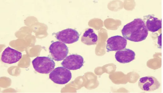
Figure 1A. L-1 stage ALL
The two purple colored cells represent L-1 ALL cells. The other cells in the Figure are RBCs. Reproduced from Hess CE and Krstic L, 2009 (reference 14) after permission.
- 4 -
Figure 1B. L-2 stage ALL
The purple colored cells represent L-2 ALL cells. These cells have more abundant cytoplasm than L1. Reproduced from the Website (reference 14).
- 5 -
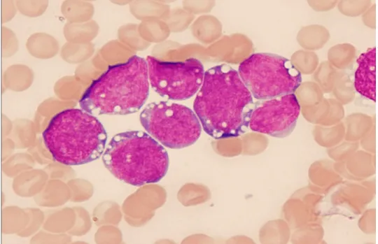
Figure 1C. L-3 stage ALL
Burkitt’s Leukemia cells are uniform in size. The cytoplasm is very basophilic (blue) and contains a variable number of lipid-laden vacuoles appearing as white spots. Reproduced from Hess CE and Krstic L, 2009 (reference 14) after permission.
- 6 -
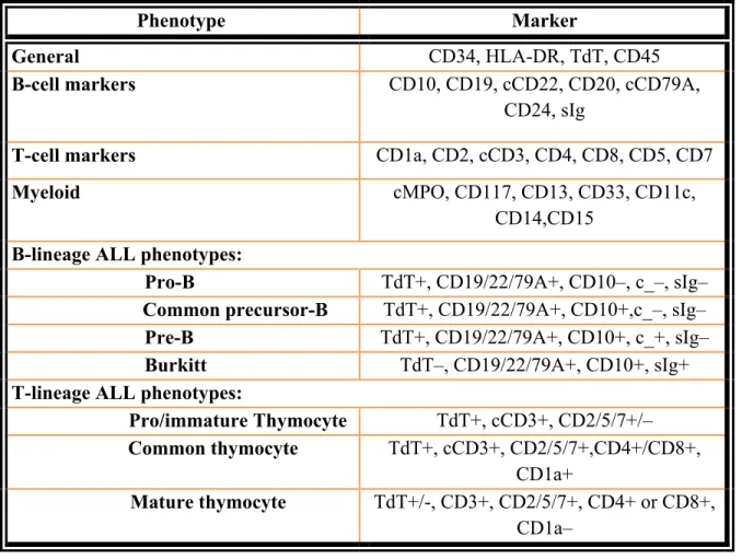
The WHO classification simply classifies ALL cases as precursor-B and precursor-T without additional categorization. The most common B-lineage ALL is the precursor-B phenotype with B-cell markers (CD19, CD22), TdT, cytoplasmic CD79A, CD34, CD10. This type has variably been called common precursor B ALL, early precursor-B ALL or simply B-ALL. The less common type, termed pro-B ALL, has a worse prognosis. Pre-B ALL is more mature than the common precursor B (16).
T lineage ALL accounts for only 15–20% of cases and can also be separated into phenotypic groups which may correspond to different stages of thymic T cell development (12). As in B-lineage ALL, the intermediate differentiated type is the most common. This common thymocyte type shows expression of the pan T-cell markers, CD2, cytoplasmic CD3 (cCD3), CD7, CD5 and characteristically shows coexpression of CD4 and CD8, and expression of CD1a. A more primitive type is called prothymocyte or immature thymocyte. Finally, a more mature phenotype than the common thymocyte type is called mature thymocyte. Again, because of a lack of conformity and variability in marker expression, the WHO classification recognizes only the precursor-T and -B groups without further immunophenotypic categorization (16). B and T lineage ALL phenotypes are listed in Table 1.
- 7 -
Table 1. Commonly used markers for immunophenotyping leukemia
Phenotype Marker
General CD34, HLA-DR, TdT, CD45
B-cell markers CD10, CD19, cCD22, CD20, cCD79A,
CD24, sIg
T-cell markers CD1a, CD2, cCD3, CD4, CD8, CD5, CD7
Myeloid cMPO, CD117, CD13, CD33, CD11c,
CD14,CD15 B-lineage ALL phenotypes:
Pro-B TdT+, CD19/22/79A+, CD10–, c_–, sIg– Common precursor-B TdT+, CD19/22/79A+, CD10+,c_–, sIg– Pre-B TdT+, CD19/22/79A+, CD10+, c_+, sIg– Burkitt TdT–, CD19/22/79A+, CD10+, sIg+ T-lineage ALL phenotypes:
Pro/immature Thymocyte TdT+, cCD3+, CD2/5/7+/– Common thymocyte TdT+, cCD3+, CD2/5/7+,CD4+/CD8+,
CD1a+
Mature thymocyte TdT+/-, CD3+, CD2/5/7+, CD4+ or CD8+, CD1a–
TdT: Terminal deoxynucleotidyl transferase, cMPO: Cytoplasmic myeloperoxidase; Modified from Larson RA and Anastasi J, 2007 (reference 16).
- 8 - 1.1.3- Signs and symptoms of ALL
As shown in Figure 2, healthy bone marrow contains stem cells, which proliferate to produce progeny cells that develop and differentiate into three types of cells found in blood:
1. Red blood cells, which oxygenate the body in the healthy state.
2. White blood cells, which are responsible for controlling infections and abnormal cells. 3. Platelets, which assist in clotting to prevent blood loss.
In acute leukemia, cancerous cells multiply very quickly and replace normal cells. They takeover normal cells, as shown in Figure 3, and become permanent cells of the bone marrow, resulting in bone marrow failure. A person with ALL is more likely to bleed and have repeated infections because of fewer platelets and abnormal WBC. Other symptoms of acute leukemia include frequent or unexplained fever, bone and joint pain (as a result of the spread of lymphoblasts to the surface of the bone or into the joint from the marrow cavity), generalized weakness and fatigue, night- sweating, shortness of breath, excessive and unexplained bruising, and skin changes, which include lumps, rashes, and paleness, etc.
1.1.4- Incidence and risk factors
With the exception of leukemia, cancer in children and adolescents is rare. In North America, about 15000 new cases of cancer are diagnosed in Caucasian children (1). Pre-B ALL is the most common form of leukemia and represents about 95% of acute leukemia cases among children younger than 12 years of age. Most cases occur in children aged 3 to 7 years.
- 9 -
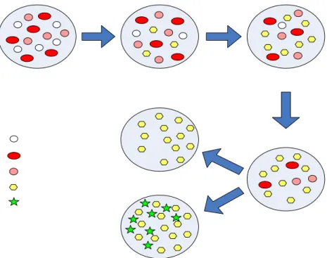
Figure 2. Normal hematopoietic stem cell proliferation
Three types of cells are produced by hematopoietic stem cells. The cells differentiate progressively as shown by the arrows. Reproduced from Winslow T, 2008 (reference 17) after permission.
- 10 -
Figure 3. Disturbances in the relative numbers of blood cell populations
occurring in leukemia.
- 11 -
Furthermore, it occurs slightly more often in boys than in girls, and the prognosis is better in girls (19). One reason for the worse prognosis in boys is the occurrence of testicular relapses among boys. The boys also appear to be at increased risk of bone marrow and CNS relapses for reasons that are not well understood (19). In contrast, acute myelogenous leukemia (AML) is more common in females. In addition, ALL occurs in Caucasian and Hispanic children more often than in black children. It may also occur in adults, but is not very common (20). Untreated acute leukemia is a fatal condition, usually because of complications that result from the infiltration of leukemic cells into bone marrow or vital organs. With treatment, the prognosis varies.
The factors that predispose children to leukemia are not fully understood. They seem to be complex and are likely to result from interplay between genetic make-up and environmental stimuli (21, 22). The risk of childhood leukemia is higher if a sibling has been diagnosed with the disease (3). Despite extensive studies on different molecules, proteomics and gene expression profiles, the exact cause of leukemia is still unknown. However, the clinicopathologic and molecular characterizations of different cytogenetic subgroups of B-ALL have been well established (23; reviewed in 24, 25). They are summarized in Table 2.
Other risk factors include exposure to very high levels of radiation, as people exposed to radioactive iodine radiation are much more likely to develop leukemia than those who have not been exposed. This is supported by the fact that the incidence of leukemia was increased in Japan and Chernobyl as a result of atomic bomb explosions and nuclear accidents, respectively (26). Internationally, the highest incidences of acute lymphoblastic leukemia (ALL) occur in Italy, the United States, Switzerland, and Costa Rica (27).
- 12 -
Table 2.Cytogenetic subgroupsof pre B-ALL and their clinicopathologic features
Cytogenetic Subgroup Frequency (%) Cytogenetic Abnormality
Fusion Gene Unique
Immunophenotypic Features Additional Molecular Abnormalities Pharmacologic Features Hyperdiploid ALL 27–29 (P) 6–8 (A) 51-65 chromosomes (+4, +14,+21, +X) NA NA uncommon BCDG mutations (13%), FLT3 mutations (21%–25%) Higher sensitivity to MTX, MP ALL with t(12;21) 22–25 (P) 1–2 (A) t(12;21) (p13;q22) TEL/AML1 (ETVX/ RUNX1) Early pre– B-ALL, My+ Monoallelic PAX5 deletions (28%) Higher sensitivity to asparaginase ALL with t(1;19) 3–6 (P) 5–7 (A) t(1;19) (q23;p13) E2A (TCF3)/ PBX1 Pre–B-ALL, CD34-/dim+, CD20-, CD9++ NA NA Philadelphia1 ALL 2–3 (P) 20–30 (A) t(9;22) (q34;q11.2) BCR/ABL (P190, P210) NA IKZF1 (Ikaros) deletions, BCDG mutations in 66% NA ALL with t(v;11q23); MLL rearranged 2–3 (P) 5–7 (A) t(4;11) (q21;q23) t(19;11) (p13;q23) AF4/MLL ENL/MLL Early pre-B, CD10-, CD15+, sCD22-, CD65+, NG2+ FLT3 mutations (18%) Increased expression of HOX genes Higher sensitivity to cytarabine Hypodiploid ALL 5%–6% (P) NA (A) <46 chromosomes (typically near haploid or low hypodiploid) NA NA BCDG mutations in 100% NA ALL with eosinophilia <1 t(5;14 q31;q32) IL3/IGH NA NA NA
Abbreviations: A and P in the Frequency column: % in adults and children, respectively, BCDG: B-cell development genes (eg, PAX5, EBF1, IKZF1, LEF1, TCF3, BLNK), FLT3: fms-related tyrosine kinase 3, MP: mercaptopurines, MTX: methotrexate, My: myeloid antigens, NA: not applicable or not known.
- 13 -
Biological risk factors for ALL include genetic conditions such as Down’s syndrome, neurofibromatosis type 1, Noonan syndrome, Fanconi anaemia and Poland syndrome. Additionally, some environmental risk factors include prenatal exposure to nicotine or alcohol, prenatal exposure to X-rays and previous chemotherapy treatment (2).
The prognosis for ALL depends on a patient’s age, white blood cell count, the degree of metastasis to the central nervous system, and the initial response to chemotherapy. Bad prognostic risk factors include age less than one year or more than ten years at the time of diagnosis, high white blood cell counts, metastasis of the leukemic cells to the central nervous system, and absent or low response to initial chemotherapy (28).
1.1.5- Treatment
The most common treatment used in ALL is chemotherapy. It can be administrated by a variety of routes including oral, intramuscular, intravenous and intrathecal (injection into the fluid surrounding the brain and spinal cord). Chemotherapy for ALL uses a combination of several anti-cancer drugs given over a long period of time (usually about 2 years; reviewed in reference 29). The most commonly used drugs and their treatment protocols are listed in Table 3.Recently, monoclonal antibodies (mAB) have been used in the treatment of haematological malignancies, including ALL. In this regard, Rituximab, which is a mAb directed against the B cell lineage antigen CD20, has been used in combination with chemotherapy for treatment of adult lymphoma. The antibody shows improvement of disease-free survival in adults with non-Hodgkin’s lymphoma (NHL) (30). It is unlikely that mAbs will have adequate activity to be effective for paediatric hematologic malignancies when used alone. However, rare cases of complete remissions in adults and children with ALL have been reported with mAbs targeting CD20, CD33, and CD52 (31, 32). Radiotherapy has also been used to kill tumour cells and prevent their growth.
- 14 -
The standard protocol for ALL treatment can be divided into three phases: remission-induction, consolidation, and maintenance. Of these, the maintenance phase is the most prolonged.
Remission-induction phase: The aim of this phase is to destroy leukemic cells, thereby placing the patient in temporary recovery, or a remission state, and restore normal hematopoiesis.
Consolidation phase: In this stage, the purpose is to eradicate any remaining inactive leukemic cells.
Maintenance and re-induction phase: Long-term maintenance therapy has been found to decrease the risk of relapses (recurrence of leukemia). It is important to prevent relapses of the disease because they are more difficult to treat. Secondary remissions, if they occur, are usually of short duration.
Bone marrow or cord-blood stem cell transplantation is commonly used to treat ALL that has not responded to chemotherapy (reviewed in 33). This transplantation consists of three steps: 1) collection of healthy stem cells from a donor without cancer or from the patient himself or herself, 2) administration of high doses of chemotherapy and possibly radiation therapy to kill any remaining leukemia cells, and 3) infusion of the healthy stem cells to produce normal, healthy blood-forming cells.
- 15 - Table 3 Typical ALL-treatment protocols.
Week Treatment Schedule Chemotherapeutic class
Continuation for high-risk patients (120 weeks)
1 Etoposide;
cyclophosphamide
Weekly Topoisomerase II inhibitor; alkylating agent 2 Methotrexate; mercaptopurine Weekly methotrexate; daily mercaptopurine Antifolate; purineantimetabolite 3 Methotrexate; cytarabine Weekly Antifolate; cytidineantimetabolite 4 Dexamethasone; vincristine Daily dexamethasone; weekly vincristine Glucocorticoid; antimicrotubule agent 5 Etoposide;
cyclophosphamide Weekly Topoisomerase alkylating agent II inhibitor;
6 High-dose
methotrexate; mercaptopurine
Weekly methotrexate;
daily mercaptopurine purineantimetabolite Antifolate; 7 Etoposide; cytarabine Weekly Topoisomerase II inhibitor;
cytidineantimetabolite 8 Dexamethasone; vincristine Daily dexamethasone; weekly vincristine Glucocorticoid; antimicrotubule agent Continuation for low-risk patients (120 weeks)
1 Methotrexate; mercaptopurine Weekly methotrexate; daily mercaptopurine Antifolate; purineantimetabolite 2 Methotrexate; mercaptopurine Weekly methotrexate; daily mercaptopurine Antifolate; purineantimetabolite 3 Methotrexate; mercaptopurine Weekly methotrexate; daily mercaptopurine Antifolate; purineantimetabolite 4 Dexamethasone; vincristine Daily dexamethasone; weekly vincristine Glucocorticoid; antimicrotubule agent 5 Methotrexate; mercaptopurine Weekly methotrexate; daily mercaptopurine Antifolate; purineantimetabolite 6 Methotrexate;
mercaptopurine Weekly methotrexate; daily mercaptopurine purineantimetabolite Antifolate;
7 High-dose
methotrexate; mercaptopurine
Weekly methotrexate;
daily mercaptopurine purineantimetabolite Antifolate; 8 Dexamethasone; vincristine Daily dexamethasone; weekly vincristine Glucocorticoid; antimicrotubule agent Adapted from Cheok M, Evans W (reference 29).
- 16 - 1.2- Human Natural Killer (NK) Cells
NK cells are low-density large granular lymphocytes. They comprise approximately 10-15% of all circulating lymphocytes in the bloodstream and are also present in tissues and lymphoid organs. NK cells have the ability to kill virus-infected or tumor cells without prior sensitization and for this reason are called Natural Killer cells (34). NK cells respond to intracellular microbes by killing the infected cell and by producing the macrophage activating cytokine IFN-γ. Phenotypically, NK cells are CD3-, CD2+, CD16+, CD56+, CD14-, and CD19-. Human NK cells can be divided into two subsets based on their cell-surface density of CD56 and CD16. These two markers are usually expressed reciprocally on two subsets of NK cells. They are divided into CD56bright CD16- and CD56dim CD16+ subsets. The cells in the two subsets differ in their proliferative potential, homing characteristics, functional capabilities and responses to different cytokines (listed in Table 4). The CD3−CD56dim CD16+ subset is more cytotoxic, and expresses intermediate affinity receptors for IL-2 and higher levels of Killer-cell Ig-like Receptors (KIR). This subset comprises more than 90% of NK cells in blood. In contrast, the CD56brightCD16- subset has greater capacity of cytokine production, but has lower ability for cytotoxicity and lacks perforin granules (35, 36).
- 17 -
Table 4. Characteristics of two major human NK cellsubsets.
Characteristic CD56brightCD16- CD56dimCD16+
IL-2R High affinity Low affinity
Cytotoxicity + +++
Cytokine productiona +++ +
Perforin Low High
CD62Lc High Low
CCR7d High Low
NKG2 Expression High Low
IL-7R High Low
ICAM-3 High Low
KIR Low High
SHIP-1e High Low
c-Kitg High Low
LFA-1 Low High
CD3 -chainf Low High
Lysozyme production Yes No
Main location Lymph nodesb Blood
ADCC Function Inefficient Efficient
Abbreviations: High and Low refer to levels of expression.
a Cytokines include IFN- , TNF- , TNF-β, IL-5, IL-13, and GM-CSF.
b In T cell-rich areas of lymph nodes and other secondary lymphoid organs and in body
tissues and organs such as liver.
c,d Lymph node homing receptors. e Needed for IFN- production.
f Signaling partner for activating receptors such as CD16a.
g c-Kit receptor tyrosine kinase needed for IL-mediated proliferation. KIR: Killer-cell
Ig-like Receptors, NKG2: NK cell group 2, CD62L: CD62 ligand. Adapted from Iannello A et al., 2008 (reference 7).
- 18 -
Some immunologists also differentiate between CD16high and CD16dim subsets. They have described NK cells expressing both CD56 and CD16 (36a).The incubation of these cells with different cytokines may change their phenotypic appearance, as well as functional and homing characteristics. For instance, IL-2, IL-12 and IL-15 have the ability to convert both CD56highCD16- and CD56lowCD16+ NK cell subsets into CD56brightCD16+ cells (7). On the other hand, TGF-β1 converts CD16+ NK cells into CD16- NK cells (7, 36a). NK cells are important effectors and regulatory cells of the immune system. Their important functions are given below:
1- Defending the host against viral infections, intracellular pathogens and malignancy: NK cells can recognize tumor cells and virus-infected cells and kill them without prior sensitization to their antigens. In addition to direct killing, they secrete anti-microbial peptides called α-defensins. Like activated T cells, NK cells can produce interferon-γ (IFN-γ), so named because of its ability to inhibit or interfere with viral replication. IFN-γ is a potent activator of macrophages, and leads to differentiation of naive CD4+ T cells into T helper type 1 (TH1) effector cells (7). In addition to IFN-γ, NK cells have also been shown to secrete TNF-α, GM-CSF, IL-5, IL-13, IL-10, TGF-ß, MIP-1α, MIP-1ß, RANTES, and nitric oxide (NO). More recently, these cells were also shown to produce IL-22 (37).
2- Role in pregnancy:
NK cells function in pregnancy through their interaction with fetal extravillous trophoblasts to remodel maternal vasculature, and increase fetal blood supply. NK cell-secreted IFN-γ plays a role in inducing placental vasculature (38). A failure of early trophoblast invasion and remodeling of the spiral arteries leads to poor blood supply to the placenta. This may result in pre-eclampsia, which is a disease that occurs in the third trimester of pregnancy (after 20 weeks). This disease is characterized by pregnancy-induced hypertension and proteinurea. Pre-eclampsia leads to maternal and fetal mortality worldwide and it is the most common cause of preterm birth. Furthermore, NK cells’ hyperactivity is suspected of being responsible for recurrent miscarriages (three or more abortions) (39). Also, some studies have shown that females with recurrent miscarriages have significantly increased numbers of NK cells expressing activating KIR receptors compared to normal fertile women (40). The women carrying ahigh content of activating
- 19 -
KIR genes have about a threefold increased risk of developing recurrent miscarriages (40).
3. Controlling leukemia:
NK cells have a significant role in controlling the development of leukemia as they can kill leukemia cells growing in culture as well as in vivo in animal models (4). In addition, blocking of the interaction between inhibitory receptors and their MHC ligands in mice enhances their anti-tumor activity and control of tumor growth (41). Moreover, leukemia cells can express ligands for many activating KIR receptors. This means that NK cells can kill tumor cells by default if they are not inhibited by the engagement of inhibitory KIR receptors. The role of NK cells in controlling leukemia is also suggested by the fact that leukemia cells do not down regulate HLA-C and HLA-Bw4, which act as ligands for several inhibitory receptors ofNK cells. On the other hand, leukemic cells down-regulate HLA-A and HLA-Bw6 (42). It is of interest that NK cells do not have any receptors that recognize these MHC antigens. In addition to the direct killing of tumor cells, the activated NK cells secrete IFN-γ, which is a potent cytostatic agent (43).
4. Role asmemory cells:
Despite being effector cells of innate immunity, NK cells have been shown to have immunological memory. This feature of NK cells was discovered in RAG-/-mice, which lack both T and B cells. The researchers discovered that recall responses to hapten-induced hypersensitivity in these mice was mediated by NK cells (44). The exact mechanisms behind NK cell dependent memory or recall responses are not well defined. Another study using mouse models of cytomegalovirus infection showed that like T cells, NK cells bearing the virus-specific LY 49H receptor undergo four phases of the adaptive immune response (45). These phases had not been previously documented in NK cells. This ability of NK cells has major implications in the generation of immunological memory against pathogens, malignancy and vaccination strategies.
- 20 -
1.2.1- NK receptors (NKR)
NK cells do not have a well-defined single molecular structure to recognize target cells. Instead, they express a variety of molecular structures that bind to MHC and non-MHC molecules on the target cells. NKRs can be divided into inhibitory and stimulatory types depending on the nature of the signal they send to NK cells after binding to their specific ligands. Each NK cell expresses inhibitory and stimulatory NKRs. The human NKR can be divided into MHC and non-MHC-binding receptors.
1.2.1.1- MHC-binding receptors
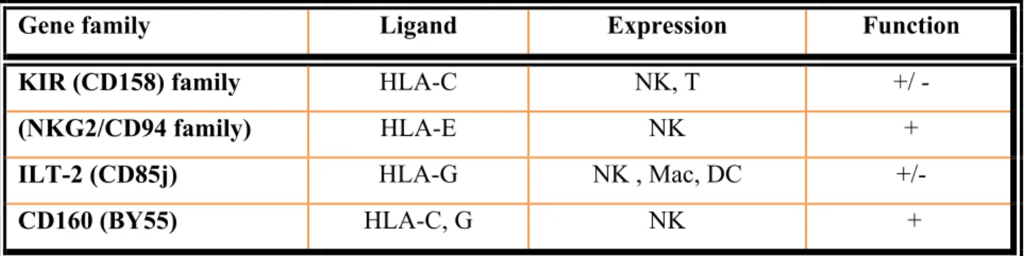
These receptors recognize and bind MHC class I molecules. They include KIR, NKG2, and Ig-like transcript (ILT) families, as shown in Table 5.
1.2.1.1.1-CD94/NKG2 killer lectin-like receptor (KLR)-C (NKG2/CD94 family) These are also known as the (NK Group 2) NKG2/CD94 family of receptors. The genes for these receptors are located on human chromosome 12p12.3–p13.2 in a region called the NK gene complex (NGC). These receptors are type II, C-type, lectin-like integral membrane glycoproteins. This family contains four receptors: A/B (KLR-C1), C (KLR-C2), E/H (KLR-C3), and F (KLR-C4). B and H represent splice variants of A and E genes, respectively (7).
Of these receptors, CD94/NKG2A carries two ITIMs in its long cytoplasmic tail. It is expressed on a subset of human NK cells having the CD56high CD16low phenotype as well as on the CD56low subset of NK cells, albeit at lower levels. This receptor has an inhibitory function. NKG2C has a short cytoplasmic tail, links noncovalently with a homodimer of DAP-12, and activates NK cells upon binding with its ligands. NKG2E has a charged amino acid (lysine) in its transmembrane region, but it does not associate with DAP-12. NKG2A and NKG2C are expressed on overlapping subsets of CD56+ NK cells. NKG2C and NKG2E act asactivating receptors. NKG2F is a unique gene within
- 21 -
Table 5. MHC-binding receptors and their characterictics.
Gene family Ligand Expression Function
KIR (CD158) family HLA-C NK, T +/ -
(NKG2/CD94 family) HLA-E NK +
ILT-2 (CD85j) HLA-G NK , Mac, DC +/-
CD160 (BY55) HLA-C, G NK +
Abbreviations: HLA: Human Leukocyte Antigen, NK: Natural Killer cells, T: T cells, Mac: macrophages, DC: dendritic cells. The signs + and – indicate activating and inhibitory functions, respectively.
- 22 -
the NKG2 family whose translated product contains a positively charged residue in its transmembrane region, an intracellular ITIM-like sequence and an extracellular domain (62 residues) that is truncated relative to other NKG2 molecules. The NKG2 receptors bind the non classical MHC class Ib molecule HLA-E (46-48).
1.2.1.1.2-ILT (CD85) family
ILT or Ig-like transcripts are also known by two other names: Leukocyte Ig-Like Receptor (LILR) and Macrophage Ig-like Receptors (MIR) (7, 49). This family consists of 13 members with either inhibitory or activating properties. They are expressed on monocytes, macrophages, dendritic cells, and some subsets of B and T cells. A single member of this family ILT2 is expressed on a subset of NK cells (49). The ILT family members bind to classical and non classical HLA class I molecules. They are located on human chromosome 19 close to the KIR gene cluster.
1.2.1.1.3-CD160 (BY55)
CD160 is a GPI anchored, Ig-like molecule expressed on the CD56dim subset of NK cells, γδ T cells, and a subset of CD8+ T lymphocytes. It binds HLA-G, HLA-C and other HLA molecules (50). CD160 positive NK cells and CTL usually accumulate in inflammatory conditions. The NK cell activation via CD160 leads to secretion of proinflammatory cytokines, which include IFN-γ, TNF- α and IL-6 (51).
1.2.1.2- Non MHC-binding receptors
These receptors recognize molecular structures other than MHC molecules. The most important members of this group are discussed below, and the list of all receptors and their characteristics is shown in Table 6.
1.2.1.2.1-NKG2D receptors (KLR-K1; CD314)
NKG2D are type II lectin-like receptors. They were first identified as a member of the NKG2 family. However, they are not typical members of this family. They are expressed on all human NK cells, and their expression is upregulated by IL-15 and IL-12. On the
- 23 -
reverse, certain other cytokines e.g., TGF-β and IL-10 decrease their expression. They are also expressed on resting and activated human CD8+ T lymphocytes (7, 52).
NKG2D receptors do not recognize and bind HLA-E, as do all members of the CD94/NKG2 family. Instead, they bind MICA, MICB and HCMV-induced ULBPs (53; reviewed in 54; shown in Figure 4). The MIC genes are highly polymorphic, and are located on human chromosome 6q25 outside the MHC locus. The ligands for NKG2D are usually not expressed under physiological conditions. However, they are induced on body cells in response to stress, DNA damage, infection and malignancy. The expression of these ligands on body cells flags them for destruction by NK cells via NKG2D (54, 55).
In addition to having different ligands, NKG2D also differs from other members of the NKG2 family; it does not need CD94 for expression on the cell surface (55).
1.2.1.2.2- Natural cytotoxicity receptors (NCRs)
There are three NCRs: NKp46 (CD335), NKp30 (CD337), and NKp44. NKp46 and NKp30 are expressed on resting and activated NK cells, whereas NKp44 is expressed only on cytokine-activated NK cells. The engagement of these receptors with their ligands triggers NK cell-mediated killing and secretion of IFN-γ. The ligands for the NCR mostlyremain unknown (7). Only NK46 and NKp30 were reported to recognize the haemagglutinin antigen (HA) of the influenza virus (56).
1.2.1.2.3- SLAM-related Receptors (SRRs)
The signalling lymphocytic activation molecule (SLAM) family of receptors is expressed by a wide range of immune cells. Through their cytoplasmic domain, the SLAM family receptors associate with SLAM-associated protein (SAP) and SAP-related molecules. SLAM (CD150) is expressed on the surface of T cells (57,58).
The SRR family includes 2B4 (CD244), NTB-A (Ly108), and CD2-like Receptor on Activated Cytoxic Cells (CRACC; CD139). These receptors are related to SLAM, as they all use similar signalling molecules, SAP or related molecules. The genes for SSRs are located on human chromosome 1q22. They are expressed on NK cells, monocytes, basophils, γδ T cells and CD8+ T cells of the effector memory phenotype (7, 57).
- 24 - 1.2.2- NK cell Co-receptors
Co-receptors usually add to the strength of activating signal. These molecules themselves are unable to trigger NK cell functions. According to this definition, some co-receptors may be able to trigger NK cell functions under certain conditions and therefore may qualify as receptors. For example, the Lymphocyte Function-associated Antigen 1 (LFA-1) can trigger NK cell-mediated lysis under appropriate conditions, and could be considered as an activating receptor (59).
NK cells express several coreceptors, which bind to their cognate ligands on target cells and send co-stimulatory signals. These signals add to the overall strength of the activating signal. Adhesion molecules, such as integrins, selectins, and several Ig-like molecules usually act as co-receptors for NK cells. Table 7 lists NK cell co-receptors, their expression and function.
- 25 - Table 6. Non MHC-binding NK cell receptors.
Receptor Expression Ligands Function
NKG2D NK,CD8+T MICA, MICB, ULBPs + NKp46 NK HA + NKp44 Activated NK HA + NKp30 NK ? + NKR-P1 (CD161; KLR-B1) NK, NKT, CTL LLT-1 +/– 2B4(CD244) NK , T, Monocytes, Basophils CD48 +/– KLR-G1 Mast cells, NK , CTL Cadherens –
FcRL B cells, NK IgG complexes –
NKp80 NK AICL +
DNAM-1 NK, B cells Nectin-2, PVR +
Four Ig-like B7 homologues (4IgB7H or B7H)
NK,T PD-1 +/–
SIGLEC-7 NK Sialic acid –
CEACAM-1 (CD66a)
NK CEA and CEA-
related Proteins
–
LAIR Leukocytes Collagen –?
CEA: Carcinoembryonic antigen; CEACAM-1: CEA-related cell adhesion molecule; FcRL6: FcR-like protein 6; SAP: Signaling Lymphocyte Activating Molecule (SLAM)-Associated Protein; SIP: Stress-induced proteins [MICA, MICB, UL16-binding protein (ULBP)]; DNAM-1: DNAX accessory molecule 1; SIGLEC-7: Sialic acid-binding Ig-like lectin 7. The designations (+), (–), and (+/–) indicate that the function is activation, inhibition, and both, respectively. Redrawn from Iannello A et al., 2008 (reference 7).
- 26 -
Figure 4. Non-MHC-binding NK Cell Receptors.
ITSM: Immunoreceptor tyrosine-based switch motif; Col: Collagens; NTB-A: NK-T-B: NK, T-B cell antigen; LAIR-1: Leukocyte-associated Ig-like Receptors. The YxxM motif, when phosphorylated, recruits PI-3K. Not drawn to scale. The question mark (?) indicates that the ligand is unknown. Reproduced from Iannello A et al., 2008 (reference 7) after permission.
- 27 -
Table7. Human NK cell co-receptors, their expression and function.
N-CAM: Neural cell adhesion molecule; VCAM: Vascular cell adhesion molecule; MAdCAM: Mucosa addressin cell adhesion molecule.
Adapted from Iannello A et al., 2008 (reference 7).
Name Ligand Expression Function
CD2 (LFA-2) D58 (LFA-3) CD48 (weakly)
All NK Costimulation, adhesion
LFA-1
(CD11a/CD18)
CD54 (ICAM-1–5) All NK Costimulation, adhesion Cytoskeleton
rearrangement
CD8 MHC class I Subset Costimulation, adhesion
CD69 Unknown Activated
NK
Costimulation
CD56 (N-CAM)
Self Subset Homotypic adhesion
CD59 C8, C9 All NK Adhesion, costimulation
CD57 Unknown Subset Marker of senescence
CD28 B-7 Fetal NK Costimulation
CD27 CD70 Subset Costimulation
CD44 Hyaluronic acid Activated NK
Costimulation, adhesion
VLA-4 Fibronectin, VCAM-1 MAdCAM-1
Subset Adhesion, diapedesis
- 28 - 1.3- KIR family of NKRs
The KIRs (Killer-cell Immunoglobulin-like Receptors) are so named because they share structural homology to immunoglobulin molecules. They are type I integral membrane glycoproteins. They are usually expressed as monomers on the surface of natural killer (NK) cells. These receptors play an important role in controlling NK cell activities. To date, fifteen KIR genes, which include two pseudo genes, have been well described. These genes are located on human chromosome 19q13.4 in a tandem head to tail fashion in a short 150 kb region called the Leukocyte Receptor Complex (LRC). The structure of a typical KIR gene and the receptor is shown in Figure 5.
Each KIR has an extracellular region, a stem, a transmembrane region and a cytoplasmic tail. The extracellular region binds the receptor ligand and consists of two or three immunoglobulin (Ig)-like domains. The cytoplasmic tail may be short or long. Each long-tail KIR has two immunoreceptor tyrosine-based inhibitory motifs (ITIM) and is inhibitory in function. When the receptor binds to its ligands, the tyrosine residues in the tail become phosphorylated and recruit SH-2 domain-containing phosphatases: SHP-1 and 2. These phosphatases dephosphorylate several substrates involved in the NK cell activation cascade. Dephosphorylation transiently inhibits NK cells from triggering their effector functions (60).
The KIR receptors with a short cytoplasmic tail possess a charged amino acid (lysine) in their transmembrane regions and associate non-covalently with an adaptor protein called KARAP/DAP-12, which has immunotyrosine-based activating motifs (ITAMs) in its cytoplasmic tail. Upon binding to the MHC-ligands, the tyrosine residues in the ITAMs become phosphorylated, recruit several kinases and ultimately trigger NK cell-mediated killing andcytokine secretion. Each KIR gene has been named according to
- 29 -
Figure 5. The structure of a typical KIR gene and the receptor.
A typical KIR gene comprises nine exons shown here on the right side of the figure. Double horizontal lines in the gene indicate introns. The schematic structure of the encoded receptor is shown on the left. The part of the receptor encoded by each individual exon is also indicated. The scissor in the figure indicates cleavage site for the signal peptide. The letters N and C designate N- and C-terminals of the protein, respectively. D0, -1, -2: Extracellular Ig-like domains; TM: Transmembrane region. Not drawn to scale. Reproduced from Iannello A et al., 2008 (reference 7) after Publisher’s permission.
- 30 -
the number of immunoglobulin-like domains in the extracellular regions and the length of the cytoplasmic tail present in its encoded receptor. In this taxonomy, 2D refers to two domain-, and 3D refers to three domain-receptor encoding gene. The letters (L) refer to long tail and (S) to short tail encoding genes. For example, KIR2DL1 means a gene that encodes a receptor having two extracellular domains and a long tail. The final number differentiates between individual KIR genes in each group.
The inhibitory KIR genes discovered so far include KIR2DL1, KIR2DL2/3(alleles), KIR2DL4, KIR2DL5a, KIR2DL5b, KIR3DL1, and KIR3DL2 (61, 62). The six activating genes include KIR2DS1-5 and KIR3DS1. All activating KIR genes represent independent loci on the chromosome. However, KIR3DS1 represents allelic variants of KIR3DL1 and encodes short tailed activating receptors (61, 63). KIR2DL4 is an unusual KIR; it is not expressed clonally on NK cells as other inhibitory KIRs are (64). It is expressed on all NK cells in all humans, and it expresses an inhibitory ITIM in its cytoplasmic tail but also expresses a charged amino acid (arginine) in its transmembrane region and interacts covalently with its signaling partner, the γ chain of FcεRI(64). In other words, it exhibits dual characteristics of an activating and an inhibitory KIR. The receptor interacts with HLA-G (a non classical MHC class Ib antigen).
1.3.1- The KIR ligands
The two-domain KIRs bind HLA-C antigens. These antigens are divided into group I and II, which are characterized by the presence of an asparagine and a lysine at position 80 in their amino acid sequences, respectively. KIR2DL1 and its allelic forms encode receptors that recognize group II HLA-C antigens (HLA-Cw1, 3, 7, 8, 13 and 14). On the other hand, KIR2DL2 and KIE2DL3 recognize group I HLA-C antigen (HLA-Cw2, 4, 5, 6, 17 and 18). It is noteworthy that all the known HLA-C molecules in humans (Cw1-18) are recognized by either KIR2DL1 or 2DL2/3 receptors (65-69). The affinity of different KIRs to bind their respective HLA-C ligands differ from one to another, e.g., KIR2DL1 binds with high affinity to their group II HLA-C ligands, whereas KIR2DL3 binds with its group I HLA-C ligands relatively weakly. The allelic variant KIR2DL2 binds with its HLA-C ligands with intermediate affinity. These binding characteristics have an important impact on NK cell functions. For example, in a person who is homozygous for
- 31 -
group II HLA-C and co-expresses KIR2DL1, NK cells will be under tight inhibition compared to a person who is homozygous for group I HLA-C and co-expresses KIR2DL3 (62-66). The three-domain KIR3DL1 and its activating allotype KIR3DS1 bind the HLA-Bw4 serospecificity molecules, which have isoleucine at position 80 (70, 71). Their binding to the HLA-Bw4 serospecific molecules that have threonine at position 80 is relatively weak. It is worth mentioning that HLA- B allotypes have two mutually exclusive serotypes, namely Bw4 or Bw6. HLA-Bw4 antigens could have threonine or isoleucine at postion 80 of their amino acid sequence. It is noteworthy that different KIR3DL1 allotypes also vary in their affinity for their HLA-Bw4 ligands. The activating short-tailed KIRs have been shown to bind HLA-ligands with very low affinities compared to the inhibitory KIR receptors (67). Table 8 lists KIRs, their distribution, signaling partners and ligands.
1.3.2- KIR haplotypes
KIR genes vary from one person to another. The degree of diversity in the KIR genes matches that found in HLA genes. There are two mechanisms that contribute to this diversity:
(1) KIR haplotypes differ in gene content. (2) KIR genes are highly polymorphic.
Because of this diversity, unrelated individuals rarely have identical KIR genotypes. Based upon their gene contents, KIR haplotypes can be divided into two distinctive groups: A and B.
- 32 - Table 8. Human KIRs (CD158) and their ligands.
Receptor Distribution
Signaling
partner
Ligand
( I ) Activating KIR
1. KIR2DS1 (p50.1)
NK
DAP-12
HLA-C II, ?
2. KIR2DS2 (p50.2)
NK DAP-12
HLA-C
I,
?
3. KIR2DS4 (p50.3)
NK DAP-12
HLA-Cw4,
?
4. KIR2DS3, 5
NK DAP-12
?
5. KIR3DS1 (p70)
NK DAP-12
HLA-Bw4-I,
?
( II ) Inhibitory KIR
1. KIR2DL1 (p58.1)
NK, CTL
SHP-1, 2
HLA-C II
2. KIR2DL2/3 (p58.2)
NK, CTL
SHP-1, 2
HLA-C I
3. KIR3DL1 (p70;
NKB1)
NK, CTL
SHP-1, 2
HLA-B Bw4
4. KIR3DL2 (p140)
NK, CTL
SHP-1, 2
HLA-A3, A11
5. KIR2DL4
NK Fc R1 -chain HLA-G
6. KIR2DL5
NK, CTL
SHP-2
HLA-G?
Activating KIRs may be expressed on CD4+ T cells in some disease conditions but are rarely expressed on CTL. HLA-Bw4-I designates Bw4 allotypes having isoleucine at position 80. The question mark (?) indicates unknown and/or controversial ligands. All KIRs are expressed clonally on overlapping subsets of NK cells except KIR2DL4, which is expressed on all NK cells. Inhibitory KIRs are also expressed on the CTL of the effector/memory phenotype. Adapted from Iannello A. et al., 2008 (reference 7).
- 33 -
Group A haplotypes are less variable. They lack stimulatory KIR genes except for KIR2DS4. Interestingly, KIR2DS4 frequently carries a 22 bp deletion in its exon 5 and encodes a non-functional receptor (72). In general, the group A haplotypes are composed of a fixed content of seven KIR genes and two pseudogenes, and are diversified through allelic polymorphism (reviewed in 73).
Group B haplotypes have a variable number of KIR genes that are not present in group A haplotypes (e.g., KIR2DS1, 2DS2, 2DS3, 2DS5, 3DS1, and 2DL5). They often, but not always, lack KIR 2DS4. B haplotypes exhibit more diversity both in terms of gene content and allelic polymorphism. There are four genes that are found in both groups of haplotypes and are called framework genes. They include KIR3DL2 on the telomeric end, 3DL3 on the centromeric end, and KIR2DL4 and KIR3DP1 in the central region (Figure 6; 62, 65, 72, 73).
1.3.3- KIR-HLA epistatic interactions and disease outcome
The genes of the KIR family are highly polymorphic. KIRs bind epitopes, which are displayed on a subset of related MHC class I antigens. The KIR genes encode either activating or inhibitory receptors. The inhibitory receptors bind with their specific ligands on the target cell and inhibit the NK cell from killing the target cell as well as from secreting chemokines and cytokines. Each inhibitory KIR has a different affinity for its MHC ligand, which means that the level of inhibition exerted by a given KIR/ligand pair is different from the other pairs. It is of interest to note that the genes for the KIR ligands (e.g., MHC class I antigens) are also highly polymorphic. Furthermore, the genes for KIR and MHC class I antigens in humans arefound on chromosomes 19 and 6, respectively,
- 34 -
Figure 6. KIR Haplotypes.
The framework genes (3DL3, 3DP1, 2DL4, and 3DL2; in violet color) are present in each haplotype. The figure shows KIR genes present in centromeric and telomeric halves of the frequently found A and B haplotypes above and below the framework genes, respectively. Each box in the figure represents a KIR gene. 3DP1 is a pseudogene. Reproduced from Iannello A et al., 2008 (reference 7) after permission.
- 35 -
meaning that during meiosis they segregate independently from one another. This would create different combinations of KIR and MHC ligand genes for each individual, which would then affect the activity of their NK cells and their ability to control tumor growth. For example, an individual with a combination of KIR and MHC ligands that tightly inhibit NK cells may be more at risk for developing leukemia (66, 73).
There is also another NK activating receptor, NKG2D, which has very low genetic variability, but whose ligands (MHC class I heavy chain-related protein A: MICA, and MICB) are very polymorphic (55). Similarly to KIR and its ligands, NKG2D has different binding affinities for various forms of MICA and MICB. Therefore, different NKG2D ligands vary in their ability to activate NK and T cells in different individuals. Because of this, an individual’s NKG2D ligands may control the level of activation of his/her NK cells, which can then affect his/her risk of developing leukemia.
1.3.4- Effects of KIR genotypes on human disease
Genetic studies on the association of KIR with diseases have been mainly carried out in viral infections and autoimmune diseases. Figure 7 shows a model for the association of KIR with different diseases. According to this model, the individuals carrying KIR and HLA gene combinations (e.g., those homozygous for KIR2DL1 and HLA-CLYS80 genes, with no activating KIR gene) that inhibit NK cells relatively tightly would be less likely to develop autoimmune diseases. Such individuals may suffer from pre-eclampsia and are likely to be more susceptible to viral infections and malignancy. On the other hand, the individuals with the KIR-HLA gene combinations (e.g., KIR2DL3 and HLA-CASP80 with one or more activating KIR genes) that inhibit NK cells to the minimum are likely to control viral infections and malignancy. However, this model predicts that such individuals would suffer more from autoimmune diseases and may experience repeated spontaneous miscarriages (73). In fact many studies concerning the associations of KIR and HLA genes with innate resistance/susceptibility to autoimmune diseases, malignancy, control of viral infections and reproductive success in humans have verified the predictions made by this model (10, 73- 78). Activating KIR genes, which encode
- 36 -
NK cell activating receptors, have also been reported to affect outcomes of many human diseases. In line with the model, several activating KIR genes have shown significant associations with autoimmune diseases. The first of these was KIR2DS2, which was associated with the development of vasculitis in rheumatoid arthritis (10). Another group found that patients with acute coronary syndrome share common features with those diagnosed with rheumatoid vasculitis (10, 79), indicating association of KIR genotypes and vascular disease.
1.3.5-KIR genes in leukemia
A few earlier studies described an association of KIR genes with leukemia (9,74, 80,81; reviewed in 82). Unfortunately all these studies were performed on adult leukemic patients. One group studied the relationship between certain inhibitory KIR-HLA interactions and chronic lymphoid and myeloid leukemia in the Belgian adult leukemic patients (9). Their study subjects mostly had other types of leukemia; only 8 cases were ALL. They found significant associations of certain inhibitory KIR haplotypes with some forms of leukemia; however the number of patients with ALL was too low to find any significant association. These workers conducted another study in Polish and German adults suffering from Myeloid or Chronic Lymphocytic Leukemia and found significant associations with certain combinations of inhibitory KIR and their HLA ligands (74). The study had no participants with ALL. Another study (80) reported significant association of the intact KIR2DS4 gene with innate resistance to CML in humans but not with resistance to ALL. The number of the leukemic patients with ALL was very low (only 21) in this study. Due to this low number of participants with ALL, the study had not enough power to detect associations between KIR2DS4 and ALL. In a relatively recent study in a Chinese population (81), researchers found that activating KIR genes were more common in CML patients than in healthy controls, although only the difference for the KIR2DS4 gene reached statistical significance. This study did not differentiate between the functional and mutant variants of the KIR2DS4 gene. The researchers also found a significantly decreased frequency of KIR2DS3 in ALL patients compared to the
- 37 -
Figure 7. Genetic model for association of KIR with human diseases.
This model is based on KIR genotypes and KIR:HLA combinations that are considered to provide different levels of inhibition and activation, and on disease studies in which an association with protection or susceptibility was associated with specific KIR genes, or specific KIR and HLA combinations.
Adapted from Williams AP et al., 2005 (reference 73). 2DL3:HLA-C ANS80 ; Homozygous
Too less inhibition
-Recurrent spontaneous abortion Too much inhibition -Pre eclampsia 2DL1:HLA-C
2DL1:HLA-C LYS 80 Homozygous AA haplotypes -Psoriasis: 2SD1 -RA vasculitis:2DS2 -Diabetes:2DS2 -Scleroderma:2DS2 -Slow progression of HIV 3DS1+HLA-Bw4 ILE80 BB haplotype Increasing inhibition Increasing activation
- 38 -
control group (81). We were unable to find any study in the literature that studied the potential association of activating or inhibitory KIR genes in childhood leukemia.
- 39 -
CHAPTER 2
AIMS AND OBJECTIVESThe aim of this study was to investigate the associations between activating KIR genes and acute lymphocytic leukemia (B ALL), which is the most common form of leukemia affecting children under 15 years of age. As stated earlier, humans differ from each other with respect to the number of inherited activating KIR genes. They may have 0-6 of these genes. Since the receptors encoded by the genes enhance NK cell activation and overall immune competence of the individual, we hypothesized that their inheritance is likely to protect individuals from developing this cancer.
The specific objectives of the study were to compare the frequencies of activating KIR genes between B-ALL patients and healthy controls in French-Canadian children, and determine whether these genes showed significant association with resistance or susceptibility to this cancer in this population.
- 40 -
CHAPTER 3
RESULTS
The results obtained from the investigations conducted on the genomic DNA obtained from B-ALL patients and control subjects were compiled into a research article. The article is reproduced in this chapter.
- 41 -
Association ofactivating Killer-cell Immunoglobulin-like Receptor genes with decreased risk for developing B cell acute lymphoblastic leukemia in children
Zaema Almalte1, Suzanne Samarani1, Alexandre Iannello1, Olfa Debbeche1, Devendra K Amre2, Daniel Sinnett2 and Ali Ahmad1
Laboratory of Innate Immunity1, CHU Sainte-Justine Research Center1,2, Department of Microbiology & Immunology1, Department of Pediatrics2, University of Montreal1,2, Montreal, Quebec, Canada
Running Title: Association between activating KIR genes and risk for B-ALL
Correspondence: Ali Ahmad
Laboratory of Innate Immunity, CHU Sainte-Justine Research Center/Department of Microbiology & Immunology, 3175 Cote Ste-Catherine, Montreal, Qc, H3T 1C5, Canada Tel: 514-345-4931 ext 6157
- 42 -
ABSTRACT
Pre-B cell acute lymphoblastic leukemia (B-ALL) is the most frequent form of leukemia affecting children under 15 years of age. Evidence is accumulating that genetic factors play an important role in conferring susceptibility/resistance to leukemia. In this regard, the Killer-cell Immunoglobulin-like Receptor (KIR) genes are of particular interest. These genes are highly polymorphic and encode receptors that control functional activities of Natural Killer (NK) cells, which are known to kill leukemia cells and regulate immune responses both in vitro and in vivo. However, little is known concerning the role of these genes and/or their variants in conferring innate susceptibility/resistance to the childhood leukemia. In this study, using a case-control design, we have addressed this issue. Our results show that harbouring activating KIR genes confers protection from this cancer in French-Canadian children. The protection increases as the number of these genes increases in an individual. These results provide novel insights concerning pathogenesis as well as for potential immunotherapy in children affected with Pre-B ALL.
- 43 -
INTRODUCTION
Each year about 15,000 new cases of leukemia are diagnosed in North America. Acute lymphoblastic leukemia (ALL) represents about 95% of these cases occurring in children under fifteen years of age. About 85% of the ALL cases in children involve Pre-B cells (1). The pre-B ALL is also the most deadly form of leukemia occurring in children. Cumulative evidence suggests that the genetic make up of an individual plays an important role in determining innate resistance/susceptibility of an individual to the development of this leukemia. In this respect, several genetic determinants that influence cell proliferation and cell cycle progression have been shown to be associated with susceptibility/resistance to develop Pre-B ALL in children (2-5; reviewed in 6). However, little is known about the role of the Killer-cell Immunoglobulin-like Receptor (KIR) genes in the development of this leukemia.
The KIR gene family comprises sixteen genes located in a head-to-tail fashion on human chromosome 19q13.4 in a 150 kb region called “Leukocyte Receptor Complex or LRC” (7, 8). The genes in the family are highly polymorphic and are rapidly evolving. Of the sixteen KIR genes, seven inhibitory genes (KIR3DL1-3, KIR2DL1, KIR2DL2/3 and KIR2DL5A and B) encode receptors with long-tailed cytoplasmic tails and inhibit the functions of the immune cells. Each inhibitory receptor binds to public epitopes present in a group of related MHC class I antigens (9, 10). The KIR family also contains six activating genes (KIR3DS1, KIR2DS1-5) that encode receptors with short cytoplasmic tails. The binding of the activating receptors to their cognate ligands on a target cell results in activation of the immune cells. In the case of Natural Killer (NK cells), this activation leads to killing of the target cell as well as secretion of soluble mediators like IFN-γ and TNF-α. It is noteworthy that KIR haplotypes may vary from one another with respect to the number of activating KIR genes and have been divided into two groups: A and B. The group A haplotypes carry only one activating KIR gene, KIR2DS4, which is often mutated and encodes a non functional receptor. The group B haplotypes usually lack KIR2DS4 but contain 1-5 other activating KIR genes. Consequently, individuals differ from each other with respect to the number of inherited activating KIR genes (7, 8). Several studies have shown that KIR genotypes as well as the number of activating KIR
- 44 -
genes inherited by an individual influence his/her resistance/susceptibility to infectious agents, development of autoimmune diseases, reproductive efficiency, graft versus host disease (GVHD) and cancer (11-16; reviewed in 17). Certain activating KIR genes have been shown to protect humans from non-Pre B ALL (18). However, little is known concerning the impact of activating KIR genes on the innate resistance/susceptibility to develop the childhood leukemia pre-B ALL. We addressed this issue in this study and show here that all activating KIR genes protect children from developing pre-B ALL. Furthermore, we also observed that the individuals who harbour higher numbers of activating KIR activating were afforded more protection from this childhood leukemia.
MATERIALS and METHODS Patient Population:
In order to investigate associations between activating KIR genes and pre-B ALL, we carried out a case-control study at the CHU Sainte-Justine Research Center, Montreal. Cases of pre-B ALL patients (N=102) were recruited from the Hemato-Oncology clinic of the hospital during 1989-2004 and included children diagnosed prior to age 18. They had the following characteristics: (a) girls to boys’ ratio of 40:60; (b) age at diagnosis: 9% between 0-1 year, 70% between 2-10 years, and 21% between 10-18 years. Diagnosis of pre-B ALL was based on established criteria that included: histological examination of the blood and bone marrow smears, immunophenotyping of the lymphoblasts.
Given the potential heterogeneity in the distribution of KIR gene frequencies even within seemingly ethnically homogeneous populations, controls (N=250) were selected from different sources to enhance population representation. These included children visiting the orthopaedic department of the study hospital for minor fractures, their siblings, a sample of population-based children. Only controls without pre-B ALL, other cancers or autoimmune disorders were included. These controls have been previously utilized for replicating or confirming associations between a number of susceptibility genes and complex diseases such as inflammatory bowel diseases (IBD) (19-21). Both cases and controls were restricted to those who described themselves as French-Canadian, and were residing in Quebec at the time of recruitment. The relevant demographic, clinical and histopathological data were extracted from the medical records of these patients or from