Université de Montréal
The roles of the somatosensory cortices in the perception of noxious and innocuous stimuli
by
Emma Gail Duerden, M.Sc.
Département de Physiologie
Faculté de Médicine
Thèse présentée à la Faculté des études supérieures
en vue de l’obtention du grade de Ph.D.
en Sciences Neurologiques
September 13, 2010
Université de Montréal
Faculté des études supérieures
Cette thèse intitulée :
The roles of the somatosensory cortices in the perception of noxious and innocuous stimuli
présentée par :
Emma Gail Duerden
a été évalué(e) par un jury composé des personnes suivantes :
_______Adriana DiPolo_____ président-rapporteur _______Gary Duncan______ directeur de recherche _____Elaine Chapman_____ membre du jury ______Robert Coghill______ examinateur externe (doctorat seulement)
______Richard Warren______ représentant du doyen de la FES
Résumé
Les premières études électrophysiologiques et anatomiques ont établi le rôle crucial du cortex somatosensoriel primaire et secondaire (SI et SII) dans le traitement de l'information somatosensorielle. Toutefois, les récentes avancées en techniques d’imagerie cérébrale ont mis en question leur rôle dans la perception somatosensorielle. La réorganisation du cortex somatosensoriel est un phénomène qui a été proposé comme cause de la douleur du membre fantôme chez les individus amputés. Comme la plupart des études se sont concentrées sur le rôle du SI, une étude plus
approfondie est nécessaire. La présente série d'expériences implique une exploration du rôle des régions somatosensorielles dans la perception des stimuli douleureux et non-douleureux chez des volontaires sains et patients avec des douleurs de membre fantôme.
La première étude expérimentale présentée dans le chapitre 3 est une méta-analyse des études de neuro-imagerie employant des stimuli nociceptifs chez des volontaires sains. En comparaison aux précédentes, la présente étude permet la génération de cartes quantitatives probabilistes permettant la localisation des régions activées en réponse à des stimuli nociceptifs.
Le rôle du cortex somatosensoriel dans la perception consciente de stimuli chauds a été étudié dans le chapitre 4 grâce à une étude d'imagerie par résonance magnétique fonctionnelle, dans laquelle des stimuli
thermiques douloureux et non-douloureux ont été administrés de manière contrebalancée. Grâce à cette procédure, la perception de la chaleur fut atténuée par les stimuli douloureux, ce qui permit la comparaison des stimuli consciemment perçus avec ceux qui ne le furent pas. Les résultats ont montrés que les stimulations chaudes perçues ont engendré
l’activation de l’aire SI controlatérale, ainsi que de la région SII.
Grâce à l’évaluation clinique de patients amputés présentant une altération de leurs perceptions somatosensorielles, il est également possible de dessiner un aperçu des régions corticales qui sous-tendent ces
modifications perceptuelles. Dans le chapitre 5 nous avons émis l'hypothèse proposant que les sensations du membre fantôme représentent un corrélat perceptuel de la réorganisation somatotopique des représentations
sensorielles corticales. En effet, la réorganisation des sensations peut donner des indices sur les régions impliquées dans la genèse des sensations référées. Ainsi, un protocole d’évaluation sensoriel a été
administré à un groupe de patients affligés de douleur au niveau du membre fantôme. Les résultats ont montré que, contrairement aux études
précédentes, les sensations diffèrent grandement selon le type et l'intensité des stimuli tactiles, sans évidence de la présence d’un modèle spatialement localisé. Toutefois, les résultats actuels suggèrent que les régions corticales à champs récepteurs bilatéraux présentent également des modifications en réponse à une déafférentation.
Ces études présentent une nouvelle image des régions corticales impliquées dans la perception des stimuli somatosensoriels, lesquelles comprennent les aires SI et SII, ainsi que l'insula. Les résultats sont pertinents à notre compréhension des corrélats neurologiques de la perception somatosensorielle consciente.
Mots clés : La douleur, la chaleur, le toucher, d'imagerie cérébrale fonctionnelle, de l'homme
Abstract
Early anatomical and single-unit recording studies established a crucial role for the primary and secondary somatosensory cortices (SI & SII) in processing somatosensory information. However, recent advances in brain imaging and analysis techniques have called into question their role in somatosensation. Findings from this recent research are relevant to the study of the reorganizational changes occurring in the somatosensory cortices that have been causally linked to the genesis of pain in amputee patients. These patients continue to perceive and experience pain in the absent limb, which is usually referred to as phantom-limb pain; but little research on this phenomenon has focused on other regions outside SI, and further study is needed. The present series of experiments involve an exploration of the roles of the somatosensory cortices in the perception of noxious and innocuous tactile stimuli in healthy volunteers and patients with phantom-limb pain.
The first experimental study in Chapter 3 is a meta-analytic review of neuroimaging studies examining noxious stimuli evoked activation in healthy volunteers. In comparison to previous reviews that have merely reported the prevalence of pain-related activation, the present study yields quantitative probabilistic maps that permit localization of the likelihood of obtaining activation in response to noxious stimuli within any brain region.
The role of the somatosensory cortices in the conscious perception of brief warm stimuli was explored in Chapter 4 using functional magnetic resonance imaging, where noxious and innocuous thermal stimuli were counterbalanced within the experimental protocol. This procedure allowed a gating of the somatosensory system in which the perception of warm stimuli was attenuated by painful stimuli, thus permitting the comparison of
detected with undetected stimuli. Results showed that detected warm stimuli significantly activated SI and SII.
It is also possible to draw insight regarding which cortical regions subserve somatosensory processing and its organization by clinical assessment of amputee patients, who demonstrate altered
somatosensation. To date, few studies have explored the relationship between referred sensations to the phantom and cortical reorganization. In Chapter 5 we hypothesized that referred sensations to phantom limbs are a perceptual correlates of a somatotopic reorganization of sensory
representations. Derangements in referred sensations can give clues to the regions involved in referred sensations genesis. Thus, a quantitative
sensory testing protocol was administered to a group of phantom-limb pain patients. Results showed that, contrary to previous reports, referred
sensations to the phantom differed greatly based on the type and intensity of the tactile stimuli applied to the body, with no evidence of a spatially localized pattern. Previous reports of referred sensations have solely
focused on plastic changes in SI. However, the present results suggest that other cortical regions with bilateral receptive fields also undergo
reorganizational changes in response to deafferentation.
These studies present an emerging picture of the cortical regions involved in the perception of somatosensory stimuli, which include SI and SII, as well as the insula. Findings are relevant to our understanding of the neural correlates of conscious perception of somatosensation and the formation of the mental representation of stimuli applied to the body.
Table of contents
RÉSUMÉ... III ABSTRACT ...V TABLE OF CONTENTS ...VII LIST OF TABLES... XVII LIST OF TABLES (CONTINUED) ... XVIII LIST OF FIGURES ... XX LIST OF ABBREVIATIONS... XXII DEDICATION... XXIII ACKNOWLEDGEMENTS...XXIV CONTRIBUTION OF AUTHORS...XXVI Introduction ... 1 Rationale... 1 Objectives ... 6 1 CHAPTER 1: BACKGROUND... 8 1.1 SOMATOSENSATION... 8 1.2 MECHANORECEPTION... 9
1.2.1 Cutaneous and Subcutaneous Mechanoreceptors... 9
1.2.2 Neuroanatomy of Tactile Processing... 10
1.2.3 Subcortical and Cortical Processing of Tactile Input ... 11
1.3.1 Thermal Receptors... 12
1.3.2 Thermosensory Spinal Pathways... 13
1.3.3 Subcortical and Cortical Temperature Processing ... 14
2 CHAPTER 2: FMRI OF PAIN... 15
2.0 ABSTRACT... 17
2.1 INTRODUCTION ... 18
2.2 BACKGROUND ... 19
2.2.1 Neuroanatomy of pain processing... 19
2.2.2 Supraspinal processing of nociceptive stimuli ... 21
2.2.3 Primary somatosensory cortex... 22
2.2.4 Secondary somatosensory cortex (SII) ... 24
2.2.5 Insular cortex... 25
2.2.6 Anterior cingulate cortex (ACC)... 26
2.2.7 Prefrontal cortex (PFC) ... 27
2.2.8 Amygdala ... 28
2.2.9 Brainstem ... 28
2.2.10 Motor cortices... 29
2.3 USE OF FMRI TO STUDY NOCICEPTIVE PROCESSING ... 29
2.3.1 Nociceptive BOLD signal... 30
2.3.2 BOLD fMRI of spinal nociceptive signals... 31
2.4 METHODS FOR FMRI PAIN EXPERIMENTS ... 33
2.4.1 Pain Assessment... 33
2.4.2.1 Conjunction analysis... 37
2.4.2.2 Connectivity Analysis... 37
2.5 FMRI AND THE STUDY OF HIGHER COGNITIVE PAIN PROCESSING ... 40
2.5.1 Pain modulation... 40
2.5.2 Pain empathy ... 42
2.6 FUTURE OF PAIN IMAGING ... 44
2.6.1 Increased sensitivity ... 44
2.6.2 Meta-analysis of functional neuroimaging data ... 45
2.6.3 Combining fMRI with morphometry ... 47
2.6.4 fMRI as a therapy for chronic pain... 48
2.7 CONCLUSION... 49
3 CHAPTER 3: LOCALIZATION OF PAIN-RELATED BRAIN ACTIVATION: A META-ANALYSIS OF NEUROIMAGING DATA ... 78
3.1 ABSTRACT... 80
3.2 INTRODUCTION... 81
3.3 STUDY 1: META-ANALYSIS OF ACTIVATION IN RESPONSE TO ALL TYPES OF NOXIOUS STIMULI... 83
3.3.1 Methods ... 83
3.3.1.1 Study selection ... 83
3.3.1.2 Quantitative analysis... 83
3.4 STUDY 2: DIFFERENTIAL BRAIN ACTIVATION IN RESPONSE TO NOXIOUS
COLD AND HEAT STIMULI... 86
3.4.1 Methods ... 87
3.4.1.1 Study selection ... 87
3.4.1.2 Quantitative analysis... 87
3.4.2 Results ... 88
3.4.2.1 Noxious cold meta-analysis ... 88
3.4.2.2 Noxious heat meta-analysis... 88
3.4.2.3 Comparison of noxious cold vs. noxious heat stimuli... 88
3.5 STUDY 3: CONTROL CONDITIONS FOR NOXIOUS HEAT... 89
3.5.1 Methods ... 91
3.5.1.1 Quantitative analysis... 91
3.5.2 Results ... 92
3.5.2.1 Noxious heat minus warm... 92
3.5.2.2 Noxious heat vs. resting baseline ... 92
3.5.2.3 Statistical comparison of noxious heat vs. baseline and noxious heat vs. warm... 93
3.6 STUDY 4: HEMISPHERIC DOMINANCE FOR ACTIVATION IN RESPONSE TO NOXIOUS STIMULI... 94
3.6.1 Methods ... 97
3.6.1.1 Study selection ... 97
3.6.1.2 Quantitative analysis... 97
3.6.1.4 Right-sided stimuli ... 98
3.6.1.5 Comparison of noxious stimuli applied to the right or left sides of the body ... 99
3.7 GENERAL DISCUSSION... 100
3.7.1 Study 1: Meta-analysis of activation evoked by all types of noxious stimuli ... 100
3.7.2 Study 2: Noxious cold compared with noxious heat ... 103
3.7.3 Study 3: Localizing activation in response to noxious heat stimuli 104 3.7.4 Study 4: Hemispheric lateralization of nociceptive processing105 3.8 STUDY LIMITATIONS... 106
3.9 CONCLUSIONS AND FUTURE WORK... 108
3.10 ACKNOWLEDGEMENTS... 109
3.11 SUPPLEMENTARY INFORMATION ... 118
3.11.1 Study 1: Meta-analysis of activation evoked by all types of noxious stimuli ... 118
3.11.1.1 Methods... 118
3.11.1.1.1 Study Selection ... 118
3.11.1.1.2 Database Variables... 119
3.11.1.1.3 Quantitative Analysis... 119
3.11.2 Study 2: Differential brain activation evoked by noxious cold and heat stimuli... 121
3.11.2.1.1 Study Selection ... 121
Study 3: Control conditions for noxious heat... 122
3.11.2.2 Methods... 122
3.11.2.2.1 Study Selection ... 122
3.11.3 Study 4: Hemispheric dominance for activation evoked by noxious stimuli ... 123
3.11.3.1 Methods... 123
3.11.3.1.1 Study Selection ... 123
3.12 REFERENCES... 189
4 CHAPTER 4: NEURAL CORRELATES OF THE CONSCIOUS PERCEPTION OF WARMTH ... 212 4.0 ABSTRACT... 214 4.1 INTRODUCTION... 215 4.2 METHODS... 219 4.2.1 Subjects ... 219 4.2.2 Stimuli ... 219 4.2.3 Experimental Paradigm ... 220
4.2.4 Functional Brain Imaging Parameters ... 222
4.2.5 Data Analysis ... 223
4.2.5.1 Behavioural Data ... 223
4.2.6 Functional Brain Imaging Data ... 223
4.2.6.1 Warm Region-of-Interest Analysis ... 223
4.3 RESULTS... 225
4.3.1 Psychophysical data... 225
4.3.2 Functional Brain Imaging Data ... 227
4.3.2.1 BOLD responses associated with detected stimulus-one presentations... 227
4.3.2.1.1 Warmth-related brain activation ... 227
4.3.2.1.2 BOLD responses to undetected innocuous stimulation 228 4.3.2.1.3 Time course for detected and undetected stimuli... 229
4.3.2.2 Detected versus Undetected Trials... 230
4.4 DISCUSSION... 231
4.4.1 Brain activation associated with detected stimulus-one presentations ... 231
4.4.2 Brain activation associated with undetected stimulus-one presentations ... 235
4.4.3 Brain activation associated with the detected vs. undetected stimulus-one presentations... 237
4.4.4 Brain activation associated with the stimulus-one and -two presentations ... 238
4.4.5 Limitations of the interpretation ... 239
4.5 CONCLUSIONS... 242
4.6 ACKNOWLEDGEMENTS... 243
4.7 REFERENCES... 267
5 CHAPTER 5: REFERRED SENSATIONS IN PHANTOM-LIMB PAIN PATIENTS PROVIDE CLUES TO CORTICAL REORGANIZATION ... 277
5.1 ABSTRACT... 279
5.2 INTRODUCTION... 280
5.3 METHODS... 284
5.3.1 Patients ... 284
5.3.2 Endogenous pain ratings... 285
5.3.3 Quantitative sensory testing ... 285
5.3.3.1 Referred sensations... 285
5.3.4 Telescoping... 288
5.4 RESULTS... 290
5.4.1 Endogenous pain ratings... 290
5.4.2 Referred sensations ... 291
5.4.3 Telescoping... 295
5.5 DISCUSSION... 297
5.5.1 Endogenous pain characteristics... 297
5.5.2 Referred sensations ... 298
5.5.3 Telescoping... 302
5.6 CONCLUSIONS... 304
5.7 ACKNOWLEDGEMENTS... 305
6 CHAPTER 6: GENERAL DISCUSSION AND FINAL CONCLUSIONS 325
6.1 GENERAL DISCUSSION... 325
6.2 LOCALIZATION OF ACTIVATION IN THE BRAIN IN RESPONSE TO NOXIOUS STIMULI... 330
6.2.1 Meta-analysis of activation in the brain in response to all types of noxious stimuli ... 330
6.2.1.1 Primary and Secondary Somatosensory Cortices (SI & SII) 330 6.2.1.2 Insula ... 333
6.2.1.3 Anterior cingulate cortex (ACC) ... 334
6.2.1.4 Prefrontal cortices... 335
6.2.1.5 Motor regions... 336
6.2.1.6 Thalamus... 337
6.2.2 Activation in the brain in response to noxious cold stimuli... 338
6.2.3 Control conditions for noxious heat stimuli ... 339
6.2.4 Hemispheric lateralization of noxious stimuli... 340
6.2.4.1 Study limitations... 341
6.3 LOCALIZATION OF WARM-EVOKED ACTIVATION IN THE BRAIN... 341
6.3.1.1 Study limitations... 342
6.4 EXPLORATION OF REFERRED SENSATIONS IN PATIENTS WITH PHANTOM -LIMB PAIN... 343
6.5 FINAL CONCLUSIONS... 348
7 REFERENCES... 352
APPENDIX I... 368
APPENDIX II... 369
List of tables
Chapter 3: Localization of pain-evoked activation in the brain: A meta-analysis of functional neuroimaging data
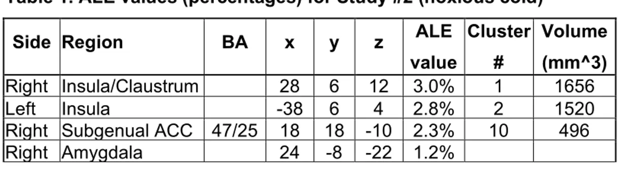
Table 1. ALE values for Study#2 (noxious cold).………112
Table 2. ALE values for Study #4 (noxious stimuli applied to the left side of the body)………..114
Table 3. ALE values for Study #4 (noxious stimuli applied to the right side of the body) ……….……….116
SUPPLEMENTARY INFORMATION Table S1. List of studies included in Study#1 (all noxious stimuli)………..130
Table S2. ALE values for Study #1 (all noxious stimuli)………143
Table S3. List of studies included in Study #2 (noxious cold)………..145
Table S4. List of studies included in Study #2 (noxious heat)………..147
Table S5. ALE values for Study#2 (noxious cold).……….149
Table S6. ALE values for Study #2 (noxious heat) ………...152
Table S7. ALE values for Study #2 (noxious cold minus heat) ………...…155
Table S8. ALE values for Study #2 (noxious heat minus cold)………157
Table S9. List of studies included in Study #3 (noxious heat vs. warm)….159 Table S10. List of studies included in Study #3 (noxious heat vs. baseline)………161
Table S11. ALE values for Study #3 (noxious heat vs. warm) ………163
Table S12. ALE values for Study #3 (noxious heat vs. baseline) ……..….165
Table S13 ALE values for Study #3 (noxious heat vs. baseline minus noxious heat vs. warm)………..……….167
Table S14. ALE values for Study #3 (noxious heat vs. warm minus noxious heat vs. baseline)……….………..….169
Table S15. List of studies included in Study #4 (noxious stimuli applied to the left side of the body)….……….………...171
Table S16. List of studies included in Study #4 (noxious stimuli applied to the right side of the body)……….………..175
List of tables (continued)
Chapter 3 – Supplementary Information
Table S17. ALE values for Study #4 (noxious heat applied to the left side of the body)……….……….……….………..……..179 Table S18. ALE values for Study #4 (noxious stimuli applied to the right side of the body) ……….……….182 Table S19. ALE values for Study #4 (left sided noxious stimuli minus the right sided stimuli) ………...185 Table S20. ALE values for Study #4 (right sided noxious stimuli minus the left sided stimuli) ..………...187
Chapter 4: Neural correlates of the conscious perception of warmth
Table 1. List of studies utilizing warm stimuli in the absence of motor responses………..251 Table 2. Temperature Tasks and Delta T calculation………253 Table 3. Numbers of detected and undetected stimuli across warm trials.255 Table 4. Numbers of detected and undetected stimuli: Individual results ………257 Table 5. Peak coordinates during time periods when stimuli were detected, undetected, and the contrast of detected vs. undetected stimuli within the warm ROI………..260 Table 6. Brain areas showing significant postive or negative BOLD-signal change outside the warm ROI that was associated with the detected or undetected stimuli, and the comparison between detected vs. undetected stimuli ………262
SUPPLEMENTARY INFORMATION
Table S1. Brain activation associated with dectected and undetected
Chapter 5: Referred sensations in phantom-limb pain patients provide clues to cortical reorganization
Table 1. Patient characteristics……….310
Table 2. Endogenous pain ratings………312
Table 3. Referred sensations in upper-limb amputees………..314
Table 4. Referred sensations in lower-limb amputees………..315
List of figures Chapter 2: fMRI of Pain
Figure 1. Time course of the BOLD nociceptive signal………51
Figure 2. Comparison of the sensitivity and resolution of fMRI and PET.…53
Chapter 3: Localization of pain-evoked activation in the brain: A meta-analysis of functional neuroimaging data
Figure 1. ALE map in response to all types of noxious stimuli…..………..110
SUPPLEMENTARY INFORMATION
Figure S1. ALE maps of painful cold (blue) versus painful heat (red)….…124 Figure S2. ALE maps demonstrating overlap between pain using either a resting baseline (yellow) or warm (blue) as a comparison………..….126 Figure S3. ALE maps of stimuli applied the right (blue) and left (red) side of the body………128
Chapter 4: Neural correlates of the conscious perception of warmth
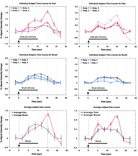
Figure 1. Stimulation protocol………244 Figure 2. Averaged warm ratings across the six scanning runs………..…246 Figure 3. Statistical parametric maps associated with the time periods for detected (left) or undetected (middle) stimuli and the comparison between detected vs. undetected stimuli (right)……….248 Figure 4. Time courses extracted from the peak positive or negative voxels in the warm ROI associated with the time periods when the stimuli were detected (blue line) or undetected (red line)………250
SUPPLEMENTARY INFORMATION
Figure S1. Time courses of the peak voxel associated with the comparison between detected (blue line) and undetected (red line) stimuli………264
Chapter 5: Referred sensations in phantom-limb pain patients provide clues to cortical reorganization
Figure 1. Pain intensity in the phantom limb ………..…………....306
List of abbreviations
ACC Anterior cingulate cortex aIC Anterior insula cortex
ALE Activation Likelihood Estimate ANOVA Analysis of Variance
CB Cerebellum
CTA Cortical thickness analysis
fMRI Functional magnetic resonance imaging FWHM Full width half maximum
LN Lentiform nucleus
LEP Laser evoked potentials MFG Middle frontal gyrus
m/DLPFC Medial/dorsolateral prefrontal cortex
MI Primary motor cortex
NC Nucleus accumbens
P Probability
PCL Paracentral lobule
PET Positron emission tomography PFC Prefrontal cortex
ROI Region-of-interest
SMA Supplementary motor area SI Primary somatosensory cortex SII Secondary somatosensory cortex STG Superior temporal gyrus
VBM Voxel based morphometry VP Ventral posterior nucleus
VPL Ventral posterior lateral nucleus VPM Ventral posterior medial nucleus
Dedication
I dedicate this thesis to my mother who constantly inspired and encouraged
Acknowledgements
I would first like to thank my supervisor, Gary Duncan, who has been my scientific mentor over the last seven years during which time he was a member of my Master’s thesis committee and my PhD supervisor. Gary has patiently taught me how to design experiments, think critically, and has done his best to try to improve my scientific writing. During these last years he has been an exceptional person to work with and I only hope that I will be able to continue to work with him in the future and to emulate his qualities with colleagues. On a personal note, he has opened his home that he shares with his amazing family (Alice, Stella and Coco) to many lab outings in Parc de la Maurice and taught us to appreciate recreational activities outside the lab.
I would also like to thank Pierre Rainville, who has served as my committee member during my doctorate degree. He has contributed to the majority of the work within this thesis and has helped me on an innumerable amount of occasions to discuss statistics and scientific ideas. Pierre’s
abilities as a scientist have continually inspired my work. I consider myself very fortunate to have had the honour to work with him and Gary during my doctoral studies.
I am indebted in gratitude to my other committee member, Elaine Chapman, for her advice and encouragement during my doctoral degree. Elaine has an extensive background in studying somatosensory processing and has helped me to improve my understanding of the neurophysiological basis of tactile processing. Additionally, she has mentored me in my
professional pursuits to pursue a postdoctoral degree.
I send a huge thank you to Marie-Claire Albanese, who has been my great friend and my colleague during these last few years. I meet Marie-Claire in 2002 during my Master’s degree. She imparted all of her knowledge to me on how to conduct a neuroimaging experiment and analyze data. During the last seven years we formed an excellent working
relationship where we could tackle projects as a team. I feel fortunate to have known her and she has contributed immeasurably to my scientific way of thinking. I know that I can rely on her for any scientific or personal
discussion and she has supported me in every decision that I have made. She is also a great driver and travel partner as was exemplified during our trips to Toronto, London, New York, Israel and Italy (and that one afternoon in Zurich).
I would also like to thank my lab mates who were always reliable when I needed a subject for the scanner or to discuss projects. In no particular order are Karin Pietruska, Danièle Laverdure-Dupont, Joshua Grant, Mathieu Roy, Mathieu Piché, Jen-I Chen, Audrey-Anne Dubé, Marianne Arseneault, Etienne Vachon-Presseu, Stéphanie Cormier, Mina Khosh Nejad, and Guoming Xie.
I am indebted in gratitude to the patients who participated in the study presented in Chapter 5. Their kindness and willingness to participate in research despite the terrible pain and suffering they have experienced taught me the importance of living life to its fullest and to be optimistic about the future.
Most of all I thank Jason, my mother, Terry, and my sister for their love and endless support during the years I have spent working on this thesis.
Contribution of Authors
Chapter 1: The introduction and background chapter is separated into two
chapters. In Chapter 1 the rationale and objectives of the research are presented followed by a brief introduction to mechnoreception and thermoreception. This background information is relevant to the subsequent experimental articles on warmth perception and the use of tactile stimuli for a quantitative sensory testing protocol.
Chapter 2: The second half of the background section of this thesis
contains a book chapter entitled “fMRI of Pain” that was published in fMRI Techniques and Protocols. This chapter provides an overview of the neuroanatomical substrates of nociceptive processing and its functional representation in the brain as revealed using brain imaging techniques. This chapter contains relevant information for the first experimental chapter on the representation of pain-evoked activation in the brain. I wrote the manuscript and Dr. Gary Duncan made revisions and comments that contributed to the final form of the manuscript.
Chapter 3: This chapter contains a manuscript in preparation entitled
“Localization of pain-evoked activation in the brain: A meta-analysis of functional neuroimaging data”. I generated the concept behind conducting the meta-analysis. I subsequently recruited and trained Joyce Fu to review articles and enter the data to create the
probabilistic maps. I reviewed the studies and data points in the meta-analysis. I subsequently performed the analysis, wrote the manuscript, prepared the figures and tables. Drs Gary Duncan and Pierre Rainville provided comments and revisions on the manuscript.
Chapter 4: This chapter contains a manuscript in preparation entitled
“Neural correlates of the conscious perception of warmth”. I
participated in the experimental design, programming of the protocol, tested all of the subjects, analyzed all of the data, wrote the manuscript and prepared all of the figures and tables. Drs. Gary Duncan, Pierre Rainville and Marie-Claire Albanese aided with the experimental design, protocol development, and analysis of the data and provided comments on the manuscript.
Chapter 5: This chapter is a manuscript in preparation entitled “Referred
sensations in phantom-limb pain patients provide clues to cortical reorganization”. I recruited and interviewed the patients included in the study, developed the quantitative sensory testing protocol, performed the testing, did the analysis, wrote the manuscript, and prepared the tables and figures. Dr. Gary Duncan aided in the development of the testing protocol and provided revisions on the manuscript.
Appendix I: This section contains the agreements of the co-authors and
publishers of the book chapter/manuscripts presented in Chapters 2, 3, 4 and 5.
Appendix II: The appendix also contains one published manuscript entitled
“Practice makes cortex” that appeared in the Journal of Neuroscience. This is a short review of an article that examined grey matter density and functional brain activation changes in response to a two week procedural learning task (Ilg et al. J Neurosci. 2008 Apr
16;28(16):4210-5). I included this manuscript as it is related to the general theme of brain plasticity discussed within this thesis. In addition, within the manuscript we used the analytic techniques
discussed in Chapter 3, whereby I conducted a meta-analysis of voxel-based morphometry studies demonstrating increased grey matter
density in response to learning induced changes. In collaboration with Danièle Laverdure-Dupont, I co-wrote the manuscript, performed the meta-analysis and prepared the figures.
Introduction Rationale
Controversy concerning the roles of the primary and secondary somatosensory cortices (SI & SII) in the perception of various types of cutaneous stimuli, including pain, temperature, and vibration, began with the first published experiments in the early 20th century and continues to the
present day. Modern advances in brain imaging techniques have made it possible to view the entire human brain in vivo, this new information has implicated other cortical regions such as the insula to be involved in
processing somatosensory information. The series of experiments described in this thesis were designed to study the involvement of SI, SII, and the insula in the perception of somatosensation. A clearer understanding of the brain regions involved in processing cutaneous stimuli could potentially improve diagnosis of chronic pain and treatment of somatosensory deficits in stroke patients.
In the early 20th century, research in somatosensation relied heavily on the clinical examination of patients who had lesions associated with brain pathology. Reports of patients with lesions to SI have produced conflicting results concerning the perception of touch and temperature information. One study found that patients with damage to SI were unable to identify objects touching their affected hand, but they retained the ability to localize painful pin-prick stimuli (Stewart 1908). Similarly, Head and Holmes
reported that patients with lesions to SI had abnormal mechanoreception but intact thermoreception (Head and Holmes, 1911). In contrast to these
findings, a more recent study found that a patient with a lesion to SI (and a portion of SII) was unable to perceive temperature and light touch but unimpaired vibration sensation (Ploner, Freund et al. 1999).
During the 1970s, a host of neurophysiological studies with non-human primates demonstrated that SI is involved in the perception of pain (Kenshalo, Jr. and Isensee 1983;Willis, Jr. 1985a;Willis, Jr. 1985b;Chudler,
Anton et al. 1990;Kenshalo, Iwata et al. 2000). This region contains a detailed somatotopic organization of nociceptive neurons, indicating that SI is involved the localization of noxious stimuli (Kenshalo, Iwata, Sholas, and Thomas 2000). Another study implicated SI in the intensity processing of painful stimuli as some of its neurons respond in an intensity-related manner (Kenshalo, Jr., Chudler et al. 1988).
While the 1990s saw advances in non-invasive brain imaging techniques, which facilitated the study of the brain in vivo, some initial studies reported inconsistent findings on activation in SI in response to noxious stimuli. Several of these modern imaging studies reported activation in SI in response to pain (Talbot, Marrett et al. 1991;Coghill, Talbot et al. 1994;Coghill, Sang et al. 1999;Chen, Ha et al. 2002), although other studies failed to find any activation in SI (Jones, Brown et al. 1991;Disbrow,
Buonocore et al. 1998). In addition to these conflicting results a series of recent studies have postulated that the pain-related activation that has been documented throughout the brain (including SI) is actually involved in the general process of magnitude estimation, rather than pain perception per se (Baliki, Geha et al. 2009).
Similarly, studies researching of the role of SII in the processing of somatosensory information have been plagued by contradictory results. For example, single-unit recording studies in animals demonstrated neuronal responses to tactile and thermal stimuli in SII (Robinson and Burton
1980a;Dong, Salonen et al. 1989). Additionally, SII has been shown to have a crude somatotopic organization indicating that this region processes spatial discrimination information (Robinson and Burton 1980b).
Corroborating these findings are clinical studies of patients with damage to SII, which demonstrate patients’ intact cutaneous and proprioceptive abilities including the ability to process temperature (Caselli 1993;Reed, Caselli et al. 1996). However, a recent conflicting report describes patients with SII lesions who exhibit intact tactile processing, with deficits only in pain and temperature perception (Kim, Greenspan et al. 2007). However, it is
important to note that some of these patients' lesions included the insula, making it difficult to dissociate the processes of either structure in cutaneous processing. Further contradictory evidence regarding SII in somatosensation comes from a single-unit recording study in nonhuman primates, which showed response suppression during attention to vibrotactile stimuli (Burton, Sinclair et al. 1997). Therefore, the role of SII in processing temperature and tactile information needs further clarification.
In addition to SI and SII, the insula is increasingly becoming the focus of somatosensory research. It receives information from and projects to parietal (including somatosensory cortices), prefrontal, and temporal cortices, making it well fitted for multisensory integration (Mesulam and Mufson 1982;Friedman, Murray et al. 1986;Preuss and Goldman-Rakic 1989). In terms of the insula’s involvement in the processing of
somatosensation, it has been implicated mainly in nociceptive processes (Apkarian, Bushnell et al. 2005;Brooks and Tracey 2007).
Electrophysiological studies in patients with epilepsy have reported painful and innocuous somesthetic responses in several regions of the insula, which follow a rough topographic organization (Penfield and Faulk
1955;Ostrowsky, Magnin et al. 2002). However, more recent reports from brain imaging studies have implicated the insula in vibration processing (Soros, Marmurek et al. 2007;Albanese, Duerden et al. 2009). Based on the known somatosensory input to the insula and the results of recent studies, in addition to SI and SII, a major focus of the current thesis is the
involvement of the insula in processing cutaneous stimuli.
Chapters 1 and 2 of this thesis provide background information on the neurophysiological basis of processing touch and temperature
information. Chapter 1 offers a review of the receptors, spinal pathways and cortical regions involved in processing tactile and temperature information. Chapter 2 focuses on both the neuroanatomy of pain processing and neuroimaging methods used to localize pain-evoked activation.
The manuscript presented in Chapter 3 describes a meta-analysis that makes a detailed survey of the pain-imaging literature over the past 20 years; using these data, four separate analyses are performed to address questions that have been difficult to resolve in isolated studies. The initial search of the pain imaging literature revealed 130 studies that satisfied the search criteria of pain, nociception, fMRI, and PET. All three-dimensional (3D) brain imaging coordinates in response to noxious stimuli were compiled and analyzed to create probability maps that can be overlaid on a standard magnetic resonance (MR) image. Each voxel in the MR image was
assigned a likelihood value denoting the absence or presence of activation in response to noxious stimuli from the individual studies. The first analysis of these data describes the creation of a general, “quantitative pain matrix” -- a 3--D interactive probability map illustrating the location and extent of the brain activation that is common across the various studies using
experimental noxious stimulation. The second analysis examines a subset of these data, searching for regions that process different types of noxious stimulation (e.g. stimuli that evoke the perception of cold pain or heat pain), irrespective of the different experimental paradigms used in the individual studies. Along similar lines, the third analysis assesses the implications of different control conditions in revealing activation related to noxious
stimulation; specifically, the location and extent of the apparent “pain-evoked” brain activation is compared for subsets of studies that have
employed either a resting baseline or an innocuous warm stimulus condition as a control for activation associated with noxious heat stimulation. This issue is important since warm stimuli are frequently used as a control condition for pain neuroimaging experiments; however, it remains unclear whether warm and noxious heat belong to the same sensory modality. Therefore, in some instances warm stimuli may not be an appropriate comparison for noxious heat. The fourth and final analysis tests for a possible hemispheric dominance for processing noxious stimuli by comparing the location and extent of brain activation across subsets of
studies that presented this type of stimuli either to the left or to right sides of the body. While targeting specific brain regions responsible for pain
perception could lead to improved diagnosis of chronic pain, it is rarely studied within the context of a single study.
The manuscript presented in Chapter 4 addresses issues that arose from the third part of the meta-analysis on the use of warm stimuli as an apprpriate control condition for painful stimuli. The third meta-analysis examined stimulus conditions used to examine “pain-related” activation, but did not focus on the brain regions associated with the perception of warmth. We were able to answer this question by using an fMRI data set in which subjects were given painful and warm stimuli presented in a counter-balanced fashion. After repeated presentations of painful stimuli, the
perception of some of the warm stimuli became attenuated. In the functional neuroimaging analysis the detected and undetected warm stimuli were entered as separate time periods that permitted the identification of brain activation in response to either condition.
Chapter 5 describes our exploration of abnormal somatosensory processing in a group of amputee patients who experience phantom-limb pain. While the underlying mechanisms remain unknown, research has shown that rapid cortical reorganization of somatotopic maps of the body in SI occurs in response to an amputation, and this has been causally
associated with phantom-limb pain intensity (Florence, Garraghty et al. 1994;Flor, Elbert et al. 1995). However, the results from the main meta-analysis from Chapter 3 would indicate that many other regions are involved in pain processing and possess a nociceptive somatotopic organization. Therefore, other regions such as SII and the insula are also likely to undergo reorganizational changes. Early case reports of patients with
upper-limb amputations inferred that the perceptual correlates of this cortical reorganization in SI manifest themselves in sensations referred to the
phantom by the touching of a patient’s face or arm (Ramachandran, Stewart et al. 1992). However, more recent reports questioned the reorganizational
changes that may occur in SI in response to deafferentaion, implying that other cortical regions that process pain also may be involved in referred sensation genesis (Grusser, Winter et al. 2001a; Sathien, 2001). Moreover, only one report has described referred sensations in lower-limb amputees that described highly localized and detailed remapping of the amputated phantom feet onto the upper thighs in these patients (Aglioti et al., 2005). Findings indicate that referred sensations are generated from body parts that lie adjacent to one another on the somatotopic map in SI. Unfortunately, only a few patients were examined, which makes it difficult to know if similar somatotopic reorganizational changes are generalisable to this population as a whole.
In the research described in Chapter 5, referred sensations were explored in a group of upper and lower-limb amputees using a variety of somatosensory stimuli to target both superficial and deep fibres in the skin. The overall goal in developing this quantitative somatosensory testing protocol was to determine if the referred sensations exhibit a somatotopic organization in order to provide clues as to which cortical regions are involved in the perception of referred sensations.
Objectives
The overall objective of the current thesis was to examine the
perception of noxious and innocuous stimuli in healthy subjects using meta-analytic and brain imaging techniques, and also by the development of a quantitative sensory testing protocol for use in patients with phantom-limb pain.
The objective of the meta-analysis presented in Chapter 3 was to investigate the common cortical regions involved in processing nociceptive information. The motivation behind performing this analysis was three-fold. Firstly, meta-analysis overcomes some of the limitations associated with conducting a single brain-imaging study, such as image artefacts or low power resulting from too few subjects. Meta-analysis can also localize
common brain regions associated with a particular task or cognitive function across studies (e.g. working memory). Secondly, the meta-analysis
permitted the exploration of a number of questions that have arisen in the pain brain-imaging community. Thirdly, the meta-analysis allowed for the creation of region-of-interest (ROI) maps, which may be used for future analysis of brain imaging data (fMRI or cortical thickness) to localize pain-processing regions in the brain.
Our objective in the manuscript presented in Chapter 4 was to identify the brain regions involved in the cogniscent awareness of innocuous warm stimuli. We explored warm-evoked activation in the brain by using data from an fMRI study we conducted in which noxious and innocuous heat stimuli were presented in a counter-balanced manner within the scanning runs. Throughout the course of the experiment, repeated presentation of painful stimuli caused peripheral fatigue of receptors on fibres transmitting pain and warmth information to the brain, and this resulted in some of the warm stimuli to be undetected by the subjects. In the functional neuroimaging analysis, we identified the time periods when subjects detected or did not detect the warm stimuli. This permitted the localization of brain areas that were activated by consciously detected or undetected stimuli.
Our objective in Chapter 5 was to document the pattern and intensity of referred sensations in phantom-limb pain patients. Such exploration could provide a greater understanding of the cortical regions affected by loss of somatosensory input. Furthermore, the results could later provide a rationale for future studies targeting the remapping of the somatotopic organization of somatosensory cortices.
1 Chapter 1: Background 1.1 Somatosensation
Somatosensation refers to the ability to perceive our body, which includes the perception of anything that comes into contact with our skin and the position of our limbs. It allows us to identify change in our pockets,
warns us against touching a hot radiator, and to perform a smooth and coordinated tennis swing. It is a sense that is largely taken for granted, as it rarely becomes noticeably impaired throughout aging, and the detrimental effects of a loss of somatosensation only become apparent after specific brain damage.
Somatosensation can be classed along two lines of perception: somatic sensations including mechanoreception (discriminative touch, vibration, light touch, movements across the skin), thermoception (cool, cold, warm), nociception (pain), and also proprioception (position and movement of the limbs). The latter is further subdivided into two
subcategories of joint position sense and kinesthesia, or knowledge of the movement of our limbs.
While each of these senses is processed by different receptors or nerve endings in the skin, and then via separate spinal pathways to the brain, they all converge outside the spinal cord in the dorsal root ganglia cells. The dorsal root ganglia cells have neurites that extend out to the periphery, to the skin or muscle and another process that enters the central nervous system (Davies and Lumsden 1990). Sensory afferent fibres enter the dorsal horn of the spinal cord and terminate either on spinal motor neurons to aid in motor reflexes, and others that ascend to the brain stem and thalamus via several fibre pathways. These pathways serve different types of somatosensation and project to the brain stem, thalamus, and cortex (Willis, 2007). An important note is that these afferents maintain a detailed spatial map of the body surface at all levels of the nervous system. The final termination point of cutaneous input is sent to SI, SII, and the superior parietal lobule (Willis, 2007). These cortical regions interact with
frontal and temporal regions to combine somatosensory information and to compare it with previous experiences (Friedman et al. 1986;Preuss and Goldman-Rakic 1989). These receptors, spinal pathways, and cortical regions provide the underpinnings of the conscious perception of somatosensation.
This thesis is primarily concerned with the study of somatic senses that include mechanoreception, thermoception, and nociception.
Proprioception is beyond the scope of the current work. The next section focuses on the receptors, spinal pathways, and cortical regions responsible for somatic sensation. The chapter concludes with an overview of brain imaging techniques with a primary focus on functional MRI of pain.
1.2 Mechanoreception
1.2.1 Cutaneous and Subcutaneous Mechanoreceptors
A variety of specialized receptors innervate the skin, viscera, muscle, joints, and bones to convey somatosensory information to the cortex
(Boulais and Misery 2008). Each receptor is classified based on the stimulus that produces the most optimal response. An important characteristic of sensory receptors is their firing pattern in response to their preferred stimuli. Receptors referred to as quickly adapting respond immediately and their firing pattern dissipates after several seconds, while slow adapting receptors maintain their initial response to a sensory stimulus, but do not have a rapid onset (Goodwin and Wheat 2004). Receptors can be further classified along three lines: those with encapsulated endings, free nerve endings, and
expanded tip endings.
Encapsulated endings are rapidly adapting receptors found in the dermis that perceive tactile information such as deep pressure,
discriminative touch, and vibration. They are aptly referred to as
tissue separated by encapsulated fluid. Pacinian corpuscles and Meissener receptors are considered encapsulated receptors (Vega et al. 2009).
Free nerve endings are responsible for primarily relaying pain and temperature information, although these receptor types also can mediate some tactile information (Fromy et al., 2008). These receptors have fine-grained, gossamer-like projections into the epidermal layer of the skin. Free nerve endings are dispersed all over the body and viscera, and for the most part have non-adapting firing patterns.
Examples of expanded tip endings are Merkel cells and Ruffini endings (Moll et al. 2005;Macefield 2005;Boulais and Misery 2007). These receptors are located in the epidermis, which suits them to be moderately adapting in their firing pattern. The receptors will fire after the application of the stimulus and will not attenuate during its presentation. The structure of the receptors is that of flattened ball-like shapes that transmit touch, pressure, and temperature information.
1.2.2 Neuroanatomy of Tactile Processing
Primary afferent fibres innervating tactile and joint receptors are located in the dorsal root ganglia (Fromy et al. 2008). These fibres are classified as either A-alpha or A-beta and are myelinated, which permits the rapid transmission of information (Provitera et al. 2007). This is true in all instances except for stretch-sensitive free nerve endings that transmit information about excessive force through A-delta fibres, which are responsible for transmitting painful information to the brain. Fibres that innervate the Pacinian corpuscles, Merkel disks, Ruffini endings, and
Meissener’s and muscle spindles will then synapse on neurons in the dorsal horn of the spinal cord (Macefield 2005). These neurons then project via the dorsolateral funiculus (DLF) and end in the lateral cervical nucleus (LCN) located in the upper cervical spinal cord segments (C1 & C2). LCN fibres then cross the midline and travel to the lower brain stem, where they join fibres in the medial lemniscus located in the medulla (Willis, 2007).
In addition to these pathways, some fibres innervating tactile
afferents will traverse through the dorsal columns to the medulla (Rustinioni et al. 1979). Fibres do not cross the midline and ascend through the spinal cord on the side ipsilateral to the site of entry. Afferent fibres from the lower limbs ascend through fasciculus gracilis and fibres from the upper part of the body travel through fasciculus cuneatus. These respective pathways
terminate in nucleus gracilis and nucleus cuneatus located at the base of the fourth ventricle on the dorsal surface of the medulla. Axons from the neurons in the nuclei cross the midline and traverse to the thalamus by way of the medial lemniscus. In the thalamus, the fibres terminate in the ventral posterior (VP) nucleus, with the fibres from the lower limbs being laterally located while the upper body are more medial (Herrero et al. 2002).
1.2.3 Subcortical and Cortical Processing of Tactile Input
Thalamocortical projections from the VP terminate in SI. Lesion
studies in higher primates have demonstrated that innocuous tactile sensory information is passed in a serial fashion from SI to SII (Pons et al.
1987;Garraghty et al. 1990). Axonal tracer studies have shown that this information is then relayed to the prefrontal cortex (Preuss and Goldman-Rakic 1989) and the mid/posterior insula and then to temporal lobe structures (Friedman et al., 1986;Mesulam and Mufson, 1982).
It has been questioned whether direct projections from VP to SII also exist, which would support parallel processing of tactile information. This has been called into question by experiments demonstrating that surgical ablation of SI renders SII unresponsive to tactile input in macaque and marmoset monkeys (Pons et al. 1987;Garraghty et al. 1990;Burton and Sinclair 1990;Murray et al. 1992). However, parallel tactile processing in SI and SII has been demonstrated in other species such as the cat (Burton and Robinson 1987) and rabbit (Murray et al. 1992). It has been postulated that differences in higher primates may be due to neurons arising from the ventral posterior inferior (VPI) nucleus projecting to SII, while those from the
VP nucleus project to SI (Garraghty et al., 1990;Krubitzer and Kaas, 1992;Friedman and Murray, 1986). Other authors have reported evidence for parallel tactile processing in SI and SII in the marmoset monkey (Zhang et al. 1996). However, these results were based on temporary inactivation by local cooling, rather than lesions of SI, and so it cannot be ruled out that SI may not have been entirely deactivated by the cooling procedure.
1.3 Thermoreception 1.3.1 Thermal Receptors
Separate receptors on the ends of sensory afferent fibres (A-delta & C-fibres) and on the cell bodies of dorsal root ganglia exist for the
perception of coolness, warmth, heat, and cold. Six different thermal transient receptor potential (TRP) channels have been identified that
mediate from noxious cold to burning heat (Patapoutian et al. 2003;Bandell et al. 2007). TRP channels are activated by changes in temperature, ligands (menthol, capsaicin, anandamide, protons), and two channels have been identified that are dependent on the cell’s membrane potential (voltage gated channels). The mechanisms by which these processes occur remain largely unknown. However, it has been suggested that temperature
fluctuation could induce ligand production and subsequent binding to TRP channels. An additional possibility may be that TRP channel proteins may undergo structural alterations in response to a change in temperature, causing the opening of the channel. A final alternative may be that TRP channels may be sensitive to alterations in membrane tension.
The cell bodies of sensory neurons are predominantly found in the dorsal root ganglia and they send neurites to the skin and muscle to transmit information about temperature. Incoming information is carried by thinly myelinated A-delta fibres and C-fibres that then terminate in the dorsal horn of the spinal cord. Other fibres that respond to temperature are A-delta fibres, which respond optimally to cool temperatures (Darian-Smith et al.
1973;Dubner et al. 1975;Dykes 1975;Kenshalo and Duclaux 1977), and a class of unmyelinated C-fibres that responds solely to stimuli in the warm temperature range (28-45oC) (Hensel and Kenshalo, 1969;Darian-Smith et
al., 1979). Additionally, a separate group of A-delta and C cutaneous afferents respond to noxious cold but not innocuous cool (Georgopoulos 1976;LaMotte and Thalhammer 1982;Simone and Kajander 1996;Simone and Kajander 1997). However, axotomized dorsal root ganglion cells can transmit temperature information indicating that the cell bodies also contain TRP channels. It is of note that the study of TRP channels is a rapidly advancing field of study and while all of the receptors have been identified, there remain many questions concerning their role in temperature
perception.
1.3.2 Thermosensory Spinal Pathways
Primary afferents that carry temperature information synapse on dorsal horn neurons. Some second-order neurons that transmit information to the brain have been found to be selectively activated by cold (Craig and Kniffki 1985) and somewhat more rarely by warm stimuli (Dostrovsky and Hellon 1978).
It was a generally held belief that these cold and warm specific second order neurons projected to the cortex through the general pain and temperature pathway, the spinothalamic tract (Craig and Dostrovsky 2001). However, this has recently come into question by a laser-evoked potentials (LEP) study that provides evidence for a warm specific spinal pathway (Iannetti et al. 2003). Short radiant heat pulses by way of a CO2 laser
stimulator were used to generate laser stimuli. These heat pulses selectively activate free nerve endings in the skin and A-delta and C-fibres in the
absence of A-beta fibre activation (Bromm and Treede 1984). In the LEP study, selective activation of C-fibres was achieved by using temperatures below those capable of being perceived by the A-delta fibres that transmit information about pain. Results showed significantly different latencies for
warm compared to painful stimuli, which the authors believed provided evidence for a warm specific pathway.
1.3.3 Subcortical and Cortical Temperature Processing
The representation of temperature processing in the thalamus and cortex has remained under dispute during the last several decades. An overwhelming amount of evidence provides proof that temperature
information is mediated by the primary sensory thalamus, the VP nucleus, and the somatosensory cortices (for review see (Willis, 2007). However, other evidence exists that the spinothalamic tract terminates in a
temperature specific nucleus, termed the ventral medial posterior nucleus (VMpo), which is located posterior to the VP nucleus. VMpo was found to contain somatotopically organized neurons that were specific to pain and temperature (Craig et al. 1994). In addition to antereograde staining studies in animals, the basis of this hypothesis was made largely from patients with lesions to VMpo who exhibited thermal sensory deficits and centrally
mediated pain. However, there is a contradictory published report in which a separate patient had exactly the same symptoms as the other group of patients, but whose lesion was localized to the VP nucleus (Montes et al. 2005).
The VMpo was found not to project to SI, but rather to the insula. These findings have been corroborated by imaging studies that found activation in the dorsal posterior insula in response to innocuous cool, but not in SI (Hua et al. 2005;Oshiro et al. 2007). However, current limitations associated with fMRI may not be able to sufficiently identify signals in the thalamus and SI, and further research in this area is needed. This is not the case for cold or warm temperatures, which, in a number of brain imaging studies have been shown to activate the somatosensory cortices, and also the prefrontal cortex and the anterior cingulate cortex (ACC) (Casey et al. 1996;Sawamoto et al. 2000;Iannetti et al. 2003;Olausson et al. 2005;Sung et al. 2007;Rolls et al. 2008).
2 Chapter 2: fMRI of Pain Preface
The following chapter is a continuation of the background chapter. It is the full text of a book chapter that I co-wrote with my supervisor Gary Duncan called “fMRI of Pain”. It has been published in “fMRI Techniques and Protocols” edited by Fillipo Massimi and published by Humana Press in 2009. I have included it as a background chapter as it reviews extensive information on pain processing. Additionally, it covers meta-analytic
techniques and the principles of fMRI, both of which are used in Chapters 3 and 4 of this thesis.
fMRI of Pain
Emma G. Duerden, M.Sc.1,2; Gary H. Duncan, D.D.S., Ph.D.1,3,4
1. Groupe de recherche sur le système nerveux central; 2. Centre de
recherche de l'Institut universitaire de gériatrie de Montréal; 3. Département
de stomatologie, Université de Montréal, Montréal, Québec, Canada; 4.
2.0 Abstract
The field of pain research has progressed immensely due to the advancement of brain imaging techniques. The initial goal of this research was to expand our understanding of the cerebral mechanisms underlying the perception of pain; more recently the research objectives have shifted towards chronic pain – understanding its origins, developing methods for its diagnosis, and exploring potential avenues for its treatment. While several different neuroimaging approaches have certain advantages for the study of pain, fMRI has ultimately become the most widely utilized imaging technique over the past decade because of its non-invasive nature, high-temporal and spatial resolution, and general availability; thus, the following chapter will focus on fMRI and the special aspects of this technique that are particular to pain research. Section 1 begins with a brief review on the spinal pathways and neuroanatomical regions involved in pain processing, and highlights the novel information that has been gained about these structures and their function through the use of fMRI and other neuroimaging techniques. Section 2 reviews a few of the aspects associated with the blood-oxygen-level-dependent (BOLD) signal commonly used in fMRI, as they apply to the particular challenges of pain research. Likewise, Section 3 summarizes some of the special considerations of experimental design and statistical analysis that are encountered in pain research and their applications to fMRI studies. Section 4 reviews special applications of fMRI for the study of higher cognitive processes implicated in pain processing, including pain empathy and cognitive reappraisal of one’s own pain perception. The chapter concludes with Section 5, exploring some of the future prospects of fMRI techniques and new applications related to pain research.
2.1 INTRODUCTION
The history of pain imaging is relatively short, although it has
advanced immensely within the last decade due to improvements in imaging techniques, statistical analysis, and specialized equipment for the delivery of painful stimuli. Initially, brain imaging studies sought simply to examine the brain areas that are involved in pain processing, to make comparisons with the long established neurophysiological studies reported in this field. Many of these initial imaging studies were prompted by electrophysiological data from patients undergoing brain surgery in the early part of the 20th century (1), which had questioned the role of the cortex in nociceptive processing. It was initially believed that the thalamus was primarily responsible for
nociceptive processing as suggested by deficits in pain perception observed in patients with thalamic lesions (2).
In the early 1990s, activation in the human brain evoked by
experimental pain stimuli was studied using positron emission tomography (PET) (3;4) and single photon emission tomography (SPECT) (5). Then in 1995 the first fMRI studies examining the cortical representation of pain (6) were conducted largely to confirm the findings of previous PET studies and to examine whether the cortical nociceptive signal could be detected using fMRI. In more recent years, the field of pain imaging has expanded
immensely, allowing researchers to answer complex questions concerning pain processing, such as how cortical regions are connected and modified during the perception of pain and, most importantly, how the cortex
responds during the modulation of pain. These experimental studies were conducted in healthy humans in order to answer broad questions regarding pain processing, with the eventual goal of applying this knowledge to a better understanding and alleviation of pain and suffering associated with chronic pain syndromes. The use of fMRI and other imaging techniques has revealed a number of cortical and subcortical changes that may occur as a
result of exposure to chronic pain (7). Indeed, with the advent of high-speed image acquisition and computational processing, not only has the
technology of fMRI revealed areas of cortical plasticity associated with chronic pain, it is now possible to use fMRI in real-time to furnish feedback to subjects (and patients) to teach them how to modulate their cortical activation in response to chronic pain (8).
This chapter reviews and discusses the various advances in our knowledge of cerebral pain processing that have been achieved using fMRI, the response properties of cortical nociceptive neurons in relation to both imaging techniques and stimuli used to evoke pain, the applications of this research to treat clinical pain in patients, and the future of pain research using fMRI.
2.2 BACKGROUND
2.2.1 Neuroanatomy of pain processing
Before describing how fMRI measures the cortical and spinal
nociceptive signal, it is important to understand how this signal is transferred to the cortex. In the periphery, a painful stimulus applied to the body is transmitted to the central nervous system (CNS) through nociceptors (9). Myelinated A-delta fibers transmit sharp pricking pain (10), while
unmyelinated C-fibers transmit slow burning pain, often referred to as second pain (11). The cell bodies of A-delta and C-fibers are located in the dorsal root ganglia, receiving afferent input from the periphery and then sending the information into the spinal cord to terminate in the dorsal horn (12;13). Axons from the second-order dorsal horn neurons rise through several ascending pathways that transmit nociceptive information to the thalamus, reticular formation, and cortex (14-19). Pain and temperature information applied to the face is relayed through cranial nerves to the spinal nucleus V terminating in the thalamus via the trigeminothalamic tract which is then relayed to the cortex. A number of spinal and cortical neurons
respond to noxious stimuli including nociceptive specific (NS) and wide dynamic range (WDR) projection neurons, the latter of which respond to both noxious and innocuous stimuli. Additionally, the dorsal horns and cortical somatosensory regions contain neurons responsive solely to innocuous stimuli called low threshold mechanical (LTM) neurons and thermoreceptive neurons responsive to temperatures in the warm and cold range. This range of responses is an important consideration when
interpreting results from fMRI studies of pain in terms of exactly what the activation pattern is reflecting.
Typically, pain-evoked brain activation is achieved by applying contact thermodes to the skin. This technique involves an increase in temperature at the rate of 1o-10oC per second. Depending on the baseline temperature it can take several seconds to reach perceived pain threshold. In addition to activating NS neurons inherently, LTM neurons respond to stimulation of the skin, and, as the temperature rises, thermoreceptors respond to the heating of the skin. Therefore, to examine pain-specific cortical activations, it is necessary to compare pain-related activations to those associated with the presentation of innocuous warm stimuli.
In addition to conductive heating of the skin using contact thermodes, nociceptive afferents can be activated using thermal radiation administered through infrared laser stimulators (20;21). Lasers can deliver heat stimuli without the need for a contact probe, thus selectively stimulating C-fibers and A-delta fibers without contaminant activation of A-beta fibers that transmit touch information. Additionally, laser stimuli can activate
nociceptive nerve endings at rapid rates for short durations (1 ms) (22;23) and are therefore well suited for rapid event-related fMRI studies. However, an important consideration associated with the use of laser stimuli is the difficulty of measuring and controlling skin temperature, which is the primary factor triggering the cascade of neural responses that culminate in the processing of heat-related nociceptive information in the brain and likewise, the assessment of pain by the subjects (24). Laser and contact heat stimuli
have been shown to produce similar patterns of
blood-oxygen-level-dependent (BOLD) activation in the secondary somatosensory cortex (SII), anterior cingulate cortex (ACC), insula, the primary motor cortex, prefrontal cortex (PFC), parahippocampal gyrus, thalamus, basal ganglia,
peri-aquaductal gray (PAG), and cerebellum. However, stronger activation in response to contact heat stimuli was noted in SII, posterior insula, posterior ACC, and regions in parietal and frontal cortices (25). Thus, these two modes of delivering noxious heat stimulation cannot be considered identical in terms of the evoked pain-related BOLD activations, and the advantages and disadvantages of each should be weighed in relation to the research questions and appropriate stimulation paradigms.
2.2.2 Supraspinal processing of nociceptive stimuli
A recent review of 68 pain neuroimaging studies using healthy subjects revealed a homogeneity of reported activations across cortical regions, thus implicating a cerebral network for pain processing (26). Regions most frequently activated by painful stimuli include primary somatosensory cortex (SI), SII, ACC, the insula, the PFC, and the thalamus.
Regions responsible for pain processing are categorized along two functional lines – the first being the sensory-discriminative (lateral pain system) component involved in the perception of temporal, intensity and localization aspects of pain processing, and the second, the affective-motivational (medial) component associated with the emotional aspects of pain (27). Dissociations between the two systems are made through
subjective reports on pain scales. After exposure to noxious stimuli, subjects are asked to quantify separately how intense and how unpleasant is the perceived pain. Subjects’ scores are recorded typically using numerical or visual analog scales (VAS) (28). Regions implicated in the lateral pain system include SI, SII, posterior insula and lateral thalamus, while the