Université de Montréal
Epithelial-Mesenchymal-Transition: a proposed mechanism in the development of Bronchiolitis Obliterans
By Areej Al Rabea
Département de Science Biomédical Faculté de medicine
Mémoire présenté à la Faculté des études supérieures en vue de l’obtention du grade de
Maître ès sciences (M.Sc.) en Science Biomédical
Avril 2011
Université de Montréal
Faculté des études supérieures et postdoctorales
Ce mémoire intitulé :
Epithelial-Mesenchymal-Transition: a proposed mechanism in the development of Bronchiolitis Obliterans
présenté par : Areej Al Rabea
a été évalué par un jury composé des personnes suivantes : Dr. Yves Berthiaume
président-rapporteur
Dre. Céline Bergeron directrice de recherché
Dr. James G Martin membre du jury
Résumé
La transplantation pulmonaire pour les patients avec une maladie pulmonaire en phase terminale est leur seul espoir de survie. Malheureusement, certains greffés du poumon rencontrent des difficultés après la transplantation du poumon, dont l'un est le rejet chronique du greffon pulmonaire également connu histologiquement comme la bronchiolite oblitérante et cliniquement comme syndrome de bronchiolite oblitérante. L'étiologie exacte de la BO reste mal comprise. Certaines hypothèses suggèrent l'implication des cellules épithéliales dans le processus de remodelage des voies respiratoires, conduisant à l'obstruction des voies aériennes. Un des mécanismes proposés est un processus de transition, connue sous le nom de transition épithéliale-mésenchymateuse (TEM). Lors de ce processus, les cellules perdent leurs propriétés épithéliales, acquièrent un phénotype mésenchymateux et deviennent plus mobiles et envahissantes. Cette transformation leur permet de participer activement au processus de remodelage bronchique dans la bronchiolite oblitérante. L’induction de la TEM peut être due à certains facteurs tels que l'inflammation et l'apoptose. Le principal objectif de ce travail de maîtrise est de détecter in vivo la présence de la TEM dans des biopsies transbronchiques obtenues chez des greffés et de l’associer à leurs conditions cliniques. Le deuxième objectif est d'induire la TEM in vitro dans les cellules épithéliales des petites voies aériennes à l'aide de milieux conditionnés apoptotiques et non apoptotiques produits par les cellules endothéliales microvasculaires humaines du poumon. D’autre part, nous avons évalué si des médiateurs connus pour participer au processus de TEM tels que le facteur de croissance du tissu conjonctif (CTGF)et le facteur de croissance transformant bêta
(TGF-beta) ainsi que le perlecan sont présents dans les milieux conditionnés utilisés. Mots-clés: transplantation pulmonaire, rejet chronique, transition épithéliale-mésenchymateuse, biopsies transbronchiques, apoptose.
Summary
For patients with end-stage lung disease, lung transplantation is their only hope for survival. Unfortunately, some of the lung transplant recipients (LTRs) might face obstacles following lung transplantation, one of which is chronic lung transplant rejection also known as bronchiolitis obliterans (BO) histologically and bronchiolitis obliterans syndrome (BOS) clinically. The exact etiology behind BO development remains poorly understood. Speculations have suggested the involvement of epithelial cells in the airway remodeling process leading to airway obstruction. One of the proposed mechanisms is a transitional process, known as epithelial-mesenchymal-transition (EMT). In this process epithelial cells lose their properties and acquire mesenchymal ones causing them to be more mobile and invasive which allow them to take part of the airway remodeling process in BO. Induction of EMT can be due to several factors such as inflammation, apoptosis. In our study we try to detect in vivo the presence of EMT in transbronchial biopsies (TBB) obtained from LTRs and associates it with their clinical conditions. We also try to manipulate and induce EMT in vitro in small airway epithelial cells (SAEC) using conditioned apoptotic (SSC4h) and non apoptotic (ZVAD) media produced from human microvascular endothelial cells (HMVEC) from lung. In addition, we worked on detecting possible mediators such as connective-tissue growth factor (CTGF), transforming growth factor-beta (TGF-β), and perlecan in produced media.
Keywords: lung transplantation, chronic rejection, epithelial-mesenchymal-transition, transbronchial biopsies, and apoptosis.
Table of Contents
Résumé …….……….……….……..…... iv Summary………….……….……… v Table of Contents……….…. vi List of Tables……….…….…... ix List of Figures………. xList of Abbreviations………..….. xii
Dedication………. xv
Acknowledgment……….. xvi
Chapter 1 ……….…1
Introduction ……….…1
1.1. Lung transplant as a therapeutic approach ………2
1.1.1. Factors influencing the rate of survival ………...… 4
1.1.2. Complications of lung transplant (LT ) ………...7
1.2. Chronic lung transplant rejection ………..………9
1.2.1. Bronchiolitis Obliterans (BO) vs. Bronchiolitis Obliterans Syndrome (BOS)……….…….……… 9
1.2.2. Risk factors contributing in the Development of Bronchiolitis Obliterans….……..……… 10
1.2.3. Clinical presentation and prognosis of Bronchiolitis Obliterans……….……….……….……… 13
1.2.4. Treatments of Bronchiolitis Obliterans………... 15
1.3. Apoptosis .………...………….…16
1.3.1. Apoptosis and Fibrosis/ Tissue Remodeling…..………. 19
1.3.2. Apoptosis and Bronchiolitis Obliterans ………. 20
1.4. Epithelial-Mesenchymal-Transition (EMT) ………....22
1.4.1. Implication of Epithelial-Mesenchymal-Transition in the pathologies of fibrosis ………...26
1.4.2. Implication of Epithelial-Mesenchymal-Transition in Lung ……….. 27
1.4.3. Epithelial-Mesenchymal-Transition and Bronchiolitis Obliterans………29
1.4.4. Mediators produced during apoptosis and Epithelial-Mesenchymal-Transition activation …..……….………. 30
1.4.5. Signaling pathways involved in the Epithelial-Mesenchymal-Transition process ………..……….. 35
1.4.6. Transcription factors involved in Epithelial-Mesenchymal-Transition process……….………..………. 39
1.5. Hypothesis and Rationale ……….…………...………... 42
Chapter 2 ……….. 44
Epithelial-Mesenchymal-Transition in Lung Transplant Recipients: Role in Bronchiolitis Obliterans Syndrome ………... 44
Context of the study and contributing authors…………...………. 45
Abstract ……….. 48
Introduction ……….... 50
Methods ……….. 53
Study subjects …..………... 53
Histochemistry staining of the transbronchial biopsies (TBB) ………. 54
Immunohistochemistry (IHC) staining ……….………. 54
Immunohistochemistry (IHC) scoring and statistical analysis……….. 55
Statistical analysis……….. 55
Results ……… 56
Analysis of demographic data ………... 56
Detection of Epithelial-Mesenchymal-Transition Expression ………. 56
Statistical analysis of gathered information ………... 57
Discussion ……….…………. 58
Conclusion ………... 60
Acknowledgment ………... 61
References ………... 70
Chapter 3………... 72
Activation of Epithelial-Mesenchymal-Transition by Lung Apoptotic Endothelial Cells Mediators: Implication in Airway Remodeling and Development of Bronchiolitis Obliterans ………... 72
Context of the study and contributing authors ………73
Abstract ………... 76
Introduction ………..………….. 79
Methods …………..………..……….. 81
Cell culture ……….……… 81
Screening for apoptosis in HMVEC-L with fluorescence microscopy ……… 82
Production of conditioned apoptotic (SSC4h) and non-apoptotic media (SSC4h-ZVAD) ……… 83
Immunoblotting ……….………. 84
Stimulation of Small Airway Epithelial Cells (SAECs) ……….……... 84
RNA extraction/Reverse Transcrption-Polymerase Chain Reaction (RT-PCR) /Quantitative-PCR (Q-PCR) …..……….….……….. 85
Immunofluorescence………... 86
Statistical analysis ….……….……….. 87
Results ……….……... 88
Produced Mediators by Apoptotic Endothelial Cells ………. 88
Expression Level of Epithelial-Mesenchymal-Transition Genes ……..….….. 88
Expression Level of Epithelial-Mesenchymal-Transition Proteins …………. 89
Discussion ……….……. 90 Conclusion .……….…... 94 References ………..…….. 106 Chapter 4...……….……….………..111 Discussion……….111 4.1. Epithelial-Mesenchymal-Transition in situ ………...113 4.1.1. Detection of Epithelial-Mesenchymal-Transition.……… 113
4.1.2. Correlations of Clinical Data with Epithelial-Mesenchymal-Transition ……….……….….. 116
4.1.3. Collagen Deposition Association with Epithelial-Mesenchymal-Transition ……….………... 117
4.2. Epithelial-Mesenchymal-Transition in vitro by Apoptotic Endothelial Cells ………. 118
4.2.1. Produced Mediators from Apoptotic Endothelial Cells ………….….……. 118
4.2.2. Change in Epithelial-Mesenchymal-Transition genes expression…….….. 120
4.2.3. Change in Epithelial-Mesenchymal-Transition Proteins Expression……. 122
Chapter 5………. 126
List of Tables:
Table I: Original and proposed classification of BOS ………. p. 10
Table II: Risk factors associated with the development of
Bronchiolitis Obliterans ……… p. 12
Table III: Clinical presentation of bronchiolitis obliterans
after lung transplantation ………. p. 13
List of Figures:
Figure 1: Survival by procedure type for adult lung transplants performed between January 1994 and June 2008 ……… p. 3
Figure 2: Number of lung transplants reported by year and procedure type .….… p. 5
Figure 3: Age distribution of adult lung transplant recipients (1/2985-6/2009) …. p. 6
Figure 4: Distribution of transplant by center volume. Lung transplants: January 1,
2000- June 30, 2009 ………. p. 7
Figure 5: Epithelial-Mesenchymal-Transition ……….. p. 23
Figure 6: Different types of Epithelial-Mesenchymal-Transition ……… p. 25
Figure 7: Mechanisms of Transforming growth factor-beta1 in inducing
Epithelial-Mesenchymal-Transition ……….. p. 32
Figure 8: Release of perlecan during endothelial cells apoptosis ………. P. 34
Figure 9: Transcriptional crosstalk between TGF-β, Wnt and Ras signaling pathways in EMT ………... p. 38
Figure 10: Proposed study hypothesis ……….. p. 43
Figure 11: Immunohistochemistry staining of E-cadherin in TBB .…….……… p. 63
Figure 12: Quantification of E-cadherin signal in the epithelium of small airways in TBB ……….………...… p. 64
Figure 13: Immunohistochemistry staining of Vimentin in TBB ……….… p. 65
Figure 14: Quantification of Vimentin signal in the epithelium of small airways in TBB ……… p. 66
Figure 15: Immunohistochemistry staining of Twist in TBB ……….…….. p. 67
Figure 16: Quantification of Twist signal in the epithelium of small airways in
TBB ………..……….…… p. 68
Figure 17: Van Gieson Stainings of obtained TBB ……….. p. 69
Figure 18: Mediators produced in apoptotic (SSC4h) and
non-apoptotic media (SSC4h-ZVAD) ……….. p. 95
Figure 19: Level of E-cadherin Expression in stimulated Small Airway Epithelial Cells (SAEC) ………. p. 96
Figure 20: Level of Collagen IA1expression in stimulated Small Airway Epithelial Cells (SAEC) ……….… p. 97
Figure 21: Level of alpha Smooth Muscle Actin (α-SMA) expression in stimulated Small Airway Epithelial Cells (SAEC) ……….….……… p. 98
Figure 22: Immunofluorescence of Epithelial markers ……….…………... p. 99
Figure 23: Immunofluorescence of Mesenchymal markers ……….…... p. 101
List of abbreviations
AEC Airway Epithelial Cells
AIDS Acquired Immune Deficiency Syndrome AP-1 Activator Protein-1
Apo2L Apo2 Ligand
ARDS Acute Respiratory Distress Syndrome
-SMA Alpha-Smooth Muscle Actin
AT1 Alveolar type I Epithelium AT2 Alveolar type II Epithelium BALF Bronchoalveolar Lavage Fluid
BALT Bronchus-Associated Lymphoid Tissue bFGF basic Fibroblast Growth Factor
bHLH basic Helix-Loop-Helix BLT Bilateral Lung Transplantation BO Bronchiolitis Obliterans
BOS Bronchiolitis Obliterans Syndrome Caspase Cysteine-Aspartic Protease
CD Cluster of Differentiation
cDNA complementary Deoxyribonucleic Acid CF Cystic Fibrosis
CMV Cytomegalovirus
COPD Chronic Obstructive Pulmonary Disorder CS Chondroitin 4-Sulfate
CTGF Connective Tissue Growth Factor DDR2 Discodin Domain Receptor 2
DISC Death Inducing Signaling Complex
DR Death Receptor
EC Endothelial Cells
ECM Extracellular Matrix
EGF Epidermal Growth Factor
ELISA Enzyme-Linked Immunosorbent Assay EMT Epithelial-Mesenchymal-Transition
FADD Fas-Associated Death Domain
FasL Fas Ligand
FEFL25-75 Mid-Expiratory Flow rate
FEV1 Forced Expiratory Volume in 1 second
FGF Fibroblast Growth Factor
FSP-1 Fibroblast-Specific Protein-1 GERD Gastroesophageal Reflux Disease
GM-CSF Granulocyte-Monocyte Colony-Stimulating Factor GSK-3β Glycogen Synthase Kinase-3 beta
HB-EGF Heparin Binding Epidermal Growth Factor
HLA Human Leukocyte Atigen
HLT Heart-Lung Transplant
HMGA2 High-Mobility Group A Protein 2
HMVEC-L Human Microvascular Endothelial Cells-Lung IGF-1 Insulin-Like Growth Factor-1
IL Interleukin
ILEI Interleukin-Like EMT Inducer IPF Idiopathic Pulmonary Fibrosis
LDLL Living Donor Lobar Lung Transplant MHC Major Histocompatibility Complex
MMPs Matrix Metalloproteinases
mRNA messenger Ribonucleic Acid
NFκB Nuclear Factor kappa-light-chain-enhancer of activated B cells
PAH Pulmonary Arterial Hypertension PDGF Platelet Derived Growth Factor
PGD Primary Graft Disfunction
PLT Primed Lymphocyte Testing
Rbm Reticular Basement Membrane
Rbm Reticular basement membrane
RTK Receptor Tyrosine Kinase
SAEC Small Airway Epithelial Cells
SLBL Split Lung Bilateral Lobar Transplant
SLT Single Lung Transplantation
SSC4h Conditioned Apoptotic Medium
SSC4h+Ab-CTGF Conditioned Apoptotic Medium + neutralizing antibody against CTGF
SSC4h-ZVAD Conditioned Non-Apoptotic Medium
TBB Transbronchial Biopsies
TGF Transforming Growth Factor
TNF Tumor Necrosis Factor
TRAIL (TNF)-Related Apoptosis-Inducing Ligand VSMC Vascular Smooth Muscle Cells
Dedication
I would like to dedicate my thesis to my supporting parents, whom without; I would not have reached this dream today. Dad, you are the greatest father in the entire world! Saying “thank you” would not be enough for what you have given me. You should know that your constant prayers, precious sacrifices, and endless support did not go to waste, and they were the hidden force that kept me going.
Mom, thank you for being there for me not only as a mother, but also as a friend. Your love, support and most importantly believing in me were my sources of strength when I was weak. Mom, people used to say “it is just a dream” that I will never be able to reach, but guess what! The dream did come true and you are the one who helped in that.
I love you both so much, and I want you both to be proud of me just as I am very proud of you both. This thesis is dedicated to you both, because only you would deserve it and no one else.
Love you,
Acknowledgment
I would like to thank my supervisor Dr. Celine Bergeron for giving me the chance and opportunity in doing my masters at her lab, and most importantly for believing in me. Dr. Bergeron you may have just given me a chance, but for me it was more than that, it was a challenge to prove what I have got. It was a hope when I was at the edge of giving up my dream. I simply cannot thank you enough for everything you have given and provided me with during my masters.
I would like to thank my colleague, Stanislaw Ptaszynski for all his support, guidance, and most importantly patience! Stan you are an awesome colleague, teacher, and of course an awesome friend who we can rely on during struggles.
I would like to thank Dr. Yves Berthiaume for his endless guidance and time, thank you so much! I would also like to thank all his team members: Andre, Francis, Jacynthe, Chantal, and Cecile. Thank you all for being there for me when I was in need for help and support, you are amazing and I will miss our good days!
Chapter 1:
1. Introduction:
1.1. Lung transplant as a therapeutical approach
Lung transplantation has been widely accepted as a therapeutic option for patients with end-stage lung disease in order to improve their survival. Up until the year of 1989 the most common form of lung transplantation was the heart-lung transplant (HLT) for patients who suffered pulmonary or cardiac combined with pulmonary end stage disease. Afterwards, single lung transplantation (SLT) and bilateral lung transplantation (BLT) started to pick up to be the most common procedures; however, nowadays the proportion of BLT is over passing the rate of SLT[1, 2] (see figure 1). In addition to previously mentioned procedures, other procedures are being considered as well such as split lung bilateral lobar transplants (SLBL) and living donor lobar lung (LDLL) transplants. According to the annual report 2010 of the International Society for Heart and Lung Transplant registry, the median survival for all adult recipients is 5.3 years, with the double lung procedure having a better survival rate compared to single lung procedure 6.6 years vs. 4.6 years, respectively (Figure 1)[2].
Figure 1: Survival by procedure type for adult lung transplants performed between January 1994 and June 2008:
Kaplan-Meier survival by procedure type for adult lung transplants performed between January 1994 and June 2008. Conditional half-life is the time to 50% survival for the sub-set of recipients who were alive 1 year after transplantation. J.D Christie 2010. J Heart Lung Transplant. 29:1104-1118 © International Society for Heart and Lung Transplantation
End-stage lung disease can be treated with lung transplant; however, types of lung transplant procedures vary and based depending on the type of disease. End-stage lung diseases include: chronic obstructive pulmonary disease (COPD), cystic fibrosis (CF), idiopathic interstitial pneumonitis, pulmonary arterial hypertension (PAH), and other rare diseases. COPD is considered to be the most common indication for lung transplantation [2]. For this type of disorder, both SLT and BLT have been suggested as procedures of lung transplant [3-9]. In the United States, the foremost cause of end-stage lung disease is cystic fibrosis (CF); it is the third most common indication for lung transplantation [10, 11]. For most adults with CF, bilateral lung transplant (BLT) would be their best option of lung transplant
procedure. The heart-lung transplant is rarely considered, depending on the patient’s needs, and association with left heart failure [12-15]. Another alternative of lung transplant procedure for CF patients would be living donor lobar lung transplant (LDLL) [16, 17]. This type of procedure is appropriate for children and young patients with CF who are small in size and would attain sufficient lung function from having two adult donor lobes [18]. Furthermore, another end-stage lung disease is idiopathic interstitial pneumonitis (fibrosis) which accounts for 20 percent of lung transplant [2, 19]. One common form of idiopathic interstitial pneumonitis is idiopathic pulmonary fibrosis (IPF) which commonly requires lung transplantation followed by nonspecific interstitial pneumonitis (NSIP) [20-22]. For patients with IPF, single lung transplant was the standard procedure; however, there is an increase in the number of patients undergoing BLT [2, 23-27]. The pulmonary arterial hypertension (PAH) accounts for less than 5 percent of lung transplant[2, 28]. It has been noted that patients with PAH have the highest 30-day mortality rate following lung transplantation In PAH patients, SLT and BLT have been considered as lung transplant procedures, where both were successful but survival comparisons revealed BLT to be more favorable over SLT. The cause and type of end-stage lung disease vary, which influences the approach and procedure of lung transplantation in leading for best results of survival for patients with end-stage lung diseases.
1.1.1. Factors influencing the rate of survival
Despite the fact that lung transplantation serves as the only therapeutic way for patients with end-stage lung diseases, however; it has limitations that might influence its successful outcome. One of the influencing factors on the survival rate of lung
transplant is the type of lung transplant that was chosen to be performed. Most common types of lung transplant include: single lung transplant (SLT), and bilateral lung transplant (BLT). The number of SLT performed annually has remained stable, though; the number of BLT has increased to surpass that of the SLT (Figure 2). As noted before, the original lung disease is a main factor in the selection of preferred lung transplant type which in turn influences the survival of LTR.
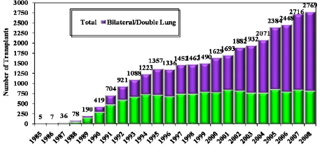
Figure 2: Number of lung transplants reported by year and procedure type:
Number of lung transplants reported by year and procedure type. J Heart Lung Transplant. 2010 Oct; 29 (10): 1083-1141[29]
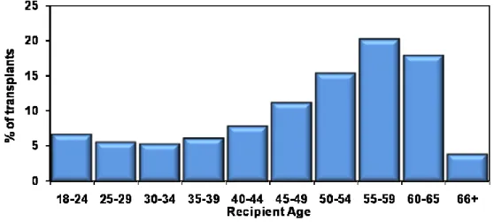
Age of the lung transplant recipient has an influence on the survival rate as well. Since younger recipients have a longer survival than older ones [2, 30], the American Thoracic Society (ATS) has proposed international guidelines to assist in selecting lung transplant candidates [31]. According to the ATS guidelines the upper age limits are as follow: approximately 55 years for HLT, approximately 65 years for
SLT, and approximately 60 years for BLT. This would help in maximizing the chances of survival (Figure 3).
Figure 3: Age distribution of adult lung transplant recipients (1/2985-6/2009):
Age distribution of adult lung transplant recipients (1/2985-6/2009). J Heart Lung Transplant. 2010 Oct; 29 (10): 1083-1141[29]
Another key factor that influences the success of lung transplantation is the involvement of transplant organizations and centers in recruiting, placing, and helping patients on the waiting list in getting lung transplant. The process of placement is based on Lung Allocation Score (LAS) in which it would help in addressing high waitlist mortality and earlier placement of patients on the waiting list [32-34]. The LAS system takes into account the urgency measure, which is defined as expected days lived without a transplant with an additional year on the waitlist. In addition, post-transplant survival measure is taken into consideration, which is defined as expected number of days lived during the first year post-transplant. These types of
measures are based and calculated using individual candidate’s clinical and physiological characteristics and statistical models [35]. Although the LAS system has been established in the year of 2005, two years following its establishment it has been noted that there was a decrease in the wait-list times and the mean LAS score of transplant recipients has increased. There was an increase in urgency for transplantation, and in the total number of transplanted patients as well [36]. The international society for heart-lung transplant registry (ISHLT) has indicated the distribution of lung transplant by center volume (Figure 4).
Figure 4: Distribution of transplant by center volume. Lung transplants: January 1, 2000- June 30, 2009:
Distribution of transplant by center volume. Lung transplants: January 1, 2000- June 30, 2009. J Heart and Lung Transplant. 2010 Oct; 29 (10): 1083-1141[29]
1.1.2 Complications of lung transplant (LT)
Despite the improvement in the lung transplantation procedure and survival, there have been reports of the occurrence of different types of complications following lung transplant. One major complication following lung transplant (LT) is primary graft dysfunction (PGD), which represents a mulitfactorial injury developing in the first 72 hours following lung transplantation. It has been referred to PGD as “ischemia-reperfusion injury”, “early graft dysfunction”, and “reimplantation edema” [37]. Characteristics of PGD include: sever hypoxemia, lung edema, and appearance of diffuse pulmonary opacities without other identifiable cause. Furthermore, diffuse alveolar damage serves as the typical pathologic pattern of PGD. It is worth mentioning that PGD is responsible for significant morbidity and mortality after lung transplantation, in spite of advances in organ preservation, surgical technique, and perioperative care [38-41].
Acute rejection is another possible complication that can occur following a lung transplant. Based on previous reports, acute cellular rejection appears less in both organs of the heart-lung transplantation compared to the rate of rejection in lungs or heart that are transplanted alone [2, 42, 43]. Furthermore, it has been noted that the rate of acute cellular rejection in the lung is higher compared to that in the heart following HLT [2, 42, 44, 45]. A proposed explanation of this increase is due to the presence of donor bronchus-associated lymphoid tissue (BALT), increase in the immunogenicity of the lungs, and frequent infectious insults with suppressed defense mechanisms [46].
Chronic rejection is also considered as one of the complications that might be faced following lung transplant. Chronic rejection of the lung is manifested histologically as bronchiolitis obliterans (BO), whereas grading via spirometry testing is known as bronchiolitis obliterans syndrome (BOS)[47] (will be discussed further in 1.2. Chronic Lung Transplant Rejection).
Infection, whether it is viral or bacterial, is considered as a possible complication following lung transplant as well. For instance, infection with cytomegalovirus (CMV) has been suggested as a risk factor for BO in patients undergoing lung transplantation [48-50].
In addition to previously mentioned complications, there are complications that might rise and are not necessarily unique to lung transplantation but rather are side effects of the immunosuppressive medications or general medical problems that are aggravated by the posttransplantation regimen [51]. One of these complications is chronic renal failure that is caused by immunosuppressant such as tacrolimus or cyclosporine [52]. Other complications include: osteoporosis [53-55], systemic hypertension [56], diabetes mellitus [57], obesity, anemia, gastroesophageal reflux disease (GERD) [58, 59], gastroparesis [60, 61], hypercholesterolemia and hypertriglyceridemia, cholecystitis, diverticulitis, weakness of respiratory and limb muscles [62-64], and pulmonary capillaritis [65].
1.2. Chronic Lung Transplant Rejection
Chronic rejection of the lung remains the major source of morbidity and mortality following lung transplant. The clinical syndrome of chronic rejection and
the infectious complications related to its treatment have been defined as major sources of late morbidity and mortality following lung transplant [66]. Chronic rejection has been classified pathologically into two types: chronic vascular rejection and chronic airway rejection [67]. The first type, less common to manifest, refers to a form of atherosclerosis developing in the pulmonary vasculature. The second type, which is more common and morbid, refers to presence of bronchiolitis obliterans (BO) histologically [68].
1.2.1 Bronchiolitis Obliterans (BO) vs. Bronchiolitis Obliterans Syndrome (BOS)
Understanding etiology and mechanism behind chronic lung transplant rejection have been puzzling researchers for several years due to its complexity in its mechanisms, diagnosis and causes of its development. As results of this confusion, a group of investigators from the International Society for Heart and Lung Transplantation has set a standardized nomenclature to help in classifying and diagnosing chronic rejection of lung transplant (see Table I)[47, 69]. They have also made a distinction between bronchiolitis obliterans (BO) and bronchiolitis obliterans syndrome (BOS). The earlier term refers to histological proofs of chronic rejection with scarring and fibrosis of the airways [70, 71]. The latter term refers to deterioration of graft function secondary to progressive airway disease with absence of histologic evidence of BO and no indication of other causes: infection, acute rejection, or anastomotic complications [69, 72].
Table I: Original and proposed classification of BOS:
Original classification Current proposition
BOS 0 FEV1 80 percent or more baseline BOS 0 FEVFEF1> 90 percent of baseline and 25-75> 75 percent of baseline BOS
0-p
FEV1 81 to 90 percent of baseline and/or FEF 25-75 ≤ 75 percent of
baseline
BOS 1 FEV1 66 to 80 percent of baseline BOS 1 FEV1 66 to 80 percent of baseline BOS 2 FEV1 51 to 65 percent of baseline BOS 2 FEV1 51 to 65 percent of baseline BOS 3 FEV1 50 percent or less of baseline BOS 3 FEV1 50 percent or less of baseline
BOS, bronchiolitis obliterans syndrome; FEF25–75, mid-expiratory flow rate; FEV1, forced expiratory volume in 1 second. Reproduced from: Estenne, M, Maurer, JR, Boehler, A et al. Bronchiolitis Obliterans Syndrome 2001: an update of the diagnostic criteria. J Heart Lung Transplant 2002; 21:297. Copyright©2006 the international Society for Heart and Lung Transplantation.
1.2.2 Risk Factors contributing in the Development of Bronchiolitis Obliterans
Although BO is a manifestation of chronic allograft rejection, other events can contribute in the development of BO. Severe acute rejection has been considered as a risk factor for BO development. Retrospective epidemiologic analyses have demonstrated that occurrence of three or more episodes of acute rejection is a major risk factor for BO development [66, 73, 74]. Moreover, cytomegalovirus (CMV) infection has been described as well as a cause of BO, where retrospective analyses have showed that it might be a risk factor for BO in patients undergoing lung transplantation; however, it was not confirmed in all studies [48-50, 75]. Primary graft dysfunction (PGD), also known as ischemia reperfusion injury, has been
associated with the development of BO, where there is a correlation between the severity of initial PGD with the risk of BO development [76-78]. This correlation can be explained due to presence of oxidative damage, impairment of nitric oxide synthesis by pulmonary endothelial cells, or by upregulation of HLA class II antigens on the allograft leading to production of anti-donor antibodies [79-82]. Another factor that may contribute to chronic allograft rejection is gastroesophageal reflux disease (GERD) which appears to be common in patients following lung transplant. Gastroesophageal dysfunction disease is common in patients with end-stage lung disease prior to lung transplantation and appears to increase following transplant [59, 83-88]. Possible mechanism underlying the risk of GERD may include injury to the vagus nerve and esophagus duringtransplant surgery [89]. Furthermore, type of lung transplant can be a key factor in the development of BO. For example, COPD patients whom underwent double lung transplant were more likely to be free of BO compared to those whom underwent single lung transplant three-years and five-years after transplantation [90]. Finally, autoimmunity has been a possible theory concerning the pathology of BO, which suggests that collagen type V epitopes resulted from ischemia/reperfusion injury or other type of an injury, cause damage of the epithelium of the allograft airway. Another key factor in the autoimmunity development of BO is human leukocyte antigen (HLA) mismatch. This process is characterized by recipient’s lymphocytes reactivity towards the donor antigen-specific class I antigens, which was reported by primed lymphocyte testing (PLT) in patients with BO [91, 92]. Production of anti-HLA class I antibodies precedes BO development, and it has been indicated that there is a correlation between an increase in anti-HLA antibodies with loss of pulmonary function [93]. Increase in HLA mismatches between graft and host,
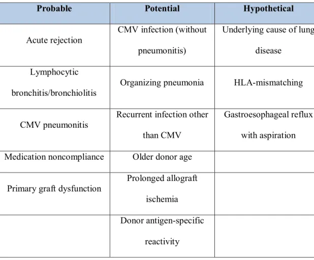
more specifically mismatches at the HLA-A locus, are associated with an enhanced risk of BO [48, 49, 94-96]. It has been mentioned as well that BOS is, for many patients, a recapitulation of the original lung disease for which the transplant was done. Risk factors contributing in the development of BO have been summarized in Table II.
Table II: Risk factors associated with the development of Bronchiolitis Obliterans:
Probable Potential Hypothetical
Acute rejection
CMV infection (without pneumonitis)
Underlying cause of lung disease
Lymphocytic bronchitis/bronchiolitis
Organizing pneumonia HLA-mismatching
CMV pneumonitis
Recurrent infection other than CMV
Gastroesophageal reflux with aspiration Medication noncompliance Older donor age
Primary graft dysfunction Prolonged allograft ischemia Donor antigen-specific
reactivity
Reproduced from: Estenne, M, Maurer, JR, Bohler, A, et al. Bronchiolitis Obliterans syndrome 2001: An update of thediagnostic criteria J Heart Lung Transplant 2002; 21: 297[47]
1.2.3. Clinical presentation and prognosis of BO
Symptoms that were associated during the development of BO still remain nonspecific, and indolent compared to those of acute rejection [97]. In Table III, the symptoms and signs at both early and late phase of BO are summarized. In usual cases, patients present a syndrome that resembles an upper respiratory tract infection. It remains unclear whether this presentation was based on a misinterpretation of the symptoms or it is further evidence of viral infection role in the etiology of BO. An increase in exertional dyspnea and decline in spirometry are usually noticed in patients. Pulmonary functioning test assists in detecting airflow obstruction. One possible early and sensitive indicator of airflow obstruction is forced expiratory flow between 25 and 75 percent of the vital capacity (FEF 25-75). This indicator appears to be more sensitive than decline in forced expiratory volume in 1 second (FEV1) [47, 98-100].
Table III: Clinical presentation of bronchiolitis obliterans after lung transplantation: Early Late Symptoms Non-productive cough; dyspnea on exertion
Productive cough; dyspnea at rest
Physical examination
Clear chest “Pops and squeaks”
Chest radiography Clear Bronchiectasis, hyperinflation Pulmonary
function tests
Obstruction; most marked in
mild flows (FEF (25-75)) Severe obstructive
Sputum culture Negative Pseudomonas
Reproduced from Reilly, JJ. Chronic lung transplant rejection: Bronchiolitis obliterans. In: UpToDate, Trulock, EP (Ed), UpToDate, Waltham, MA, 2011.
During early stages of BO, physical examination and radiography help in excluding other potential explanation for symptoms. Some of the early BO characteristics include: normal physical examination, clear chest radiograph, sterile sputum cultures or “oral flora” growth. Once BO reaches advanced stages the following characteristics are demonstrated: abnormal chest examination, and end inspiratory pops and squeaks. In addition, brnochiectasis and hyperinflation might be revealed by chest radiograph and computed tomography (CT) [101, 102]. Bronchiectasis presents the symptom at advanced stages with chronic productive
cough, breathlessness, and severe airflow obstruction on pulmonary function testing (Table III).
In the diagnosis of BO two approaches have been set: diagnosis by exclusion and definitive proof. It has been demonstrated in part 1.2.1 that histological proof refers to BO, whereas BOS refers to deterioration of the lung function secondary to an airway disease. In the histological part transbronchial biopsies (TBB) are used in confirming the diagnosis of BO. In one study, it has been reported a sensitivity of 17 percent and specificity of 94.5 percent of a single set of TBB [103]. In a second study, it has been reported a 15 percent histologic confirmation in patients clinically diagnosed with BOS[98]. Furthermore, a third study where TBB were used demonstrated a diagnosis confirmation in 82 percent of patients who developed clinical BOS [74]. Finally, in a fourth study it was noted that among 77 patients who were diagnosed with chronic rejection 52 percent of them had decline in FEV1 [70]. In this study, 9 percent of patients (7 out of 77) had diagnostic biopsies without accompanying physiological abnormalities, whereas 39 percent of patients (30 out of 77) revealed both positive histology and decline in spirometry. Other than the use of TBB and clinical data, several markers have been suggested as potential markers of early BOS. These markers include: neutrophil-predominant alveolitis with an increased levels of interleukin-12 (IL-12) in bronchoalveolar lavage fluid (BALF) [104-107], elevated levels of exhaled nitric oxide [108-110], evidence of air trapping on chest CT scan [111-114], bronchial hyperresponsiveness [115], and soluble CD 30 levels [116].
It is worth mentioning as well that bronchiolitis obliterans (BO) might also be idiopathic. It is characterized by a progressive airflow obstruction that leads to dyspnea, hypercapnia, and death. The importance and severity of BO rises due to its irreversible condition which limits the chances of survival in lung transplant recipients. According to the ISHLT registry[29], BO is considered as a leading cause of mortality with 25.4% in period between 1-3 years posttransplant, and 29.2% in period between 3-5 years posttranstplant.
1.2.4. Treatments of BO
Various approaches have been proposed in the treatment of BO, however; there is no well-established protocol in treating BO. For instance, in one study it was demonstrated that 32 patients with BO presented spirometric stabilization after switching from cyclosporine to tacrolimus over 12 months of follow-up [117]. In another study, similar results were observed when mycophenolate mofetil was introduced[118]. Other data revealed photopheresis to help in stabilizing some BO patients [119]. A report provided by a single center demonstrated possible benefit from aerosolized cyclosporine usage [120]. In an open study, it was noted that substitution of sirolimus by azathioprine was likely to lead of BOS progression in 37 subjects receiving either cyclosporine or tacrolimus [121]. Preliminary reports assessed the value of prolonged oral azithromycin therapy in a total of 34 patients with BOS [122-124]; demonstrating an association with significant improvements in FEV1 for some of the patients. In a larger observational study, 24 out of 81 patients
showed an improvement in FEV1 [125]. Another study evaluating the use of anti-CD 52 antibody Alemtuzumab for BOS revealed stabilization in BOS grade, but not FEV1 in 7 out of 10 patients [126]. Furthermore, according to limited evidence it was suggested that high-dose inhaled glucocorticoids are not effective in slowing or preventing BOS development [127]. Finally, retransplantation has been considered as a treatment of BO. However, early experiences suggest that BO tends to recur in retransplant recipients in an accelerated fashion.
1.3. Apoptosis
All cells have a finite life span which is terminated by cell death that occurs either through passive necrotic processes or as result of an active process of programmed cell death, also known as “apoptosis”[128, 129]. Apoptosis is an important key factor in the maintenance of human embryonic development and adult tissue homeostasis [129]. Apoptosis is a complex and organized machinery that functions in eliminating damaged or unneeded cells in the body [128, 130]. Characteristics of cells undergoing apoptosis include: cell shrinkage, condensation, fragmentation of the nucleus and bubbling of the plasma membrane, known as “blebbing,” and chromatin condensation and nucleosomal fragmentation [131]. Furthermore, resulting membrane-bound apoptotic bodies get consumed by either neighboring cells or by macrophages. In normal event, initiation of apoptosis occurs as a response to developmental stimuli such as a decrease in the local concentration of a particular tissue morphogen or growth factor. Other stimulating factors include: severe stress or damage to vital cellular components, which can result from ionizing radiation, heat shock, toxins, cell detachment from surrounding tissue, bacterial or
viral infection, and/or oncogenic signaling [132, 133]. Well functioning apoptotic pathways are essential for tissue homeostasis where dysregulation of it has been implicated in multiple diseases. Increase in apoptosis exacerbates many disorders such as: acquired immunodeficiency syndrome (AIDS), neurodegenerative disorders such as Alzheimer’s disease and Huntington’s disease, cardiac ischemia, and renal damage [133]. On the other hand, inadequate rate of apoptosis leads to development of cancer and autoimmune diseases [120]. Malfunction of apoptosis is a hallmark in cancer and essential in cancer development and tumor cell survival[134]. This suggests that targeting and manipulating apoptosis can serve as a therapeutic approach in treating cancer and other disorders [133].
Regulation of apoptosis is done via two main pathways: the intrinsic pathway, and the extrinsic pathway, both of which are anticancer therapeutic targets [135-137]. As the name implies, the intrinsic pathway is initiated from within the cell. This usually occurs as a response to cellular signaling due to DNA damage, defective cell cycle, detachment from the extracellular matrix (ECM), hypoxia, loss of cell survival factors, or other types of severe cell stress. Moreover, this pathway involves release of pro-apoptotic proteins that work on activating cysteine-aspartic protease (caspase) enzymes. Activation of the caspase process ultimately triggers apoptosis [135, 138-140]. The intrinsic apoptotic pathway hinges on the balance of activity between pro-apoptotic and anti-pro-apoptotic members of the Bcl-2 superfamily of proteins which work on regulating the permeability of the mitochondrial membrane. Some of the pro-apoptotic proteins include: BIK, BAD, and BIM; and anti-apoptotic include: Bcl-2, Bcl-XL, and BCLW [138].
The extrinsic pathway starts outside the cell at the activation of specific pro-apoptotic receptors on the cell surface. This activation occurs by binding of specific molecules known as pro-apoptotic ligands which include: CD95L/Fas ligand (FasL), and Apo2 ligand/tumor necrosis factor (TNF)-related apoptosis-inducing ligand (Apo2L/TRAIL), where these ligands bind to their cognate receptors CD95/Fas; and death receptor 4(DR4) and death receptor 5 (DR5), respectively [132, 135, 137, 141]. Unlike the intrinsic pathway, the extrinsic pathway triggers apoptosis independently of p53 protein (tumor suppressor protein 53) [142, 143]. Binding of the ligand to its receptor induces receptor clustering and recruitment of the adaptor protein Fas-associated death domain (FADD) and the initiator caspases 8 or 10 as procaspases forming a death-inducing signaling complex (DISC) [144-147]. The DISC formation results in bringing the procaspase molecules into close proximity to one another, leading them to be auto-catalytically processed and released into the cytoplasm where they activate effector caspases 3, 6, and/or 7; and thus, stimulating the intrinsic pathway [136, 148, 149]. The dimerization of caspase 8 is a key factor in its activation, and clustering of the receptors with associated DISC molecules enhance its activation [149]. Once DISC gets activated, the extrinsic pathway follows and adopts same machinery as the intrinsic pathway. Furthermore, it has been known that extrinsic pathway activation through binding of CD95L/FasL to CD95/Fas can result in two apoptotic programs, termed type I and type II. In the former type, cells are able to overcome the need for mitochondrial amplification of the death signal in CD95-mediated process by producing sufficient amounts of caspase 8 at the DISC which results in direct cleavage and activation of effector caspases and executes cell death [150]. Therefore, in type I cells bypass the mitochondrial involvement in
CD95-mediated apoptosis, expression of Bcl-2 or Bcl-XL has no inhibitory effect on their apoptotic program. On the other hand, in type II cells active caspase 8 is produced at a minimal amount at the DISC and requires the mitochondrial amplification of the CD95 signal [150]. Amplification of this signal might be through the pro-apoptotic BH3 domain, which only contains the Bcl-2 family member, Bid [150]. Bid gets cleaved by caspase 8 resulting in its translocation to the mitochondria where it initiates the release of mitochondrial factors, leading to increase in cell death. Since type II cells depend on the apoptotic function of mitochondria, expression of Bcl-2/Bcl-XL confers protection from apoptosis mediated by CD95 [150]. Differences between type I and type II cells remain unclear and need to be further studied and investigated.
1.3.1. Apoptosis and Fibrosis/Tissue Remodelling
Apoptosis of endothelial cells (EC) has been recognized as an early pathogenic event in fibrosis [151]. Increase in apoptotic EC has been associated with several fibrogenic disorders such as systemic sclerosis [152, 153], graft-versus-host disease [154, 155], and chronic rejection of solid allografts [156, 157]. The involvement of apoptotic EC was explained by its role in recruiting professional phagocytes such as macrophages [158]. As a result of apoptotic cell engulfment by the macrophages, transforming growth factor-beta1 (TGF-β1) gets produced [159]. The produced TGF-β1 stimulates myofibroblast differentiation and resistance to apoptosis in fibroblasts and myofibroblasts[57]. Furthermore, apoptotic endothelial cells have been suggested to have a direct impact in fibrogenesis by producing
paracrine mediators, such as TGF-β, connective tissue growth factor (CTGF), and perlecan (will be discussed in 1.4.4) that have been suggested to stimulate differentiation and resistance to apoptosis in fibroblasts [160, 161].
In the event of an injury, the process of wound healing is activated, such as endothelial damage; fibroblasts accumulate at the site of injury and differentiate into myofibroblasts, a fibroblast type characterized by presence of stress fiber and alpha-smooth-muscle actin (α-SMA)[162]. Normally, myofibroblasts undergo apoptosis once the healing process is terminated. Fibrosis follows the same pattern of wound healing; however, myofibroblasts develop resistance towards apoptosis which prevents their clearance, leads to accumulation of myofibroblasts and tissue contraction, which results in deformation and loss of function [163, 164]. Alteration in apoptosis’ rate, whether it is in epithelial or endothelial cells, is a feature that has been implicated in several lung pathogenesis such as idiopathic pulmonary fibrosis (IPF) [165], acute respiratory distress syndrome (ARDS) [166], and bronchiolitis obliterans organizing pneumonia [167].
1.3.2 Apoptosis and Bronchiolitis Obliterans
In lung transplantation, the implication of apoptosis was investigated in ischemia-reperfusion injury which was associated with endothelial apoptosis [168-171] and in transbronchial biopsies obtained from patients undergoing acute or chronic lung allograft rejections have been associated with epithelial and macrophages apoptosis[172-174]. In BO after lung transplantation, it has been
mentioned that the main target of rejection is the bronchial epithelium [175], where apoptosis is suggested as the mode of cell death. Apoptotic cells and their role in allograft rejection and development of BO are still under investigation [176].
Previous studies demonstrated the importance of airway epithelial cells (AEC) as immunologic targets during the process of acute or chronic lung allograft rejection [177-179]. Activated epithelial cells result in production of various growth factors including: epidermal growth factor (EGF) [180], heparin binding EGF (HB-EGF) [179], basic fibroblast growth factor (bFGF) [179], granulocyte-monocyte colony-stimulating factor (GM-CSF) [181], insulin-like growth actor1 (IGF-1) [182], platelet-derived growth factor (PDGF), and TGF-β [179]. Studies have noted the involvement of these growth factors in inducing proliferation of fibroblasts and smooth muscle cells indicating their potential role in fibrogenic activity in vivo [182-189]. In addition, studies have reported elevated levels of PDGF, TGF-β, and IGF-1 during the development of BOS after lung transplantation [190-198]. However, cellular sources and stimuli for fibrogenic growth factor production during the process of BOS development remain unknown. Studies have reported development of anti-HLA class I antibodies as a predisposing factor in BOS development after lung transplantation [93]. In addition, the association of anti-HLA class I antibodies has been linked with the development of transplant atherosclerosis and graft loss after kidney and heart allograft transplantation [199, 200]. Since binding of anti-HLA induce proliferation and apoptosis of AEC, binding of anti-major histocompatibility complex (MHC) has been associated with apoptotic cell death of activated human T
and B lymphocytes [201, 202], and of cardiovascular origin cells such as endothelial cells, smooth muscle cells, fibroblasts and monocytes [203]. The contribution of apoptosis has been noted in acute and chronic rejection of heart, lung, kidney, and liver allograft [172, 204-206]. Furthermore, studies have revealed increased levels of AEC apoptosis in lung allografts of patients with BOS [173]. In experimental lung transplantation (LT), an association between ischemic-reperfusion and endothelial cells was noted [169-171]. In one of our recent studies [207], we demonstrate the involvement of airway endothelial and epithelial apoptosis in the pathogenesis of BO, where the triggering factor of apoptosis initiation needs to be further investigated in order to help and improve the outcome of lung transplantation.
1.4 Epithelial-Mesenchymal-Transition (EMT)
The mystery behind the origin of mesenchymal cells that participate in tissue repair and pathological processes, tissue fibrosis, tumor invasiveness, and metastasis, is poorly understood. An important providing source that has been proposed for the generation of mesenchymal cells is epithelial-mesenchymal-transition (EMT). This process is defined as a transdifferentiating process where it allows an intact polarized epithelial cell, which has specific interaction with the basement membrane via its cell surface, to undergo multiple biochemical changes that enable it to assume properties of mesenchymal cells. The properties of mesenchymal cells include: enhanced
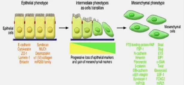
migratory capacity, invasiveness, elevated resistance to apoptosis, and increase the production of extracellular matrix (ECM) components [208]. Once the EMT process has been completed, it signals degradation of the underlying basement membrane, the newly formed mesenchymal cell migrates away from its original epithelial layer. Initiation and activation of the EMT process takes an orchestrated manner where number of complex molecular processes are involved in it. Such an organized process involves ordered steps: activation of transcription factors, expression of specific cell-surface proteins, reorganization and expression of cytoskelatal proteins, production of ECM-degrading enzymes, and changes in the expression of specific microRNAs. The involved factors can be used as biomarkers to help in assisting the passage of a cell through an EMT (Figure 5) [209].
Figure 5: Epithelial-Mesenchymal-Transition:
An EMT involves a functional transition of polarized epithelial cells into mobile and ECM component–secreting mesenchymal cells. The epithelial and mesenchymal cell markers commonly used by EMT researchers are listed. Colocalization of these two sets of distinct markers defines an intermediate phenotype of EMT, indicating cells that have passed only partly through an EMT. Detection of cells expressing both sets of markers makes it impossible to identify all mesenchymal cells that originate from the epithelia via EMT, as many mesenchymal cells likely shed all epithelial markers once a transition is completed. For this reason, most studies in mice use irreversible epithelial cell–lineage tagging to address the full range of EMT-induced changes. ZO-1, zona occludens 1; MUCZO-1, mucin ZO-1, cell surface associated; miR200, microRNA 200; SIP1, survival of motor neuron protein interacting protein 1; FOXC2, forkhead box C[209]. Kalluri, R. and R. A. Weinberg (2009). "The basics of epithelial-mesenchymal transition." J Clin Invest 119(6): 1420-1428
Based on previous studies done on developmental biology, during embryogenesis and organ development, certain epithelial cells appear to be plastic and thus able to move back and forth between the epithelial and mesenchymal states via the processes of EMT and MET (mesenchymal-epithelial-transition) [210]. This suggests that occurrence of such transdifferentiation allows conversion of epithelial cells to mesenchymal derivatives is needed during embryo development and adulthood. Activation of EMT has been also associated with tissue repair and
pathological stresses. This leads to recognizing EMT as a key player in dispersing cells in embryos, forming mesenchymal cells in injured tissues, and initiating the invasive and metastatic behavior of epithelial cancers (R. Kalluri) [209].
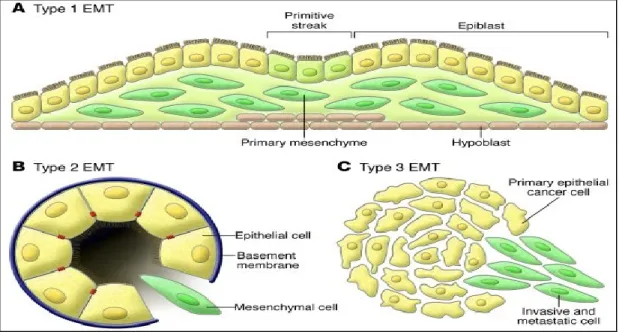
The epithelial-mesenchymal-transition (EMT) has been classified into three distinct biological classes leading to different functional consequences. The first class of EMTs, type I (Figure 6), represents the process that is associated with implantation, embryo formation, and organ development which leads to an organized generation of diverse cell types that share common mesenchymal phenotypes [211]. This class of EMTs is neither involved in fibrosis nor induces an invasive type which spreads out via the circulation system. In addition, the produced mesenchymal cells can switch back to epithelial cells through MET, leading to generation of secondary epithelial cells.
Figure 6: Different types of Epithelial-Mesenchymal-Transition:
(A) Type 1 EMT is associated with implantation and embryonic gastrulation and gives rise to the mesoderm and endoderm and to mobile neural crest cells. The primitive epithelium, specifically the epiblast, gives rise to primary mesenchyme via an EMT. This primary mesenchyme can be re-induced to form secondary epithelia by a MET. It is speculated that such secondary epithelia may further differentiate to form other types of epithelial tissues and undergo subsequent EMT to generate the cells of connective tissue, including astrocytes, adipocytes, chondrocytes, osteoblasts, and muscle cells. (B) EMTs are re-engaged in the context of inflammation and fibrosis and represent the type 2 EMTs. Unlike the type 1 EMT, the type 2 EMT is expressed over extended periods of time and can eventually destroy an affected organ if the primary inflammatory insult is not removed or attenuated. (C) Finally, the secondary epithelia associated with many organs can transform into cancer cells that later undergo the EMTs that enable invasion and metastasis, thereby representing type 3 EMTs.[209] Kalluri, R. and R. A. Weinberg (2009). "The basics of epithelial-mesenchymal transition." J Clin Invest 119(6): 1420-1428.
The second class of EMTs, type II (Figure 6), represents EMTs that are associated with wound healing, tissue generation and organ fibrosis. In this type of EMTs, the process starts out as a normal “repair” process that needs to generate
fibroblasts and other coupled cells in order to help in reconstructing the damaged area following an insult or inflammation. However, in contrast to type I EMTs, type II EMTs are activated as a response to inflammation and cease once the inflammation is attenuated, this is noted in wound healing and tissue repair. In the case of fibrosis, type II EMTs continue on as a response to ongoing inflammation, which leads to destruction of organs. In other words, tissue fibrosis is an unabated form of wound healing process due to persistent inflammation. The third class of EMTs (Figure 6) represents EMTs that occur in neoplastic cells that have already undergone through genetic and epigenetic changes that result in clonal outgrowth and localized tumors. These outcomes are due to alterations of oncogenes and tumor suppressor genes, thus the result is different from those of the other two types of EMTs.
.
1.4.1 Implication of Epithelial-Mesenchymal-Transition in the pathologies of fibrosis
Since our work explore the implication of EMT in the formation and development of bronchiolitis obliterans (BO), we will be focusing on type II of EMTs which is associated with tissue regeneration and organ fibrosis. The organ fibrosis is triggered by inflammatory cells and resident fibroblasts that function to release a variety of inflammatory signals and extracellular matrix (ECM) components that include collagens, laminins, elastin, and tenacins [209]. The type II EMTs which leads to organ fibrosis has been associated with fibrosis occurring in kidney, liver, lung and intestine [212-215]. Such an association was proved by studies done on
transgenic mice that bear germ-line reporter genes whose expression was driven by specific promoters. Follow up of the expression of these reporters provided evidence for the involvement of epithelial cells as key promoter and generator of fibroblasts in organ fibrosis, via EMT [216-218]. Several biomarkers, such as fibroblast-specific protein 1(FSP-1), α-SMA and collagen I, are generated by the EMT process which leads to fibrosis of several organs [9, 10, 219]. In addition to previously mentioned biomarkers, other markers such as discodin domain receptor tyrosine kinase 2 (DDR2), vimentin, and desmin have been studied in identifying the epithelial cells of kidney, liver, lung and intestine that are mid-way through EMT associated with inflammation. What was noted is that cells at this stage not only show epithelial-specific morphology and molecular markers such as cytokeratin and E-cadherin, but also express mesenchymal markers FSP-1 and α-SMA. Possible explanation of such behavior is that these cells are likely to be at an intermediate phase of EMT, where epithelial markers are still expressed but mesenchymal ones are being acquired as well. This behavior serves as an early indication or prediction of epithelium being exposed to an inflammatory stress. Once these cells “leave their epithelial layer, negotiate their way through the underlying basement membrane, and accumulate in the interstitium of the tissue, they shed all of their epithelial markers and gain fibroblastic ones”[220].
1.4.2 Implication of Epithelial-Mesenchymal-Transition in lung
As mentioned before, EMT has been proposed as a possible contributor in the fibrosis of kidney, liver, and intestine [5-8]. This had lead to propose the possible involvement of EMT in the pathogenesis of lung fibrosis. The exact role of EMT as a
response to an injury and pathogenesis of fibrosis in the adult lung remains to be further studied. Evidence has suggested participation of EMT as a major source of pathogenic mesenchymal cells, such as myofibroblasts, that lead to the development of pulmonary fibrosis. EMT was identified in both the alveolar epithelium and airway epithelium. Alveolar epithelial cells have been proposed as a key pathogenic intermediary of idiopathic pulmonary fibrosis (IPF) [221-223]. The importance of alveolar epithelial cells rises due to its regulatory functions that involve: production and response to profibrotic mediators, regulation of fibroblasts’ functions and differentiation through release of mediators, and modification of cell morphology and gene expression in response to injury [224-232]. Alveolar epithelial cells in IPF demonstrate the following features: morphological abnormality, pneumocytes hyperplasticity, and reactivity of elongated cells overlying the fibroblastic foci, which is presumed to be the site of active fibrogenesis [221, 233-235]. In addition, expression patterns of cytokeratin have been altered [236], and apoptosis of alveolar epithelial cells adjacent to fibroblastic foci has increased [237-239]. Upon activation of the alveolar epithelial cells in IPF synthesis of several procoagulant factors [229], and fibrogenic cytokines, such as PDGF [224], TGF-β [226-228], TNF-α [240], endothelin-1 [225], and CTGF [232] get produced which allows for a bidirectional signaling between alveolar epithelial cells and fibroblasts. The alveolar epithelial cells also stimulate production of matrix metalloproteinases (MMPs), which suggests contribution of alveolar epithelial cells in the extracellular matrix remodeling [241, 242].
Studies done on lung explants of transplanted mice models revealed that lung fibrosis can be initiated with epithelial injury and irregular repair mechanisms even in the absence of inflammation, and the presence of an intact epithelial layer has suppressed fibroblast proliferation and matrix deposition [243, 244]. Further confirmation was done by in vivo studies which provide evidence of the importance of EMT in fibrosis. These studies have used Cre-LOX system with β-galactosidase (β-gal) tagging, alveolar type II epithelium (AT2) has shown an expression of vimentin and undergo EMT when exposed to overexpression of TGF-β1 [215]. It has been noted that vimentin-positive cells within injured lungs were all β-gal positive, which suggests epithelial cells to be a reservoir of mesenchymal cells. Further reports done on AEC obtained from mice fibrotic lung overexressing insulin-like growth factor- binding protein-5 (IGFBP-5) coexpresses the expression of epithelial markers and α-SMA, suggesting EMT [245]. Airway epithelium has been investigated as potential contributor of intrapulmonary fibroblasts and myofibroblasts as a response to injury. Fibrotic obstruction of small and large airways is a key pathologic contributor in a variety of disorders, such as asthma [246], and obliterative bronchiolitis [247]. For instance, asthma is characterized with airway remodeling that can cause the disease and occur independently of inflammation [248, 249]. In recent studies, it has been suggested that abnormal epithelial-mesenchymal response to environmental challenges has a major role in the pathology of airways and physiology of asthma [250]. An increase in the deposition of collagen, fibronectin, and other ECM proteins was observed in asthma, which leads to subepithelial fibrosis and airway hyeperresponsiveness [251]. Possible explanation of the production of these
proteins is due to the presence of fibroblasts and myofibroblasts, where the number of these cells showed a correlation with the magnitude of subepithelial thickness [252].
1.4.3 Epithelial-Mesenchymal-Transition and Bronchiolitis Obliterans
Since understanding the pathogenesis of airway remodeling is needed for therapeutic development. One of the unresolved and poorly understood airway disorders is bronchiolitis obliterans (BO). BO is defined pathologically as airway response to chronic allograft rejection, and physiologically as bronchiolitis obliterans syndrome (BOS). This disorder is characterized by being an irreversible process which leads the patient to direct morbidity and mortality [47, 253]. Etiology behind BO is still poorly understood and still under constant investigation, but it is suggested to be a result of epithelial response upon an injury by an immunological or non-immunological events [254]. As mentioned earlier, remodeling and fibrotic obstruction of the small airways are key pathological factors in BO [34, 35]. Not only the precise mechanism of BO remains mysterious, but also the origin of fibroblasts responsible for airway fibrosis is unknown and serves as an important role in knowing the basic mechanisms. Up to the present date, only one study of its kind, Ward et al.[255], has suggested a link between lung transplant recipients (LTRs) and EMT. In this study it has been noted that airway epithelial cells obtained from stable LTRs have exhibited features of EMT. They were able to detect positive staining of fibroblast specific protein-1 (FSP-1), a marker of mesenchymal phenotype, in 15% of the sampled epithelium sampled. In addition, stimulation of obtained epithelium with