Université de Montréal
The Roles of TL1A and Pno1 in the Pathogenesis of
Rheumatoid Arthritis
par Xuehai Wang
Département de Médecine Faculté de Médecine
Thèse présentée à la Faculté des études supérieures en vue de l’obtention du grade de
Philosophiae Doctor (Ph.D.) en Sciences Biomédicales
Octobre 2013
Université de Montréal
Faculté des études supérieures et postdoctorales
Cette thèse intitulée:
The Roles of TL1A and Pno1 in the Pathogenesis of Rheumatoid Arthritis
Présentée par : Xuehai Wang
a été évaluée par un jury composé des personnes suivantes :
Dr. Mohit Kapoor Président-rapporteur Dr. Jiangping Wu Directeur de recherché Dr. Hongyu Luo Codirectrice
Dr. Marie Sarfati Membre du jury Dr. Joyce Rauch Examinateur externe
RÉSUMÉ
La polyarthrite rhumatoïde (PR) est une maladie auto-immune chronique. Elle est caractérisée par une inflammation persistante touchant de multiples petites articulations, causant douleurs, rougeurs, gonflements et déformations. Des études menées auprès de patients et d’animaux ont démontré que certains auto-anticorps, cytokines et enzymes tissue-déstructives sont des médiateurs importants dans le développement de la PR. Au cours des deux dernières décennies, les traitements de fond (DMARDs en anglais) ont été démontrés très efficaces pour traiter la PR. D'autre part, des effets secondaires ont été rapportés pour ces traitements, par exemple l'augmentation du risque d'infections opportunistes. L’objectif de ce travail est d’acquérir des connaissances sur le rôle du TL1A (TNF-like molécule 1 A; TNFSF15) et son partenaire Nob1 (Pno1 ; YOR145c) dans la pathogenèse de la PR afin de découvrir de nouveaux médicaments contre ces molécules dans l'avenir.
TL1A est un membre de la famille du TNF. Il déclenche des signaux co-stimulateurs via le récepteur de mort 3 (DR3) et induit la prolifération ainsi que la production des cytokines pro inflammatoires par les lymphocytes. Des données multiples suggèrent l'implication de la cascade TL1A-DR3 dans plusieurs maladies auto-immunes. Donc, nous avons proposé les hypothèses suivantes:1) la production locale de TL1A dans les articulations est un composant d’un cercle vicieux qui aggrave la PR; 2) dans la PR, la production de TL1A dans les organes lymphoïde augmente la production d’auto-anticorps pathogénique. Au cours de ce travail, nous avons démontré que la TL1A aggrave la maladie chez les souris où l’arthrite a été induite par le collagène (AIC). Par ailleurs, nous avons constaté que l’expression de TL1A est élevée dans les tissus atteints de PR ainsi que dans les ganglions lymphatiques drainant de la souris AIC. Mécaniquement, nous avons découvert que la TL1A est induite par le TNF-α et IL-17 produits par les cellules T in vitro. Ces résultats montrent directement que les TL1A-DR3 jouent un rôle essentiel dans la pathogenèse de la PR. De plus, afin de poursuivre notre étude, la TL1A a été génétiquement supprimée dans les souris (TL1A KO). Nous avons montré que les souris TL1A KO n’ont aucune anomalie apparente et aucun dysfonctionnement du système immunitaire dans des conditions normales. Cependant, ces souris manifestent des AIC améliorées et une réduction significative des niveaux d'anticorps, anti-collagène du type II
dans le sérum. Nous avons trouvé que les ganglions lymphatiques de drainage (dLNs) de souris KO étaient plus petites avec une cellularité inférieure comparativement aux souris WT de 14 jours après l’immunisation. De plus, nous avons découvert que le DR3 a été exprimé par les cellules plasmatiques dans l’étape de la différenciation terminale et ces cellules surviennent mieux en présence de TL1A. La conclusion de cette étude apporte des nouvelles connaissances sur le rôle de TL1A qui amplifie les réponses humorales d’AIC. Nous avons suggéré que TL1A pourrait augmenter la réponse d’initiation d'anticorps contre collagène II (CII) ainsi que prolonger la survie des cellules plasmatiques.
Une autre molécule qui nous intéresse est Pno1. Des études antérieures menées chez la levure ont suggéré que Pno1 est essentielle pour la néogénèse du protéasome et du ribosome Le protéasome étant crucial pour la différenciation terminale des cellules plasmatiques pendant les réponses humorales chez les mammifères, nous avons donc supposé que Pno1 joue un rôle dans la production d'anticorps pathogenique dans la PR via la voie du protéasome. Nous avons donc généré des souris génétiquement modifiées pour Pno1 afin d’étudier la fonction de Pno1 in vivo. Cependant, une mutation non-sens dans le Pno1 provoque une létalité embryonnaire à un stade très précoce chez les souris. D'autre part, une réduction de 50% de Pno1 ou une surexpression de Pno1 n’ont aucun effet ni sur le fonctionnent des cellules T et B, ni sur les activités du protéasome ainsi que sur la réponse humorale dans l’AIC. Ces résultats suggèrent que Pno1 est une molécule essentielle sans redondance. Par conséquent, il n’est pas une cible appropriée pour le développement de médicaments thérapeutiques.
En conclusion, nos études ont révélé que la TL1A n’est pas essentielle pour maintenir les fonctions du système immunitaire dans des conditions normales. En revanche, il joue un rôle critique dans la pathogenèse de la PR en favorisant l'inflammation locale et la réponse humorale contre des auto-antigènes. Par conséquent, une inhibition de la TL1A pourrait être une stratégie thérapeutique pour le traitement de la PR. Au contraire, Pno1 est essentiel pour la fonction normale des cellules. Une délétion totale pourrait entraîner des conséquences graves. Il n’est pas une cible appropriée pour développer des médicaments de la PR.
Mots-clés: l'arthrite rhumatoïde, TL1A, DR3, inflammation, les cytokines, les cellules plasmatiques, Pno1, protéasome
ABSTRACT
Rheumatoid Arthritis (RA) is a chronic autoimmune disease characterized by persistent inflammation of multiple small joints, which manifests pain, redness, swelling, and deformation. Studies with patients and animal models have found that autoantibodies, cytokines and tissue-destructive enzymes are important mediators of the pathogenesis of RA. In the past two decades, biologic disease-modifying antirheumatic drugs (DMARDs) have achieved great success in the treatment of RA. On the other hand, they are also associated with adverse effect like increasing the chance of opportunistic infections. The aim of present work was to investigate the roles of TNF-like molecule 1A (TL1A; TNFSF15) and partner of Nob1 (Pno1; YOR145c) in the pathogenesis of RA for developing novel drugs based on these molecules in the future.
TL1A is a member of the TNF superfamily. It triggers costimulatory signals though death receptor 3 (DR3) and induces the proliferation and pro-inflammatory cytokine production in lymphocytes. Multiple lines of evidence suggest the implication of TL1A-DR3 signaling in several autoimmune diseases. Therefore, We hypothesized that 1) local TL1A production in the joints is a component of a vicious circle aggravating RA; 2) in RA, TL1A production in lymphoid organs enhances pathogenic autoantibody production. We demonstrated that the TL1A aggravates disease in murine collagen-induced arthritis (CIA). Moreover, we found elevated TL1A expression in RA-affected tissues, as well as in the draining lymph nodes (dLNs) of CIA mice. Mechanistically, we discovered that TL1A induces TNF-α and IL-17 production by T cells in vitro. These findings provided direct evidence that TL1A-DR3 signaling plays a critical role in the pathogenesis of RA. TL1A knockout (TL1A KO) mice were generated to further our study. We showed that TL1A KO mice have no visual anomaly, and no malfunction of immune system under a normal circumstance. However, they display ameliorated CIA and significantly reduced anti-Collagen II antibody levels in sera. We found that the draining lymph nodes (dLNs) from KO mice were smaller in size and lower in cellularity compared with their WT counterparts 14 days after immunization. Furthermore, we discovered that terminally differentiated plasma cells express DR3 and they survive better in the presence of TL1A. Our findings in this study present novel knowledge about the role of
TL1A promoting the humoral responses in CIA; we suggest that TL1A could elevate the initial Ab response against Collagen II (CII), as well as prolong the survival of plasma cells producing such pathogenic Abs.
Another molecule we were interested in present study is Pno1. Previous studies conducted in yeast suggest that Pno1 is essential to the proteasome and ribosome neogenesis. Since proteasome is crucial for the terminal differentiation of plasma cells during the humoral response in mammals, we hypothesized that Pno1 plays a role in the pathogenic Ab production in RA by affecting the proteasome assembly. For this purpose, we generated pno1 gene-modified mice to investigate the function of Pno1 in vivo. However, null-mutation in pno1 causes embryonic lethality in mice at a very early stage. On the other hand, a half amount reduction or overexpression of Pno1 is neither harmful nor useful to the T and B cell function, proteasome activities as well as humoral immune responses in CIA. These findings suggest that Pno1 is a vital molecule with no redundancy and is absolutely required for cell function, but animals can function normally with a small fraction of the normal Pno1 expression level. Thus, it might not be an appropriate target for developing therapeutic drugs.
In conclusion, our studies suggest that TL1A seems not essential in maintaining the immune functions under normal circumstances, but plays critical roles in the pathogenesis of RA by promoting local inflammation and humoral immune responses against autoantigens. Therefore, inhibiting TL1A could be a propitious therapeutic strategy for treating RA. In contrast, Pno1 is vital to the normal cell function, and its disruption could cause disastrous consequences. Thus, it might not be a good drug target for treating RA.
Keywords: Rheumatoid arthritis, TL1A, DR3, inflammation, cytokine, plasma cells, Pno1, proteasome
TABLE OF CONTENTS
RÉSUMÉ ... i
ABSTRACT ... iii
TABLE OF CONTENTS ... v
LIST OF TABLES ... x
LIST OF FIGURES ... xi
ACKNOWLEGEMENTS ... xiv
STATEMENT OF AUTHORSHIP ... xv
LIST OF ABBREVIATIONS ... xvii
Chapter 1 INTRODUCTION ... 1
1.1 Overview of Rheumatoid Arthritis ... 1
1.1.1 Diagnosis and Classifications of RA ... 1
1.1.2 Epidemiology of RA ... 2
1.1.3 Risk Factors of RA ... 3
1.1.3.1 Genetic Factors ... 3
1.1.3.2 Gender and Hormonal Factors ... 3
1.1.3.3 Environmental Factors ... 4
1.1.4 Management and Challenges in the Treatment of RA ... 5
1.2 Collagen-Induced Arthritis (CIA) ... 7
1.3 Mechanisms of RA ... 8
1.3.1 Synovial Architecture of Normal Joint ... 8
1.3.2 Architecture Changes of RA Joint ... 8
1.3.3 Autoantigens, Autoantibodies and B cells ... 9
1.3.3.1 Rheumatoid Factor ... 10
1.3.3.2 Anti-Citrullinated Protein Antibody (ACPA) ... 10
1.3.3.3 Anti-Collagen II (CII) Antibody ... 11
1.3.3.4 Anti-Glucose-6-Phosphate Isomerase (GPI) Antibody ... 12
1.3.3.5 Other Autoantigens and Autoantibodies ... 13
1.3.3.6 Functions of B cells Other Than Producing Autoantibodies ... 14
1.3.4.1 Th1 and Th2 Cells ... 15 1.3.4.2 Th17 Cells ... 17 1.3.4.3 CD4Treg cells ... 19 1.3.4.4 Tfh Cells ... 21 1.3.4.5 CD8 T Cells ... 21 1.3.5 Monocytes/Macrophages ... 23 1.3.6 Synovial Fibroblasts (SF) ... 25
1.3.7 Mast Cell, Dendritic Cell and Neutrophil ... 27
1.4 TNF-Like Ligand 1A and Death Receptor 3 ... 28
1.4.1 Characterization of DR3 and TL1A ... 29
1.4.2 Expression of TL1A and DR3 ... 31
1.4.3 Signaling Pathway of TL1A-DR3 Interaction ... 32
1.4.4 Relationship Between TL1A-DR3 and Human Diseases ... 32
1.4.5 TL1A and T Cell Functions ... 33
1.4.5.1 Proliferation ... 33
1.4.5.2 Cytokine Production ... 34
1.4.6 TL1A and Macrophages ... 35
1.4.7 TL1A and Dendritic Cells ... 36
1.4.8 TL1A Induced Apoptosis ... 37
1.4.9 TL1A-DR3 in Animal Disease Models ... 37
1.4.9.1 Inflammatory Bowel Disease ... 37
1.4.9.2 Experimental Allergic Asthma ... 38
1.4.9.3 Experimental Autoimmune Encephalomyelitis (EAE) ... 39
1.5 Pno1 ... 39
1.5.1 Subcellular Location ... 40
1.5.2 Pno1 and Ribosome ... 41
1.5.3 Pno1 and Proteasome ... 42
1.5.4 Proteasome and Plasma Cells ... 43
1.6 Rational for Present Studies ... 45
Chapter 2 ARTICLE-1 ... 47
Abstract ... 49
Introduction ... 50
Materials and methods ... 53
Results ... 58 Discussion ... 63 Reference ... 65 Figure legend ... 70 Figures ... 74 Chapter 3 ARTICLE-2 ... 80
3.1 TNF-like ligand 1A (TL1A) gene knockout leads to ameliorated collagen-induced arthritis in mice: implication of TL1A in humoral immune responses ... 81
Abstract ... 82
Introduction ... 83
Materials and methods ... 85
Results ... 90 Discussion ... 96 Reference ... 100 Figure legends ... 105 Figures ... 111 Supplementary materials ... 117 Chapter 4 ARTICLE-3 ... 123
4.1 Pno1 tissue-specific expression and its functions related to the immune responses and proteasome activities ... 124
Abstract ... 125
Introduction ... 126
Materials and methods ... 128
Results ... 133
Discussion ... 139
Reference ... 142
Figure legends ... 145
Figures ... 153
Chapter 5 DISCUSSION ... 164
5.1 Summary of the Novel Findings in this Thesis ... 164
5.2 Significance of the Major Findings in this Thesis ... 165
5.2.1 The implication of TL1A in the RA ... 165
5.2.1.1 TL1A promotes the immune cell expansion in RA ... 165
5.2.1.2 TL1A promotes the RA-related pro-inflammatory cytokines production ... 166
5.2.1.3 TL1A and bone erosion in RA ... 167
5.2.1.4 TL1A promotes the humoral immune responses in RA ... 167
5.2.1.5 TL1A and Treg cells ... 170
5.2.2 Unidentified ligand(s) and receptor(s) of TL1A/DR3 pair ... 171
5.2.3 Exogenous TL1A vs endogenous TL1A ... 172
5.2.4 Implication of Pno1 in RA ... 173
5.3 Conclusions and Future Perspectives ... 174
References ... 175
APPENDIX ... i
Appendix-1 Article: Investigation of tissue-specific expression and functions of MLF1-IP during development and in the immune system ... ii
ABSTRACT ... iii
INTRODUCTION ... iv
MATERIALS AND METHODS ... vi
RESULTS ... x
DISCUSSION ... xv
REFERENCES ... xvii
FIGURE LEGENDS ... xix
TABLES ... xxiv
FIGURES ... xxvii
Appendix-2 Article: To investigate the necessity of STRA6 upregulation in T cells during T cell immune responses ... xxxiv
ABSTRACT ... xxxv
MATERIALS AND METHODS ... xxxix RESULTS ... xliv DISCUSSION ... xlviii REFERENCES ... lii FIGURE LEGENDS ... lvii TABLES ... lxii
LIST OF TABLES
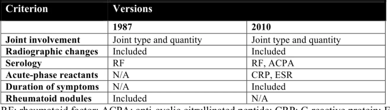
CHAPTER 1Table 1.1 Comparison of 1987 and 2010 versions of RA criteria ... 2
Table 1.2 DMARDs recommended by ACR 2008 and ACR 2012 ... 6
Table 1.3 Autoantigens and autoantibodies found in rheumatoid arthritis ... 13
Table 1.4 Monocyte/macrophage cytokines and their potential functions ... 23
CHAPTER 4 Table 4.1 Genotypic analysis of embryos from Pno1+/- × Pno1+/- mating ... 152
APPENDIX Table 0.1 Sequences of siRNA specific to MLF1-IP and control siRNA ... xxiv
Table 0.2 Summary of MLF1-IP mRNA expression in various tissues and organs ... xxv
Table 0.3 Genotyping of embryos from MLF1-IP+/- × MLF1-IP+/- mating ... xxvi
LIST OF FIGURES
CHAPTER 1Figure 1.1. Synovial architecture joint ... 9
Figure 1.2. The pathogenesis of RA. ... 28
Figure 1.3. Isoforms of TL1A and DR3 . ... 30
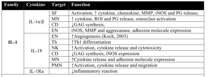
Figure 1.4. Role of TL1A in the inflammatory responses ... 39
Figure 1.5. Comparison of yeast, human and mouse Pno1 amino acid sequences. ... 40
CHAPTER 2 Figure 2.1 ... 74 Figure 2.2 ... 75 Figure 2.3 ... 76 Figure 2.4 ... 77 Figure 2.5 ... 78 Figure 2.6 ... 79 CHAPTER 3 Figure 3.1 ... 111 Figure 3.2 ... 112 Figure 3.3 ... 113 Figure 3.4 ... 114 Figure 3.5 ... 115 Figure 3.6 ... 116 Figure 3.7 ... 119 Figure 3.8 ... 120 Figure 3.9 ... 121 Figure 3.10 ... 122 CHAPTER 4 Figure 4.1 ... 153
Figure 4.2 ... 154 Figure 4.3 ... 155 Figure 4.4 ... 156 Figure 4.5 ... 157 Figure 4.6 ... 158 Figure 4.7 ... 159 Figure 4.8 ... 160 Figure 4.9 ... 161 Figure 4.10 ... 162 Figure 4.11 ... 163 APPENDIX Figure 0.1 ... xxvii Figure 0.2 ... xxviii Figure 0.3 ... xxix Figure 0.4 ... xxx Figure 0.5 ... xxxi Figure 0.6 ... xxxii Figure 0.7 ... xxxiii Figure 0.8 ... lxiii Figure 0.9 ... lxiv Figure 0.10 ... lxv Figure 0.11 ... lxvi Figure 0.12 ... lxvii Figure 0.13 ... lxviii Figure 0.14 ... lxix Figure 0.15 ... lxx
This thesis is dedicated to:
ACKNOWLEGEMENTS
First of all, I would like to express my sincerest appreciation to my supervisor, Dr. Jiangping Wu and co-supervisor Dr. Hongyu Luo, for giving me the opportunity to further my study here and sharing their passion for science. My doctoral researches and this thesis could never have been done without their invaluable support, constant encouragement and untiring guidance.
I would also like to thank all my colleagues; Dr. Terra Rafik, Dr. Diep Pham, Dr. Hector Valderrama, Dr. Bing Han, Dr. Jianning Mao, Dr. Zenghui Wu, Wei Jin, Yujia Wang and Yan Hu, for their warmhearted help, suggestions and joyful crazy ideas in every lab meetings.
Many thanks to Dr. Jun Zhang, Dr. Hassan Fahmi, Dr. Martin Marcinkiewicz, Dr. Shijie Qi, Dr. Tao Wu, Dr. Alain Lamarre, Dr. Yaned Gatain, Dr. Maxime Bouchard, Dr. Michel Tremblay, Dr. Noriko Uetani, Dr. Silva Hanissian and all for their splendid collaborations. Finally, I would like to extend my deepest gratitude to my jury members for evaluating this thesis and for their insightful comments.
STATEMENT OF AUTHORSHIP
Here is a statement regarding the contribution of coauthors and myself to the three papers included in this thesis.
Chapter 2:
Zhang J, Wang X (Equal contributor), Fahmi H, Wojcik S, Fikes J, Yu Y, Wu J and Luo H. (2009) Role of TL1A in the Pathogenesis of Rheumatoid Arthritis. The Journal of
Immunology 183, 5350–5357
Conceived and designed the experiments: JZ XW HL JW. Performed the experiments: JZ XW SW JF YY HL. Analyzed the data: JZ XW YY HL. Contributed reagents/materials/analysis tools HF Wrote the paper: XW JW. (XW conceived, designed, performed and analyzed all the experiments in Figure 4a, 4b, 5 and 6)
Chapter 3:
Wang X, Hu Y, Charpentier T, Lamarre A, Qi S, Wu J, Luo H. (2013) TNF-like ligand 1A (TL1A) gene knockout leads to ameliorated collagen-induced arthritis in mice: implication of TL1A in humoral immune responses. The Journal of Immunology 191, 5420-5429.
Conceived and designed the experiments: XW HL JW. Performed the experiments: XW YH TC SQ. Analyzed the data: XW AL WJ HL. Wrote the paper: XW JW.HL
Chapter 4:
Wang X, Wu T, Hu Y, Marcinkiewicz M, Qi S, et al. (2012) Pno1 Tissue-Specific Expression and Its Functions Related to the Immune Responses and Proteasome Activities. PLoS ONE 7: e46093.
Conceived and designed the experiments: XW HL JW. Performed the experiments: XW TW YH MM SQ HV HL. Analyzed the data: XW TW YH MM HV HL. Wrote the paper: XW JW.
Wang X, Marcinkiewicz M, Gatain Y, Bouchard M, Mao J, Tremblay M, Uetani N, Hanissian S, Qi S, Wu J, Luo H. (2013) Investigation of Tissue-Specific Expression and Functions of MLF1-IP during Development and in the Immune System. PLoS
ONE 8:e63783
Conceived and designed the experiments: XW MB MT NU JW HL. Performed the xperiments: XW MM YG JM SQ. Analyzed the data: XW MM YG JM JW HL. Contributed
reagents/materials/analysis tools: SH. Wrote the paper: XW JW HL. Appendix-2:
Terra R, Wang X (first co-author), Hu Y, Charpentier T, Lamarre A, Zhong M, Sun H, Mao J, Qi S, Luo H, Wu J. (2013) To Investigate the Necessity of STRA6 Upregulation in T Cells during T Cell Immune Responses. PLoS ONE 8:e82808
Conceived and designed the experiments: RT XW YH SH HL JW. Performed the experiments: RT XW YH JM SQ. Analyzed the data: RT XW AL MZ HS JM JW HL. Wrote the paper: RT XW YH HL JW. (XW conceived, designed, performed and analyzed all the experiments in Figure 2,4 and 5)
LIST OF ABBREVIATIONS
ACR American College of Rheumatology
ACPA Anti-cyclic citrullinated peptidea antibody
AIA Antigen-induced arthritis
ADAMTS A Disintegrin And Metalloproteinase with Thrombospondin Motifs
APRIL, TNFSF13 A proliferation-induced ligand BAFF, TNFSF13B B cell-activation factor
BCMA B cell maturation Ag
BMDC Bone marrow-derived dendritic cell
CD Chron's disease
CFA Complete Freund’s adjuvant
cFib Citrullinated fibrinogen
CHOP C/EBP homologous protein
CHX Cycloheximide
CIA Collagen-Induced Arthritis
CII Collagen II
COX Cyclooxygenase
CP Core particle
CRP C-reactive protein
CTLA4 Cytotoxic T-lymphocyte antigen 4
DCs Dendritic cells
dLN Draining lymph node
DR3, TNFRSF25 Death receptor 3
DSS Chronic-dextran sodium sulfate
EAA Experimental allergic asthma.
EAE Experimental Autoimmune Encephalomyelitis
EAMG Experimental autoimmune myasthenia gravis
ER Endoplasmic reticulum
ESR Erythrocyte sedimentation rate
EULAR European League Against Rheumatism
FADD Fas-associated death domain-containing molecule
FDA Food and drug administration
FDC Follicular dendritic cell
FGL Fibrinogen-like protein
FLICE FADD-like ICE
FLIP FLICE-inhibitory protein
G-CSF Granulocyte-colony-stimulating factor
GAG Glycosaminoglycans
GC Germinal center
GPI Glucose-6-phosphate isomerase
HUVEC Human umbilical vein endothelial cell
HVEM Herpesvirus entry mediator
IBD Inflammatory bowel disease
IC Immune complexes
ICE Interleukin-1β-converting enzyme
iNOS Inducible nitric oxide synthase
KO Knockout
LAG Lymphocyte activation gene
LCMV Lymphocytic choriomenigitis
LIF Leukemia inhibitory factor
LLPC Long-lived PCs
LP Lamina propria
LPMC Lamina propria mononuclear cells
LT Lymphotoxin
MCF Mononuclear cell factor
MCMV Murine cytomegalovirus MCs Mast cells MDC Monocyte-derived DC mDC Myeloid DCs MM Multiple myeloma MMP Matrix metalloproteinase
mRNA Messenge RNA
mTOR Mammalian target of rapamycin
MTX Methotrexate
NASIDs Non-steroidal anti-inflammatory drugs
NF-κB Nuclear factor κB
NO Nitro oxide
NTPDase-1, CD39 Triphosphate diphosphohydrolase-1
OSM Oncostatin M
PAC Proteasome-assembling chaperones
PADI4 Encoding type IV peptidylarginine deiminase
pDCs Plasmacytoid DCs
PDGF, Platelet-derived growth factor
PNA Peanut agglutinin
PG Proteoglycan
PGE2 Prostaglandin E2
PGIA Proteoglycan induced arthritis
PI Proteasome inhibitors
PIGF Placenta growth factor
PMN, Polymorphonuclear
Pno1 Partner of Nob1
PTPN22 Protein tyrosine phosphatase, non receptor 22
RANKL Receptor activator of NF-κB ligand
RF Rheumatoid factor
RF Rheumatoid factor
RIP Receptor interaction protein
ROI Reactive oxygen intermediate
RP Regulatory particle
SF Synovial fibroblast
SLPCs Short-lived PCs
SNPs Single nucleotide polymorphisms
SUMO Small ubiquitin-like modifier
SZW Streptococcal cell wall
TACI Transmembrane activator and calcium modulator ligand interactor
TCR T cell receptor
TEC Tubular epithelial cell
Tfh T follicular helper cell
TL1A TNF-like ligand 1A
TLR4 Toll-like receptor 4
TRADD TNFR-1-associated death domain protein
UC Ulcertive colitis
UMP1 Ubiquitin-mediated proteolysis 1
UPR Unfolded protein response
VEGF Vescular endothelial cell growth factor
VEGI Vascular endothelial growth inhibitor
CHAPTER 1 INTRODUCTION
1.1 Overview of Rheumatoid Arthritis
Rheumatoid arthritis (RA) is the most common inflammatory arthritis, manifesting redness, swelling, tenderness and destruction of the joint tissues (Aletaha et al., 2010). It is caused by chronic immune system disorder, which primarily affects the joints and their surrounding tissues, but also affects many other parts of our body (Firestein, 2003). Tissues and organs involved in RA complications include: the skin (rheumatoid nodules, ulcers and rash), eyes (scleritis, Sjogren’s syndrome), cardiovascular system (pericarditis, heart attack and stroke), circulation system (vasculitis, anemia, Felty’s syndrome), kidney (renal amyloidosis) and lung (collapsed lungs, plural effusion and pulmonary hypertension). There is no cure for RA, but current treatment can delay the disease progression and prevent the joint damage (Upchurch and Kay, 2012).
1.1.1 Diagnosis and Classifications of RA
The original diagnostic criteria for RA were published by the Committee of the American Rheumatism Association in 1956, and quickly updated by a revised version after two years (Committee of the American Rheumatism Association, 2008; Ropes et al., 1957). The criteria was again revised and published in 1987, formulated from computerized analysis of RA patients and healthy controls, with fewer criteria, improved sensitivity and specificity (Arnett et al., 1988). In 2010, a joint group of American College of Rheumatology (ACR) and European League Against Rheumatism (EULAR) published new classification criteria, aiming to differentiate RA patients at early-stage (Aletaha et al., 2010). It is noteworthy that the criteria in this version were designed for better classification of RA patients for clinical studies, rather than for a diagnostic purpose (Aletaha et al., 2010). Several studies have compared them with the 1987 diagnostic criteria and found that the 2010 criteria are more robust and sensitive, but slightly less specific (Britsemmer et al., 2011; Radner et al.; Van der Linden and Knevel, 2011). More evidence remain to be collected to determine whether these new criteria are appropriate to be used for diagnosis.
Table 1.1 Comparison of 1987 and 2010 versions of RA criteria
Criterion Versions
1987 2010
Joint involvement Joint type and quantity Joint type and quantity
Radiographic changes Included Included
Serology RF RF, ACPA
Acute-phase reactants N/A CRP, ESR
Duration of symptoms N/A Included
Rheumatoid nodules Included N/A
RF: rheumatoid factor; ACPA: anti-cyclic citrullinated peptide; CRP: C-reactive protein; ESR: erythrocyte sedimentation rate
1.1.2 Epidemiology of RA
RA occurs in 0.5-1% of the general population (Firestein, 2003). Several epidemiological studies have shown variations of disease incidence across populations. One half to 1.1% of the population in North America and Northern Europe are affected by RA, while lower prevalence of 0.3-0.7% is found in Southern Europe (Tobón et al., 2010). The prevalence of RA in East Asia is similar to those in Southern Europe. Developing countries in South Asia, Middle East and Africa have lowest prevalence of RA, which is around 0.38% (Hammoudeh et al., 2013; Tobón et al., 2010; Zeng et al., 2008). The low prevalence of RA found in developing countries may not reflect the actual situations due to the limited prevalence studies and incomplete data collection (Alamanos et al., 2006). Prevalence and incidence of RA also display a gender-specific disparity, with approximately 2-4 fold more RA patients and higher incidence among women than men in different populations (Alamanos et al., 2006; Hammoudeh et al., 2013). Multiple studies have suggested that the average onset age of RA is rising (Gabriel et al., 1999; Imanaka et al., 1997; O et al., 1996). At the same time, there are progressive decreases in both incidence and prevalence of RA across several different regions (Doran et al., 2002; Jacobsson et al., 1994; Seppanen et al., 1996; Kaipiainen-Seppanen and Kautiainen, 2006; O and K, 2000; Shichikawa et al., 1999). On the other hand, despite our better knowledge of pathogenic mechanisms and great improvement of the RA management, the mortality rate in RA patients seems not decreasing in the last few decades (Myasoedova et al., 2010).
1.1.3 Risk Factors of RA
1.1.3.1 Genetic Factors
Various evidence from migration studies, familial clustering studies and twin studies have indicated the importance of genetic factors to the susceptibility, severity and responsiveness to treatment of RA (Criswell et al., 1998; John and Worthington, 2001; Lin et al., 1999; Plant et al., 2011). From genetic association studies, growing numbers of RA genetic risk factors have been uncovered in the past decades (Huizinga, 2003; McInnes and Schett, 2007; van der Helm-van Mil et al., 2005). The first and most well-established disease locus is HLA-DRB1, alleles in HLA-DRB1 region containing “shared epitope” confer disease risk, and account up to one third of the total genetic risk of susceptibility of RA (Cornélis et al., 1998; Gregersen et al., 1987). To date, most potential risk genes are found in the genes encoding molecules important to immune regulations, such as NF-κB, PTPN22 (protein tyrosine phosphatase, non receptor 22), CTLA4 (cytotoxic T-lymphocyte antigen 4), STAT4 and CD40, as well as those encoding pro-inflammatory cytokines (IL-10, IL-1β, IL-2 and IL-21) (Begovich et al., 2004; Cantagrel et al., 1999; Daha et al., 2009; Eskdale et al., 1998; Li et al., 2012b; Orozco et al., 2010; Plenge et al., 2007; Remmers et al., 2007). Moreover, single nucleotide polymorphisms (SNPs) discovered in sequence encoding type IV peptidylarginine deiminase (PADI4), an enzyme believed to produce autoantigens by posttranslationally converting arginine residues to citrulline, are also reported to have association with susceptibility to RA(Suzuki et al., 2003). It is worth mentioning that many of risk genes listed above are also related to other autoimmune diseases, such as Type 1 diabetes and systemic lupus erythematosus (SLE), implicating the shared mechanisms in these common autoimmune disease (Begovich et al., 2004; Remmers et al., 2007). In addition, polymorphisms proved to be the risk factors in one population are not necessary to be the risk factors in other populations (Barton et al., 2004; Suzuki et al., 2003).
1.1.3.2 Gender and Hormonal Factors
Gender is probably the most important risk factors for RA. Since females have higher risk of developing RA, it is conceivable that sex hormones might also have a role in RA. Early study has found that RA patients have decreased levels of serum testosterone; the reduction of serum
testosterone levels occur prior to the onset of RA (Pikwer et al., 2013; Spector et al., 1988). The negative association of testosterone levels and risk of disease indicate the protective function of testosterone, and its potential to be a pre-clinical marker to predict the RA risk. No consensus has been achieved regarding to the effect of estrogen on RA. Serum levels of estrogen are normal in men with RA, but elevated local concentration of estrogen is discovered in Synovial Fluid (SF), which is believed to aggravate the disease (Cutolo et al., 2003). Interestingly, women who take oral contraceptives containing high concentration of estrogen have a moderate decrease of RA risk (Doran et al., 2004). A possible explanation could be the different biological functions of systematically administered exogenous estrogen versus local endogenous estrogen. Additionally, female patients could have a “temporary exemption” from RA during the pregnancy due to the hormones changes in that period (Silman et al., 1992)
1.1.3.3 Environmental Factors
Several studies have indicated that smoking has a strong association with RA incidence as well as the disease activity and severity (Papadopoulos et al., 2005). Besides smoking, other forms of bronchial stress, such as exposure to silica and traffic pollution have also been confirmed as novel environmental risk factors for RA (Hart et al., 2009; Yahya et al., 2013). Other environmental factors associated with RA include obesity, blood transfusion, virus infection and stress (Cutolo and Straub, 2006; Meron et al., 2010; Symmons et al., 1997). On the other hand, coffee, tea and caffeine consumption don’t seem to be risk factors of RA, at least to women (Karlson et al., 2003; Mikuls et al., 2002).
The development of RA is believed to depend on the interactions between multiple risk factors. Substantial evidence indicated that the interaction between the genetic background and environmental factors could have synergistic effect that greatly increases the risk of RA. For example, individuals who are genetically susceptible and cigarette smoking at the same time are at higher risk to develop RA than those with either of these risk factors alone (Costenbader et al., 2008; Lundström et al., 2009; Padyukov et al., 2004; van der Helm-van Mil et al., 2007; Verpoort et al., 2007). Taken together, understanding the role of various risk factors and the
interactions among them could help us to have a better knowledge in etiology, pathogenesis, as well as prevention and the treatment of RA.
1.1.4 Management and Challenges in the Treatment of RA
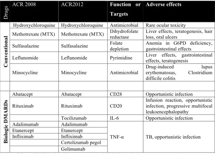
The goals of RA management and the treatment are relieving the swelling and pain, slowing down the progression of the disease, and preventing joint deformity and other extra-articular complications (American College of Rheumatology Subcommittee on Rheumatoid Arthritis Guidelines, 2002; Wasserman, 2011). Current RA treatment mainly relies on medications. However, invasive measures such as joint replacement surgery is also carefully considered in patients with unacceptable pain, lost joint motions or end-stage joint damage (American College of Rheumatology Subcommittee on Rheumatoid Arthritis Guidelines, 2002; Scott et al., 2010). Medications adopted in RA treatment fall into two main classes: non-steroidal anti-inflammatory drugs (NASIDs) and disease-modifying anti-rheumatic drugs (DMARDs). In the first half of 19th century, NASIDs were first introduced to RA treatment for their symptomatic benefits as reducing the pain and stiffness of the affected joints. However, drugs of this class have limited impact on reducing disease activity and slowing down the disease progression (Upchurch and Kay, 2012). DMARDs were wildly accepted as the key therapeutic agents in the past two decades as they are able to modify the disease outcome such as, decelerating the progression of the disease, improving joints’ functions and reducing the disability (Scott et al., 2010; Wasserman, 2011). DMARDs could be further divided into non-biologics and biologics. The first non-biologic DMARDs, gold salt, was reported on 1929, and many others were discovered thereafter (Hartung, 1943; Upchurch and Kay, 2012). Most of the early DMARDs are not recommended anymore for use today because of the high incidence of adverse events or poor tolerance (Saag et al., 2008). Currently, only five non-biologic DMARDs are included in the recommendations for the treatment of RA (Table 2) (Saag et al., 2008; Singh et al., 2012). The biologic DMARDs are antibodies and cell surface receptor inhibitors that target key cytokines and molecules regulating the inflammatory responses in RA (McInnes and Schett, 2007). According to clinical studies and meta-analysis, it is safe to conclude that these biologic DMARDs are generally effective and safe, but whether they are superior to the conventional DMARDs is still inconclusive (Aaltonen et al., 2012; Breedveld et al., 2005; Keystone et al., 2004). However, the combination use of biologics and MTX is better tolerated
and more efficacious than the use of either MTX or biologics alone (Aaltonen et al., 2012; Ash and Emery, 2012; Chen et al., 2006; Klareskog et al., 2004; Lipsky et al., 2000; Weinblatt et al., 1999). As immunosuppressive agents, elevated infection risk is the main adverse drug reaction observed in patients taking biologics to treat RA (Singh et al., 2011). There are currently five TNF-α blockers, one IL-1 receptor antagonist, one IL-6 blocker, one T cell co-stimulation blocker, and one B cell co-stimulation blocker, approved by FDA and recommended by ACR for treatment of RA (Table 2) (Saag et al., 2008; Scott, 2012; Singh et al., 2012; 2011).
Table 1.2 DMARDs recommended by ACR 2008 and ACR 2012
Dr
ugs
ACR 2008 ACR2012 Function or
Targets Adverse effects C on ve n ti on al DMARDs
Hydroxychloroquine Hydroxychloroquine Antimicrobial Rare ocular toxicity
Methotrexate (MTX) Methotrexate (MTX) Dihydrofolate reductase Liver effects, teratogenesis, hair loss, oral ulcers Sulfasalazine Sulfasalazine Folate depletion Anemia in G6PD deficiency, gastrointestinal effects Leflunomide Leflunomide Pyrimidine Liver effects, gastrointestinal
effects, teratogenesis Minocycline Minocycline Antimicrobial
Drug-induced lupus erythematosus, Clostridium difficile colitis Bi ol ogi c D M A R D s
Abatacept Abatacept CD28 Opportunistic infection Rituximab Rituximab CD20
Infusion reaction, opportunistic infection, progressive multifocal leukoencephalopathy
Tocilizumab IL-6 Opportunistic infection Adalimumab Adalimumab TNF-α TB, opportunistic infection Etanercept Etanercept Infliximab Infliximab Certolizumab pegol Golimumab
Adapted from Singh et al. and Wasserman et al. (Singh et al., 2012; Wasserman, 2011)
Historically, newly diagnosed RA patients were first treated with NSAIDs and followed by DMARDs. But growing evidence have suggested that the early intervention of RA by DMARDs achieves significant reduction of radiographic progression in RA patients (Finckh et al., 2006). Since the past decade, more aggressive strategy has been adopted to treat early RA
(Upchurch and Kay, 2012). The ACR suggests patients to start DMARDs therapy within 3 months of diagnosis, and with periodically reassessment of disease progression and update of treatment regimen accordingly (American College of Rheumatology Subcommittee on Rheumatoid Arthritis Guidelines, 2002). Single (monotherapy) or combination conventional with DMARDs (dual-therapy or triple therapy) is recommended to patients with different levels of disease and features of prognosis, regardless of disease duration. However, biologic DMARDs are used with more caution; early RA patients are not recommended to, except those one with high disease activity and poor prognosis; for established RA, biologic DMARDs are only considered after the failure of combination DMARDs therapy (American College of Rheumatology Subcommittee on Rheumatoid Arthritis Guidelines, 2002). In addition, tuberculosis has to be treated before using the biologic DMARDs (Saag et al., 2008; Singh et al., 2012).
1.2 Collagen-Induced Arthritis (CIA)
In 1977, Trentham et al. first reported that rat immunized with type II collagen in either complete Freund’s adjuvant (CFA) or incomplete Freund’s adjuvant (IFA) developes a chronic inflammatory arthritis, which they designated as Collagen-Induced Arthritis (CIA) (Trentham et al., 1977). A subsequent study hypothesized that there are two stages of disease development. In the first stage, as early as day 12 after the immunization, hyperplasia and fibrin deposition could be seen in the synovium; the second stage starts earliest on day19, it is characterized by the inflammatory cells infiltration, which form the pannus and cause the destruction of cartilage and bone eventually (Caulfield et al., 1982). Similar to the rat model, the CIA mouse model starts with edema and synovial hyperplasia, and is followed by infiltration of polymorphonuclear and mononuclear cells. As the disease progresses, pannus forms in the affected joins causing cartilage damage, and finally bone erosion (Courtenay et al., 1980).
Since CIA resembles many pathological features found in RA, it is still the most wildly used model for studying the pathogenesis of human RA in animals, as well as a gold standard for testing the therapeutic drugs for RA in the pharmaceutical industry (Brand et al., 2007). During the past three decades, CIA model made substantial contributions to understand the
role of individual cell types (T cells, B cells, master cells, etc) and cytokines (TNF-α, IL-1β, IL-17, etc) in RA (Brahn et al., 1992; Hom et al., 1992; Kadowaki et al., 1994; Morgan et al., 2005; Nakae et al., 2003b; Nigrovic and Lee, 2005; Tada et al., 1996; Yanaba et al., 2007). Furthermore, the arthritogenicity of autoantibody was also revealed by the fact that the arthritis could be transferred by serum from rodents immunized with Collagen II (Stuart et al., 1984). Of course, there are also certain discrepancies between CIA animal models and human RA. For example, vasculitis which often could be found in RA, is not presented in CIA; on the other hand, periostitis develops in CIA but not in RA(Stuart et al., 1984).
1.3 Mechanisms of RA
1.3.1 Synovial Architecture of Normal Joint
The synovial joints are the most common type of joints in our body, providing precise and smooth movement and maintain the stability and strength as well. The major components of synovial joints are synovial capsule and articulating bones; the outer layer of synovial capsule is a fibrous layer, uniting the articulating bones by attaching their periosteum linings; the inner layer is a one to three cell layer called synovial membrane which contains fibroblast-like synoviocytes and macrophages, secreting synovial fluid to fill up the synovial cavity. The synovial cavity is enclosed by the synovial capsule(Perlman and Pope, 2010). Synovial fluid contains high concentrations of hyaluronic acid and albumin; it absorbs shocks, and lubricates and nourishes the joint. The articulating bones are covered by a thin layer of connective tissue called cartilage, which is made up with a well-organized network of type II collagen fibers. These collagen fibers are produced by chondrocytes scattered in the cartilage. Importantly, there is no blood supply to articular cartilage, while its nourishment depends exclusively on synovial fluid. The synovial fluid also contains phagocytes which are responsible for cleaning up the tissue debris generated from wear and tear of cartilage in our daily life (Rogers, 2010).
1.3.2 Architecture Changes of RA Joint
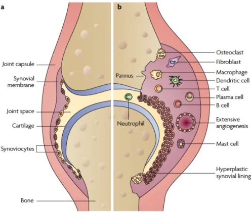
The normal joint structure goes through a series of changes in RA. It involves synovial hyperplasia and lymphocyte infiltration. In RA, synovium increases its thickness, due to the synovial lining cell proliferation and blood-derived lymphocytes influx trough the vessel wall
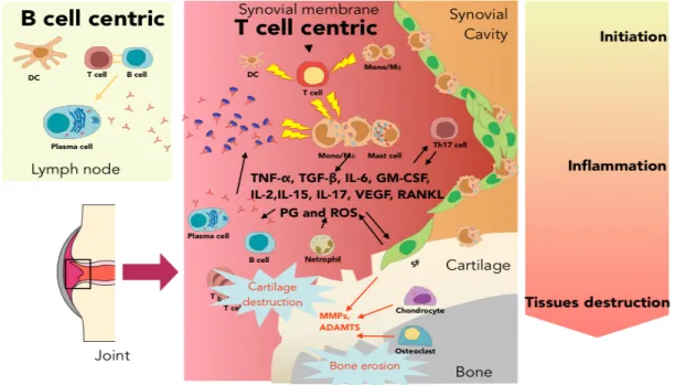
into the synovium (Figure 1.1) (Konisti et al., 2012). Moreover, the increased cell numbers in the affected joint contribute to hypoxia and ischemia, posing a signal for angiogenesis; Neovessels exuberate the synovial inflammation by transporting more inflammatory cells and delivering nutrients and oxygen to the RA synovium (Marrelli et al., 2011). As inflammatory cells accumulate in the synovial membrane, where they protrude and form a unique structure called pannus. Pannus invades the cartilage and bone, and causes the cartilage degradation and bone erosion (McInnes and Schett, 2007; Neumann et al., 2010).
Figure 1.1. Synovial architecture joint. (a) normal joint; (b) joints affected by RA. Adapted from Strand V. et al. (Strand et al., 2007)
1.3.3 Autoantigens, Autoantibodies and B cells
The presence of autoantibodies in the serum and synovial fluids of RA patients has been known for a long time. Since the first description of “a factor” that agglutinate sheep blood cells (late designated as rheumatoid factor (RF)), growing number of autoantibodies have been discovered, and some of them are utilized in clinical diagnostics (Bläss et al., 1999; Waaler, 1940). While most attention to the autoantibodies is focused on their diagnostic value, less contemplation has been given to their role in the pathogenesis of RA. In fact, how those
normal molecules turn into “autoantigens”, and stimulate the production of autoantibodies is the fundamental question that needs to be answered. The followings are four most prominent pairs of autoantigen and autoantibodies related to RA.
1.3.3.1 Rheumatoid Factor
Rheumatoid factor (RF) was first discovered in 1940s and later identified as antibodies that bind the Fc region of Ig (FRANKLIN et al., 1957; Waaler, 1940).It was quickly included as a criterion for clinical diagnosis of RA in 1958, and remains the exclusive diagnostic autoantibody for more than 50 years (Aletaha et al., 2010; Arnett et al., 1988; Committee of the American Rheumatism Association, 2008). Healthy humans or animals are reported to produce a transient wave of RFs after vaccination or immunization, while 80% of RA patients have persistent increased level of RF in sera(Coulie and Van Snick, 1983; M J Welch, 1983; Nell et al., 2005). The physiological roles of RFs are 1) promoting complement fixation, 2) helping the clearance of immune complexes (IC), and 3) increasing the avidity and specific of functional antibodies(Carson et al., 1987). RF is believed to be pathogenic in RA as it induces the RA-related cytokines production. Early studies have shown that RF induces monocytes to produce prostaglandin E2 and mononuclear cell factor (MCF), two potent stimulator of bone resorption (Gowen et al., 1983; Nardella et al., 1983). Moreover, IC formed by RF and immunoglobulin (Ig) from arthritic synovial tissue has been demonstrated to stimulate the production of TNFs from monocytes (Ishikawa et al., 1975; Mathsson et al., 2006).
1.3.3.2 Anti-Citrullinated Protein Antibody (ACPA)
Citrullination is a biological process of converting amino acid arginine into citrulline; it is conducted by peptidylarginine deminase enzymes (PAD), which have 5 isoforms (PAD1, 2, 3, 4/5 and 6)(Vossenaar et al., 2003). Many essential physiological activities require PADs, such as embryo development, electrical insulation in the nervous system and transcriptional control for DNA, and so on (Vossenaar et al., 2003). Citrullination may also occur under certain pathogenic conditions, notably in RA, producing autoantigens. Both PADs (2 and 4) and citrullinated protein (fibrinogen, fibronectin, vimentin and Collagen II) are found in affected RA joints, but not healthy joints (Baeten et al., 2001; Kinloch et al., 2008; Tabushi et al.; Vossenaar et al., 2004; Yoshida et al., 2006). Anti-citrullinated protein antibodies (ACPAs)
are antibodies against citrullinated proteins of various origins. They are present in around 70% of RA patients, and included in the RA classification criteria, due to their high specificity for RA (90-95% compare to 40-90% of RF) and strong association with a severe disease status (Aggarwal et al., 2009; Aletaha et al., 2010; Schellekens et al., 2000). Generally, citrullination of protein and their further conjugation with their autoantibodies increase their immunogenicity and arthritogenicity (Lundberg et al., 2005; Shelef et al., 2012; Sokolove et al., 2011). For examples, citrullinated fibrinogen (cFib) and myelin binding protein, but not their natural forms, are capable of activating basophils from ACPA+ RA patients (Schuerwegh et al., 2010). Both cFib and cFib-IC are more potent in inducing TNF production from macrophages compared to the natural form of fibrinogen (Clavel et al., 2008; Sokolove et al., 2011). In vivo, immunizing rat with citrullinated Collagen II (cCII) in incomplete Freund’s adjuvant (IFA) induces earlier arthritis with higher incidences than with natural CII (Lundberg et al., 2005). Immunization with cFib but not natural fibrinogen in CFA or intra-articular administration of cFib plus transferring of cFib-specific T cells induces arthritis in mice carrying a RA susceptible MHC-II allele (Hill et al., 2008). The ACPAs might not be pathogenic under a healthy condition, but they could play a role in aggravating the disease after the onset of arthritis (Kuhn et al., 2006). A novel study discovered that ACPAs could bind to the citrullinated vimentin on osteoclast precursor cells and promote their differentiation into osteoclasts, leading to the bone erosion and joint destruction (Harre et al., 2012).These results are in accordance with the clinical finding that the presence of ACPAs in serum is usually related to the more advanced bone erosion condition in RA (Aggarwal et al., 2009).
1.3.3.3 Anti-Collagen II (CII) Antibody
Type II collagen (collagen II, CII) is the major component of articular cartilage in joints. The association between RA and CII antibodies has been found in the several clinical studies. The presence of anti-CII antibodies in serum is observed in about 30% of RA patients with indications of an early inflammatory/destructive disease phenotype (Beard et al., 1980; Cook et al., 1996; Fujii et al., 1992; Mullazehi et al., 2007; 2012; Raza et al., 2007; S et al., 1988). Anti-CII antibodies are also found in the synovial fluids of some RA patients who are genetically HLA-DR4 positive (Rönnelid et al., 1994). Both CII and anti-CII antibody are
arthritogenic: immunization of CII emulsified with either IFA or CFA induces arthritis in rat and mice (Courtenay et al., 1980; Trentham et al., 1977; 1978). The pathogenic role of anti-CII antibodies has been demonstrated by successful induction of arthritis in naïve rats/mice passively transferred with anti-CII antibodies-containing sera from CIA rats/mice; it has been further confirmed by the fact that direct administration of combination of anti-CII monoclonal antibodies caused full-blown arthritis (HOLMDAHL et al., 1990; Stuart and Dixon, 1983; Stuart et al., 1982; Terato et al., 1992). Moreover, anti-CII antibodies have been reported to cause thickening, aggregation and disorganization of CII fibrils in newly synthesized extracellular matrix (ECM), as well as alter the secretion rate of matrix metalloproteinase (MMPs) in culture, by interfering chondrocytes (Takagi and Jasin, 1992). Furthermore, CII-ICs have been shown to activate PBMCs and induce RA-like disease pro-inflammatory cytokine (TNF-α, IL-1β and IL-8) production (Mullazehi et al., 2006).
1.3.3.4 Anti-Glucose-6-Phosphate Isomerase (GPI) Antibody
The pathogenic role of GPI and its antibody in RA was discovered by chance. When transgenic mice overexpressing the TCR that recognizes peptide of bovine pancreas ribonuclease (RNase) in the context of Ak (KRN TCR) are crossed with NOD mice which bear a unique MHC-II (I-Ag7), the transgenic offspring (K/BxN) exhibits spontaneous inflammatory arthritis (Kouskoff et al., 1996). GPI, a ubiquitously expressed enzyme converting glucose-6-phosphate into fructose-6-phosphate, is later identified as the autoantigen in this arthritis murine model (Matsumoto et al., 1999). In the joints of arthritic K/BxN mice, thick linings of GPI molecules, accompanied by the deposition of IgG and C3 complement fragments could be seen on the surface of cartilage, synovium and pannus (Matsumoto et al., 2002). Accordantly, extremely high level of anti-GPI antibody is presented in the serum of K/BxN mice (Matsumoto et al., 2002). The mechanistic study has revealed that the joint inflammation in this particular arthritic model is caused by the GPI-IC formed in the joints which triggers the alternative complement pathway activation (Maccioni et al., 2002; Matsumoto et al., 2002). In the clinical study, deposition of GPI molecules is also observed in the synovial tissues from RA patients. However, most studies have shown anti-GPI antibodies are either rarely or non-specifically detected in RA serum (Kassahn et al., 2002; Matsumoto et al., 2003; Schaller et al., 2001; Schubert et al., 2002). Collectively, although the anti-GPI
antibody is proved to be athrogenic in mice, whether it is involved in the initiation and development of human RA is still unclear.
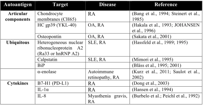
1.3.3.5 Other Autoantigens and Autoantibodies
In addition to those mentioned above, many other autoantigens and autoantibodies have been found related to RA. Some of them have potential to be novel diagnostic markers, while others attract less attention due to the inconsistent data and low disease specificities. Some of them are listed in Table 1.3.
Table 1.3 Autoantigens and autoantibodies found in rheumatoid arthritis
Autoantigen Target Disease Reference
Articular components
Chondrocyte
membranes (CH65) RA
(Bang et al., 1994; Steinert et al., 1985)
HC gp39 (YKL-40) OA, RA (Hakala et al., 1993; JOHANSEN et al., 1996)
Osteopontin OA, RA (Sakata et al., 2001)
Ubiquitous Heterogeneous nuclear
ribonucleoprotein A2 (Ra33 or hnRNP A2)
SLE, RA (Hassfeld et al., 1989; 1995)
Calpstatin SLE, RA (Mimori et al., 1995) BiP (Bläss et al., 1995; 2001) α-enolase Autoimmune
retinopathy, RA
(Kurz et al., 2011; Saulot et al., 2002)
Cytokines B7-H1 (PD-L1) RA (Dong et al., 2003)
IL-1α RA (Hansen et al., 1994) IL-8 Myasthenia gravis,
RA
(Burbelo et al.; Peichl et al., 1992)
In summary, there are three possibilities for the pathogenic roles of a pair of autoantigen and its autoantibody. First, autoantigen is pathogenic by itself. Natural or modified proteins could become autoantigens and involved in the pathogenesis of RA by inducing the pro-inflammatory cytokine production, angiogenesis and synoviocyte proliferation (Sokolove et al., 2011; Yoo et al., 2012). Importantly, these autoantigens are not necessarily to be the joint exclusive proteins. However it is crucial for them to be present in the joint tissues of individuals with certain genetic background, given that immunization of these autoantigens is only able to induce arthritis in mice with the susceptible genetic background. Second, free Abs could be pathogenic. Certain autoantibodies such as anti-CII or anti-calpastatin Abs are able to
interfere with the interactions among cartilage proteins in joints, and cause degradation of cartilage components (Amirahmadi et al., 2005; Ménard and el-Amine, 1996). Third, autoantigen and its autoantibody contribute to the RA by forming immune complexes (IC). Transferring sera (or IgG fraction but not IC fraction) from CIA or K/BxN arthritic mice to naive mice causes arthritis (HOLMDAHL et al., 1990; Korganow et al., 1999; Matsumoto et al., 1999; Stuart and Dixon, 1983; Stuart et al., 1982; Terato et al., 1992). In accordance, anti-GPI autoantibodies have been reported to locate to distal joints within minutes after intravenous injection, whereas preformed ICs have difficulties to get into the joints (Wipke et al., 2002). It is believed that the autoantibody penetrates into the joints, forms articular ICs with pre-deposited autoantigens (like GPI or cCII), and initiates the disease (Matsumoto et al., 1999). These ICs then bind to the FcγR on the surface of monocytes/macrophages and induce the production of TNF, IL-1β, and IL-8 (Mathsson et al., 2006; Mullazehi et al., 2006; Sokolove et al., 2011). Besides FcγR, ICs are also reported to interact with toll-like receptor 4 (TLR4), which works synergistically with FcγR and elevates the TNF production by macrophage (Sokolove et al., 2011). More detailed in vivo studies using knockout mice have revealed that FcγRI and FcγRIII are responsible for the successful induction of CIA or antibody-induced arthritis (AIA) (Kleinau et al., 2000; Nandakumar et al., 2003a).
1.3.3.6 Functions of B cells Other Than Producing Autoantibodies
In addition to producing autoantibodies, several novel roles B cells playing in RA have been discovered by recent studies. Firstly, B cell is able to serve as an APC for autoreactive T cells. In a proteoglycan induced arthritis (PGIA) model, when transferring T cells from proteoglycan (PG)-immunized Ig-deficient mice to SCID mice, only those T cells that received adequate priming from cognate B cells are able to elicit the disease (O'Neill et al., 2005). A later study from the same group has shown that CD80/CD86, two costimulatory molecules expressed on B cells, are essential for the activation of autoreactive T cells in PGIA (O'Neill et al., 2007). Accordant with animal studies, a human study has also reported that B cell is essential for the activation of synovial follicular T cells, since adoptively transferred follicular CD4 T cells fails to get activated and secret pro-inflammatory cytokines in SCID mice chimeras with human RA tissues lacking B cells (Takemura et al., 2001). Secondly, B cell could affect the pathogenesis of RA by secreting cytokines. The B cells are reported to secrete various
cytokines to promote inflammation (TNF, IL-1, IL-6, G-CSF, GM-CSF and IL-12) and angiogenesis (VEGF-A), two prominent features in RA (Angeli et al., 2006; Pistoia, 1997). On the other hand, a novel subset of B cells called B regulatory cells have been discovered to attenuate the harmful immune responses by secreting IL-10 and TGF-β (Mizoguchi and Bhan, 2006). Two studies have shown that regulatory B cells ameliorate CIA in DBA mice through the secretion of IL-10 (Evans et al., 2007; Mauri et al., 2003).
1.3.4 T cells
1.3.4.1 Th1 and Th2 Cells
Th1 cells produce IFN-γ, which primarily activates macrophages and mediates the immune responses against intracellular infections (Kindt et al., 2007; Szabo et al., 2000). Th2 cells secrete IL-4, 5, 10 and 13 and are responsible for humoral responses to protect against infections from extracellular pathogens (Kindt et al., 2007). Early studies on RA patients found mRNA expression of IL-12 and IFN-γ in RA synovial fluid mononuclear cells, and identified Th1 cells as the predominant T cell subsets in synovial fluid (SF) from RA patients (Bucht et al., 1996; Kusaba et al., 1998; Morita et al., 1998). Th2 cytokine-producing cells are also detectable in the synovial tissues and peripheral blood of RA patients, but at a significantly lower frequency comparing to cells producing Th1 cytokines (Kusaba et al., 1998; Morita et al., 1998; Yudoh et al., 2000). Messenger RNA levels of IL-4 and IL-10 in synoviocytes become significantly lower as RA progresses (Miyata et al., 2000). It was believed that Th1 responses are generally associated with inflammation, whereas Th2 responses are protective to some extent. Based on this belief and clinical findings, a Th1/Th2 imbalance hypothesis is proposed for the pathogenesis of RA. However, emerging evidence from animal studies have shown complicated role of Th1 and Th2 cells in RA.
IFN-γ receptor deficient mice are more susceptible to CIA, and this is accompanied by accelerated and exacerbated disease, but less anti-CII antibodies (Manoury-Schwartz et al., 1997; Vermeire et al., 1997). More detailed studies by the same group and other groups have been performed to understand the protective role of this cytokine. The possible mechanisms are as follows: 1) the presence of IFN-γ signaling suppresses the arthritogenic myelopoiesis of the splenic Mac-1+ cells elicited by the mycobacterial component of CFA (Matthys et al.,
2001). Using IFA instead of CFA for immunization or inhibition of CFA-induced splenic Mac-1+ cell expansion abolishes the exacerbated CIA in IFN-γ receptor KO mice (Matthys et al., 1999). 2) It is suggested by the same group that deficiency in IFN-γ signaling diminishes the function of Treg cells by bringing down Foxp3 transcription levels in Treg, and also dampens the function of APC cells (Kelchtermans et al., 2005). This finding is in accordance with recent discovery of regulatory role of IFN-γ (Wood and Sawitzki, 2006). 3) Two studies using streptococcal cell wall (SCW)-induced arthritis in rat have shown IFN-γ inhibits leukocyte chemotactic migration to the synovium, and prostaglandin E2 (PGE2) production and synoviocytes proliferation, indicting IFN-γ could suppress the arthritis through suppressing the mesenchymal cells (Allen et al., 1991; Wahl et al., 1991). 4) IFN-γ suppresses the Th17 cells. It is known that IFN-γ suppresses the in vitro differentiation of IL-17-producing Th17 cells (Bettelli et al., 2008). In vivo, neutralization of IFN-γ in CIA increases circulating and articular levels of IL-17, which causes accelerated disease (Sarkar et al., 2009). IFN-γ knockout mice in antigen-induced arthritis (AIA) have similar phenotypes as IFN-γ receptor deficiency mice in CIA; Articular administration of IFN-γ or treatments with mAb neutralizing IL-17 alleviates the disease (Irmler et al., 2007). It is suggested that deficiency of IFN-γ releases the suppression of IL-17 and leads to the aggravated disease (Irmler et al., 2007). A controversial result has been reported recently showing reduced arthritis in IFN-γ receptor KO mice in glucose-6-phosphate isomerase (G6PI)-induced arthritis (Frey et al., 2011). Doodes et al. has shown similar results using IFN-γ KO mouse models of proteoglycan-induced arthritis model (Doodes et al., 2010). Although IFN-γ is implied to exacerbate the arthritis in their findings, the suppressions of IL-17 production by IFN- γ in these studies are still obvious and negatively correlate with the disease (Doodes et al., 2010; Frey et al., 2011). Collectively, either IFN-γ or IL-17 alone is pathogenic in RA. The latter is much more avid, but is negatively regulated by the former. When the disease condition is in favor of IL-17 production, IFN-γ exhibits a protective role. Otherwise, IFN-γ would be pathogenic for the disease.
Administration of exogenous IL-4 reduces the histological score, pathogenic anti-CII antibodies’ level and pro-inflammatory cytokine secretion by joint tissues in CIA mice (Horsfall et al., 1997). Similar results are obtained when treating the mice with IL-4 or IL-10,
or both, in PGIA and SCW models (Finnegan et al., 2003; Lubberts et al., 1998). More specifically, either systemic administration or articular overexpression of IL-4 appears to protect against bone destruction of CIA (Joosten et al., 1999; Lubberts et al., 2000a). The protective effect of IL-4 against bone destruction is through the suppression of IL-6 and LIF, and inhibition of osteoclasts (Miossec et al., 1994). Conversely, depletion of IL-4 or IL-10 has caused elevated disease in several different arthritis models (Finnegan et al., 2003; 2002; Hata et al., 2004). The change of disease caused by adding or removal of IL-4 is closely associated with the levels of specific arthritogenic IgG2a, indicating the IL-4 may prevent the class switching towards the pathogenic IgG isotope (Brand et al., 2003; Finnegan et al., 2002; Horsfall et al., 1997; Joosten et al., 1999; Kim et al., 2001b; Myers et al., 2002). IL-10 also confers a protective role in RA. It reduces TNF-α and IL-1 secretion from cultured synovial membrane tissues of RA patients, and costimulatory molecule expression of synovial cells(Katsikis et al., 1994; Kawakami et al., 1997). A Korean group has reported that the IL-10 suppresses the arthritis by diminishing IL-17 expression and inducing the Treg cells generation (Heo et al., 2010). Furthermore, IL-10 has an additive effect, or even a synergistic effect with IL-4 on the inhibition of proinflammatory cytokine production by activated mononuclear cells from RA patients (van Roon et al., 1996). In the SCW arthritis model, administration of IL-10 with IL-4 reduces both IL-1β and TNF-α levels in the synovium, while IL-10 alone only brings down the latter cytokine (Lubberts et al., 1998). In addition, IL-10 and IL-4 were also reported to have additive effect on stimulating the cartilage proteoglycan synthesis (van Roon et al., 1996). While most studies acknowledge the protective role of IL-4 in RA, there is a recent study implying that IL-4 can be pathogenic. In the K/BxN model, IL-4 has been demonstrated to be essential in initiating the arthritis by activating B cells (Ohmura et al., 2005). This result indicates that different settings of the arthritis models and genetic backgrounds could change the disease phenotypes drastically.
1.3.4.2 Th17 Cells
Th17 is a T cell lineage, producing a distinct cytokine profile including 17, 21 and IL-22. It is the major source of IL-17 (Weaver et al., 2006). IL-17, also known as IL-17A, belongs to the IL-17 family, which contains 7 different members. It is a potent proinflammatory cytokine and plays an essential role in autoimmune diseases (Gaffen, 2008).
Elevated IL-17 levels are observed in sera and synovial fluids from RA patients (Hussein et al., 2008; Kotake et al., 1999; Ziolkowska et al., 2000). The target cells of IL-17 in RA include synovial fibroblasts, macrophages, chondrocytes and osteoblasts, all of which are directly related to the pathogenesis of RA (van den Berg and Miossec, 2009). IL-17 alone, or synergistically with IL-1β or TNF-α, induces the production of IL-6, IL-8, granulocyte-colony-stimulating factor (G-CSF), prostaglandin E2 (PEG2) and leukemia inhibitory factor (LIF) by synoviocytes (Chabaud et al., 1998; Fossiez et al., 1996). In a Th17 cell and synovial fibroblast co-culture system, IL-17 increases the secretion of matrix metalloproteinase-1, -3 (MMP-1, -3), as well as IL-6 and IL-8, by synovial fibroblasts. Synovial fibroblasts, on the other hand, increase autocrine IL-17 production by Th17 cells through the cyclooxygenase (COX)/PEG2 pathway. Together, they built up a proinflammatory loop contributing to the pathogenesis of RA (Paulissen et al., 2013; van Hamburg et al., 2010). In addition, IL-17 has been reported to promote the survival of synovial fibroblasts (Lee et al., 2013). A similar proinflammatory loop is suspected to exist between 17 and synovial macrophages too. IL-17 stimulates macrophages and upregulates their arthritogenic cytokine (such as IL-1β and TNF-α) production, and synovial macrophages could, in turn, support the generation of Th17 cells (Egan et al., 2008; Jovanovic et al., 1998). Moreover, IL-17 was also suggested to promote the migration of macrophages (Shahrara et al., 2009; 2010). Chondrocytes are believed to be the target of IL-17 in RA as well. Chondrocytes stimulated with IL-17 elevate the production of nitro oxide (NO), inducible nitric oxide synthase (iNOS) and COX-2, as well as IL-1β, IL-6 and stromelysin; NO and IL-1β then cause cartilage proteoglycan (PG) loss and inhibit its synthesis in chondrocyte (Lubberts et al., 2000b; Shalom-Barak et al., 1998). IL-17’s function on osteoclastogenesis was first demonstrated by a mouse hematopoietic cell and primary osteoblast co-culture system. Adding IL-17 stimulates PEG2 synthesis and elevates osteoclast differentiation factor (ODF) mRNA levels of osteoblasts. This then promotes the differentiation of osteoclasts in a cell-cell contacting fashion (Kotake et al., 1999). Mechanistically, it was suggested that IL-17 breaks the balance of the receptor activator of NF-κB ligand (RANKL)/RANK pathway, leading to osteoclastogenesis (Lubberts et al., 2003). Various arthritis murine models have been used in the in vivo studies of the essential roles of IL-17 in RA. Intra-articular injection of IL-17 could induce cartilage degradation in naive mice (Chabaud et al., 2001). Articular-specific adenoviral overexpression of IL-17