Primary sarcoma of an abdominal aortic
aneurysm
O. D. Defawe,
1,3A. Thiry,
2C. M. Lapiere,
3R. Limet,
1N. Sakalihasan
1 1Department of Cardiovascular Surgery, University of Liege, CHU Sart-Tilman, B-4000 Sart-Tilman, Liege, Belgium
2
Department of Pathology, University of Liege, CHU Sart-Tilman, B-4000 Sart-Tilman, Liege, Belgium
3
Department of Connective Tissue Biology, University of Liege, CHU Sart-Tilman, B-4000 Sart-Tilman, Liege, Belgium
Abstract
Primary tumors of the aorta are extremely rare and the diagnosis is made most often after surgery or autopsy. Because clinical symptoms of abdominal sarcoma are similar to those of occlusive or aneurysmal disease, aortic sarcomas are frequently mistaken for these lesions. The imaging findings are frequently nonspecific and therefore do not allow a definitive preoperative diagnosis. We re-port a case of an epithelioid angiosarcoma in the vessel wall of an abdominal aortic aneurysm.
Key words: abdominal ortic aneurysm—Sarcoma—Aor-tography—Diagnosis
Primary tumors of the aorta are extremely rare and al-most always malignant. Most aortic tumors are often confused for occlusive or aneurysmal atherosclerotic le-sions and are diagnosed mainly after surgery or autopsy. We report a case of an epithelioid angiosarcoma in the vessel wall of an abdominal aortic aneurysm (AAA).
Case report
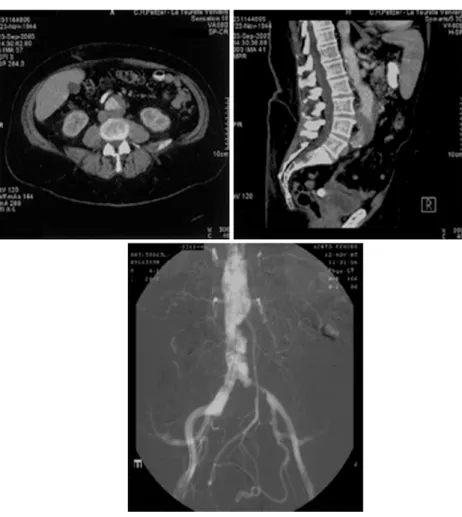
A 50-year-old man was referred to our department with a known AAA that was 46 mm in diameter and a wall thrombus diagnosed by abdominal computed tomogra-phy (CT; Fig. 1A,B). The patient complained of left intermittent claudication, lumbar pain, sexual impo-tence, and significant weight loss (25 kg) in the preceding year. These physical problems were associated with psychological disorders. Aortography confirmed the diagnosis of a small aneurysm with subocclusion of the left common iliac artery (Fig. 1C). An irregular plaque of the infrarenal aorta was also observed. Aortic surgery was done to alleviate the occlusive lesions of the iliac
artery and the small AAA. Aortotomy revealed an unusual cystic wall thrombus at the level of the terminal aorta and left common iliac artery. A sample of the thrombus including the aortic wall was excised for his-tologic analysis. An aortobiiliac bifurcated graft was inserted.
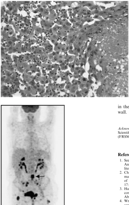
The histologic investigation showed irregular, poly-morphic large cells grouped in poorly cohesive sheets (Fig. 2). Atypical mitotic figures were observed. These cells showed positive immunohistologic staining for vi-mentin, CD31, CD34, and factor VIII but were negative for epithelial markers and smooth muscle actin. This pattern was suggestive of an epithelioid angiosarcoma.
Postoperatively, a whole-body positron emission tomography, scintigraphy and CT were performed to search for a primary lesion and potential metastasis. Intense fluorine 18 fluorodeoxyglucose (18F-FDG) up-take was observed at the level of the terminal aorta and at the level of right and left femurs, iliac artery, and vertebral body (Fig. 3). CT and scintigraphy demon-strated several lytic lesions in the bones of the lower part of the body (vertebral body L4, right and left iliac bones, sacrum, neck of the right femur, patella, and condyle of the left iliac bone). These osteoclastic lesions and some erosions were suggestive of metastases.
Discussion
Fewer than 140 aortic sarcomas have been reported in the literature, with only 10 defined as angiosarcoma of the infrarenal aorta [1–3]. The median survival rate of patients with aortic sarcomas has been reported to be 7 months, which was explained by a late diagnosis com-bined with inadequate surgical resection [1]. The imaging findings are frequently nonspecific and in most published cases did not allow a definitive preoperative diagnosis. This requires immunohistologic examination of the tu-mor or peripheral emboli. These procedures help also to establish a differential diagnosis between malignant and
Correspondence to:N. Sakalihasan; email: [email protected]
ª Springer Science+Business Media, Inc. 2005 Published online: 28 November 2005
A
bdominal
I
maging
Abdom Imaging (2006) 31:117–119 DOI: 10.1007/s00261-005-0366-9
reactive processes. In our case, expression of CD31, CD34, and factor VIII confirmed the endothelial differ-entiation of the tumoral cells. Despite these techniques, histologic definition of large vessel tumors remains challenging. As a consequence, large vessel neoplasms have been classified according to their localization within the aortic wall [4]. Tumors of the media or adventitia, also called mural tumors, may appear like aneurysms or masses. Tumors involving the intima grow into the aortic lumen and are seen as vascular occlusions, whereas in-traluminal tumors grow as polypoidal masses with em-bolic potential.
Patients with abdominal aortic tumors may develop claudication, fatigue, back or abdominal pain, weight loss, or decreased peripheral pulses. Because some of these symptoms are similar to those of occlusive or aneurysmal disease, primary aortic tumors are frequently mistaken for these lesions [4, 5]. In our case, we related the intermittent claudication and sexual impotence to the iliac occlusion and the weight loss to the psychological problems related to sexual impotence. In retrospect, we realized that the AAA appearance on aortography was atypical. Intraluminal protrusive vegetations without signs of atherosclerotic plaque were observed at the level of the terminal aorta and left common iliac artery (Fig. 1C), but CT showed no calcification of the aortic
wall (Fig. 1A,B). Angiographic distinction of an intra-luminal malignancy from thrombus or atheroma can be difficult. We failed to recognize this rare entity preop-eratively and intraoppreop-eratively, so our surgical approach was inappropriate. Ideally, adequate management is wide excision of the aortic wall and of the periaortic soft tissues. It should be kept in mind that, when a thrombus is inhomogeneous with protrusive vegetations and there is no atherosclerosis, a neoplastic lesion should be con-sidered.
Ultrasound, CT, magnetic resonance imaging, and magnetic resonance angiography have been used to nose aortic sarcomas [1, 6–9]; however, the correct diag-nosis has rarely been made. To our knowledge, this is the second report to document the use of conventional aor-tography for diagnosing an intimal sarcoma of the aorta before autopsy. Silverman et al. [10] reported a case of primary intraluminal myxoma at the level of the ascending aorta. However, there was no reference to any protrusive vegetation in absence of atherosclerotic plaque. Recently, using contrast-enhanced CT angiography, Hagspiel et al. [3] described a primary intimal abdominal aorta sarcoma. Ideally, adequate management is wide excision of the aortic wall and of the periaortic soft tissues. Unfortu-nately, most aortic sarcoma have already metastasized at diagnosis.
Fig. 1. Transverse (Top left) and sagittal (Top right) CT scans show an aneurysm of the infrarenal aorta with parietal thrombus. Bottom Aortogram displays a small aneurysm of the abdominal aorta with subocclusion of the left common iliac artery. An irregular stenotic plaque was also observed at the level of the terminal aorta and left common iliac artery.
Primary sarcoma in the wall of an AAA is extremely rare but the prognostic importance of this finding supports routine examination of vascular pathology. The finding, on conventional aortography, of an inho-mogeneous thrombus with protrusive vegetations with-out signs of atherosclerotic plaque should raise suspicions as to the nature of the lesion. In selected cases, additional imaging, such as PET, may be helpful
in the diagnosis of such neoplastic lesions of the aortic wall.
Acknowledgments. This study was supported by the Belgian Fonds Scientifique de la Recherche Me´dicale and Fondation L. Fredericq (FRSM 3.4566.99).
References
1. Seelig MH, Klingler PJ, Oldenburg WA, Blackshear JL (1998) Angiosarcoma of the aorta: report of a case and review of the literature. J Vasc Surg 28:732–737
2. Chiche L, Mongredien B, Brocheriou I, Kieffer E (2003) Pri-mary tumors of the thoracoabdominal aorta: surgical treatment of 5 patients and review of the literature. Ann Vasc Surg 17:354–364
3. Hagspiel KD, Hunter YR, Ahmed HK, et al. (2004) Primary sar-coma of the distal abdominal aorta: CT angiography findings. Abdom Imaging 29:507–510
4. Wright E, Viramini R, Glick A, Page D (1985) Aortic intimal sarcoma with embolic metastases. Am J Surg Pathol 9:890– 897
5. Espada R, Iliopoulos D, Weilbaecher D (1966) Sarcoma presenting as thoracoabdominal aortic aneurysm: case reports and literature review. Contemp Surg 48:135–139
6. Bohner H, Luther B, Braunstein S, et al. (2003) Primary malignant tumors of the aorta: clinical presentation, treatment, and course of different entities. J Vasc Surg 38:1430–1433
7. Higgins R, Posner MC, Moosa HH, et al. (1991) Mesenteric infarction secondary to tumor emboli from primary aortic sar-coma. Guidelines for diagnosis and management. Cancer 68:1622– 1627
8. Sekine S, Abe T, Seki K, et al. (1995) Primary aortic sarcoma: resection by total arch replacement. J Thorac Cardiovasc Surg 110:554–556
9. Pollock JG, Moody AR, Ludman CN, et al. (2000) Postirradiation aortic sarcoma demonstrated by magnetic resonance angiography. J Vasc Surg 31:798–801
10. Silverman JF, Wexler L (1972) Primary intraluminal tumor of the aorta. Case report with preoperative angiographic diagnosis. Radiology 102:581–582
Fig. 2. Irregular polymorphic cells grouped in poorly cohesive sheets. Atypical mitotic figures are also observed. Hematoxin and eosin, original magnification 250·.
Fig. 3. PET scan shows intense18F-FDG uptake at the level of the terminal aorta (arrow) and at the level of the right and left femur and iliac artery.