Université de Montréal
Characterization of Actinobacillus pleuropneumoniae antiviral effect against porcine reproductive and respiratory syndrome virus in porcine alveolar macrophages.
par
Yenney Hernandez Reyes
Département de pathologie et microbiologie Faculté de médecine vétérinaire
Mémoire présenté à la Faculté de médecine vétérinaire en vue de l’obtention du grade de maître ès sciences (M. Sc.)
en sciences vétérinaires option microbiologie
Aout, 2014
Ce mémoire intitulé
Characterization of Actinobacillus pleuropneumoniae antiviral effect against porcine reproductive and respiratory syndrome virus in porcine alveolar macrophages.
présenté par Yenney Hernandez Reyes
a été évalué par un jury composé des personnes suivantes
Dr Marie Archambault, présidente-rapporteuse Dr Carl A. Gagnon, directeur de recherche
Dr Mario Jacques, codirecteur Dr Marcelo Gottschalk, membre du jury
i RÉSUMÉ
Le syndrome reproducteur et respiratoire porcin (SRRP) est la maladie infectieuse la plus économiquement importante de l’industrie porcine. Une étude récente a démontré que le surnageant de culture d’Actinobacillus pleuropneumoniae (App) inhibe l’infection du virus SRRP (VSRRP) in vitro dans des cellules de singe. L’objectif de cette étude est de démontrer l’effet antiviral d’App contre le VSRRP dans les cellules cibles du virus in vivo: les macrophages alvéolaires porcins (MAPs) et d’étudier les mécanismes spécifiques impliqués lors de l’inhibition virale. Les MAPs ont été traités avec App, avant et après l’infection avec le VSRRP. À différents temps post-infection, la réplication et la transcription du génome viral ont été quantifiées. L’expression des interférons (IFN) type I et II, ainsi que le profil protéomique en présence ou absence d’App ont été évalués. L’expression de certaines protéines a été confirmée par immunobuvardage et immunofluorescence (IF). Les résultats ont démontré que l’effet antiviral d’App n’est pas via l’induction des IFN type I et II. App inhibe l’infection virale dans MAPs avant la réplication et la transcription du génome viral, ce qui indique qu’App inhibe précocement le cycle réplicatif viral. Le profil protéomique a révélé qu’App augmentait l’expression de la cofiline, une protéine qui provoque la dépolymérisation de l’actine. De plus, ce phénomène de dépolymérisation a été confirmé par IF. Le traitement des MAPs avec la cytochalasin D (un composé qui provoque la fragmentation des microfilments) a démontré que comme pour App, cette drogue inhibe la réplication virale. Les résultats obtenus suggèrent que l’effet antiviral d’App est via l'activation de la cofiline et dépolymérisation de l’actine, affectant probablement l’endocytose du VSRRP.
Mot clés : SRRP/ VSRRP/ MAPs/ réplication du VSRRP / cytosquelette d'actine/cofiline/A. pleuropneumoniae
ii ABSTRACT
Porcine reproductive and respiratory syndrome (PRRS) is the most economically important infectious disease of swine production. A recent study has demonstrated that the culture supernatant of Actinobacilus pleuropneumoniae (App) inhibits PRRS virus (PRRSV) infection in vitro in a monkey cell line. Following this finding, the objective of this study was to demonstrate the antiviral effect of App in the primary target cells of PRRSV in vivo: porcine alveolar macrophages (PAM) and to elucidate how App inhibits PRRSV replication in PAM. Cells were treated with App before and after PRRSV infection. At different times post-infection, viral genome replication and transcription were measured in the presence of App. mRNA expression of type I and II interferon (IFN) and the proteomic profile of infected cells treated with App were evaluated. The expression of selected proteins was confirmed by immunofluorescence (IFA) and Western blot assays. Results showed that App antiviral effect against PRRSV is not via the induction of type I and II IFN expression. Moreover, it was observed that App inhibits PRRSV infection in PAM before its genome replication and transcription, indicating that App antiviral effect takes place early in PRRSV replication cycle. Proteomic results revealed that App increases cofilin, a protein that induces actin filaments depolymerisation in its active form. This depolymerisation phenomenon was further confirmed by IFA. Interestingly, a microfilament-disrupting compound (cytochalasin D) induced the same effect on PRRSV replication than App suggesting that App antiviral effect against PRRSV takes place via the activation of cofilin and thus actin depolymerisation, which probably affects PRRSV endocytosis.
Key words: PRRS/ PRRSV/ PAM/ PRRSV replication/ actin cytoskeleton/ cofilin/ A. pleuropneumoniae
iii
TABLE OF CONTENTS
RÉSUMÉ ... i
ABSTRACT ... ii
TABLE OF CONTENTS ... iii
LIST OF FIGURES ... vi
ABREVIATIONS AND SIGLES ... viii
DEDICATION ... xiii
ACKNOWLEDGMENTS ... xiv
INTRODUCTION ... 1
CHAPTER I: LITERATURE REVIEW ... 5
1. ACTINOBACILLUSPLEUROPNEUMONIAE ... 6 HISTORY ... 6 CLASSIFICATION ... 6 VIRULENCE FACTORS ... 6 Lipopolysaccharides ... 6 Capsular polysaccharides ... 7 Apx toxins ... 7 Iron-uptake systems ... 8 Biofilm formation ... 8
Other outer membrane proteins ... 8
Secreted proteases ... 9
IMMUNE RESPONSES ... 9
2. PORCINEREPRODUCTIVEANDRESPIRATORYSYNDROMEVIRUS ... 10
HISTORY ... 10
TAXONOMY ... 10
MORPHOLOGY ... 10
iv
PRRSV NON-STRUCTURAL PROTEINS ... 13
PRRSV STRUCTURAL PROTEINS ... 14
CELLULAR TROPISM ... 16
PRRSV LIFE CYCLE IN CELLS ... 17
PRRSV entry ... 17
PRRSV uncoating ... 19
Genome replication and transcription ... 19
Virion assembly ... 21
Virus released ... 22
VIRAL PATHOGENESIS IN VIVO ... 22
IMMUNE RESPONSES ... 23
Innate immune response ... 23
Adaptive response ... 24
PRRSV CONTROL AND ELIMINATION ... 25
3. BACKGROUNDOFTHISTHESIS ... 26
CHAPTER II: Actinobacillus pleuropneumoniae blocks porcine reproductive and respiratory syndrome virus replication prior to its genome replication and transcription. ... 27
GENERAL DISCUSSION ... 68
GENERAL CONCLUSION ... 73
REFERENCES ... 75
ANNEXES ... xv
ANNEXEI:Actinobacillus pleuropneumoniae possesses an antiviral activity against porcine reproductive and respiratory syndrome virus. ... xvi ANNEXEII:Curriculum vitae ... xlvii
v
LIST OF TABLES Chapter I: Literature review
Table 1: PRRSV non structural proteins characteristics and functions. ... 13 Table 2: PRRSV structural proteins characteristics and functions. ... 15
Chapter II: Actinobacillus pleuropneumoniae blocks porcine reproductive and respiratory syndrome virus replication prior to its genome replication and transcription.
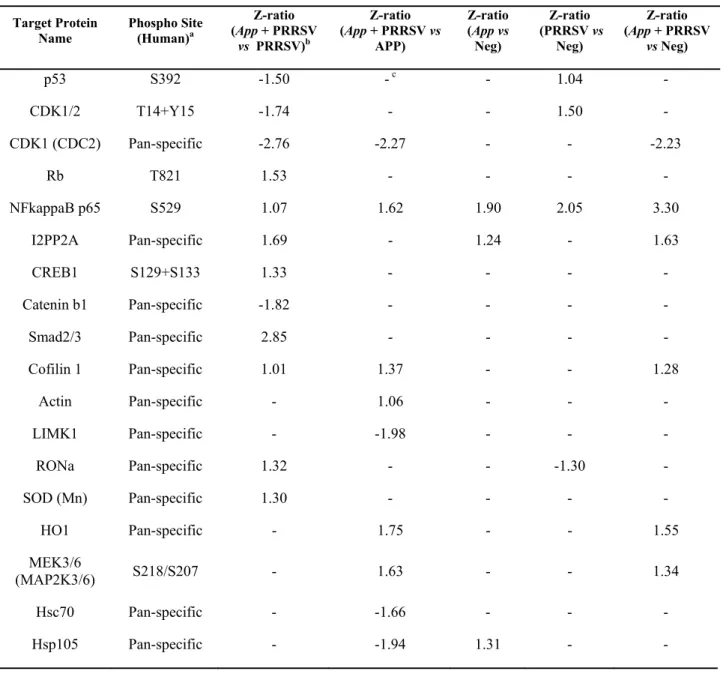
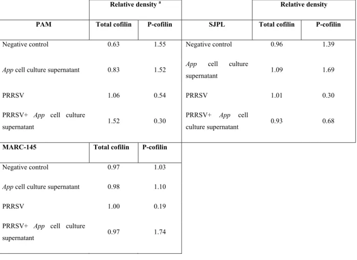
Table 1: Z-ratios of the most modulated cell proteins by PRRSV and App supernatant. ... 37 Table 2: Total cofilin and P-cofilin relative densities in PAM, SJPL and MARC-145 cells. .. 41
Annexe I: Actinobacillus pleuropneumoniae possesses an antiviral activity against porcine reproductive and respiratory syndrome virus.
Table 1: Antiviral activity of AppΔapxIΔapxIIC supernatant against several animal DNA and RNA viruses in SJPL infected cells. ... xxix
vi
LIST OF FIGURES Chapter I: Literature review
Figure 1: Schematic representation of PRRSV genome organization. ... 12 Figure 2: Arterivirus replication cycle inside cells. ... 18 Figure 3: Model for Arterivirus genome replication and transcription. ... 21
Chapter II: Actinobacillus pleuropneumoniae blocks porcine reproductive and respiratory syndrome virus replication prior to its genome replication and transcription.
Figure 1: PAM viability and mortality in the presence of App cell culture supernatant. ... 32 Figure 2: Relative expression of type I and II IFN mRNAs in PAM cells treated with App cell
culture supernatant. ... 33 Figure 3: PRRSV genome replication and transcription kinetics assays in infected cells treated
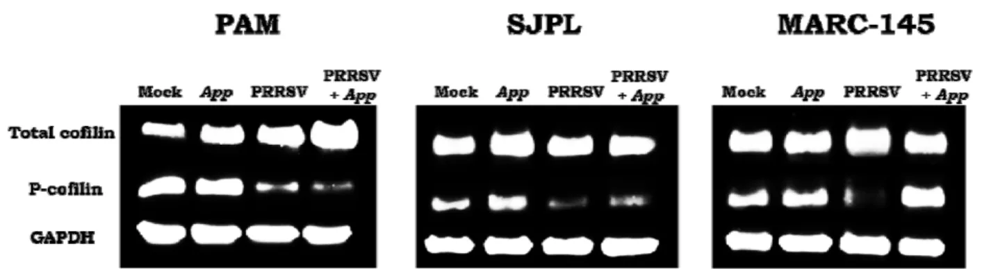
with App cell culture supernatant. ... 35 Figure 4: Cofilin expression level in PRRSV infected cells treated or not with App cell culture
supernatant. ... 40 Figure 5: F-actin expression in the presence of App cell culture supernatant. ... 42 Figure 6: Infectious viral particles production in PRRSV-infected PAM, SJPL and MARC-145
cells treated with cytochalasin D. ... 44 Figure 7: App supernatant antiviral activity against others PRRSV strains in MARC-145 cells. ... 46 Supporting information
Figure S1: β-actin protein and mRNA expression in the presence of App cell culture supernatant. ... 67
vii
Annexe I: Actinobacillus pleuropneumoniae possesses an antiviral activity against porcine reproductive and respiratory syndrome virus.
Figure 1: Bacterial adherence over time of Appwt or AppΔapxIΔapxIIC in PRRSV co-infected SJPL and MARC-145 cells... xxi Figure 2: Cytotoxicity over time of Appwt or AppΔapxIΔapxIIC in PRRSV co-infected SJPL and MARC-145 cells. ... xxiii Figure 3: PRRSV antigen detection in SJPL, MARC-145 and PAM cells co-infected with
AppΔapxIΔapxIIC. ... xxvi Figure 4: PRRSV titer in App treated SJPL, MARC-145 and PAM cells. ... xxvii Figure 5: AppΔapxIΔapxIIC cell culture supernatant < 1 kDa fraction antiviral activity against PRRSV. ... xxviii Figure 6: AppΔapxIΔapxIIC cell culture supernatant and PRRSV effects on mRNA
quantification of type I (IFNα, IFNβ) and type II (IFNγ) interferons. ... xxx Supporting information
Figure S1: NOD1 and NOD2 inhibition effect on PRRSV replication. ... xlv Figure S2: Antiviral activities of AppΔapxIΔapxIIC cell culture supernatant fractions against
PRRSV. ... xlvi
viii
ABREVIATIONS AND SIGLES
ADE Antibody-dependent enhancement
AMP Cyclic adenosine monophosphate
App Actinobacillus pleuropneumoniae
Appwt App wild type strain
AppΔapxICΔapxIIC App mutant strain producing non-active ApxI and ApxII toxins
Apx App toxins
ATCC/VR-2332 American Type Culture Collection/PRRSV reference strain for genotype II BALF Bronchoalveolar lavage fluid
BAV3 Bovine adenovirus 3
BHI Brain heart infusion
BHV-1/4 Bovine herpes virus type 1 and 4 BVDV-1 Bovine viral diarrhea virus type 1 cAMP Cyclic adenosine monophosphate CD4+/CD8+/ CD4+
-CD8+ T cells CD4
+/CD8+/CD4+-CD8+ double positives
CD151 Cluster of Differentiation 151 (PRRSV cellular receptor) CD163 Cluster of Differentiation 163 (PRRSV cellular receptor) CFU/mL Colony-forming units per milliliter
ix
ClpP Protein member of the caseinolytic protease family 3CLpro 3C-like proteinase domain
CPE Cytopathic effect
CPV Canine parvovirus
CREB cAMP response element-binding DMEM Dulbecco’s modified Eagle’s medium DMVs Double membrane vesicles
dpi Days post-infection
dsRNA Double-stranded ribonucleic acid EHV-1 Equine herpes virus type 1
ER Endoplasmic reticulum
F-actin Actin filaments
FBS Foetal bovine serum
Fhu A/B/C/D Cellular proteins responsible for the transport of ferric hydroxamate FITC Fluorescein isothiocyanate
GP Glycoprotein
gRNA Genomic RNA
H1N1/H3N2 Influenza A virus subtypes HPIV-3 Human parainfluenza virus 3
x IFN α/β/γ Interferon alpha/beta/gamma IgG/IgA Immunoglobulin G and A
IL Interleukin
IRF3 IFN regulatory factor 3 ISG15 interferon-stimulated gene 15 ISP-1 IFN-beta promoter stimulator 1
JAK-STAT Janus kinase- Signal Transducer and Activator of Transcription (Type I IFN signaling pathway)
KINEX TM Antibody microarray proteomic test
LDH Lactate dehydrogenase
LDV Lactate dehydrogenase-elevating virus
LPS Lipopolysaccharide
LV Lelystad virus
M PRRSV membrane protein
MARC-145 African green monkey kidney cell line derived from MA-104 MBHPP147 Name of App mutant
MDCK Madin-Darby canine kidney cells
MEM Minimum essential medium
MHC Major histocompatibility complex class
MLV Modified-live virus
xi
N PRRSV nucleocapsid protein
NAbs Neutralizing antibodies NVSL PRRSV genotype II strain
β-NAD Beta-nicotinamide adenine dinucleotide NendoU Nidovirus endoribonuclease
NFκB Transcriptional regulator nuclear factor-κB
NOD1/NOD2 Nucleotide-binding oligomerization domain-containing protein 1/2 NPTr Newborn pig trachea cells
nsps Non-structural proteins
ORFs Open reading frames
PAM Porcine alveolar macrophages
PBS Phosphate buffer saline solution
PCP α/β Papain-like cysteine proteinase domains alpha/beta PCV2 Porcine circovirus 2
PFA Paraformaldehyde
PGA Poly-N-acetylglucosamine
PL2pro Chymotrypsin-like cysteine protease
Poly I:C Polyinosinic–polycytidylic acid potassium salt PRDC Porcine respiratory disease complex
xii
PRRS Porcine reproductive and respiratory syndrome PRRSV Porcine reproductive and respiratory syndrome virus
RdRp RNA-dependent RNA polymerase
RTC Replication-transcription complex S4074 App serotype 1 reference strain
SD Standard deviation
sg mRNA Sub-genomic messenger RNAs SJPL St-Jude porcine lung cells
ssRNA Single-stranded RNA
TCID50/mL Tissue culture infectious dose 50 % per mL
TNF-α Tumor necrosis factor alpha TRSs Transcription regulatory sequences
xiii
Dedicated to my God, my husband, my son and my parents
for their incomparable love, patience, help and encouragement
xiv
ACKNOWLEDGMENTS
I would like to recognize the FQRNT and the NSERC for supporting this research. I would like to thank my director Dr. Carl A. Gagnon and my co-director Dr. Mario
Jacques for giving me the opportunity to make my studies in their laboratories and also for their support, advices and availability.
Thanks to Dr. Chantale Provost for its incomparable help and the excellent scientific knowledge that she shared with me.
I really appreciated the opportune scientific advices of Dr. Christian Savard and Dr. Christian Bellehumeur and also their humour sense. Besides, I want to thanks Fernando for his help and also Daniel for being really nice and kind persons.
Thanks to Isabel and Carolina because I found in them real friends that encourage and advise me during my studies and for the beautiful moments we spent together.
To my parents because they fight so hard to make me the person that I am today. Thanks for being your entire lives ready to encourage and loving me.
To my lovely husband for its patience and unconditional love because as it’s written love is patient and kind; love it is not irritable or resentful; love bears all things, believes all things, hopes all things, endures all things.
To my biggest treasure, my son, because he is able to convert each hard or exhausted day in a hopeful and colourful day, only with his smile, hugs and stories.
Thanks to my God for its incredible and incomparable love and mercy. I will love you, trust you and I will have faith in you forever. I know that you will never leave me or forsake me.
1
2
Porcine reproductive and respiratory syndrome (PRRS) is a worldwide endemic infectious disease which causes significant economic losses in the swine industry (1). The etiological agent, PRRS virus (PRRSV), is an enveloped and single-stranded positive sense RNA virus of approximately 15 kb that encodes for at least 10 open reading frames (ORFs) (2-5). PRRSV is classified in the order Nidovirales, family Arteriviridae and genus Arterivirus, which also includes the lactate dehydrogenase-elevating virus of mice, equine arteritis virus and simian hemorrhagic fever virus (6, 7). PRRSV isolates are divided into two genotypes, where the Lelystad virus in Europe and ATCC VR-2332 in North America are the reference strains for genotype I and II, respectively (6, 8, 9).
PRRSV has a very narrow cell tropism both in vivo and in vitro. In vivo, PRRSV has preference for cells of monocyte/macrophage lineage, especially the fully differentiated macrophages of lungs, lymphoid organs and placenta (10-12). Porcine alveolar macrophages (PAM) constitutes the main in vivo target cells of PRRSV and primary PAM has been extensively used for in vitro study of host cell infection (6, 11, 13, 14). Two continuous cell lines, from monkey origin, are the only cells able to fully support PRRSV replication in vitro: the African green monkey kidney cell line MA-104 and derivatives such as MARC-145 and CL2621 (15) and the newly reported St-Jude porcine lung (SJPL) cells (16, 17).
Following PRRSV entry and release of the viral genome into the cytoplasm, the PRRSV ORF1 is translated and the resulting non-structural proteins (nsps) trigger the formation of the replication-transcription complex, which is associated with double membrane vesicles and supports genome replication and transcription process (18-20). The genome replication is produced by the continuous synthesis of negative (-) full-length RNA strands using as template the genomic RNA (gRNA), then the (-) RNA strands will lead the formation of new gRNAs (21). The genome transcription process is named to the synthesis of a nested set of six sub-genomic mRNAs (sg mRNAs). According with a model proposed by Sawicki and colleagues (22), the generation of these sg mRNAs is through a discontinuous RNA synthesis process, where (-) sg RNA strands are produced and then are used as template for the synthesis of the sg mRNAs.
3
The actin cytoskeleton plays an important role in PRRSV life cycle inside cells. It has been reported that PRRSV entry into PAM is via clathrin-mediated endocytosis and that this process is dependent (13). The use of cytochalasin D (a microfilament-disrupting compound) inhibited PRRSV primary and secondary infection in MARC-145 cell line (23). Moreover, it was observed that in PRRSV infected cells there were less actin filaments (F-actin) expression, than in the adjacent untreated cells suggesting that PRRSV can modulate the actin cytoskeleton to favor cell infection and that higher F-actin expression correlated with PRRSV resistance (23).
Current management strategies to control PRRS, which include surveillance, severe biosecurity measures, whole herd depopulation and repopulation, herd closure and vaccination, seem to be partially efficient for the control of the disease (24-26). This phenomenon has stimulated the research of novel strategies to successfully control PRRSV infection. Several studies have found natural compounds with antiviral activity against PRRSV such as macrolides (27), N-acetylpenicillamine (28), Cryptoporus volvatus extracts (29), morpholino oligomer (30, 31), matrine (32), sodium tanshinone IIA sulfonate (33). However, for the moment there are no effective commercially available drugs to prevent PRRSV infection.
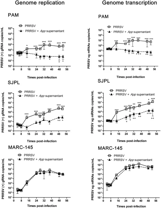
Actinobacillus pleuropneumoniae (App) is the etiological agent of porcine pleuropneumonia a worldwide endemic disease (34). App is divided into two biotypes, the biotype 1 which is dependent on exogenous beta-nicotinamide adenine dinucleotide (β-NAD) and the biotype 2 which is NAD-independent (35). App is divided also into 15 serotypes (1-4, 5a, 5b and 6-15) (34, 36). A recent study performed in our laboratory demonstrated that the culture supernatant of a mutant App strain (AppΔapxICΔapxIIC), which produces inactive Apx I and II toxins, has a potent antiviral effect against PRRSV (37). This phenomenon of inhibition was observed in the monkey SJPL cell line. Since, PAM are the main in vivo target cells of PRRSV, the first objective of this study was to demonstrate the App supernatant antiviral effect against PRRSV in primary cultures of PAM. Results corresponding to this objective were already published [(37), Annexe I] and demonstrated that App supernatant efficiently inhibits PRRSV infection in PAM. Based on the literature and according with preliminary results obtained in our laboratory, it is hypothesised that App cell culture supernatant modulates cellular(s)
4
component(s) and by consequence PRRSV infection is blocked. Then, the second objective of this study is to determine the possible mechanisms involved in the viral inhibition.
5
6
1. ACTINOBACILLUS PLEUROPNEUMONIAE
HISTORY
In 1957, the first case of porcine pleuropneumonia was reported in United States by Pattison and colleagues (38) and the bacteria associated with the pneumonic lesions was firstly classified in the genus Haemophilus. Afterwards, in 1983 it was reclassified, since DNA studies revealed that this pathogen belonged to the genus Actinobacillus of the Pasteurellaceae family together with the bacteria of the genus Haemophilus, Pasteurella and Mannheimia. Thus, Actinobacillus pleuropneumoniae (App) is the etiologic agent of porcine pleuropneumonia a worldwide endemic disease, which affects pigs of all ages and causes considerable economic losses (34).
CLASSIFICATION
App is a non-motile and a facultative anaerobic Gram-negative encapsulated coccobacillus (39). App strains are classified into two biotypes, where the biotype 1 is dependent on exogenous beta-nicotinamide adenine dinucleotide (β-NAD), whereas the biotype 2 is able to synthesize this component by itself (35). Based on the surface polysaccharides this bacterium has been divided into 15 serotypes (1-4, 5a, 5b and 6-15) (34, 36). All serotypes can induce the disease but with differences in virulence (40). App is also positive to the CAMP (Christie Atkins Munch-Petersen) test (41).
VIRULENCE FACTORS
The lower respiratory tract is the preferential site of infection of App, since it binds the ciliated cells of the terminal bronchiole and alveolar epithelial cells. There are three important steps during App infection that allow the apparition of the disease: the colonization, the evasion of the host’s defense mechanisms and host tissue damages (42). Different virulence factors have been identified to participate in each of these stages.
Lipopolysaccharides
Several studies have confirmed that the lipopolysaccharide (LPS) is necessary in App adherence to the respiratory epithelium (43-45). However, others have postulated that this stage in bacterial pathogenesis is probably LPS-independent (46). The LPS is formed of three
7
regions: the lipid A, the LPS core and the O-antigen (47). Provost and collaborators demonstrated that a LPS core mutant decreased adherence to host cells, showing its critical role in adhesion (48). According with a proposed multiple-step adhesion mechanism, firstly the O-antigen of the LPS may interact with the phosphatidylethanolamine (a host membrane phospholipid) by a low-affinity binding, then, a stronger union to the respiratory tract is made by the interaction of the LPS core and/or surface proteins (a 55 kDa protein, type 4 fimbriae (will be discussed below)) to other host cell receptors (49). The lipid A of the LPS is able to bind the porcine hemoglobin and by this way the bacteria acquires the iron for its growth (50). The LPS is being associated also to the formation of lesions, since it was demonstrated that the LPS outer core interacts with ApxI and ApxII toxins and this interaction may enhance App cytotoxicity and virulence (51).
Capsular polysaccharides
Cruijsen and colleagues demonstrated that App reduces the phagocytic activity of porcine alveolar macrophages (PAM) in vitro by inducing cell lysis, which causes viable bacteria liberation (52). Among the factors that may contribute to App survival, the capsular polysaccharides should play an important role. It was demonstrated that encapsulated App strains were resistant to complement-mediated killing, whereas non-capsulated strains were killed (53, 54). Moreover, there is a direct association between the type and the amount of App capsular polysaccharides and its virulence in vivo (55). Capsular polysaccharides are not involved in App adherence, since it was observed that a capsule deficient mutant adheres more efficiently to cells than the wild type strain. However, the capsule can mask the adhesins, at least in part, affecting indirectly the adherence (56).
Apx toxins
App repeats-in-toxins (RTX) exoproteins (ApxI, ApxII, ApxIII and Apx IV) are involved in the induction of pulmonary lesions (57, 58). ApxI is the most haemolytic and cytotoxic toxin for alveolar macrophages and neutrophils and induces apoptosis in PAM cells (57, 59-61), ApxII is weakly haemolytic and moderately cytotoxic (34, 60, 62), ApxIII is non-haemolytic, highly cytotoxic and has a pro-apoptotic activity (63) and Apx IV is only secreted in vivo and is essential for App full virulence (64, 65).
8 Iron-uptake systems
The lower respiratory tract is limited in supplying the essential nutrients for bacterial growth (34). However, App is able to use the host transferrin (66-68) and hemoglobin (50, 69) and exogenous siderophores (70) as sources of iron for its growth. App binds to transferrin through two proteins present on its surface of approximately 60 and 100 kDa, where the 100 kDa protein is a transmembrane protein that may form a channel allowing the transport of iron across the outer membrane (67, 71). The hemoglobin receptors are two outer membrane proteins of approximately 75 and 105 kDa, where the 75 kDa protein can also bind hemin (47). The last iron-acquisition system is mediated by the uptake of exogenous siderophores such as the hydroxamate siderophore ferrichrome. There are four genes implicated in the ferric hydroxamate uptake, which are located in a single operon. These genes encode for the outer membrane protein FhuA, which is the receptor for ferrichrome, the FhuD protein is responsible for the translocation of ferric hydroxamate from the outer to the inner membrane and FhuC and FhuB proteins are cytoplasmic-membrane-associated proteins, which are components of an ABC transporter that internalizes the ferric hydroxamate (72).
Biofilm formation
Several studies have demonstrated that App has the ability to form biofilm. It is believed that biofilm is necessary for bacteria colonization (34, 73-76). The polysaccharide poly-N-acetylglucosamine (PGA) was observed to be involved in biofilm formation and probably functions as the major biofilm adhesin in App (73, 76). Moreover, Buettner and collaborators showed that a mutant App strain, deficient in biofilm formation, had a reduction in its virulence (77). A transcriptomic study revealed that, after contact of App with the newborn pig trachea (NPTr) cell line, genes involved in biofilm biosynthesis were up-regulated (78). Other outer membrane proteins
Fimbriae is a bacterial surface structure that is believed to be involved in App adhesion since it was demonstrated that the type 4 fimbriae was induced by contact of App with the host cells in vitro and in vivo (79). A 55 kDa outer membrane protein was identified and is postulated to be implicated in the adhesion to the host alveolar epithelial cells (80). Additionally, another
9
surface protein of 60 kDa was identified, which is able to adhere to porcine collagen and fibrinogen (81).
Secreted proteases
The App secreted proteases can be considered as another virulence factor because it was demonstrated that they can degrade the immunoglobulin A and G (IgA, IgG) and the hemoglobin from porcine, human and bovine origin (82, 83). It was observed in another study that a 24 kDa App zinc metalloprotease can degrade actin protein in vitro (84). In addition there is another App protease that was described, the ClpP (member of the Clp (caseinolytic protease family)), to be important in virulence regulation (85).
IMMUNE RESPONSES
App pathobiology includes pulmonary lesions which are characterized by the presence of macrophages, granulocytes, lymphocytes, hemorrhages, necrosis, etc (42). Cruijsen and colleagues compared PAM and polymorphonuclear leukocytes (PMN) abilities to phagocytize and kill App in vitro (52). It was observed that both cells were able to phagocytize the bacteria. However, PAM was unable to kill the intracellular bacteria compared to PMN, which killed 95% of the ingested App. There are two possible explanations to this phenomenon in PAM 1) cytolysin produced by App might affects the cellular killing mechanisms or 2) the phagocytised App can cause the impairment of reactive oxygen species synthesis (which have a potential bactericidal capacity), allowing then, the releasing of viable bacteria.
Cytokines such as tumor necrosis factor alpha (TNF-α), interleukin (IL)-1beta (IL-1β), IL-1α, IL-6 and IL-8 were produced in experimentally infected pigs with App (86, 87). The overexpression of these proinflammatory mediators in response to App infection are probably involved in the pulmonary lesions associated with the disease (88). Additionally, the expression of IL-10 and IL-12 was detected also in experimentally infected pigs and it was suggested that they are involved in App pathogenesis (89). Benga and colleagues detected different amounts of interferon gamma (IFN-γ) in plasma and in bronchoalveolar lavage fluid (BALF) during the infection (90). Moreover, it was observed that the increase of IFN-γ was associated with an increase in the severity of the disease. However, this increase of IFN-γ was
10
dependant on pig breeds, where the porcine breeds not showing any increase of IFN-γ were more resistant to the disease (90).
2. PORCINE REPRODUCTIVE AND RESPIRATORY SYNDROME VIRUS HISTORY
In the United States, in 1987, a new emerging “mystery swine disease” of unknown etiology causing reproductive failure and neonatal severe pneumonia was reported (91, 92). A similar syndrome was after recognized in Europe in 1990 (93, 94). The causative agent was firstly isolated in the Netherlands using porcine alveolar macrophages and designated as Lelystad virus (LV) (8). Shortly after, it was isolated in North America using gnotobiotic pigs and designated as American Type Culture Collection (ATCC) VR-2332 virus (9). In 1992, the disease was named porcine reproductive and respiratory syndrome (PRRS), according with the symptoms and the observed clinical signs. The PRRS etiological agent, PRRS virus (PRRSV), has spread worldwide in the last decades. In 2006, a highly pathogenic PRRS virus (PRRSV) strain emerged in China and Vietnam, which caused an atypical PRRS outbreak (95). At present, PRRS is a worldwide endemic disease causing significant economic losses in swine production, since it can provoke a severe pneumonia in growing and finishing pigs (1).
TAXONOMY
PRRSV is an enveloped and single-stranded (ss) positive sense RNA virus classified in the order Nidovirales, family Arteriviridae and genus Arterivirus, which also includes the lactate dehydrogenase-elevating virus (LDV) of mice, equine arteritis virus (EAV) and simian hemorrhagic fever virus (6, 7). PRRSV isolates are divided into two genotypes, where LV and ATCC VR-2332 are considered the reference strains for PRRSV genotypes I and II, respectively (6, 8). The two genotypes share approximately 60% nucleotide identity (96, 97). Additionally, within the same genotype exists high genetic variabilities (98-101).
MORPHOLOGY
PRRSV virions are pleomorphic with form varying from spherical to oval shape. As observed by cryoelectron tomography, virions size range between 50 to 65 nm with an internal core of around 40 nm in diameter (102). The virion outer membrane is smooth and is formed by a
11
lipid bilayer, which has protrusions that probably correspond to ectodomains formed by the envelope proteins (102). The nucleocapsid core is formed by the nucleocapsid protein (N) and the viral RNA and it’s been suggested to have an asymmetric and helical organization (102, 103). Virions survival and stability are dependent on the pH and temperature. For instance, it was demonstrated that at pH 7.5, the virus was stable for a long period at -20°C and -70°C. The virions half-life is 50 hours at 4°C and pH 6.25 while at a high temperature (37°C) and pH 6.0, it is 6.5 hours (104). Besides, the addition of lipid solvents such as chloroform reduced the virion infectivity from 105 TCID50/mL (tissue culture infectious dose 50 % per mL) to < 101 TCID50/mL (6).
GENOME ORGANIZATION
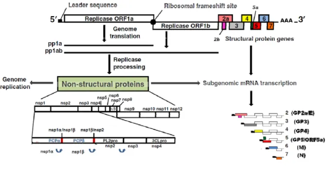
PRRSV genome is approximately 15 kb in length, encodes for at least 10 open reading frames (ORFs) and is capped at the 5' end and polyadenylated at the 3' tail (3-5, 105) (Figure 1). The ORF1a and ORF1b, which comprise almost the three-quarters of the total genome, encodes for 14 non-structural proteins (nsps) (103). The nsps are synthesized as polyproteins (pp1a and pp1ab) obtained from ORF1a and ORF1b, respectively (20). The pp1ab is expressed from a −1 programmed ribosomal frameshifting (PRF) in the ORF1a/ORF1b overlap region (106). The polyproteins are then processed to lead the formation of the 14 nsps: nsp1α, nsp1β, nsp2-6, nsp7α, nsp7β and nsp8-12 (39, 107). The nsp1α, nsp1β, nsp2 and nsp4 encode the viral proteases responsible for polyprotein processing (20, 108, 109). The nsps are also implicated in viral RNA replication, sub-genomic (sg) mRNA transcription and translation. Recently, other PRF (not illustrated in Figure 1) was found that allows the access to a short transframe (TF) ORF, that overlaps the nsp2-encoding region of ORF1a in the +1 frame and it is translated by −2 PRF, yielding the expression of nsp2TF protein (105). The PRRSV structural proteins encoded by the ORF 2 to 7 are synthesized from a nested set of six sg mRNAs by a process of discontinuous RNA synthesis (110). The sg mRNAs are structurally polycistronic with the exception of the sg mRNA7, but are presumed to be functionally monocistronic, with the exception of the sg mRNAs 2 and 5, that are believed to be bicistronic (4, 5, 111). All the sg mRNAs are 3' co-terminal and also shares a 5' leader sequence, which is identical to the 5' end of the genome (108, 112, 113). ORFs 2a, 3 - 5 encode for the glycoproteins (GP) 2a, 3, 4 and 5, respectively. ORF6 and ORF 7 encode for the membrane protein (M) and the N protein,
12
respectively (2, 108). ORF2b is fully inside the ORF2a and encode for the non-glycosylated protein E (114). The recently discovered ORF5a, which overlaps the 5' end of the ORF5, encode for the ORF5a protein (4, 5).
Figure 1: Schematic representation of PRRSV genome organization.
Adapted from Music and Gagnon (115). The top of the figure represents the PRRSV complete genome from ORF1 to ORF7. The leader sequence is represented by a black rectangle and the ribosomal frame shift (between ORF1a and ORF1b) is illustrated as a black circle. The 14 nsps resulted from the proteolytic cleavage of the two polyproteins (pp1a and pp1ab) are represented as well as the four proteases responsible for it: the papain-like cysteine proteinase domains (PCPα and PCPβ) located in nsp1α and nsp1β, respectively, the chymotrypsin-like cysteine protease domain (PL2pro) presented in nsp2 and the main serine proteinase, 3C-like proteinase domain (3CLpro), located in nsp4 (108, 116). PCPα, PCPβ and PL2pro cleave the junction between nsp1α/nsp1β, nsp1β/nsp2 and nsp2/nsp3, respectively and 3CLpro is responsible for the liberation of the remainder nsps (nsp3 to nsp12). The sg mRNAs (2-7) are 3’ co-terminal and also contain a common leader sequence in the 5’ end. The translated proteins form each sg mRNA are represented between parenthesis.
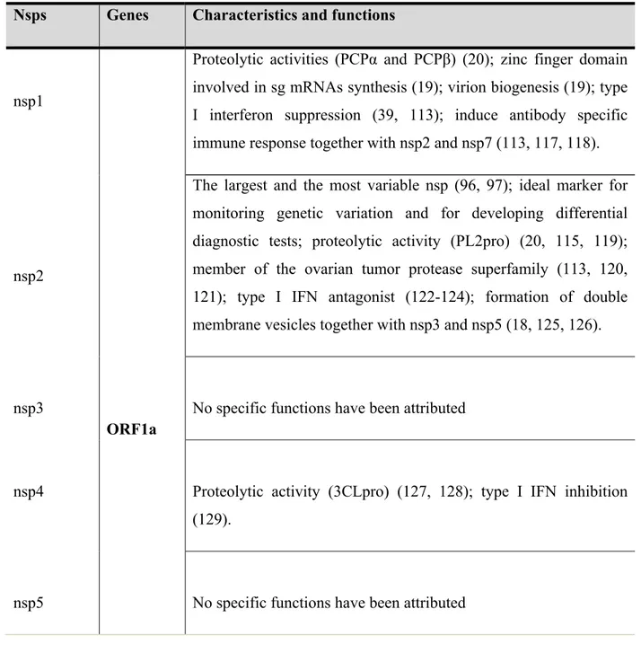
13 PRRSV NON-STRUCTURAL PROTEINS
Table 1 describes the principal functions and the most important generalities of the non-structural proteins.
Table 1: PRRSV non-structural proteins characteristics and functions. Adapted from Music and Gagnon (115).
Nsps Genes Characteristics and functions
nsp1
Proteolytic activities (PCPα and PCPβ) (20); zinc finger domain involved in sg mRNAs synthesis (19); virion biogenesis (19); type I interferon suppression (39, 113); induce antibody specific immune response together with nsp2 and nsp7 (113, 117, 118).
nsp2
The largest and the most variable nsp (96, 97); ideal marker for monitoring genetic variation and for developing differential diagnostic tests; proteolytic activity (PL2pro) (20, 115, 119); member of the ovarian tumor protease superfamily (113, 120, 121); type I IFN antagonist (122-124); formation of double membrane vesicles together with nsp3 and nsp5 (18, 125, 126).
nsp3
ORF1a
No specific functions have been attributed
nsp4 Proteolytic activity (3CLpro) (127, 128); type I IFN inhibition (129).
14
nsp6 No specific functions have been attributed
nsp7
Cleaved by 3CLpro in nsp7α and nsp7β (109); genome synthesis and translation of viral proteins (130).
nsp8 No specific functions have been attributed
nsp9 RNA-dependent RNA polymerase (RdRp); virus transcription and replication (131, 132).
Helicase; zinc-binding domain (133, 134); ATPase activity in vitro (135, 136).
nsp10
ORF1b
Nidovirus endoribonuclease (NendoU), which is considered the genetic marker of Nidovirales order (133, 137, 138); type I IFN inhibition (139).
nsp11
nsp12 No specific functions have been attributed
PRRSV STRUCTURAL PROTEINS
The PRRSV structural proteins are classified into major or minor structural proteins, based in their abundance into the virion. GP2a, E, GP3 and GP4 are considered the minor structural envelope proteins and GP5 and M are the major structural envelope components. N protein is the sole component of the viral nucleocapsid. Table 2 summarises the main characteristics and functions of PRRSV structural proteins.
15
Table 2: PRRSV structural proteins characteristics and functions. Adapted from Music and Gagnon (115).
Structural
proteins Genes Characteristics and functions
GP2a ORF2a
Contains 2 two highly conserved putative N-linked glycosylation sites (140); incorporated into virions as a multimeric (GP2a, E, GP3 and GP4) complex; essential for virus infectivity (141); interacts with the cellular receptor CD163 (142, 143); involved in PRRSV uncoating; apoptosis inhibition (144).
E ORF2b
Unglycosylated and myristoylated structural protein (111); incorporated into virions as a multimeric complex (141); essential for virus infectivity (141, 145); possesses ion-channel like properties and may function as a viroporin in the envelope (145); involved in genome released into the cytoplasm.
GP3 ORF3
One of the most variable PRRSV proteins; highly glycosylated that contains seven predicted N-glycosilation sites (2); its membrane topology is strain dependent (146); highly antigenic and involved in the glycan shielding process (147); incorporated into virions as a multimeric complex; essential for virus infectivity (141).
GP4 ORF4
Highly glycosylated protein; key protein in the formation of the multimeric complex incorporated into virions (141); essential for virus infectivity; mediates interaction between the multimeric complex and GP5 (142); interacts with the cellular receptor CD163 (142, 143); involved in PRRSV uncoating; induce neutralizing antibodies and cell-mediated immune responses (148-151).
GP5 ORF5 The major PRRSV GP and the most variable structural protein with a variable number of potential N-glycosylation sites (140);
16
covalent association of GP5 and M is crucial for virus assembly (152, 153); involved in virus entry into the cells and in apoptosis; neutralizing antibodies are predominantly directed to GP5 (154, 155); involved in glycan shielding process (147).
ORF5a ORF5a Overlaps the ORF5 in its 5' end; essential for virus viability (4, 5, 156); cannot protect animals from PRRSV infection (157).
M ORF6
Unglycosylated and the most conserved structural protein; involved in virus assembly and budding (2, 115, 152); GP5/M heterodimer is crucial for virus infectivity (152, 153).
N ORF7
Unglycosylated, small and highly basic protein (115, 140); the sole component of the viral capsid and interacts with itself by covalent and non covalent interactions to form a homodimer (2, 152); highly immunogenic and is used in diagnostic procedures to detect the presence of the disease (149, 158); localised in the cytoplasm and in the nucleus and nucleolus (159); type I IFN inhibition (160).
CELLULAR TROPISM
PRRSV has a very narrow cell tropism both in vivo and in vitro. In vivo, PRRSV has high preferences for cells of monocyte/macrophage lineage, especially the fully differentiated macrophages of lungs, lymphoid organs and placenta (10-12). It was also reported that porcine dendritic cells support PRRSV infection, however in those studies monocyte-derived dendritic cells and bone marrow-derived dendritic cells were used, those may differ from the primary dendritic cells (161-164). In naturally infected pigs PRRSV antigens were found in bronchiolar epithelial cells (165). However this finding is contradictory with the result obtained by Teifke and collaborators, which demonstrated that PAM are the only pulmonary target cells of PRRSV (166). In fact, PAM constitutes the main in vivo target cells of PRRSV and primary PAM has been extensively used for in vitro study of host cell infection. These
17
cells are of myeloid origin, which circulate in the blood as monocytes and are differentiated into macrophages that reside in tissues (167). PAM cells are members of the mononuclear phagocyte system of the lung and they are able to protect the respiratory tract from invasion of foreign pathogens (by phagocytosis; bactericidal activity; cytotoxicity; cytokines production; activation of T cells) (168, 169). However, ingestion of virus (ex. PRRSV) by PAM allows viral infection and the subsequent functional impairment of the cells (161, 170, 171).
In addition, there are only two continuous cell lines, from monkey origin, that are able to fully support PRRSV replication: the African green monkey kidney cell line MA-104 and its derivatives such as MARC-145 and CL2621 (15) and the newly reported SJPL cells (16, 17). In the literature, it has been reported that non permissive continuous cell lines were able to support PRRSV replication after the introduction of the PRRSV receptors: CD163 or sialoadhesin (172, 173).
PRRSV LIFE CYCLE IN CELLS
In this part, each stage of PRRSV replication cycle inside cells and the cellular components involved in it will be described. Figure 2 summarizes all the steps of the virus replication cycle.
PRRSV entry
PRRSV entry may differ between PAM and MARC-145 cells since the cellular receptors required for it are different. In PAM, a recent review has proposed a possible model for PRRSV entry by integrating the major findings about PRRSV entry into PAM (164). According with the model proposed PRRSV first interacts with the heparan sulphate on the macrophage surface. Then, PRRSV GP5/M heterodimer interacts with PAM sialoadhesin in a much stable way, through the sialic acid-binding domain present in the macrophage sialoadhesin and the sialic acids present on the heterodimer. This is followed by internalization of the virus-receptor complex via a process of clathrin-mediated endocytosis. This process was demonstrated to be dependent on actin cytoskeleton, since the use of cytochalasin D, a microfilament-disrupting compound, blocked virus entry (13). Subsequently, the viral genome is released (will be explained below), from the early endosome, into the cytoplasm (164),
18
showing its ability to escape prematurely from the endocytic pathway, by evading its degradation in the lysosome (174). In MARC-145 cells, the sialoadhesin is not present (14) and the sialic acids on the virion surface are not essential for the entry (175). It’s believed that the virus firstly bind to a heparin-like molecule (176), then will be internalized also via a mechanism of clathrin-mediated endocytosis, since it was demonstrated that cytochalasin D can inhibit PRRSV primary and secondary infections in this cell type (23). However recent studies demonstrated that cholesterol was involved in virus entry and release and also suggested that PRRSV entry in MARC-145 cells could be via a lipid-raft-dependent endocytosis (177, 178). Moreover, the vimentin protein, an intermediate filament, can interact with the PRRSV N protein and is suggested to mediate transport of virus inside the cells, together with other components of the intermediate filaments (179, 180).
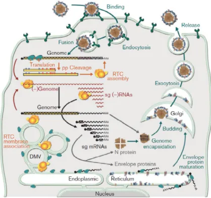
Figure 2: Arterivirus replication cycle inside cells.
Taken from Snijder and collaborators (113). Followed entry via receptor-mediated endocytosis the viral genome is released from the early endosome to the cytoplasm. There the ORF1a and ORF1b are translated to lead the formation of the polyproteins and the subsequent mature nsps are formed, which trigger the formation of the replication-transcription complex (RTC), which
19
is associated with double membrane vesicles (DMVs). RTC supports genome replication and transcription. The sg mRNAs are translated to obtain the different structural proteins. Once the new genomic RNAs (gRNAs) and the structural proteins are synthesised, the virus is assembled. First, genome encapsidation is triggered and then, the nucleocapsid buds to the smooth ER/Golgi complex (where the envelope proteins are retained) to get the viral envelope. Then, the new viral particles are accumulated into vesicles and are released by exocytosis. PRRSV uncoating
This stage is when the viral RNA genome is released from the early endosome to the cytoplasm. According with the model proposed by Van Breedam and colleagues (164), this process is critically dependent on the acidic pH of the endosome and on the interaction with the CD163 receptor (13, 173). GP2 and GP4 are the structural proteins responsible for this interaction with the scavenger receptor (142). Additionally, it was demonstrated that aspartic protease cathepsin E is involved in PRRSV uncoating stage (181). In MARC-145, PRRSV uncoating was clearly demonstrated to be also dependant on endosome acidification (182). CD151, a host cellular protein, interacts with PRRSV 3’ untranslated region (UTR) RNA and may be involved in viral envelope fusion with the endosome (183). CD163 is also necessary for PRRSV infection in MARC-145 cells (173). However, the exact action mechanism of both receptors is yet unknown.
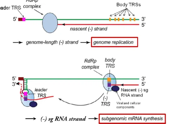
Genome replication and transcription
This stage of PRRSV replication cycle is produced in double membrane vesicles (DMVs) present in the cytoplasm and recently was suggested that probably they are autophagosome-like DMVs (184, 185). Then, once the gRNA is released into the cytoplasm, it will act as an mRNA and the host translational machinery will translate the ORF1a and ORF1b to obtain the polyproteins, which are then cleaved by the internal proteases to obtain the mature nsps. Subsequently, these nsps trigger the formation of the RTC that is associated with the DMVs, which are the presumably site of PRRSV replication and transcription (18, 19). The RTC directs both genome replication and transcription. The replication includes the continuous synthesis, by the RNA-dependent RNA polymerase (RdRp), of negative (-) full-length RNA strands using as template the gRNA and then these (-) RNA strands will lead the formation of
20
new gRNAs (Figure 3). The transcription process is the synthesis of a nested set of six sg mRNA, where all these sg mRNAs are 5’- and 3’-coterminal with the gRNA (112, 186). Then, the question of how these sg mRNAs are formed, has been subjected to many hypotheses and the most accepted is the model proposed by Sawicki and Sawicki (22), which is probably common within Nidovirales order. This model proposes that the sg mRNAs are synthesized from (-) sg RNA strands, which are produced by a discontinuous RNA synthesis process (Figure 3). Conserved transcription regulatory sequences (TRSs) are found preceding each structural protein ORFs termed as body TRSs in the gRNA. The same sequence is also present at the 3’ end of the leader sequence (5’ end of gRNA) and is denominated as leader TRS (187). The synthesis of (-) sg RNA strands begin at the 3’ end of the gRNA, then the elongation (by the RdRp) of the nascent (-) sg RNA strand will follow until the first body TRS appears. Subsequently, the synthesis is attenuated and the nascent (-) sg RNA strand, which carry in its 3’ end the complementary sequence to the body TRS, is relocated to the 5’ end of the gRNA. There, the complementary body TRS sequence of the nascent (-) sg RNA strand and the leader TRS sequence will be complementary and the nascent (-) sg RNA strand will be elongated by copying the 5’ end of the gRNA. Finally, the complete (-) sg RNA strands will serve as template for the synthesis of the sg mRNAs. These sg mRNAs are then translated to form the different structural proteins.
21
Figure 3: Model for Arterivirus genome replication and transcription.
Adapted from Nedialkova and colleagues (21). The genome replication (top of the figure) occurs by a continuous RNA synthesis process (leaded by the RdRp complex), where the new gRNAs are obtained from (-) full-length RNA strands. The second part of the figure represents the discontinuous synthesis of the sg mRNAs, which are obtained from (-) sg RNA strands. The extension of the (-) sg RNA strands begins at the 3’ end of the gRNA and is attenuated when the body TRSs appear (yellow rectangle). The nascent (-) sg RNA strand, porting in its 3’ end the complementary sequence of the body TRS (violet rectangle), is transferred to the 5’ end of the gRNA and will form complementary base pairs with the leader TRS (pink rectangle). Following this union, the elongation of the nascent (-) sg RNA strand is finished and is used as template for the synthesis of the sg mRNAs.
Virion assembly
Virion assembly is believed to begin in the replication site, where de genome and the N protein should interact to form the nucleocapsid (188) and finished in the endoplasmic reticulum (ER) or Golgi complex, where the envelope proteins are retained (113, 189, 190). Therefore, the preformed nucleocapsid is wrapped by the smooth ER/Golgi complex to acquire the viral envelope, a process known as budding. Although the exact mechanism of
22
virus assembly is not known yet, the formation of GP5/M heterodimer is believed to be determinant in this stage (153, 191).
Virus released
It is beleived that PRRSV virions leave the infected cells by exocytosis (2, 192). The new formed viral particles are accumulated into intracellular vesicles and finally are released by exocytosis (193). This last stage in PRRSV life cycle was demonstrated to be also dependant on cellular cytoskeleton (23).
VIRAL PATHOGENESIS IN VIVO
PRRSV can be transmitted horizontally (between infected and naïve animals) and vertically (from sows to the fetuses) and also via the semen of infected boars (194-198). When PRRSV enters the organism (via intranasal, oro-nasal or intramuscular route), it first replicates in the respiratory tract, probably in PAM (198). Afterwards, viremia is developed, as observed in inoculated young pigs (1 to 2 months old), between 3 to 14 days post infection (dpi). After this time, PRRSV persistence (though a “smoldering” infection, where the virus replicates at a low level) was detected in lung lymph nodes and tonsil tissues up to 156 dpi (199). In most cases, the infection is cleared by 156 dpi or shortly after. In young or growing and finishing pigs, the clinical sigs are mainly anorexia, lethargy, cutaneous hyperemia, dyspnea, reduced weight gain and an increase in mortality from secondary infections (200). In infected pregnant sows, PRRSV probably enters into the endometrium during viremia, which probably passes through the placenta and then, can infect fetuses (197, 201). It was demonstrated that the congenital infection is mainly restricted to the end of gestation, probably because there are high number of PRRSV susceptible cells in placenta, late in the gestation (202). The reproductive failure is characterized by late-term abortions, premature farrowings, stillborn fetuses, mummified fetuses and live weak born pigs (203). The molecular bases of PRRSV pathogenesis are not clear at all. However, several studies have demonstrated that PRRSV replication induces apoptosis in infected and in bystander uninfected cells both in vivo and in vitro. For instance, it was demonstrated that PRRSV induces cell death by apoptosis in the endometrium and placenta in late gestation (201), which probably can justify the reproductive failure associate with the disease. Moreover, PRRSV induces apoptosis in PAM and in pulmonary intravascular
23
macrophages (PIM) and is able to interfere with the macrophage phagocytic activity, leading the organism susceptible for opportunistic secondary infections (204). In infected MARC-145 cells, it was demonstrated that PRRSV induces, early in infection, antiapoptotic mechanisms, probably to favor its replication, but later cells die by apoptosis (144, 205).
IMMUNE RESPONSES Innate immune response
During PRRSV infection in PAM, it has been suggested that the virus and the toll-like receptors (TLR) interact. These receptors constitute an early host defense against invading pathogens, since they recognize specific molecular patterns present in the microbes. Stimulation of TLR3, TLR7, TLR8 and TLR9 lead the induction of the type I interferon (IFN) (206), which constitute key cytokines against viruses infections (207). TLR3 is activated by double-stranded (ds) RNA and is well known that during PRRSV genome replication there is formation of dsRNA, then, it is believed that PRRSV eventually interacts with this receptor (149). It has been proposed that PRRSV is able to evade TLR3 signaling pathway in PAM, since it was clearly demonstrated that the induction of the TLR3 using a dsRNA synthetic molecule (poly I: C) increased the level of IFN-alpha (IFN-α), which suppressed PRRSV infection (208), however its susceptibly to IFN-α differ among isolates (209). In contrast, it was observed that PRRSV suppress type I IFN expression in poly I:C treated MARC-145 cells (210). Therefore, PRRSV has developed different strategies to evade the antiviral effects of type I IFN. To date, PRRSV is able to inhibit type I IFN synthesis [by interfering with the functions of ISP-1 (IFN-beta promoter stimulator 1) (139), IRF3 (IFN regulatory factor 3) (129), NFκB (transcriptional regulator nuclear factor-κB) (124, 211) and CREB ((cyclic AMP responsive element binding)-binding protein (CBP)) (212)] and type I IFN signaling [by affecting JAK-STAT signaling pathway (39) and IFN-stimulated response elements such as the ISG15 (interferon-stimulated gene 15 ) (122, 123)]. Different studies have revealed that the pro-inflammatory cytokines such as TNF- α (213, 214) and IL-6 (215, 216) can be up or downregulated during PRRSV infection and IL-8 is highly expressed (217, 218). Most of the studies, in vivo or in vitro, demonstrated that PRRSV can induce the mRNA and protein expressions of the pleiotropic IL-10, which is a potent immunosuppressive cytokine that is
24
believed to play a key role in the immunopathogenesis of PRRSV (170, 219). Moreover, it was recently discovered that PRRSV IL-10 induction depends on NFκB activation and P38 mitogen-activated protein kinase (220).
Adaptive response
PRRSV induces high antibody responses which started at around 5 dpi and can last until 56 dpi and all challenged animals are seroconverting at 14 dpi (215, 221, 222). The antibodies are predominately directed against the glycoproteins, M, N and nsps (nsp1α, nsp1β, nsp2 and nsp7), where N protein induces the strongest response (117, 118, 149, 158). However, most of these are non-neutralizing antibodies (Non-NAbs) and is proposed that they (mainly the antibodies directed against GP5 and N protein ) may enhance viral infection by a phenomenon termed as antibody-dependent enhancement (ADE) (223, 224). In ADE, the opsonised virus, by the Non-NAbs, is delivered into the macrophages, which allow virus replication. NAbs appear late in PRRSV infection, around the fourth week pi and the titers are usually low (158, 215). The NAbs are generally directed against the GP3, GP4, GP5 and M, but is believed that GP5 possess the major neutralizing epitope in its ectodomain (148, 149, 225). In vitro studies demonstrated that NAbs are able to block PRRSV internalization (14, 226) , however it is not clear why in vivo they appear late and theirs titers remain lows. Several hypotheses have been postulated to explain it and one of the most accepted phenomenon is the presence of a decoy epitope in the GP5 ectodomain (225). Two epitopes were detected in GP5 ectodomain, named A and B. A is the immunodominant epitope and B has neutralizing activity and it was proposed that A may act as a decoy epitope, which interferes with the immune response against B and then cause a delay in the apparition of NAbs. The other proposed hypothesis is related with the number of N-glycosylations residues around the neutralizing epitope in GP5, which interfere with the recognition of the epitope by NAbs, a phenomenon known as glycan shielding (147).
The cell-mediated immune response face to PRRSV infection has not been well explored. CD4+, CD8+ and CD4+/CD8+ T cells have been detected during PRRSV infection and their responses are directed mainly against GP4, GP5, M and N (113, 149). IFN-γ-inducing epitopes have been identified into these structural proteins in addition to nsp2, nsp5, nsp9 and
25
nsp10 (149, 227, 228). IFN-γ produced by T-cells against PRRSV appears around 8-10 weeks pi and increase gradually after 3-4 months pi or post-vaccination (221). The induced IFN-γ seems to be insufficient to reduce the infection in vivo. However, a pre-treatment of MARC-145 and PAM cells with IFN-γ clearly reduced PRRSV infection, probably by the induction of cellular protective immunity (229, 230). Also, it was demonstrated that following PRRSV infection, the expression of MHC II (major histocompatibility complex class II) is decreased (163).
PRRSV CONTROL AND ELIMINATION
PRRSV current management strategies include surveillance, for instance: avoid introduction of contaminated semen into the herd, pig’s clinical examination and blood samples analysis, surveillance of pig’s production to detect possible reproductive problems, the implantation of severe biosecurity measures within the herd. Once the virus is already inside the farm, different measures have been described to eliminate it, such as: test and removal, whole herd depopulation and repopulation, herd closure and vaccination (25). At present, vaccination partially prevents PRRSV infection. There are two types of commercially available vaccines, the modified-live virus (MLV) vaccine and the killed-virus vaccine (24, 26). Adaptive response against MLV vaccines is weak and late (24, 149). However, they can offer an effective protection in reducing the reproductive and respiratory sigs and lesions associated with the disease (24, 231, 232). Nevertheless, the MLV vaccines efficacy has been questioned since they are genotype-specific vaccines or even strain-specific vaccines, which make them partially ineffective face to heterologous field strains (233). Another aspect that put in doubt the MLV vaccines is their safety, since their reversion to virulence it was clearly proved, through recombination with field isolates (234). The killed-virus vaccines are safe, but less effective or ineffective in inducing protection (26, 235). Additionally, another problem found was that vaccinated pigs cannot be differentiated from pigs naturally infected (133). For these reasons, there are continuous efforts in order to find the perfect safe and effective PRRSV vaccine. In this sense, several alternative vaccines have been created such as bacterial vector vaccines (236), DNA vaccines (237), plant-derived vaccines (238), multistrain vaccines (239), autogenous inactivated PRRSV vaccines (240) and others.
26 3. BACKGROUND OF THIS THESIS
Together with the current novel vaccine strategies against PRRSV, other researchers have been focusing in finding PRRSV antiviral compounds, which can be an alternative and also effective strategy to prevent or control PRRSV infection. Accordingly, recent published works showed a few natural compounds with antiviral activities against PRRSV, as glycosides, terpenoids, coumarins, isoflavones, peptolides, alkaloids, flavones, macrolides (27), N-acetylpenicillamine (28), sodium tanshinone IIA sulfonate (33), morpholino oligomer (30, 31), flavaspidic acid AB (241), Matrine (32), dietary germanium biotite (242), Cryptoporus volvatus extracts (29), etc. Each of these compounds inhibits PRRSV replication differently. For instance, the flavaspidic acid AB inhibits PRRSV internalization and the cell-to-cell transmission, probably by the induction of type I IFN (241). Sun and colleagues demonstrated that Matrine inhibits N protein expression and has antiapoptotic functions (32). Moreover, Cryptoporus volvatus extract was demonstrated to inhibit PRRSV infection in vitro and in vivo, probably by the direct inhibition of PRRSV polymerase (RdRp) activity (29). Despite all these efforts, there are no effective commercially available antiviral drugs to prevent PRRSV infections.
A recent research performed in our laboratory demonstrated that the culture supernatant of a mutant App (AppΔapxICΔapxIIC) strain has a potent antiviral activity against PRRSV ((37), Annexe I).This strong antiviral effect was observed in the newly discovered SJPL permissive cell line, but was almost ineffective in MARC-145 infected cells. Interestingly, this phenomenon was also observed in the primary target cells of PRRSV, the porcine alveolar macrophages (results corresponding to the first objective of this project). Since it is believed that the bacterial antiviral effect against PRRSV is via the modulation of cellular(s) component(s), this thesis has as second goal to identify the possible mechanisms used by App to inhibit PRRSV infection in PAM.
27
CHAPTER II: Actinobacillus pleuropneumoniae blocks porcine reproductive and respiratory syndrome virus replication prior to its genome replication and transcription.
Yenney Hernandez Reyes, Chantale Provost, Carolina Kist Traesel, Mario Jacques, Carl A. Gagnon*
28
AUTHOR CONTRIBUTIONS
As mentioned in the introduction section, this thesis has two objectives. The results from the first objective, which is to demonstrate the App cell culture supernatant antiviral effect against PRRSV in porcine alveolar macrophages (PAM), were already published by Levesque and coworkers (24) (see annexes section, specifically Annexe I: Figures 3 and 4).
The results concerning the second objective of my work that is to identify the possible mechanisms used by App cell culture supernatant to inhibit PRRSV infection in PAM cells in vitro will be discussed in this article. Also, we have included the SJPL and MARC-145 cells in order to compare the results.
My contribution to this study was in performing almost all tests described in this paper, which include: cell viability and mortality tests, type I and II IFN and β-actin mRNA expressions by qRT-PCR, PRRSV genome replication and transcription kinetics assay by qRT-PCR, western blot, IFA and viral titer determination. Dr. Chantale Provost helped me in interpreting and analysing the Kinex™ Antibody Microarray results and in performing the PAM collecting technique from lungs. I followed the technical advices of Dr. Chantale Provost and my professor’s suggestions.
29 ABSTRACT
Current management strategies are inadequate for long term control of PRRS, which justifies the search of novel strategies to control the disease. Recently, a strong antiviral acitvity of Actinobacillus pleuropneumoniae (App) cell culture supernantant against PRRSV in PAM and SJPL infected cells was discovered. Following this finding, the objective of the present study was to understand how App inhibits PRRSV replication. First, cells were treated with App before and after PRRSV infection. At different times post-infections, viral genome replication and transcription were measured in the presence of App. Type I and II interferon (IFN) mRNA expression and proteins expression modulation of PRRSV infected PAM cells treated with App were evaluated using qRT-PCR and the KINEX™ Microarrays assays, respectively. The expression of some modulated proteins were subsequently, confirmed by immunofluorescence (IFA) and western blot assays. Results showed that type I and II IFN mRNA expressions were not modulated in the presence of App. Moreover, it was observed that App inhibits PRRSV infection before the first cycle of genome replication and transcription, indicating that App antiviral effect against PRRSV take place at an early step during PRRSV infection. The proteomic experiments revealed an increase of cofilin expression (a protein that regulates actin cytoskeleton dynamics) in the presence of App, which was further confirmed by western blot. Subsequently, a diminution of actin filaments was demonstrated by IFA. Interestingly, the treatment with cytochalasin D (an actin polymerization inhibitor) revealed the same effect on PRRSV replication than App suggesting that App antiviral effect against PRRSV may take place via the activation of cofilin which provokes actin depolymerisation and subsequently, probably affects PRRSV endocytosis.
30 INTRODUCTION
Porcine reproductive and respiratory syndrome (PRRS) is considered a worldwide endemic disease which causes significant economic losses in pig-producing countries. The causative agent, PRRS virus (PRRSV), belongs to the family Arteriviridae of the Nidovirales order. PRRSV is an enveloped single-stranded positive sense RNA virus of approximately 15 kb in length that encodes for at least 10 open reading frames (ORFs) (1, 2). PRRSV has a strongly restricted cell tropism for the monocyte–macrophage lineage in vivo. The primary target cells for PRRSV infection in vivo are the fully differentiated porcine alveolar macrophages (PAM), which are often used for in vitro study of host cell infections (3-6). In the literature, the only two continuous cell lines non-genetically modified able to fully replicate PRRSV are: African green monkey kidney cell line MA-104 and its derivatives such as MARC-145 (7) and the newly reported SJPL cells (8, 9).
Following PRRSV entry and release of the viral genome into the cytoplasm, the PRRSV ORF1 is translated and the resulting non-structural proteins trigger the formation of the replication-transcription complex, which is associated with double membrane vesicles and supports genome replication and transcription processes (10-12). The genome replication is produced by the continuous synthesis of negative (-) full-length RNA strands using as template the positive genomic RNA [(+) gRNA], then the (-) RNA strands will lead the formation of new (+) gRNAs (13). The genome transcription process is the synthesis of a nested set of six sub-genomic mRNAs (sg mRNAs). According to a model proposed by Sawicki and colleagues (14), the generation of these sg mRNAs is through a discontinuous RNA synthesis process, where (-) sg RNA strands are produced and then are used as template for the synthesis of the sg mRNAs.
Current management strategies, which focus on the prevention of PRRSV infection (ex. surveillance and removal, whole herd depopulation and repopulation, herd closure (15), etc.) and vaccination using commercially available modified live-attenuated vaccines or autogenous killed vaccines, have usually been demonstrated to be inadequate for long-term control of PRRS (16). This supports the search of novel strategies to control PRRSV infection. Recent published works have reported the discovery of natural compounds that possess antiviral