Dendritic Cell Differentiation and Immune Tolerance to
Insulin-Related Peptides in Igf2-Deficient Mice
1
Isabelle Hansenne,
2* Chantal Renard-Charlet,* Roland Greimers,
†and Vincent Geenen*
There is some evidence that insulin-like growth factor 2 (IGF-2) may intervene in the control of T cell differentiation. To further study the immunoregulatory function of this growth factor, we analyzed the immune system of Igf2ⴚ/ⴚmice. Phenotypically, some immunological parameters such as lymphoid organ morphology and cellularity were unaltered in Igf2ⴚ/ⴚmice, but an increase of CD8ⴙcells and a decrease of B220ⴙcells were observed in spleen. In vitro, the development of bone marrow-derived dendritic cells was affected by the absence of Igf2 expression. After maturation, a higher percentage of immature dendritic cells was observed in Igf2ⴚ/ⴚpopulation, together with a secondary decrease in allogenic T cell proliferation. Activation of T cells was also affected by the lack of expression of this growth factor. The profile of B cell response in mutant mice immunized with IGF-2 evidenced a T-dependent profile of anti-IGF-2 Abs that was absent in Igf2ⴙ/ⴙmice. The influence of IGF-2 upon tolerance to insulin was also assessed in this model, and this showed that IGF-2 also intervenes in tolerance to insulin. The presence of a T-dependent response in Igf2-deficient mice should allow cloning of specific “forbidden” T CD4ⴙlymphocytes directed against IGF-2, as well as further investigation of their possible pathogenic properties against insulin family. The Journal of Immunology, 2006, 176: 4651– 4657.
I
nsulin-like growth factors 1 and 2 (IGF-1/2)3are members ofthe insulin hormone family that exert a prominent role in fetal and postnatal development (1, 2). Null mutant mice for Igf1 show a marked growth deficiency with 60% of normal body weight, a variable neonatal lethality frequency depending on ge-netic background, and they are infertile (3). The absence of Igf2 induces similar growth failure, but Igf2⫺/⫺mice are viable and fertile (4, 5).
With regard to the implication of IGF in immune physiology and development, most of the studies have focused on the growth hormone/IGF-1 axis and type 1 IGF receptor (IGF-1R), a trans-membrane tyrosine kinase homologous to the insulin receptor that mediates most of the biological effects of IGF-1 and IGF-2 (6, 7). Normal development and ex vivo activation of T and B cells are observed in chimeric Rag2-deficient C57BL/6 mice reconstituted with fetal liver cells from Igf1r⫺/⫺ mice. However, this model revealed an unexpected decrease of the T-independent B cell re-sponse which is important in bacterial defense mechanisms (8).
So far, very few studies have investigated the function of IGF-2 in immune development and physiology. This growth factor is the dominant peptide of the insulin family expressed in the thymus epithelium of different species (9, 10). Thymic IGF-2 influences thymic development and T cell differentiation as evidenced by analysis of IGF-2 transgenic dwarf mice, which develop a thymic hyperplasia (11) with an increased number of thymocytes (and CD4⫹T lymphocytes in particular) (12, 13). This increase of T cells is also observed in the spleen compartment of IGF-2 trans-genic mice, but there is no significant effect on B cell development. In vitro, T cell differentiation and proliferation are also impaired in fetal thymic organ culture treated with anti-IGF-2, anti-IGF-1R, or anti-IGF-2R. In these experimental conditions, T cell differentia-tion is inhibited at early CD4⫺CD8⫺stage, whereas anti-IGF-2 induces an increase in thymic CD8⫹T cells suggesting a role for IGF-2 in T cell final commitment to CD4 or CD8 lineage (14). Finally, a role of thymic IGF-2 in central immune self-tolerance of the insulin family has been suggested by the observation of a de-fect of Igf2 transcription in the thymus of Bio-Breeding (BB) rats, an animal model of type 1 diabetes (15).
In this study, we further investigate the role played by IGF-2 in the immune function through the use of Igf2-deficient mice. Al-though Igf2 deficiency does not interfere with the development of major lymphoid structures, bone marrow (BM)-derived dendritic cells (DC) from Igf2-deficient mice show a defect of maturation as evidenced by a higher persistence of immature DC (iDC) com-pared with wild-type (wt) mice. This is associated with a decrease of ability of Igf2⫺/⫺DC to activate allogenic T lymphocytes in MLR. T cell proliferation assessed by MLR is also severely im-paired. The profile of the immune responses against IGF-2 and insulin in mutant mice was also evaluated and shows that tolerance to IGF-2 depends on Igf2 expression, but that IGF-2 also contrib-utes to tolerance to insulin.
Materials and Methods
Animals
Igf2⫹/⫺mice were provided by Dr. A. Efstratiadis and were bred at the Animal Department of Liege University under conventional conditions
*Center of Immunology and†
Department of Anatomy and Cytopathology, University of Liege, Liege-Sart Tilman, Belgium
Received for publication October 17, 2005. Accepted for publication February 3, 2006.
The costs of publication of this article were defrayed in part by the payment of page charges. This article must therefore be hereby marked advertisement in accordance with 18 U.S.C. Section 1734 solely to indicate this fact.
1
This work was supported by the Fonds pour la Recherche en Industrie et en Agrono-mie (Belgium) and by the Fondation Le´on Fredericq pour la Recherche Biome´dicale (Liege Medical School and Liege University Hospital). These studies were supported by the National Fund of Scientific Research (Belgium), by the Fondation Vaugrenier pour la Recherche en Tole´rance (Geneva), by the European Association for the Study of Diabetes (Du¨sseldorf), by TOLEDIAB (Walloon Region), and by the European Union (FP6 Integrated Project Euro-Thymaide LSHB-CT-2003-503410).
2
Address correspondence and reprints requests to Dr. Isabelle Hansenne, Center of Immunology, Institute of Pathology CHU-B23, B-4000 Liege-Sart Tilman, Belgium. E-mail address: Isabelle.Hansenne@ulg.ac.be
3
Abbreviations used in this paper: IGF-1/2, insulin-like growth factor-1/2; IGF-1R, type 1 IGF receptor; IGF-2R, type 2 IGF receptor; DC, dendritic cell; iDC, immature DC; wt, wild type; mDC, mature DC; BM, bone marrow; TEC, thymic epithelial cell; rm, recombinant murine.
biopsies. BALB/c mice were also bred under the same conditions. Male and female mice were 5– 8 wk old at the time of experiments, which were performed in agreement with the local ethic committee.
Phenotype of mice
Igf2⫺/⫺and wt mice were weighed and were sacrificed by cervical dislo-cation. The thymus and the spleen were removed and weighed. The number of living cells was estimated by trypan blue dye exclusion.
Generation of BM-derived DC
BM-derived DC were isolated and differentiated according to the method developed by Lutz et al. (16). Briefly, Igf2⫺/⫺and wt mice were sacrificed by cervical dislocation and femur/tibiae were removed and freed of mus-cles and tendons. Both ends were cut with scissors and BM cells were flushed with DPBS without Ca2⫹and Mg2⫹(Cambrex) using a syringe with a needle 0.45 mm in diameter. Cell suspension was filtered through a 70-m cell strainer (BD Biosciences) to remove remaining bone frag-ments. At day 0, the cells were seeded at 2⫻ 105cells/ml in 100-mm petri
dishes (Falcon no. 1029; VWR International) and 10 ml per dish. The medium used during differentiation (10 days) to obtain iDC was RPMI 1640 supplemented with penicillin (100 U/ml), streptomycin (100g/ml),
L-glutamine (2 mM) (Cambrex), 2-ME (50M), 10% heat-inactivated FCS (Invitrogen Life Technologies) and recombinant murine (rm) GM-CSF (20 ng/ml, personal production). The vector pcDNA GM-CSF was a gift from J.-C. Renauld (Universite´ Catholique de Louvain, Brussels, Belgium) and rmGM-CSF was produced by COS-7 transfection using FuGENE 6 trans-fection reagent (Roche) according to the manufacturer’s instructions. At day 3, an additional 10 ml of complete medium containing rmGM-CSF (20 ng/ml) per dish were added, and at day 6 and 8, 10 ml were collected, centrifuged at 200⫻ g for 7 min. The cell pellet was resuspended in fresh complete medium with rmGM-CSF (20 ng/ml) and was plated again. At day 10, the maturation to mDC was performed in 100 mm tissue culture petri dishes (Falcon no. 3003; VWR International) with the same medium containing rmGM-CSF (10 ng/ml) and LPS (2.5g/ml, Escherichia coli O26:B6; Sigma-Aldrich) for 24 or 48 h. For this step, the cells were seeded at 1.5⫻ 105cells/ml and 10 ml per plate.
RNA isolation
Total RNA was extracted from BM cells, iDC, mDC, and 16-day-old fetal brains using RNeasy Mini kit (catalog no. 74104; Qiagen) according to the manufacturer’s instructions. Pancreas and liver total RNA was isolated using Tripure isolation reagent (Roche). DNA contamination was removed by treatment of samples with RNase-free DNase (Roche). RNA was dosed with the RiboGreen RNA quantitation kit (Molecular Probes).
Actin, Insulin 1 (Ins1), Insulin 2 (Ins2), Igf1, Igf2, Igf1r, Igf2r RT-PCR and sequencing
Total RNA (250 –500 ng) was reverse transcribed in a total of 20l by the 1st Strand cDNA synthesis kit for RT-PCR (AMV; Roche) using oli-go(dT)primer. Reverse-transcription products (1:20) were used directly for PCR using FastStart TaqDNA polymerase (Roche) according to the man-ufacturer’s instructions in a UNO II rapid thermocycler (Biometra). After
cycles comprising 94°C for 45 s, variable temperature depending on prim-ers for 45 s and 72°C for 45 s. To finish, an additional elongation step was performed at 72°C for 7 min. PCR products were analyzed in 2% agarose ethidium bromide-staining gels. The sequence of primers and the respec-tive temperature are presented in Table I. The PCR products were se-quenced (Genome Express) using their respective primers.
Flow cytometry
Immature DC, mDC, fresh thymocytes, BM cells, and splenic cells (5⫻ 105-106) were centrifuged at 200⫻ g for 5 min and prepared in 100l of
DPBS (Cambrex) for labeling. Cell preparations were stained for 20 min at 4°C and washed one time in DPBS.
Flow cytometric analyses were performed with FACStarPlus(BD
Bio-sciences). Abs (BD Biosciences) were CD86-PE clone GL1 (rat IgG2a), CD80-PE clone 16-10A1 (Armenian hamster IgG), Gr-1/Ly6G-PE clone RB6-8C5 (rat IgG2b), I-A/I-E-PE clone M5/114.15.2 (rat IgG2b), CD11c-FITC clone HL3 (Armenian hamster IgG), CD8␣-FITC clone 53-6.7 (rat IgG2a), CD45R/B220-PE clone RA3-6B2 (rat IgG2a), and CD4-PE clone RH4-5 (rat IgG2a). The data analysis was performed using CellQuest soft-ware (BD Biosciences).
The MLR
The capacity of DC from wt and Igf2⫺/⫺ mice on T cell activation was evaluated in a primary allogenic MLR. DC were harvested after 24 h of maturation with LPS and used as T cell stimulators after 3000 rad of gam-ma-irradiation. BALB/c spleen cell responders were prepared by mechan-ical disruption, erythrocyte lysis (Hybrid-Max; Sigma-Aldrich), and filtration through a 70-m cell strainer (BD Biosciences). The MLR was performed with 106responders⫻ 105irradiated stimulators/ml in RPMI
1640 supplemented with 100 U/ml penicillin, 100g/ml streptomycin, 2 mML-glutamine, 1 mM sodium pyruvate, 1% MEM nonessential acid, 10 mM HEPES (Cambrex), 50M 2-ME (Invitrogen Life Technologies), and 10% heat-inactivated FCS (Invitrogen Life Technologies). The cells were incubated in 5% CO2at 37°C.
Igf2⫺/⫺and wt T cell proliferation was also assessed by MLR. BALB/c BM-derived DC were used as stimulators after 3000 rad of gamma-irra-diation. Igf2⫺/⫺and wt T cell responders were prepared by mechanical disruption, erythrocyte lysis (Hybrid-Max; Sigma-Aldrich), and filtration through 70m cell strainer (BD Biosciences). The MLR was performed with 106responders⫻ 105gamma-irradiated stimulators/ml in RPMI 1640
supplemented with 100 U/ml penicillin, 100g/ml streptomycin, 2 mM
L-glutamine, 1 mM sodium pyruvate, 1% MEM nonessential acid, 10 mM HEPES (Cambrex), 50M 2-ME (Invitrogen Life Technologies), and 10% heat-inactivated FCS (Invitrogen Life Technologies). The cells were incu-bated in 5% CO2at 37°C.
After 0, 48, and 96 h, 800l of culture were collected and distributed in triplicate on 96-well flat-bottom (200l/well). Then, 0.5 Ci of [3
H]thy-midine (Amersham Biosciences) was added to each well for an additional 18 h of culture. Cellular DNA was harvested using a Titertek cell harvester (Flow Laboratories), and scintillation counting was performed in a Beck-man liquid scintillation counter (BD Biosciences). wst-1 tests were per-formed with 10 l of wst-1 Cell Proliferation Reagent (Roche) for an additional 4 h of culture. The absorbance was measured at 450 – 620 nm.
Table I. Synthetic primers selected for RT-PCR and sequencing
Genes Strand Primer Sequence
Annealing T°
PCR Products
Size (bp) Reference
Ins1 Sense AGACCATCAGCAAGCAGGTC 65°C 324 17 Antisense CTGGTGCAGCACTGATCCAC
Ins2 Sense GTGGAGGACCCACAAGTGG 65°C 198 17 Antisense ATTCATTGCAGAGGGGTAGGCT
Igf1 Sense GCTGAGCTGGTGGAATGCTCTTCAGTTC 55°C 215 18 Antisense CTTCTGAGTCTTGGGCATGTCAGTGTG
Igf2 Sense GAGCTTGTTGACACGCTTCAGTTTGTC 55°C 356 18 Antisense ACGTTTGGCCTCTCTGAACTCTTTGAG
Igf1r Sense GACATCCGCAACGACTATCAG 50°C 395 19
Antisense GTAGTTATTGGACACCGCATC
Igf2r Sense CTGGAGGTGATGAGTGTAGCTTGGC 55°C 235 20
Antisense GAGTGACGAGCCAACACAGACAGGTC
Actin Sense TAAAGACCTCTATGCCAACACAGT 54°C 250 21
Mice immunization
Igf2⫺/⫺and wt mice were injected i.p. with 5g of rmIGF-2 (R&D Sys-tems) or with 5g of human insulin (Roche) to study the humoral response and with 50g of murine IGF-2-derived B11-25 (NEOSYSTEM) to an-alyze the cellular response. CFA (Sigma-Aldrich) (v/v) is used for the first injection and IFA (Sigma-Aldrich) (v/v) for the other injections. For hu-moral response, three injections separated by 20 days were performed. Blood test was performed 15 days after each injection in agreement with the Liege University ethic committee. For cellular response, two injections separated by 15 days were performed.
Dosage of anti-IGF-2 and anti-insulin Abs
Abs against IGF-2 and insulin were detected and quantified using a specific ELISA procedure. Microplates (Nunc Maxisorp 468667; VWR
Interna-tional) were incubated overnight at 4°C with 2 g/ml rmIGF-2 (R&D Systems) or 5g/ml human insulin (Roche) in 100 l of 0.1 M NaHCO3
(pH 8.4). From this step, the procedure was identical between the two kinds of ELISA. SL buffer (0.01 M phosphate buffer, pH 7.4) was used as the basic buffer, and all reactions were incubated at 37°C under agitation. To block nonspecific binding, 200l of SL buffer containing 1% of BSA (Sigma-Aldrich) was added per well for 2 h. Four washes with 0.01 M phosphate buffer with 0.5‰ of Tween 20 (washing buffer) were performed, and the sera were diluted in SL buffer containing 0.5% BSA and 0.1% Tween 20 (VWR International) and incubated to 100l/well for 2 h. After four washes, the samples were incubated with peroxidase-labeled anti-mouse Abs diluted in the same buffer at a different concentration: 1/4000 for anti-IgG (Prosan), 1/2000 for anti-IgG1, 1/2000 for anti-IgG2a, 1/2000 for anti-IgG2b, 1/2000 for anti-IgG3, and 1/3000 for anti-IgM (Serotec). The incubation was performed in 100l for 1 h. The microplates were washed 4 times and 200l/well of substrate buffer (TMB; Biosource Eu-rope) were added for 20 min at room temperature under agitation. The reaction was stopped with 50l of 0.4 N HCl, and the absorbance was measured at 450 – 620 nm with a spectrophotometer.
Specific T cell culture
After immunization with murine IGF-2-derived B11-25, spleen cells were prepared by mechanical disruption, erythrocyte lysis (Hybrid-Max; Sigma-Aldrich), and filtration through a 70-m cell strainer (BD Biosciences). The culture was performed with 3 ⫻ 106spleen cells without or with
murine IGF-2-derived B11-25 (20g/ml) in Ex vivo-15 medium (Cam-brex) supplemented with 100 U/ml penicillin, 100g/ml streptomycin, 2 mML-glutamine, 1 mM sodium pyruvate, 1% MEM nonessential acid, 10
Table II. Comparison of surface phenotype on Igf2⫹/⫹and Igf2⫺/⫺ mDC (n⫽ 5 of each genotype) Igf2⫹/⫹ Igf2⫺/⫺ A CD11c⫹Ly6G⫺ 76.79⫾ 9.191 67.76⫾ 6.224* CD11c⫹Ly6G⫹ 9.10⫾ 3.660 19.18⫾ 6.116* B CD80low/int 21.45⫾ 12.85 37.22⫾ 13.94* CD80high 78.82⫾ 13.10 63.17⫾ 13.97* CD86low/int 19.60⫾ 12.01 39.21⫾ 16.62* CD86high 80.60⫾ 12.22 61.19⫾ 16.58* MHC-IIlow/int 21.34⫾ 17.56 38.94⫾ 15.45* MHC-IIhigh 78.68⫾ 17.53 61.16⫾ 15.34* *, p⬍ 0.05; mean (%) ⫾ SD.
FIGURE 1. Analysis of Igf2⫹/⫹
and Igf2⫺/⫺phenotype. A, Thymic and splenic weights of Igf2⫹/⫹and Igf2⫺/⫺ mice (n⫽ 4 of each genotype). Tissue weights were calculated in percentage of total body weight. B, Cellularity of
Igf2⫹/⫹(filled histogram) and Igf2⫺/⫺ (hatched histogram) in BM, thymus, and spleen. The number of Igf2⫺/⫺ cells was expressed in percentage of
Igf2⫹/⫹cells (100%) (n⫽ 8 of each genotype). C, Lymphoid cell sub-classes (%) in spleen of the two kinds of mice. These subclasses were ana-lyzed by flux cytometry (n⫽ 8 of each genotype). Mean ⫾ SD; *,
p⬍ 0.05.
FIGURE 2. Expression of insulin family genes and their receptors in BM (lane 1), iDC (lane 2), and mDC (lane 3) by RT-PCR. H2O served as
negative control (lane⫺). Liver and pancreas served as positive control (lane⫹) for Igf1 and Igf2, and Ins1 and Ins2, respectively. Fetal brain was the positive control (lane⫹) for Igf1r and Igf2r. M represented the m.w. marker (lane M). One experiment representative of three individual exper-iments is shown.
mM HEPES (Cambrex), and 50M 2-ME (Invitrogen Life Technologies). The cells were incubated in 5% CO2at 37°C.
IL-2 production
After 48, 72, and 96 h of MLR or after 24 and 48 h of specific T cell culture, 800l were removed of each condition and IL-2 production re-leased by Igf2⫺/⫺and wt T cells in MLR was assessed by specific ELISA (Biosource Europe).
Statistical analyses
One way ANOVA was used for MLR experiments (Newman-Keuls post-test) and t test was used to compare Igf2⫺/⫺and wt mice in cytometric experiments, IL-2 production, and Ab production after immunization. The level of significance was p⬍ 0.05.
Normal phenotype of lymphoid organs in Igf2-deficient mice Igf2⫺/⫺mice presented a significant reduced body weight (61.6% of normal body weight) when compared with wt mice. Thymus and spleen from mutant mice were not significantly affected by the deletion of this gene because they presented a similar size and morphology compared with wt organs (Fig. 1A). The cellularity of different lymphoid organs such as thymus, spleen, and BM, was also analyzed and did not show any significant difference between wt and Igf2⫺/⫺ mice. The number of Igf2⫺/⫺ cells in the three studied organs was reduced in the same proportion than the size reduction (60%) (Fig. 1B). In vivo analyses of lymphoid compart-ment (T and B lymphocytes) showed no significant difference in thymocyte subpopulations (data not shown). In spleen, T cell anal-ysis revealed no difference for CD4⫹CD8⫹ cells, CD4⫺CD8⫺ cells and CD4⫹CD8⫺cells but the CD4⫺CD8⫹population was significantly higher in mutant mice compared with wt mice. Igf2⫺/⫺mice also presented a significant decrease of B cell pop-ulation (B220⫹cells) (Fig. 1C).
Influence of IGF-2 on DC differentiation and maturation
Insulin-related genes and their receptors expressed by BM, iDC, and mDC. RNA expression of Igf1, Igf2, Ins1, Ins2, and their
receptors Igf1r and Igf2r was assessed by RT-PCR with specific primers. The positive controls were liver for Igf1 and Igf2, fetal brain for Igf1r and Igf2r, and pancreas for Ins1 and Ins2. Negative control was H2O. Igf1, Igf2, Igf1r, and Igf2r transcripts (215, 356, 395, and 235 bp, respectively) were detected in wt BM cells, wt iDC, and wt mDC extracts but, on the other hand, neither Ins1, nor Ins2 transcripts (324 and 198 bp, respectively) were found in wt DC samples contrary to positive controls (Fig. 2). The semiquan-titative PCR pattern suggested that Igf2 expression is up-regulated between iDC and mDC. Sequencing of Igf1, Igf2, Ins1, Ins2, Igf1r, and Igf2r PCR products confirmed the specificity and the selec-tivity of different primers. Each product presented 97–100% ho-mology compared with their respective GenBank sequence. All genes detected in wt samples were found in Igf2⫺/⫺ BM cells, Igf2⫺/⫺iDC and Igf2⫺/⫺mDC except Igf2 for which the expres-sion was severely decreased.
Surface phenotype of iDC and mDC is affected by deficiency of Igf2 expression. DC population was determined by analysis of
CD11c expression in flow cytometry. From day 0 to day 11, wt
FIGURE 3. Comparison of surface expression of MHC-II, CD80, and CD86 by iDC (A) and mDC (B) from wt (filled histograms) and Igf2⫺/⫺ mice (open histograms). Flow cytometric analysis of gated CD11c⫹cells represent the three markers implicated in Ag presentation and T cell acti-vation. After 10 days with rmGM-CSF, BM cells were differentiated to iDC. Maturation was performed with rmGM-CSF and LPS for 24 h. One experiment representative of five individual experiments is shown.
FIGURE 4. Implication of IGF-2 in DC activation capacity and in T cell proliferation. A, Igf2⫹/⫹and Igf2⫺/⫺DC capacity to stimulate BALB/c spleen cells in MLR. B, wt and Igf2⫺/⫺spleen cell proliferation in MLR with BALB/c DC stimulators. Proliferations was assessed by [3H]thymidine uptake and
measured after 48 and 96 h. Data are expressed as growth percentage vs time (h). Mean⫾ SD; *, p ⬍ 0.01, one experiment representative of three individual experiments is shown, each performed in triplicate (n⫽ 3 of each genotype). C, IL-2 production of Igf2⫹/⫹and Igf2⫺/⫺spleen cells after 48 h of culture (n⫽ 8 of each kind of mice).
CD11c⫹ cells increased in the same way than Igf2⫺/⫺CD11c⫹ cells to reach 84.75⫾ 10.75% and 86.94 ⫾ 10.46%, respectively, at mature stage. The percentage of CD11c⫹cell population from Igf2-deficient mice was identical than the CD11c⫹ population from wt mice. These CD11c⫹cells showed no CD8␣ expression during differentiation and maturation. Among mature CD11c⫹ cells, two populations were analyzed: CD11c⫹Ly6G⫺cells and CD11c⫹Ly6G⫹cells. The distribution of these classes was mod-ulated according to the origin of cells: the wt CD11c⫹Ly6G⫺ pop-ulation was significantly higher than Igf2⫺/⫺CD11c⫹Ly6G⫺ pop-ulation, and this was the opposite for CD11c⫹Ly6G⫹cells (Table II, part A). This difference was not observed at day 10 but only after maturation. CD11c⫺Ly6G⫹ population was also analyzed and showed no difference between wt and mutant mice after ferentiation and maturation (data not shown). Expression of dif-ferent molecules involved in T cell activation such as class II MHC (MHC-II), CD80, and CD86 was assessed by flow cytometry on CD11c⫹cells. After differentiation (Fig. 3A), wt iDC expressed a low or intermediate level of the three markers, with no significant difference with Igf2⫺/⫺iDC. In wt mice, LPS treatment promoted overexpression of these molecules during DC maturation (Fig. 3B). The persistence of iDC population with a lower expression of MHC-II, CD80, and CD86 was higher in Igf2⫺/⫺ culture com-pared with wt culture (Table II, B). This immature population in Igf2⫺/⫺culture was observed after 24 and 48 h of maturation.
Higher iDC population influences T cell activation. The influence
of a higher iDC population in Igf2⫺/⫺ mice on T cell activation was studied using [3H]thymidine incorporation assays (Fig. 4A).
After 48 h of MLR, a significant decrease in T cell proliferation with Igf2⫺/⫺DC was observed when compared with wt DC. The decreased proliferation with Igf2⫺/⫺DC was time persistent and still observed after 96 h of MLR.
IGF-2 influence on T cell proliferation and activation
The absence of Igf2 expression in T lymphocytes caused a signif-icant decrease of proliferation of spleen cells as shown by [3
H]thy-midine incorporation assays (Fig. 4B). A similar decrease was ob-served with wst-1 tests but did not reach the level of significance (data not shown). This reduction of proliferation was persistent
until 120 h of MLR. However, no significant difference in IL-2 production was observed between wt and Igf2⫺/⫺culture along the time of culture (Fig. 4C).
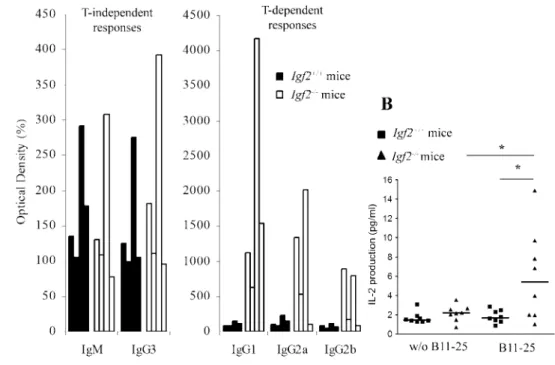
Characterization of anti-IGF-2-specific immune responses Igf2⫺/⫺and wt mice were immunized with IGF-2 to characterize anti-IGF-2-specific immune responses. The analysis of humoral response (Fig. 5A) showed that immunization of Igf2-deficient mice rapidly induced anti-IGF-2 Abs. This response was charac-terized by a T-independent response (both IgM and IgG3) and after the second injection, a T-dependent response with IgG1, IgG2a, and IgG2b production. Contrary to Igf2⫺/⫺ mice, wt mice pre-sented only IgM and IgG3 production with a similar titer, but no T-dependent response was observed.
FIGURE 5. Production of IGF-2-specific Abs and T cells by wt and
Igf2⫺/⫺mice after i.p. immunization.
A, Analysis of different isotypes: IgM
and IgG3 present the T-independent responses and IgG1, IgG2a, and IgG2b the T-dependent responses. Se-rial dilutions were performed, and data represent the percentage of the OD in preimmune serum at the dilu-tion 1/200 for IgM and IgG3, 1/500 for IgG2a and IgG2b, and 1/10,000 for IgG1. These histograms represent four representative wt mice and four representative Igf2⫺/⫺mice (n⫽ 8 of each kind of mice). B, IL-2 produc-tion by IGF-2-specific T cells from wt and Igf2⫺/⫺mice after two i.p. injec-tions of IGF-2-derived B11-25 and one in vitro stimulation; *, p⬍ 0.05 (n⫽ 8 of each kind of mice).
FIGURE 6. Insulin-specific humoral response in wt and Igf2⫺/⫺mice after i.p. immunization. The analysis of Ig production following injection was performed in mice with Ig production at day 0 lower than 1 in OD. Serial dilutions were performed, and data represent the percentage of the OD in preimmune serum at the dilution 1/100. Mean⫾ SEM; *, p ⬍ 0.05 (n⫽ 6/8 of each kind of mice).
tivated after immunization was characterized by IL-2 production with or without in vitro stimulation by IGF-2-derived B11-25 (Fig. 5B). In culture with murine IGF-2-derived B11-25, spleen cells from Igf2⫺/⫺mice present a significant increase of IL-2 produc-tion when compared with wt mice. This IL-2 producproduc-tion was spe-cific of B11-25 because cultures without B11-25 did not show any IL-2 secretion.
Characterization of anti-insulin specific humoral response Igf2⫺/⫺ and wt mice were immunized with insulin to evaluate anti-insulin-specific humoral response. We observed that Ig re-sponse following immunization was dependent of anti-insulin Ig level in preimmune serum. Mice with high insulin-specific Ig pro-duction before immunization did not display any response to in-jections (data not shown). We decided to consider mice with anti-insulin Ab level at day 0 lower than 1 in OD. Igf2⫺/⫺ mice presented a higher anti-insulin Ig-specific humoral response fol-lowing the three injections compared with Igf2⫹/⫹mice (Fig. 6).
Discussion
The main objective of this study was to evaluate the importance of IGF-2 in immune development and physiology. The absence of Igf2 expression does not influence lymphoid organ development as shown by a similar growth of thymus and spleen, as well as a cellularity in both organs and BM, which is identical in wt and Igf2⫺/⫺ mice. In Igf2-deficient mice, thymocyte development is not affected but, in the spleen, a significant increase of CD4⫺CD8⫹cells and a lower B220⫹population is observed.
We decided in a second step to investigate the effect of Igf2 deficiency on the maturation and function of DC, the most potent APC (22) implicated in polarization of the immune response, i.e., immunity vs tolerance (23, 24). It has been previously reported that IGF-1 promotes maturation and inhibits apoptosis of immature cord blood monocyte-derived DC (25). The switch from iDC to mDC stage reduces the IGF-1 mRNA level as shown by DNA array (26). In normal mice, BM-derived DC express IGF and IGF-R genes at all differentiation stages (BM cells, iDC, and mDC), but our RT-PCR conditions failed to detect any Ins1 and Ins2 transcripts in all DC subtypes. All genes detected in wt samples were found in Igf2⫺/⫺ BM cells, Igf2⫺/⫺ iDC, and Igf2⫺/⫺mDC except Igf2 for which the expression was severely decreased. Sequencing of PCR products confirmed the specificity of these Igf2⫺/⫺ transcripts. With a high number of PCR cycles, some products can be detected from the turnover of read-through primary transcript. However, these transcripts cannot generate a functional protein because the deleted exon 4 encodes the signal peptide and 28 residues of mature IGF-2 (4, 27). After having checked that DC express the genes of different factors implicated in a potential action of insulin-related factors, we analyzed the implication of Igf2 deficiency in DC development and acquirement of molecules implicated in T-DC interactions. In wt mice, BM-derived DC express CD11c and the majority of these cells are CD11c⫹Ly6G⫺CD8␣⫺myeloid DC as described by other groups with this method using GM-CSF only for DC differentiation (16, 28 –30). However, a CD11c⫹Ly6G⫹CD8␣⫺population was also identified in our culture conditions. This population could be due to the source of GM-CSF generated by COS-7 transfection be-cause the conditioned medium is used in BM cell culture. Other cytokines present in this medium could also influence DC differ-entiation. This second DC subset in culture exhibits the character-istics of plasmacytoid DC (pDC) expressing CD11c and Ly6C/ Ly6G (clone RB6-8C5 recognized Ly6G/Gr1 and Ly6C Ag) (31, 32), and this population is higher in deficient mice compared with
iDC population, which persisted 24 to 48 h after LPS maturation suggesting that this effect was not due to a delay of maturation but to its absence. The increased immature population as well imma-ture myeloid DC as pDC in Igf2⫺/⫺ mice is considered to be tolerogenic DC (33, 34), which are not effective in T cell activa-tion. Our results concur with this functional status because we observed a decreased allogenic T cell proliferation in Igf2⫺/⫺ MLR. The implication of IGF-2 is only observed in DC maturation (no significant difference is observed after 10 days of differentia-tion); this factor is not implicated in DC development to obtain cell population with DC characteristics because the percentage of CD11c⫹ population after culture is identical between the two kinds of mice. The role of IGF-2 in maturation is further confirmed by semiquantitative PCR showing a higher Igf2 transcription in wt mDC than in wt iDC. The mechanisms by which IGF-2 intervenes in maturation is not determined, but in our culture conditions, BM-derived DC differentiated and maturated in medium supplemented with 10% FCS (which contains a significant amount of IGF-2). Igf2⫺/⫺DC could easily use external IGF-2 to promote their mat-uration if only the absence of IGF-2 production was implicated. Igf2 expression deficiency seems to be responsible for the defect observed in DC maturation, and a deficiency in an IGF-2-mediated intracrine signaling could be speculated in our experimental model.
With regard to the role of IGF-2 in T cell development, a thymic hyperplasia was observed in Igf2 overexpression transgenic mice, with an increased number of thymocytes and CD4⫹cells (11, 12). In our experiments, we observed a significant defect of prolifera-tion as shown by a decrease in [3H]thymidine uptake, but an early
event in T cell activation such as IL-2 production was not affected. A recent study based on the inhibitory implication of NK cells on T cell proliferation suggests that this effect could be due to a direct impact on p21 and on the cellular cycle without affecting an early event in T cell activation including IL-2 secretion and IL-2R up-regulation (35). IGF-2 could also influence the apoptotic phenom-enon to promote T cell survival in the same way as IGF-1, which influences T cell survival via Akt and JNK pathway (36). The role played by IGF-2 could be mediated through IGF-1R, which binds both IGF-1 and IGF-2, and is regulated during T-lymphocyte ac-tivation (37). These different ways are under current investigation. Central self-tolerance of the immune system is established in the thymus through the MHC presentation of self-antigens transcribed by thymic stromal cells to immature T cells and through the gen-eration of self-antigen-specific regulatory T cells (for review, see Refs. 38 and 39). With regard to the insulin gene family, all mem-bers of this family are transcribed in the murine thymus according to a precise hierarchy and topography of their expression profile: Igf2 (cortical (c) and medullary (m) thymic epithelial cells (TEC))⬎ Igf1 (macrophages) ⬎⬎ Ins2 (mTEC) ⬎ Ins1 (mTEC) (9, 40). A thymus-specific defect of Igf2 transcription has been observed in BB rats, an animal model of type 1 diabetes, strongly suggesting some impaired tolerance to IGF-2 in this disease (15). As expected, Igf2⫺/⫺mice are completely intolerant of IGF-2 as confirmed by our immunization experiments. Indeed, only Igf2-deficient mice immunized with IGF-2 develop IgG1, IgG2a, and IgG2b following an isotypic switch that requires the help of IGF-2-specific CD4⫹T cells. These T cells were detected by IL-2 pro-duction in culture stimulated by IGF-2-derived B11-25. In wt mice immunized with IGF-2, these switch and cellular responses were not observed confirming a complete tolerance to IGF-2. In the same mouse model, influence of IGF-2 upon tolerance to insulin was also assessed. This study showed that Igf2⫺/⫺mice present a lower tolerance to insulin than Igf2⫹/⫹mice. The insulin-specific
Ab level in preimmune serum is correlated with the immune re-sponse suggesting the capture of insulin by these specific Abs. This phenomenon would influence the Ag availability for immune re-sponse. We can conclude that IGF-2 is able to mediate its own tolerance and also contributes to insulin tolerance. Insulin is the only -cell-specific autoantigen in this disease and was recently demonstrated to be the primary target recognized by the immune system and initiator of the autoimmune response (41, 42). Theo-retically, immunization of Igf2⫺/⫺mice with IGF-2 should allow cloning of IGF-2-specific CD4⫹ lymphocytes and to investigate the pathogenic properties of these “forbidden” self-reactive clones. This will further provide significant help in understanding the con-sequences of a thymic Igf2 defect in the establishment of immune self-tolerance to the whole insulin family and in type 1 diabetes pathogenesis.
Acknowledgments
We thank Dr. A. Efstratiadis and Dr. C. Graham for providing Igf2⫺/⫺ mice and Dr. J.-C. Renauld for the gift of the GM-CSF vector.
Disclosures
The authors have no financial conflict of interest.
References
1. O’Dell, S. D., and I. N. Day. 1998. Insulin-like growth factor II (IGF-II). Int.
J. Biochem. Cell Biol. 30: 767–771.
2. Allan, G. J., D. J. Flint, and K. Patel. 2001. Insulin-like growth factor axis during embryonic development. Reproduction 122: 31–39.
3. Liu, J. P., J. Baker, A. S. Perkins, E. J. Robertson, and A. Efstratiadis. 1993. Mice carrying null mutations of the genes encoding insulin-like growth factor I (Igf-1) and type 1 IGF receptor (Igf1r). Cell 75: 59 –72.
4. DeChiara, T. M., E. J. Robertson, and A. Efstratiadis. 1991. Parental imprinting of the mouse insulin-like growth factor II gene. Cell 64: 849 – 859.
5. DeChiara, T. M., A. Efstratiadis, and E. J. Robertson. 1990. A growth-deficiency phenotype in heterozygous mice carrying an insulin-like growth factor II gene disrupted by targeting. Nature 345: 78 – 80.
6. Clark, R. 1997. The somatogenic hormones and insulin-like growth factor-1: stimulators of lymphopoiesis and immune function. Endocr. Rev. 18: 157–179. 7. Kooijman, R. 2004. The growth hormone/insulin-like growth factor axis and the immune system. In Immunoendocrinology in Health and Disease. Marcel Dek-ker, New-York, pp. 163–192.
8. Baudler, S., J. Baumgartl, B. Hampel, T. Buch, A. Waisman, C. M. Snapper, W. Krone, and J. C. Bruning. 2005. Insulin-like growth factor-1 controls type 2 T cell-independent B cell response. J. Immunol. 174: 5516 –5525.
9. Geenen, V., I. Achour, F. Robert, E. Vandersmissen, J. C. Sodoyez, M. P. Defresne, J. Boniver, P. J. Lefebvre, and P. Franchimont. 1993. Evidence that insulin-like growth factor 2 (IGF2) is the dominant thymic peptide of the insulin superfamily. Thymus 21: 115–127.
10. Martens, H., B. Goxe, and V. Geenen. 1996. The thymic repertoire of neuroen-docrine self-antigens: physiological implications in T-cell life and death.
Immu-nol. Today 17: 312–317.
11. van Buul-Offers, S. C., K. de Haan, M. G. Reijnen-Gresnigt, D. Meinsma, M. Jansen, S. L. Oei, E. J. Bonte, J. S. Sussenbach, and J. L. Van den Brande. 1995. Overexpression of human insulin-like growth factor-II in transgenic mice causes increased growth of the thymus. J. Endocrinol. 144: 491–502. 12. Kooijman, R., S. C. van Buul-Offers, L. E. Scholtens, R. G. Reijnen-Gresnigt,
and B. J. Zegers. 1997. T and B cell development in pituitary deficient insulin-like growth factor-II transgenic dwarf mice. J. Endocrinol. 155: 165–170. 13. Kooijman, R., S. C. van Buul-Offers, L. E. Scholtens, H. J. Schuurman,
L. J. Van den Brande, and B. J. Zegers. 1995. T cell development in insulin-like growth factor-II transgenic mice. J. Immunol. 154: 5736 –5745.
14. Kecha, O., F. Brilot, H. Martens, N. Franchimont, C. Renard, R. Greimers, M. P. Defresne, R. Winkler, and V. Geenen. 2000. Involvement of insulin-like growth factors in early T cell development: a study using fetal thymic organ cultures. Endocrinology 141: 1209 –1217.
15. Kecha-Kamoun, O., I. Achour, H. Martens, J. Collette, P. J. Lefebvre, D. L. Greiner, and V. Geenen. 2001. Thymic expression of insulin-related genes in an animal model of autoimmune type 1 diabetes. Diabetes Metab. Res. Rev. 17: 146 –152.
16. Lutz, M. B., N. Kukutsch, A. L. Ogilvie, S. Rossner, F. Koch, N. Romani, and G. Schuler. 1999. An advanced culture method for generating large quantities of
highly pure dendritic cells from mouse bone marrow. J. Immunol. Methods 223: 77–92.
17. Heath, V. L., N. C. Moore, S. M. Parnell, and D. W. Mason. 1998. Intrathymic expression of genes involved in organ specific autoimmune disease.
J. Autoimmun. 11: 309 –318.
18. Clawson, T. F., W. H. Lee, and M. C. Yoder. 1996. Differential expression of insulin-like growth factor binding proteins in murine hematopoietic stromal cell lines. Mol. Cell Endocrinol. 120: 59 – 66.
19. Rho, O., D. K. Bol, J. You, L. Beltran, T. Rupp, and J. DiGiovanni. 1996. Altered expression of insulin-like growth factor I and its receptor during multistage car-cinogenesis in mouse skin. Mol. Carcinog. 17: 62– 69.
20. Szabo, P. E., and J. R. Mann. 1995. Biallelic expression of imprinted genes in the mouse germ line: implications for erasure, establishment, and mechanisms of genomic imprinting. Genes Dev. 9: 1857–1868.
21. Pleau, J. M., A. Esling, J. F. Bach, and M. Dardenne. 1996. Gene expression of pancreatic glutamic acid decarboxylase in the nonobese diabetic mouse. Biochem.
Biophys. Res. Commun. 220: 399 – 404.
22. Banchereau, J., F. Briere, C. Caux, J. Davoust, S. Lebecque, Y. J. Liu, B. Pulendran, and K. Palucka. 2000. Immunobiology of dendritic cells. Annu.
Rev. Immunol. 18: 767– 811.
23. Steinman, R. M. 2003. Some interfaces of dendritic cell biology. Apmis 111: 675– 697.
24. Moser, M. 2003. Dendritic cells in immunity and tolerance— do they display opposite functions? Immunity 19: 5– 8.
25. Liu, E., H. K. Law, and Y. L. Lau. 2003. Insulin-like growth factor I promotes maturation and inhibits apoptosis of immature cord blood monocyte-derived den-dritic cells through MEK and PI 3-kinase pathways. Pediatr. Res. 54: 919 –925. 26. Chen, Z., J. R. Gordon, X. Zhang, and J. Xiang. 2002. Analysis of the gene expression profiles of immature versus mature bone marrow-derived dendritic cells using DNA arrays. Biochem. Biophys. Res. Commun. 290: 66 –72. 27. Rotwein, P., and L. J. Hall. 1990. Evolution of insulin-like growth factor II:
characterization of the mouse IGF-II gene and identification of two pseudo-exons. DNA Cell Biol. 9: 725–735.
28. Petersen, M. S., H. E. Toldbod, S. Holtz, M. Hokland, L. Bolund, and R. Agger. 2000. Strain-specific variations in the development of dendritic cells in murine bone-marrow cultures. Scand. J. Immunol. 51: 586 –594.
29. Inaba, K., M. Inaba, N. Romani, H. Aya, M. Deguchi, S. Ikehara, S. Muramatsu, and R. M. Steinman. 1992. Generation of large numbers of dendritic cells from mouse bone marrow cultures supplemented with granulocyte/macrophage colo-ny-stimulating factor. J. Exp. Med. 176: 1693–1702.
30. Shortman, K., and Y. J. Liu. 2002. Mouse and human dendritic cell subtypes. Nat.
Rev. Immunol. 2: 151–161.
31. Asselin-Paturel, C., G. Brizard, J. J. Pin, F. Briere, and G. Trinchieri. 2003. Mouse strain differences in plasmacytoid dendritic cell frequency and function revealed by a novel monoclonal antibody. J. Immunol. 171: 6466 – 6477. 32. Fleming, T. J., M. L. Fleming, and T. R. Malek. 1993. Selective expression of
Ly-6G on myeloid lineage cells in mouse bone marrow. RB6-8C5 mAb to gran-ulocyte-differentiation antigen (Gr-1) detects members of the Ly-6 family. J.
Im-munol. 151: 2399 –2408.
33. Kuwana, M. 2002. Induction of anergic and regulatory T cells by plasmacytoid dendritic cells and other dendritic cell subsets. Hum. Immunol. 63: 1156 –1163. 34. Steinman, R. M., D. Hawiger, and M. C. Nussenzweig. 2003. Tolerogenic
den-dritic cells. Annu. Rev. Immunol. 21: 685–711.
35. Trivedi, P. P., P. C. Roberts, N. A. Wolf, and R. H. Swanborg. 2005. NK cells inhibit T cell proliferation via p21-mediated cell cycle arrest. J. Immunol. 174: 4590 – 4597.
36. Walsh, P. T., L. M. Smith, and R. O’Connor. 2002. Insulin-like growth factor-1 activates Akt and Jun N-terminal kinases (JNKs) in promoting the survival of T lymphocytes. Immunology 107: 461– 471.
37. Segretin, M. E., A. Galeano, A. Roldan, and R. Schillaci. 2003. Insulin-like growth factor-1 receptor regulation in activated human T lymphocytes. Horm.
Res. 59: 276 –280.
38. Kyewski, B., J. Derbinski, J. Gotter, and L. Klein. 2002. Promiscuous gene ex-pression and central T-cell tolerance: more than meets the eye. Trends Immunol. 23: 364 –371.
39. Pugliese, A. 2004. Central and peripheral autoantigen presentation in immune tolerance. Immunology 111: 138 –146.
40. Chentoufi, A. A., and C. Polychronakos. 2002. Insulin expression levels in the thymus modulate insulin-specific autoreactive T-cell tolerance: the mechanism by which the IDDM2 locus may predispose to diabetes. Diabetes 51: 1383–1390. 41. Kent, S. C., Y. Chen, L. Bregoli, S. M. Clemmings, N. S. Kenyon, C. Ricordi,
B. J. Hering, and D. A. Hafler. 2005. Expanded T cells from pancreatic lymph nodes of type 1 diabetic subjects recognize an insulin epitope. Nature 435: 224 –228.
42. Nakayama, M., N. Abiru, H. Moriyama, N. Babaya, E. Liu, D. Miao, L. Yu, D. R. Wegmann, J. C. Hutton, J. F. Elliott, and G. S. Eisenbarth. 2005. Prime role for an insulin epitope in the development of type 1 diabetes in NOD mice. Nature 435: 220 –223.