Université de Montréal
Co-encapsulation of enzymes and antibodies for chemical
deactivation of pathogens on paper
par
Arash Atashi
Département de chimie Faculté des arts et des sciences
Mémoire présenté à la Faculté des études supérieures et postdoctorales en vue de l’obtention du grade de Maître ès sciences (M.Sc.)
en chimie
Décembre, 2015
Université de Montréal
Faculté des études supérieures et postdoctorales
Ce mémoire intitulé :
Co-encapsulation of enzymes and antibodies for chemical deactivation of
pathogens on paper
Présenté par :
Arash Atashi
a été évalué par un jury composé des personnes suivantes :
Prof. Jean-François Masson, président-rapporteur Prof. Dominic Rochefort, directeur de recherche
Résumé
Le papier bioactif est obtenu par la modification de substrat du papier avec des biomolécules et des réactifs. Ce type de papier est utilisé dans le développement de nouveaux biocapteurs qui sont portables, jetables et économiques visant à capturer, détecter et dans certains cas, désactiver les agents pathogènes. Généralement les papiers bioactifs sont fabriqués par l’incorporation de biomolécules telles que les enzymes et les anticorps sur la surface du papier. L’immobilisation de ces biomolécules sur les surfaces solides est largement utilisée pour différentes applications de diagnostic comme dans immunocapteurs et immunoessais mais en raison de la nature sensible des enzymes, leur intégration au papier à grande échelle a rencontré plusieurs difficultés surtout dans les conditions industrielles. Pendant ce temps, les microcapsules sont une plate-forme intéressante pour l’immobilisation des enzymes et aussi assez efficace pour permettre à la fonctionnalisation du papier à grande échelle car le papier peut être facilement recouvert avec une couche de telles microcapsules.
Dans cette étude, nous avons développé une plate-forme générique utilisant des microcapsules à base d’alginate qui peuvent être appliquées aux procédés usuels de production de papier bioactif et antibactérien avec la capacité de capturer des pathogènes à sa surface et de les désactiver grâce à la production d’un réactif anti-pathogène. La conception de cette plate-forme antibactérienne est basée sur la production constante de peroxyde d’hydrogène en tant qu’agent antibactérien à l’intérieur des microcapsules d’alginate. Cette production de peroxyde d’hydrogène est obtenue par oxydation du glucose catalysée par la glucose oxydase encapsulée à l’intérieur des billes d’alginate. Les différentes étapes de cette étude comprennent le piégeage de la glucose oxydase à l’intérieur des microcapsules d’alginate, l’activation et le renforcement de la surface des microcapsules par ajout d’une couche supplémentaire de chitosan, la vérification de la possibilité d’immobilisation des anticorps (immunoglobulines G humaine comme une modèle d’anticorps) sur la surface des microcapsules et enfin, l’évaluation des propriétés antibactériennes de cette plate-forme vis-à-vis l’Escherichia coli K-12 (E. coli K-12) en tant qu’un représentant des agents pathogènes. Après avoir effectué chaque étape, certaines mesures et observations ont été faites en utilisant diverses méthodes et techniques analytiques telles que la méthode de Bradford pour dosage des protéines, l’électroanalyse d’oxygène, la
microscopie optique et confocale à balayage laser (CLSM), la spectrométrie de masse avec désorption laser assistée par matrice- temps de vol (MALDI-TOF-MS), etc. Les essais appropriés ont été effectués pour valider la réussite de modification des microcapsules et pour confirmer à ce fait que la glucose oxydase est toujours active après chaque étape de modification. L’activité enzymatique spécifique de la glucose oxydase après l’encapsulation a été évaluée à 120±30 U/g. Aussi, des efforts ont été faits pour immobiliser la glucose oxydase sur des nanoparticules d’or avec deux tailles différentes de diamètre (10,9 nm et 50 nm) afin d’améliorer l’activité enzymatique et augmenter l’efficacité d’encapsulation.
Les résultats obtenus lors de cette étude démontrent les modifications réussies sur les microcapsules d’alginate et aussi une réponse favorable de cette plate-forme antibactérienne concernant la désactivation de E. coli K-12. La concentration efficace de l’activité enzymatique afin de désactivation de cet agent pathogénique modèle a été déterminée à 1.3×10⁻² U/ml pour une concentration de 6.7×10⁸ cellules/ml de bactéries. D’autres études sont nécessaires pour évaluer l’efficacité de l’anticorps immobilisé dans la désactivation des agents pathogènes et également intégrer la plate-forme sur le papier et valider l’efficacité du système une fois qu’il est déposé sur papier.
Mots-clés: Papier antibactérien / Encapsulation de la glucose oxydase / Microcapsules d’alginate / Inhibition de croissance de E. coli
Abstract
Bioactive paper is obtained through the modification of paper substrate with biomolecules and reagents. It is used in the development of novel biosensors that are portable, disposable and inexpensive, aimed at capturing, detecting and in some cases deactivating pathogens. Generally bioactive papers are made by incorporating biomolecules such as enzymes and/or antibodies on to paper. The immobilization of such biomolecules on solid surfaces is widely used for different diagnostic applications such as in immunosensors and immunoassays but due to the sensitive nature of enzymes, their large scale incorporation into paper has faced several difficulties especially under industrial papermaking conditions. The functionalization of paper at large scale is possible because paper can be easily coated with a layer of microcapsules, which have proven to be an efficient immobilization platform for enzymes and to allow.
In this study, we developed a generic alginate-based platform incorporating microcapsules that can be applied to current paper production processes to prepare antibacterial bioactive paper with the ability to capture pathogens on its surface and to deactivate them by producing an anti-pathogenic agent. The design of the antibacterial platform is based on constant production of hydrogen peroxide as the antibacterial agent inside the alginate microcapsules. Hydrogen peroxide production is achieved through oxidation of glucose, catalyzed by the enzyme glucose oxidase encapsulated inside the alginate beads. The different steps of development included the entrapment of glucose oxidase inside alginate microcapsules, the reinforcement and surface activation of microcapsules by adding an additional layer of chitosan, investigating the possibility of immobilization of antibodies (human immunoglobulin G as a model antibody) on the surface of microcapsules and, finally, verifying the antibacterial properties of the system against Escherichia coli K-12 (E. coli K-12) as a representative pathogen. During development, certain measurements and observations were made using various analytical methods and techniques such as Bradford protein assay, oxygen electroanalysis, optical and confocal laser canning microscopy (CLSM), matrix assisted laser desorption/ionization- time of flight mass spectrometry (MALDI-TOF-MS), etc. Appropriate tests were performed to validate the successful modification of microcapsules and to ensure that glucose oxidase is still active after each modification. It was found that the encapsulated glucose oxidase maintained the specific enzymatic activity of 120±30 U/g. Subsequent efforts were made to immobilize glucose oxidase
on gold NPs of two different diameters (10.9 nm and 50 nm) to enhance the enzymatic activity and increase the encapsulation efficiency.
The results obtained during this study demonstrate successful modifications on alginate microcapsules and also a successful response of such antibacterial platform regarding deactivation of the pathogen representative, E. coli K-12. The threshold for the enzymatic activity was found to be 1.3×10⁻² U/ml for E. coli K-12 growth inhibition of 6.7×10⁸ cells/ml. Further studies are needed to assess the efficiency of immobilized antibody in the capture of pathogens and also to incorporate the platform onto paper and to validate the efficiency of the system once it is coated on paper.
Keywords: Antibacterial paper/ Glucose oxidase encapsulation/ Alginate microcapsules/ E. coli growth inhibition
Table of Contents
Résumé ... iii
Abstract ... v
Table of Contents ... vii
List of Figures ... xii
List of Tables ... xvi
List of Abbreviations, Initials and Acronymes ... xvii
Acknowledgements ... xx
Chapter 1. Introduction ... 1
1.1. Project design and objectives ... 2
1.2. Bioactive papers ... 3
1.2.1. Functionality ... 5
1.3. Immobilization of biomolecules on paper ... 5
1.3.1. Physical immobilization ... 5
1.3.2. Covalent and affinity-based attachment ... 6
1.3.3. Immobilization on carriers ... 7 1.3.4. Entrapment ... 7 1.4. Enzyme encapsulation ... 8 1.4.1. Alginate-chitosan microcapsules ... 9 1.4.2. Encapsulation methods ... 12 Dispersion methods ... 12 Prilling methods ... 13 Nebulization ... 14
Emulsification ... 14
Microdispersion ... 14
Stabilization methods ... 14
1.5. Enzymatic activity ... 15
1.5.1. Glucose oxidase overview ... 16
1.5.2. Glucose oxidase applications ... 17
1.5.3. Enzyme kinetics ... 18
1.6. Escherichia coli (E. coli) ... 21
1.6.1. Bacterial growth ... 22
1.7. Structure of the thesis ... 25
Chapter 2. Instruments and experimental methods ... 27
2.1. Enzyme encapsulation ... 28
2.1.1. Laminar jet break-up encapsulation ... 28
Method ... 28
Materials ... 30
Protocol ... 30
2.1.2. Encapsulation Efficiency ... 31
Method for protein assay ... 31
Materials ... 32
Protocol ... 32
Method ... 32 Materials ... 33 Protocol ... 33 2.3. Microcapsule characterization ... 33 2.3.1. Imaging methods ... 34 Optical imaging ... 34
Confocal laser scanning microscopy imaging ... 34
Materials ... 34 Method ... 34 2.3.2. Elemental analysis ... 36 2.4. Antibody Immobilization ... 36 2.4.1. Method ... 36 2.4.2. Materials ... 38 2.4.3. Protocol ... 38
2.5. Antibody immobilization confirmation ... 39
2.5.1. Method ... 39 2.5.2. Protocol ... 39 2.6. Activity measurements ... 40 2.6.1. Method ... 40 2.6.2. Materials ... 42 2.6.3. Protocol ... 42
2.7.1. Bacterial growth on petri dish ... 42
Method ... 42
Materials ... 43
Protocol ... 43
2.7.2. Optical density measurements for bacterial growth in Luria-Bertani broth ... 43
Method ... 43
Materials ... 44
Protocol ... 44
Chapter 3. Enzyme encapsulation ... 45
3.1. General objectives ... 46
3.2. Size distribution of microcapsules ... 46
3.3. Encapsulation efficiency ... 47
3.4. Chitosan modification ... 48
3.5. Antibody immobilization ... 50
3.6. Activity measurements ... 52
3.7. Summary ... 55
Chapter 4. Bacterial growth inhibition ... 56
4.1. General objectives ... 57
4.2. Inhibition of bacterial growth in a petri dish ... 57
4.3. Growth inhibition threshold ... 58
Chapter 5. Immobilization of glucose oxidase on gold nanoparticles ... 65
5.1. General objectives ... 66
5.2. Introduction ... 66
5.3. Methods ... 67
5.3.1. Preparation of thiol-modified gold NPs ... 67
5.3.2. Immobilization of glucose oxidase on gold NPs ... 67
5.4. Enzymatic activity measurements ... 69
5.5. Results and discussion ... 71
5.5.1. Preparation of gold NPs ... 71
5.5.2. Immobilization of glucose oxidase on gold nanoparticles with average diameter of 10.9 nm ... 74
5.5.3. Immobilization of glucose oxidase on gold nanoparticles with average diameter of ≈50 nm ... 76
5.6. Summary ... 81
Chapter 6. Conclusion and future work ... 82
Chapter 7. References ... 86
List of Figures
Figure 1. Schematic view of the proposed antibacterial platform. ... 2 Figure 2. Different sequences (blocks) found in a linear chain of Alginic Acid. M =
β-D-mannuronic acid. G = α-L-guluronic acid. ... 10 Figure 3. "Egg box” model of gel formation resulting from the crosslinking of alginate chains
using calcium ion (adapted from references 31 and 32 ). ... 10 Figure 4. Chitosan formation from the deacetylation of its natural source, chitin. ... 11 Figure 5. Ionotropic affinity between alginate and chitosan. ... 12 Figure 6. a) Schematic operation view of a laminar flow breaking up using an electrostatic
potential. b) Laminar flow breaking up using a rotating disk. (reproduced from reference 38) ... 13 Figure 7. a) GOx from Aspergillus niger (adapted from reference 41) and b) GOx from
Penicillium amagasakiense (adapted from reference 42). ... 16 Figure 8. Details of the oxidation reaction of glucose(adapted from reference 44). ... 17 Figure 9. Concentration change of the species in a single substrate enzymatic reaction. S:
substrate, E: enzyme, P: product, ES: enzyme-substrate complex (reproduced from reference 45). ... 19 Figure 10. The plot of the initial rate of the reaction versus the concentration of substrate
(reproduced from reference 45). ... 21 Figure 11. Scanning electron micrograph of Escherichia coli, grown in culture and adhered
Figure 12. Different steps of a binary fission process in E. coli cell (reproduced from reference 52). ... 23 Figure 13. Life cycle of a bacterial growth (reproduced from reference 52). ... 24 Figure 14. Inotech Encapsulator® IE-50 R. ... 28 Figure 15. Schematic representation of Inotech Encapsulator® IE-50 R(reproduced from
reference 54). ... 29 Figure 16. Coomassie® Brilliant Blue G-250. The protein complexation dye in Bradford
protein assay. ... 31 Figure 17. Fluorescein isothiocyanate (FITC) reaction with amino groups. ... 33 Figure 18. Confocal laser scanning microscope Leica TCS SP5(reproduced from reference
58). ... 35 Figure 19. Reactions of glutaraldehyde with proteins under acidic or neutral conditions. The
labels I, IV and V refer to different forms of glutaraldehyde in the original document. I: Monomer (single molecular from). IV: Cyclic hemiacetal form. V: Polymeric species of cyclic hemiacetal form (reproduced from reference 60). ... 37 Figure 20. Different steps of antibody immobilization on alginate-chitosan microcapsules. .. 38 Figure 21. Schematic view of Clark-Cell oxygen electrode(adapted from reference 64). ... 41 Figure 22. Optical microscopy image of alginate microcapsules. ... 46 Figure 23. Histogram of microcapsules size distribution. ... 47 Figure 24. A typical Bradford assay calibration curve using BSA as the standard. The
concentration values refer to the total protein concentrations in spectrophotometric cell. ... 48
Figure 25. CLSM transmission images (a) and fluorescent images (b) of alginate microcapsules covered with FITC-labeled chitosan. ... 50 Figure 26. MALDI-TOF-MS spectra of human IgG (a), alginate microcapsules (b) and
human IgG immobilized on alginate microcapsules (c). ... 51 Figure 27. The oxygen content evolution in Clark-Cell oxygen electrode in the presence of
alginate microcapsules. ... 53 Figure 28. Bacterial Growth inhibition on petri dish with microcapsules containing glucose
oxidase (right) and microcapsules without glucose oxidase (left) applied to a 2 cm2 paper disc placed at the centre of the petri dish. ... 58 Figure 29. Effective enzymatic activity threshold inhibiting bacterial growth for six different
concentrations (in U/ml) of free GOx. ... 59 Figure 30. Linear regression of the exponential growth phase from the graphs in Figure 29
for free GOx. ... 61 Figure 31. Reproducibility and comparison between antibacterial microcapsules (orange)
and free glucose oxidase (yellow). The error bars show SD of three measurements. ... 62 Figure 32. Initial concentration of bacteria threshold for a constant level of free GOx of
1.9×10⁻² U/ml enzymatic activity. ... 63 Figure 34. Schematic illustration of enzyme immobilization on gold nanoparticles (adapted
from reference 69). ... 68 Figure 33. Mechanism of the reactions resulting in immobilization of glucose oxidase on
thiol-modified gold nanoparticles. ... 68 Figure 35. The reaction scheme of peroxidase oxidation of o-dianisidine (reproduced from
Figure 36. Enzymatic activity measurement in HRP/o-dianisidine- a sample plot. ... 70 Figure 37. TEM image of gold NPs of d=10.9 nm. ... 71 Figure 38. UV-Vis extinction spectra of gold NPs (d=10.9 nm) and GOx-immobilized-gold
NPs(d=10.9 nm). The absorbance values are normalized to the value of the maximun absorbance of each curve. ... 72 Figure 39. Histogram of gold NPs’ size distribution with average diameter equal to 10.9 nm
as determined by TEM image treatment. ... 72 Figure 40. UV-Vis extinction spectra of gold NPs (d≈50 nm) and GOx-immobilized-gold
NPs(d≈50 nm). The absorbance values are normalized to the value of the maximun absorbance of each curve. ... 73 Figure 41. TEM images of gold nanoparticles with MUA (a) and gold nanoparticles with
immobilized glucose oxidase (b). ... 78 Figure 42. Magnified TEM image of glucose oxidase- immobilized gold nanoparticles (a)
and EDS of the specified region (b) . ... 78 Figure 43. Cylindrical laboratory coater -CLC 7000 (reproduced from reference 79). ... 85
List of Tables
Table 1. Commonly used biopolymers in microencapsulation process (reproduced from reference 28). ... 9 Table 2. Glucose oxidase encapsulation parameters. ... 30 Table 3. Elemental analysis result on alginate and alginate-chitosan microcapsules. .... 49 Table 4. Activity measurements after each modification step on microcapsules. ... 54 Table 5. Immobilized enzyme content on gold NPs of d=10.9 nm. ... 74 Table 6. Enzymatic activity measurements for free glucose oxidase and different
nanoparticle samples of d=10.9 nm. ... 75 Table 7. Immobilized enzyme content on gold NPs of d≈50 nm. ... 76 Table 8. Enzymatic activity measurements for free glucose oxidase and different
nanoparticle samples of d≈50 nm. ... 77 Table 9. Zeta potential measurements of different gold NPs samples of d≈50 nm. ... 79 Table 10. Zeta potential and enzymatic activity of glucose oxidase immobilized- gold
List of Abbreviations, Initials and Acronymes
ACS American chemical society
AIDS Acquired immune deficiency syndrome
BSA Bovine serum albumin
CBM Cellulose-binding module
CLSM Confocal laser scanning microscopy
DNA Deoxyribonucleic acid
DPSS Diode-pumped solid state E. coli Escherichia coli
EDC N-ethyl-N’-(3-dimethylaminopropyl) carbodiimide EDS Energy-dispersive X-ray spectra
Enz Enzyme
FAB Fast atom bombardment
FAD Flavine adenine dinucleotide (quinone form) FADH2 Flavine adenine dinucleotide (hyrdoquinone form)
FD Field desorption
FITC Fluorescein isothiocyanate
G α-L-guluronic acid
GOx Glucose oxidase
HRP Hydrogen peroxidase
IgG Immunoglobulin G
IR Infra-red
LB Luria-Bertani
LDI Laser desorption/ ionization
M β-D-mannuronic acid
MALDI Matrix assisted laser desorption/ionization MC-LR Microcystin-LR
MUA Mercaptoundecanoinc acid
MW Molecular weight
NHS N-hydroxy-succinimide
NPs Nanoparticles
NSERC Natural Sciences and Engineering Research Council of Canada O.D. Optical density
o-dia o-dianisidine
o-dia (ox) o-dianisidine oxidized form
PD Plasma desorption
PE Polyethylene
PLGA Poly(lactic-co-glycolic) acid PMT Photomultiplier tube
POC Point-of-care
RDS Rate-determining step
RNA Ribonucleic acid
SARS Severe acute respiratory syndrome
SD Standard deviation
SPR Surface plasmon resonance SWNTs Single-walled carbon nanotubes TEM Transmission electron microscopy TFA Trifluoroacetic acid
TOF Time of flight
UV Ultra violet
Vis Visible
w/o/w Water-in-oil-in- water
I dedicate this dissertation to my
family; especially, to my dearest:
my Mother.
Mamani, I know you’d always be happy to
see my accomplishments. This one’s to you.
Acknowledgements
I would like to express my deepest appreciation and sincere gratitude to my supervisor, Prof. Dominic Rochefort for giving me the opportunity to pursue my studies in his group and for providing me with his invaluable scientific guidance during this research and most importantly his constant patience and encouragement throughout this tedious path. Also, I gratefully acknowledge SENTINEL Bioactive Paper Network for granting the financial support to this project.
Moreover, I would like to thank Prof. Joelle Pelletier and Dr. David Charbonneau for their collaboration in this research and also their comments and ideas which enlightened my understanding regarding bacterial growth.
Very special thanks to Prof. Karen Waldron for her advice and support whenever I needed her knowledge or access to her laboratories. Many appreciations to all faculty and staff of the Chemistry department at Université de Montréal specially, Prof. Jean-François Masson, Prof. Pierre Chaurand, Prof. Kevin Wilkinson and Prof. Andreea Schmitzer for granting access to their laboratories and instruments and also Dr. Hélène Yockell-Lelièvre in Prof. Masson’s research group for the preparation of gold nanoparticles.
I would like to thank my committee members Prof. Jean-François Masson and Prof. Karen Waldron for their expertise, precious time and extensive comments on the thesis.
I wish to thank all current and previous members in Prof. Rochefort and Prof. Waldron’s research group, Bruno Gélinas, Han-Jin Xie, Valentyn Skrypik, Soumia El Khakani, Imène Benrazek, Dr. Cedric Lousteau, Dr. Ahmad Zohrevand, Solmaz Taghavikani, Dr. David Lepage, Dr. Yvon Dougassa, Dr. Golfam Ghafourifar, Vincent Dumont, etc. for their friendship, sympathy and for creating a lovely atmosphere to work in. Also I would like to thank my friend and companion, Rehda Chérif, for his contributions to this project.
On a more personal note, I would like to express my deepest feelings to my parents, my sister, Azin and my brother-in-law, Saviz, for their love, encouragement, moral support, for believing in my capabilities and for being around in my moments of joy and sorrow.
1.1. Project design and objectives
The objective of the project, as a part of Canada’s SENTINEL Bioactive Paper Network, is to develop a generic platform that can be applied to current paper production processes to prepare antibacterial bioactive paper that has the ability to capture pathogens on its surface and to deactivate them by producing an anti-pathogenic agent. As schematically demonstrated in
Figure 1, this platform is based on alginate microcapsules carrying an active enzyme, glucose
oxidase (GOx). While the enzyme is active inside microcapsules, in the presence of the substrate (glucose), a constant production of hydrogen peroxide occurs which can be used as an anti-pathogenic agent. The pathogen capturing agent is an antibody specific to a given pathogen. The antibody needs to be covalently conjugated to the external surface of microcapsules to be able to capture the pathogens and with the production of hydrogen peroxide diffusing out of microcapsules, the pathogens are decomposed and therefore deactivated. The proposed microcapsule-based platform (which carries the enzymes inside and the antibody on its surface) will possess antibacterial properties and can be applied easily to industrial conventional paper production processes, either by coating the paper or by incorporating it into the paper pulp.
The specific steps in this project are as follows and the activity of the enzyme was measured after each step to evaluate possible losses;
- Encapsulation of GOx in alginate microcapsules to prepare an enzymatically active platform.
- Surface preparation and functionalization of alginate microcapsules using chitosan to add –NH2 functional groups on microcapsules.
- Immobilization of IgG on the external surface of the alginate-chitosan microcapsules. - Testing the bacterial growth in the presence of the prepared system to verify its
antibacterial properties and to determine the optimal conditions and the durability of the system.
- GOx was immobilized on gold nanoparticles (NPs) and the effect of this immobilization on enzymatic activity was studied.
1.2. Bioactive papers
In modern times, mankind’s wellbeing is subject to various biological threats. Susceptibility against severe acute respiratory syndrome (SARS), water quality problems and incidents of tainted beef, for instance, underscore our vulnerability to biological hazards which is why we, as everyday water and food consumers, demand a safe and hygienic intake. Providing reliable food products and safety of the citizens have always been a concern for food production companies and regulatory authorities. To achieve such goals, paper-based sterile packaging, food wrap, face masks and protective clothing have been produced employing novel technologies and playing an important role in protecting us from pathogens1.
Bioactive paper is a broad term used to define paper-based, portable, disposable and inexpensive biosensors aimed at capturing, detecting and in some cases deactivating pathogens. The research on bioactive paper is quite multidisciplinary as it lies at the interface of bioanalytical chemistry, paper chemistry and nanobiotechnology2. The history of bioactive paper dates back to 1952 when Martin and Syne won the Noble Prize in chemistry for the invention of paper chromatography3. Then in 1957, for the first time a paper-based bio assay was introduced by Free et al.4 for detection of glucose in urine samples. This paper based bio
assay was made by the incorporation of an enzyme, GOx, using a simple dipping procedure. Work has also been done to prepare a bacteria-based biosensor to detect low amounts of arsenic in potable water5. The color change in such biosensors indicated the presence of arsenic and it is visually detectable. The application then spread to Point-Of-Care (POC) tests, some quick and easy to do clinical tests which are used to diagnose diabetes, pregnancy and the presence of pathogens and infectious diseases such as AIDS, Malaria and Syphilis6. The incorporation of single-walled carbon nanotubes (SWNTs) and antibodies on to paper was described by Wang et
al.7 to detect a water contaminant, microcystin-LR (MC-LR). Dip-coating of a strip of paper with carbon nanotubes and the antibody to MC-LR renders it conductive, and the change in conductivity of the paper strip was correlated to the presence of MC-LR in water samples. The possibility of printing antibodies on paper strips for blood type determination was demonstrated by Khan et al.8 and developed for inexpensive and portable blood-typing purposes. Savolainen
et al.9 fabricated bioactive papers using three techniques, namely screen printing, rod coating, and flexo-printing. These methods were used to coat a paper with polyethylene (PE) microcapsules containing laccase. This bioactive paper, which contains laccase, was further improved by Virtanen et al10. Yu et al.11 have also designed a microfluidic paper-based chemiluminescence analytical device (μPCAD) for simultaneous detection of glucose and uric acid in urine samples. This lab-on-paper biosensor implements some enzymatically catalyzed oxidation reactions (GOx and urate oxidase) as well as the chemiluminescence reaction between a rhodanine derivative and generated hydrogen peroxide. In recent years some bioactive paper strips were invented for rapid detection of pathogens like E. coli based on intracellular enzyme (β-galactosidase or β-glucuronidase) activity12. Similarly, for rapid self-diagnosis of bacterial vaginosis Zhang and Rochefort13 developed an effective paper-based spot assay technique emphasizing its fast response and long-term storage stability. At the same time, some sensing platforms were designed for detection of phenols14 and pesticides15,16 which can potentially be used in the preparation of paper-based sensors. Another platform based on gold NPs, suitable for point-of-care applications, was developed to detect food-borne pathogens based on sensing of RNA markers17.
1.2.1. Functionality
Certain advantages are found in using paper instead of plastic to prepare a bioactive surface. Biodegradability is an important characteristic of paper since it is made from naturally abundant cellulose and also it is more energy efficient. Its cellulosic structure is proven to be more compatible with proteins and biomolecules. The porous structure of paper facilitates a lateral flow assay and plays both roles of filtering and pumping by capillary forces and therefore, even in more complex paper-based microfluidic devices, there is no need to use an external power source to exert an interaction between the sample and the bioactive agent immobilized on the paper. Moreover, such paper substrates are inexpensive so they can be used to decrease the cost of diagnostic devices that are handy for on-site detection and can easily get to market since they are a proven technology to control the bacterial contamination in food samples.2
1.3. Immobilization of biomolecules on paper
There are certain structural and surface chemical properties of paper that affect the immobilization of biomolecules. Structural properties such as the porosity, mass distribution, the degree of crystallinity of fibers, their preferred orientation and accessible area of paper for immobilization influence the maximum quantity of biomolecules that can be immobilized. Surface chemical properties of paper such as the surface energy, surface sizing additives, cellulose grafting additives and plasma treatments of paper surface are factors that influence the facility of biomolecule immobilization and the reporting strategies2. Functionalization of paper is feasible either during the paper making process or on the finished paper. The goal in biomolecule immobilization on paper is to retain active biomolecules with maximal density and minimal leaching. In general, there are four methods available for this purpose: physical immobilization, covalent and affinity-based attachment, immobilization on carriers and entrapment18.
1.3.1. Physical immobilization
In the physical immobilization method, the biomolecule adheres to the surface of the paper spontaneously via van der Waals and electrostatic interactions. This method is extensively
used in contact printing or non-contact paper printing (i.e. inkjet printing). These interactions can be improved by pre-coating of paper with cationic polymers, which graft well onto cellulose and render the surface highly cationic. Or in cases of neutral biomolecules, pre-coatings with a layer of hydrophobic molecules will enhance such interactions19. One of the potential problems of this method arises in certain cases of biomolecules that are not highly cationic thus their interaction with the paper surface (which is weakly anionic) is rather weak and results in the leaching of those biomolecules during assays in which the ionic strength of the media is high or the pH values are above 7. Another challenge would be the orientation of adhered biomolecules, which is essential to maintain their bioactivity. For instance, in the case of immobilizing bacteriophages on paper, a polyvinylamine pre-coating is required to render the surface of the paper positively charged so that the bacteriophage’s head (with a net negative charge) can be attached to the paper surface while the tail (with a net positive charge) remains still active to interact with the bacteria20. One drawback in using such modifications to increase electrostatic or hydrophobic interactions is that there is a high chance of encountering some non-specific binding onto paper which can be reduced by using blocking agents such as Tween 20 surfactant or bovine serum albumin (BSA) after the adsorption of the species of interest, but adding another step to the manufacturing process can be costly and in some cases difficult21.
1.3.2. Covalent and affinity-based attachment
In this approach a direct covalent bond acts as a bridge between biomolecules and paper. Since cellulose does not contain a significant chemically active group on its structure, some chemical modifications are needed to activate the surface of paper. These chemical modifications usually involve multiple steps, which adds to the manufacturing cost of the final product. One of the proposed solutions to resolve such difficulty is to fuse a cellulose-binding module (CBM) – generally a protein– to the biomolecule and then immobilize the biomolecule via such CBMs which attach to cellulose spontaneously. But seemingly, to synthesize such CBMs requires extra protein engineering which limits the versatility in large scale productions.18
1.3.3. Immobilization on carriers
In the method using carriers, the biomolecules are first attached to a carrier molecule which is then printed onto paper surface. For instance, it was demonstrated that antibodies and DNA aptamers can be covalently attached to carboxylic poly(N-isopropylacrylamide) microgels and then printed on paper stripes to be used for paper chromatography22. The microgels are mechanically entrapped on paper and those biomolecules retain their recognition capabilities. After applying the microgels on paper strips it is necessary to let them dry before rewetting the paper. The drying will force the microgels to penetrate the fiber network of the paper as a result of capillary forces22. Using water-soluble carriers provides the possibility to incorporate them into water-based inks, but this method still suffers from the added complexity to the manufacturing process18.
1.3.4. Entrapment
Entrapment of biomolecules is carried out by entangling them in a network of cross linked polymers through a sol-gel process while maintaining the activity of the biomolecule. This network should be tight enough to prevent the leaching of biomolecules and at the same time it has to be porous enough to let smaller molecules such as reactants, substrates and target species pass through the gel and have access to active sites of biomolecules. Among all polymers, sol-gel based silica and alginate-silica sol-sol-gel hybrid are the most popular matrices for entrapment of biomolecules.23
Encapsulation of biomolecules and microorganisms is another type of entrapment. In fact, this general term refers to a class of technologies which forms a polymeric matrix or shell around solids, liquids or gases, which traps them inside. The formation of encapsulated species is usually done is a single step and the particle size that is created using the encapsulation technique usually ranges from 1µm to 1000 µm; therefore, the particles are called “microcapsules” and the process is termed “microencapsulation”. Microcapsules have extensive applications as drug delivery systems in pharmaceutical and medical technology to protect the drug from environmental adverse effects, to eliminate incompatibilities, to stabilize sensitive drugs and also to mask their bad taste24. Apart from medical and pharmaceutical industries, microcapsules
have vast applications in other fields like in cosmetics, electronics, waste treatments, detergents, photography, graphics and paint industry, agriculture and food industry, etc25.
1.4. Enzyme encapsulation
As mentioned in Section 1.3, the biomolecule should maintain a high activity after the immobilization process. In the case of enzyme encapsulation, often the enzymatic activity is reduced during the encapsulation process26 (i.e. once it is trapped inside a microcapsule). There could be losses in enzyme quantity during the encapsulation process. Also, the heterogeneous confined space inside a microcapsule imposes some conformational changes in the structure of the enzyme resulting from interactions between the protein and the cross linked polymeric network of the microcapsule. These interactions, such as Lifshitz-van der Waals forces, dipolar or hydrogen bonding, conformational entropy, electrostatic forces, coulomb and hydrophobic dehydrations may stabilize certain conformations of the enzyme that are not favorable for the enzymatic activity. Also the diffusion rate of the substrate and other reactants across the microcapsules’ wall, which is controlled by the porosity, is a limiting factor for enzymatic activity because it reduces the access of the substrates to the enzyme’s active site26. However, in a very few reported cases, researchers were able to maintain the enzymatic activity. As demonstrated by Montalvo-Ortiz et al.27 the use of hydrogen peroxidase (HRP) NPs instead of a lyophilized protein formulation encapsulated in poly(lactic-co-glycolic) acid (PLGA) microspheres through the standard water-in-oil-in- water (w/o/w) methods, will enhance the encapsulation efficiency as well as reducing burst release and enhancing the enzymatic activity during release from PLGA microspheres.
In recent years, biopolymers such as polysaccharides, proteins and lipids have been widely used as the matrix of microcapsules in microencapsulation process. There are five criteria that limit our choice of biopolymers for a given application: being a natural emulsifier for proteins and lipids, permeability for gases, the chemical composition and existing functional groups, the possibility of surface modification and finally its large scale availability in market with a reasonable price. A combination of all these criteria allows us to choose a compatible biopolymer for a certain application by granting them favorable properties such as mechanical
stability for storage, elasticity, biocompatibility, biodegradability and non-toxicity, etc28. A list of such biopolymers conventionally used as the matrix of microcapsules is given in Table 1.
Table 1. Commonly used biopolymers in microencapsulation process (reproduced from reference 28).
1.4.1. Alginate-chitosan microcapsules
Among all biopolymers commonly employed as the membrane material and the matrix for encapsulation, alginate is the most conventional and successful one because of its biocompatibility. It is also common to cover negatively charged polyamino acid polylysine microcapsules with an extra layer of alginate in order to improve biocompatibility of such membranes29. Alginate is the anion of alginic acid, which is a linear copolymer of β-D-mannuronic acid (M) and α-L-guluronic acid (G) covalently linked together in different sequences or blocks. The M and G monomers are arranged in the alginate chain, to form three different sequences namely M-block (two M monomers attached together), G-block (two G monomers attached together) and alternating GM-block (a G monomer and an M monomer attached to each other). These different blocks composing the alginate linear body are illustrated in Figure 2. This anionic biopolymer is extensively found in cell walls of brown algae.30
Divalent cations, such as calcium can crosslink alginate chains to form an “egg box model” structure. The formation of such structures is a result of an interaction between the divalent cation and the G-blocks of alginate, which creates a gel network (Figure 3)31,32. The ratio
Source Polysaccharide-based Protein-based Lipid-based
Plants Cyclodextrine, Starch, Pectine, etc.
Gluten,
Protein extracts from green beans, soy beans, lupine, etc.
Hydrogenated palm oil, Vax, Lecithin (soy), etc. Aquatics Carrageenans, Alginate,
Agarose. Animals or microbes Xanthan, Dextrose, Chitosan, Gellan. Gelatin, Collagen,
Caseins, Albumins Lecithin (egg) Genetics Recombination proteins
between the monomers and their sequential composition is the factor that determines the physical properties of the gel such as strength and the porosity of the gel33. To prepare calcium-alginate microcapsules, a solution of calcium-alginate is poured in the form of tiny droplets into a bath of calcium chloride. When each droplet arrives into the calcium chloride bath the gelation process happens and the droplet will be solidified while maintaining its spherical shape and thus becomes a microcapsule34.
2+
Figure 2. Different sequences (blocks) found in a linear chain of Alginic Acid. M = β-D-mannuronic acid. G = α-L-guluronic acid.
Figure 3. "Egg box” model of gel formation resulting from the crosslinking of alginate chains using calcium ion (adapted from references 31 and 32 ).
The encapsulation method using calcium alginate gels was chosen in our research because of its mild encapsulation conditions for biomolecules, non-toxicity, low cost and its ease of use. The characteristics of microcapsules such as permeability and wall thickness can also be easily controlled by changing the encapsulation parameters. Some of the drawbacks associated with this method are low stability in the presence of citrate, lactate and phosphate (most likely because of their affinity for calcium ion) and also high porosity of the membrane which may result in leakage of the immobilized biomolecule35. The porosity of such microcapsules allows low molecular weight substrates such as glucose and oxygen molecule (in this study) to pass through the microcapsule’s wall; therefore, they can diffuse in and out of the microcapsule.
Chitosan is a linear polysaccharide composed of β-(1-4)-linked D-glucosamine (de-acetylated unit) and N-acetyl-D-glucosamine ((de-acetylated unit). Chitosan is derived from its natural source, chitin, which is a long-chain polymer of a N-acetylglucosamine abundantly found in exoskeletons of crabs, lobsters, shrimps and insects36. The structure of these two biopolymers are shown in Figure 4.
There are several methods to prepare alginate-chitosan microcapsules. Basically chitosan has an ionotropic affinity for alginate (Figure 5). A one-stage preparation process involves droplet addition of alginate solution into an aqueous solution of chitosan which results in microcapsule formation comprising an alginate core and a chitosan layer. The opposite process is also possible to prepare chitosan core microcapsules with an alginate layer by pouring chitosan solution in the form of small droplets into an alginate bath33. In a two-stage procedure,
calcium-alginate microcapsules are prepared (Section 2.1) followed by incubation in a solution of chitosan to cover them with an extra chitosan layer for which the procedure will be discussed in Section 2.2.
1.4.2. Encapsulation methods
In general microencapsulation procedures are performed in three steps37. The first step is the addition of the biomolecule, or the species which is to be encapsulated, to the encapsulation matrix. This step normally involves the dissolution of the species to be encapsulated into the matrix. The second step includes liquid/liquid or liquid/air mechanical dispersion of encapsulation matrix and the third step is to stabilize microcapsules either by chemical methods such as polymerization or by physical methods such as evaporation, solidification, coalescence or by physicochemical methods such as gelification or coacervation.
Dispersion methods
The common event in all dispersion methods is that the encapsulation matrix containing the target species (which are meant to be encapsulated) must be dispersed in the form of droplets in the stabilizing media to form microcapsules. Prilling, nebulization, emulsification and micro-dispersion are different methods of micro-dispersion, which are explained briefly in the following sections.
Prilling methods
Basically, in prilling methods, a laminar flow of the encapsulating matrix and the biomolecule target is created by passing it through a narrow nozzle and this flow is broken up in to small droplets having a relatively small dispersion in size (i.e. with a maximum standard deviation of 10 % of the average particle size). The breaking up of such laminar flow is done by applying an electrostatic potential that reduces the surface tension of the flow (having flow rates of a few milliliters per hour) resulting in formation of small droplets. A schematic operating view of such nozzle is shown in Figure 6a38. It can also be performed using a vibrating nozzle which works well with low viscosity solutions and higher flow rates (a few liters per hour). Other prilling methods involve cutting off the laminar jet using a rapidly rotating disk (Figure
6b38) or grid, which are effective for higher viscosities (more than 200 mPa·s).
In our study we used the vibrating nozzle prilling method because it gives narrow size dispersion of the beads and also because of its compatibility to operate with the viscosity of sodium alginate solutions.
Figure 6. a) Schematic operation view of a laminar flow breaking up using an electrostatic potential. b) Laminar flow breaking up using a rotating disk. (reproduced from reference 38)
Nebulization
Nebulization or spray-drying, is a technique in which a flow of the encapsulation matrix is passed through a liquid/air nozzle or sometimes it is jetted to the surface of a spinning disk at high speed. The droplets formed using this method normally have a relatively wide dispersion,
i.e. standard deviation of 30 – 50 % of the average particle size. The source of such a wide
dispersion is the use of turbulent flow instead of a laminar flow in comparison with prilling methods. The advantage of this method is the availability of its commercialized equipment in industry for large scale productions.
Emulsification
In this method the encapsulation matrix containing the target species is dispersed in another immiscible liquid and the small droplets are formed with the help of violent agitation using a turbine or stirrer. The droplets may have a wide particle size dispersion (standard deviation of 30-50 %) with the high level of agitation there is a possibility of denaturing the target species; for instance, in case of living cells, they may get disrupted under strong agitation. For industrial large scale productions (tons per hour), a system of continuous flow has been designed using static mixers.
Microdispersion
The principle of microdispersion is the same as emulsification, which is described in Section 1.4.2.1.3, but the dispersion of two immiscible liquids is done with the help of a surfactant. The average droplet size is relatively small (less that one micrometer) and thermodynamically speaking, such micro-emulsions are generally more stable than normal emulsions.
Stabilization methods
After creating small droplets of the encapsulation matrix in the previous dispersion step, a stabilization method is required so that they are solidified to form microcapsules while trapping the target species inside them. The solidification of hot matrices (previously melted)
can easily be achieved by cooling them to the temperature where they become solid once more. Sometimes it is possible to evaporate the solvent of the encapsulation matrix using dry hot air and the technique is therefore called evaporation. Many of the polymers used as the matrix of encapsulation can be jellified in a jellification process either by using low temperature or certain chelating ions. For instance, agarose can be jellified by lowering the temperature and alginate can be crosslinked using calcium ions. In the polymerization method, a polymeric network is formed by the polymerization of the monomeric units in the presence of the agent to be encapsulated. For instance, in an emulsion we may incorporate two different monomers, one in each phase. These two monomers can react with each other to create a polymer at the interface of the two phases i.e. around each droplet. In the coacervation method, the precipitation of a polymer is induced by changing the physico-chemical parameters of the media such as acidity, ionic strength, etc.
In our study we used the jellification process of alginate using calcium ions because of the mild conditions for stabilization of microcapsules in the presence of biomolecule targets (GOx in this case) without adding a reactive agent or altering the temperature, which could be devastating to the biomolecule and its functionality.
1.5. Enzymatic activity
Enzymes are catalysts in biochemical reactions essential to the functionality of living systems. Each enzyme has an active site that interacts with a substrate to catalyze the latter’s transformation to the products. Despite the similarity of the catalysis principles between the enzymes and non-protein based catalysts, enzymes have unique properties in their performance. An enzymatically catalyzed reaction rate is usually enhanced to the order of 106 to 1012 times39. The condition for optimal activity of most of enzymes is quite moderate, i.e. operating temperature of below 100˚C, atmospheric pressure and a pH value almost neutral. The substrates and the reactants of each enzyme are specific to a great extent. The activity of an enzyme is tunable by adjusting the regulatory parameters such as allosteric control (modification of an enzyme to induce a change in the shape of the enzyme to change the interaction of the active site with the substrate)39 and covalent modification of the enzyme, for instance covalent bioconjugation of the enzyme and silver NPs40. The choice of GOx in this study was based on
the fact that this enzyme is well known and characterized to a high extent and also it catalyzes a reaction whose product (hydrogen peroxide) has antibacterial properties.
1.5.1. Glucose oxidase overview
GOx (β-D-glucose: oxygen 1-oxireductase) is a flavoprotein that catalyzes the oxidation reaction of β-D-glucose. This enzyme can be produced and purified from different fungi among which Aspergillus niger (Figure 7a41) and Penicillium species (Figure 7b42) are the most common sources of this enzyme. Perhaps Aspergillus niger is the most conventional source of GOx meanwhile the enzyme produced from Penicillium species has a more efficient enzymatic activity. Different types of GOx from different sources have different detailed compositions but all of them have in common a co-factor, flavine adenine dinucleotide (FAD), which is tightly entangled in the polypeptide chain but not covalently attached to the protein. This unit plays an important role in the activity of GOx43.
As mentioned before, GOx catalyzes the oxidation reaction of β-D-glucose to D-glucono-δ-lactone using molecular oxygen as an electron acceptor (Equation 1). As shown in Figure 844, the FAD co-factor plays an important role in the catalytic activity of the enzyme by participating in the oxidation-reduction reaction. In the reductive half-reaction, glucose is oxidized to glucono-lactone (which is subsequently hydrolyzed to gluconic acid) while FAD is reduced to
a) b)
Figure 7. a) GOx from Aspergillus niger (adapted from reference 41) and b) GOx from Penicillium amagasakiense (adapted from reference 42).
FADH2. In the oxidative half-reaction, FADH2 transforms back to FAD while giving an electron to molecular oxygen and reducing it to hydrogen peroxide.
Equation 1
1.5.2. Glucose oxidase applications
GOx has been widely used in biotechnology and the pharmaceutical, food and beverage, electronics and textile industries43. In pharmaceutical and clinical chemistry, GOx is the enzyme used in nearly all types of glucose biosensors. The glucose biosensor for diabetes monitoring is an important field of application for this enzyme and it is even in some cases lifesaving. These biosensors are extensively used to monitor and detect the blood sugar fluctuations for patients having diabetes who are prone to hyperglycemia (high blood glucose content) or to hypoglycemia (low blood glucose content). This enzyme also can be used to prepare oral care products thanks to bacteriocide properties of H2O2 which is produced during the oxidation reaction.43
Also the presence of glucose in some food may boost the growth of harmful microorganisms which reduces the food’s shelf time and produces unwanted bad taste and
inconvenient color. Therefore, in food and beverage industry GOx is successfully used to remove the residual glucose content from food or beverages to increase their durability and shelf time. To remove the H2O2 produced in food products, the use of catalase is favorable to transform H2O2 to water and oxygen. For instance, during the production of egg powder, using the combination of the two enzymes, GOx and catalase, during the drying process prevents the appearance of the brown color. Also it is used to remove the oxygen from the top of the bottled beverages and food packing. Moreover, the gluconic acid (produced form hydrolysis of D-glucono-δ-lactone) can act as a food additive for acidity regulation purposes.43
In the electronics industry, bio-electronic devices need biofuel cells as a power supply. GOx is one of the biocatalysts that is used in biofuel cells to convert biochemical energy to electrical energy. In one of the approaches, enzymatically catalyzed oxidation reaction of glucose can be used in anode, coupled with the enzymatically catalyzed reduction of dioxygen in cathode using dioxygen-reducing enzyme such as laccase. All material used in such biofuel cells are biocompatible and biodegradable.43
In textile industry, the produced H2O2 can be used as the bleaching agent for scoured woven cotton fabric also the gluronic acid can act as the stabilizer in the process so there is no need to use a stabilizing agent. Also the combination GOx/ hydrogen peroxidase (HRP) can be used to oxidize the colored components since H2O2 is produced and then it is rapidly consumed to oxidize and subsequently bleaches the natural fibers.43
In this project, the production of H2O2 from the reaction of glucose with oxygen will be used to deactivate pathogens.
1.5.3. Enzyme kinetics
As mentioned before, enzymes are biological catalysts but their function is much more complicated than normal catalysts. Some enzymes conduct a reaction on a single substrate while others may act on two or more substrates39. In the simplest case where the enzyme deals with one single substrate, it has been found that in low concentrations of the substrate, the rate of the reaction varies linearly with the initial concentration of the substrate which means the reaction is following first-order kinetics while at high concentrations of the substrate, the reaction rate
becomes independent of the initial substrate concentration which means the kinetics of the reaction has a zero-order pattern. The change in the concentrations of the different species involved in a single substrate enzymatic reaction is demonstrated in Figure 945.
The behavior of the enzymes was first studied and defined in 1913 by Leonor Michaelis (1875-1949) and Maud L. Menten (1879-1960) as Equation 246:
Equation 2 ⇄ →
where E, S and P are the enzyme, substrate and the product, respectively and ES is the enzyme-substrate complex, k values are rate constants for each reaction. In Equation 2, when the concentration of the substrate is much bigger than the concentration of the enzyme, it can be assumed that all the enzyme is transformed to the enzyme-substrate complex so the second step of the reaction is the rate-determining step (RDS) so the rate of the reaction (υ) can be expressed as:
Equation 3
Figure 9. Concentration change of the species in a single substrate enzymatic reaction. S: substrate, E: enzyme, P: product, ES: enzyme-substrate complex (reproduced from
reference 45).
k1 k2
The steady state assumption which allows us to assume that the change in the concentration of ES in time equals zero, provides an estimation for the concentration of the enzyme-substrate complex:
Equation 4
Where [E]0 is the initial concentration of the enzyme and at any time point and it equals the concentration of free enzyme plus the concentration of the enzyme in the form of the complex with substrate ([E]0 = [E] + [ES]). By substitution of Equation 4 in Equation 3 and also considering the fact that the maximum rate of the reaction occurs when the substrate exists at high concentrations which converts all of the enzyme to ES (i.e. Vmax=k2[E]0), further rearrangement of the Equation 3 results in:
Equation 5
By defining as Michealis-Menten constant, Equation 5 can be written as:
Equation 6
Two boundaries of substrate concentration can be imagined, one where [S] is much bigger than Km, as can be seen in Figure 1045, the rate of the reaction reaches its maximum value and independent of [S] which means it is zero-order. The other extreme is met when [S] is much lower than Km, so the rate of the reaction becomes first-order (Equation 7) and it is preferably said that no rate determining step exists in the kinetics of the reaction46.
Equation 7 υ
It can be easily perceived from Equation 6, when the rate of reaction is at one-half of Vmax, the value of [S] equals the value of Km. Smaller values of Km indicates that the saturation of the enzyme (total conversion of the enzyme to enzyme-substrate complex) occurs at lower substrate concentrations and thus, the affinity of the enzyme for its substrate is relatively high. Greater values of Km, similarly means that the affinity of the enzyme for the substrate in relatively low
so higher concentrations of the substrate are needed to saturate the enzyme. In our study, we will make sure to operate at Vmax to be able to measure the enzymatic activity of GOx.
1.6. Escherichia coli (E. coli)
In our study, E. coli was selected as a representative model of pathogens. E. coli is an abbreviation for Escherichia coli (Figure 11)47 that is attributed to a group of bacteria that reside in the intestines of humans and animals. They are mostly found in contaminated food or water so disinfecting processes on food and water such as proper cooking of meat and raw vegetables, water treatments, pasteurization of dairy products and juices as well as controlled contact with animals in petting zoos and farms can highly reduce the risk of spreading such bacteria. Infections of E. coli are both gastrointestinal and extraintestinal. Severe stomach cramps, diarrhea, vomiting, a mild fever for certain strand of bacteria kidney failure are the symptoms and adverse effects of E. coli on human body48. Since the term biotechnology first emerged during a meeting at a Hawaiian delicatessen in 1972, E. coli has been extensively used for research purposes in microbiology and biological engineering research specially as a host for propagating and cloning engineered plasmids49. In our study, E. coli K-12 was used as a
Figure 10. The plot of the initial rate of the reaction versus the concentration of substrate (reproduced from reference 45).
representative pathogen because there are non-pathogenic strains of this bacteria which can be safely manipulated in laboratories for research purposes.
1.6.1. Bacterial growth
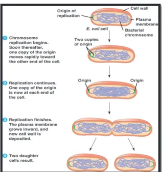
Growth is a characteristic of all living organisms and is a general definition used when a living organism increases its cellular mass or the number of cells. About half a century ago (or even earlier), researchers were able to understand the general laws of bacterial growth50. This growth is greatly affected by environmental factors such as pH, temperature, osmotic pressure, the content of nutrients, etc. Bacteria are unicellular microorganisms and in favorable conditions for their growth, they start consuming the nutrients in the media and attain certain size and consequently, they divide into two cells through a binary fission process. In this process a parent bacteria cell is divided into two new daughter cells while its genetic material (DNA) duplicates and each daughter cell receives one copy of DNA51. Different steps of such reproduction process for an E. coli bacteria cell is schematically illustrated in Figure 1252. Generally, prokaryotic organisms have chosen binary fission as their primary method of reproduction.
Figure 11. Scanning electron micrograph of Escherichia coli, grown in culture and adhered to a cover slip (reproduced from reference 47).
The growth pattern for each bacteria is varied in different ecological conditions. The number of cells (Nt) after a certain time (t) can be predicted from Equation 8 where N0 is the initial number of bacteria cells and τ is the time required for a single cell to grow and duplicate53.
Equation 8 2
The bacterial growth curve is an indication of a bacteria’s population by plotting the binary logarithmic scale of the number of cells at each time point versus time. The resulting graph is illustrated in Figure 1352. This graph exhibits four distinctive stages of the bacterial life cycle provided that bacteria are cultured in an isolated culture media where no additional nutrients or space are added and also no waste or dead cells are removed from the culture media. The first stage is the lag phase which occurs after the bacteria are added to the culture medium. In fact, this time elapse before the growth phase (second stage) is the period of time required for the organisms to adapt themselves to the new environment so the population growth at this period close to 0. The second stage is the growth phase and the binary logarithm of the number of cells grows linearly with time according to Equation 9 obtained by rearranging Equation 8.
Equation 9
Figure 12. Different steps of a binary fission process in E. coli cell(reproduced from reference 52).
This growth phase which also called log phase is the period of optimal population growth depending of the growth conditions. Eventually, however, when the growing population approaches the upper limit, the media runs out of nutrients and space. At this point, the bacterial population levels off and population growth becomes nearly 0 again. This stage, called the
stationary phase and may last for a long period of time. The last stage of this life cycle is called
the death phase. The waste and dead cells begin to accumulate and the population declines because of the lack of nutrients. Some species that are able to form spores can persist beyond this stage and can regenerate a population if conditions once again become favorable. In this study, the effect of H2O2 generated by the enzymatically catalyzed reaction of glucose on the lag phase of the bacterial growth were studied.
1.7. Structure of the thesis
As mentioned in Section 1.1, the goal of this research is to develop a generic antibacterial platform that relies on the use of immobilized enzymes and can be applied to current paper production processes. The design, preparation and characterization of such a platform and its impact on the enzymatic activity is presented in the current study. This antibacterial platform was designed to trap GOx in microcapsules consisting of an alginate-calcium matrix. The enzymatic activity of GOx in the presence of its substrates (glucose and O2) produces an antibacterial agent, H2O2, which leaches out of the microcapsules and decomposes pathogens in the media. In this regard, non-pathogenic bacteria, E. coli K-12, was proposed as a model. Since this platform is destined to be used in a laboratorial papermaking process (in future research) and it will be incorporated onto paper, it has to be structurally robust against mechanical stress. Therefore, a layer of chitosan was used to cover the surface of alginate microcapsules. Also, to be able to capture low amounts of pathogens on the surface of microcapsules and to increase the efficiency of the system, the immobilization of a given antibody (selective to a certain pathogen) was considered. Since antibodies against E. coli are expensive and available only in small amounts, human Immunoglobulin G (human IgG) was selected as a model antibody. It was assumed that developing a successful immobilization method for this model antibody, while maintaining the enzymatic activity of encapsulated GOx, could be applied to other antibodies with similar structure. After the preparation of this platform, its antibacterial properties against our model pathogen were investigated. It is important to note that the application of this antibacterial platform onto paper was not pursued in this research and will be the subject of the future studies.
As will be presented in following sections of this research, the encapsulation of GOx resulted in a reduction in its enzymatic activity. Therefore, we came up with the idea that immobilization of GOx on gold nanoparticles (NPs), before encapsulation, could possibly increase its enzymatic activity. The goal of this sub-project (Chapter 5) was to compensate the enzymatic activity loss during the encapsulation process and also to increase the encapsulation efficiency.
This thesis is composed of seven chapters. The current chapter includes an introduction to the scientific notions of this research and provides background knowledge for readers to be able to understand this project. The second chapter presents the detailed description of all materials, protocols, methods and the instruments with which the research was carried out. All the observations, measurements and data regarding the preparation of encapsulated GOx are brought to the reader in chapter three. The results and discussion regarding the study on antibacterial properties of our microcapsules are presented in chapter four along with the interpretations and discussions to justify those obtained results. Chapter five contains the data regarding the immobilization of GOx on gold NPs and the effect of this immobilization on the enzymatic activity. Since this part of the project is considered as a sub-project (additional to the main stream of the project), the introduction and experimental procedures for this part of the study are presented in the same chapter as the corresponding results and discussion. The sixth chapter presents the conclusion of this project and opens a window to the future possible works which lie ahead. Finally, the seventh chapter of this thesis provides all the implemented bibliographic references to support the ideas discussed in the thesis.
2.1. Enzyme encapsulation
The encapsulation of enzyme is achieved by ionotropic jellification of small droplets of alginate solution, which contain GOx. This process also involves laminar jet break-up of the solution. The encapsulation efficiency is then determined to evaluate the amount of encapsulated enzyme.
2.1.1. Laminar jet break-up encapsulation
Method
For the preparation of encapsulated enzyme, a laminar jet break-up54 with a vibrational nozzle method of dispersion was used in combination with a further gelification of the encapsulation matrix as a stabilization method using the Inotech Encapsulator® IE-50 R (Figure
14) which is a semi-automated instrument for encapsulation of biomolecules and pharmaceutics.
Setting different instrumental parameters helps us to control and determine the size of microcapsules (beads). The bead size varies from 100 µm to > 1000 µm in diameter according to the encapsulation parameters and the size of the nozzle with size dispersion of about 5 % in