The BTB/POZ Transcription Factor Miz-1 Is Required To Regulate The Commitment, Survival And Differentiation Of Early B And T Cell Lineages
par Ingrid Saba
Département de microbiologie et immunologie Faculté de médecine
Thèse présentée à la Faculté des études supérieures (FES) en vue de l’obtention du grade de Philosophiæ Doctor (Ph.D.)
en microbiologie et immunologie
Janvier 2012
Faculté des études supérieures (FES)
Cette thèse intitulée :
The BTB/POZ Transcription Factor Miz-1 Is Required To Regulated The Commitment, Survival And Differentiation Of Early B And T Cell Lineages
présentée par : Ingrid Saba
a été évaluée par un jury composé des personnes suivantes :
Dr Sylvie Lesage, président-rapporteur Dr Tarik Möröy, directeur de recherche Dr Martin Guimond, membre du jury Dr Yves St-Pierre, examinateur externe Dr Claude Perreault, représentant du doyen
Résumé
Les lymphocytes B et T sont issus de cellules progénitrices lymphoïdes de la moelle osseuse qui se différencient grâce à l’action de facteurs de transcription, cytokines et voies de signalisation, dont l’interleukine-7 (IL-7)/IL-7 récepteur (IL-7R). Le facteur de transcription c-Myc est exprimé par les cellules lymphoïdes et contrôle leur croissance et leur différenciation. Cette régulation transcriptionnelle peut être coordonnée par le complexe c-Myc/Myc-Interacting Zinc finger protein-1 (Miz-1). Le but de ce projet était de comprendre les mécanismes qui impliquent Miz-1 et le complexe c-Myc/Miz-1 dans le développement des lymphocytes B et T. Pour réaliser ce projet, des souris déficientes pour le domaine de transactivation de Miz-1 (Miz-1ΔPOZ) et des souris à allèles mutantes pour c-MycV394D, mutation qui empêche l’interaction avec Miz-1, ont été générées.
La caractérisation des souris Miz-1ΔPOZ a démontré que l’inactivation de Miz-1 perturbe le développement des lymphocytes B et T aux stades précoces de leur différenciation qui dépend de l’IL-7. L’analyse de la cascade de signalisation IL-7/IL-7R a montré que ces cellules surexpriment la protéine inhibitrice SOCS1 qui empêche la phosphorylation de STAT5 et perturbe la régulation à la hausse de la protéine de survie Bcl-2. De plus, Miz-1 se lie directement au promoteur de SOCS1 et contrôle son activité. En plus de contrôler l’axe IL-7/IL-7R/STAT5/Bcl-2 spécifiquement aux stades précoces du développement afin d’assurer la survie des progéniteurs B et T, Miz-1 régule l’axe EBF/Pax-5/Rag-1/2 dans les cellules B afin de coordonner les signaux nécessaires pour la différenciation des cellules immatures. La caractérisation des souris c-MycV394D a montré, quant à elle, que les fonctions de Miz-1 dans les cellules B et T semblent indépendantes de c-Myc.
Les cellules T des souris Miz-1ΔPOZ ont un défaut de différenciation additionnel au niveau de la β-sélection, étape où les signaux initiés par le TCR remplacent ceux induits par IL-7 pour assurer la prolifération et la différenciation des thymocytes en stades plus
matures. À cette étape du développement, une forme fonctionnelle de Miz-1 semble être requise pour contrôler le niveau d’activation de la voie p53, induite lors du processus de réarrangement V(D)J du TCR. L’expression de gènes pro-apoptotiques PUMA, NOXA, Bax et du régulateur de cycle cellulaire p21CIP1 est régulée à la hausse dans les cellules des souris Miz-1ΔPOZ. Ceci provoque un débalancement pro-apoptotique qui empêche la progression du cycle cellulaire des cellules TCR-positives. La survie des cellules peut être rétablie à ce stade de différenciation en assurant une coordination adéquate entre les signaux initiés par l’introduction d’un TCR transgénique et d’un transgène codant pour la protéine Bcl-2.
En conclusion, ces études ont montré que Miz-1 intervient à deux niveaux du développement lymphoïde: l’un précoce en contrôlant la signalisation induite par l’IL-7 dans les cellules B et T, en plus de l’axe EBF/Pax-5/Rag-1/2 dans les cellules B; et l’autre tardif, en coordonnant les signaux de survie issus par le TCR et p53 dans les cellules T. Étant donné que les thymocytes et lymphocytes B immatures sont sujets à plusieurs rondes de prolifération, ces études serviront à mieux comprendre l’implication des régulateurs du cycle cellulaire comme c-Myc et Miz-1 dans la génération des signaux nécessaires à la différenciation non aberrante et à la survie des ces cellules. Enfin, les modèles expérimentaux, souris déficientes ou à allèles mutantes, utilisés pour ce travail permettront de mieux définir les bases moléculaires de la transformation maligne des lymphocytes B et T et de révéler les mécanismes conduisant au lymphome.
Mots-clés : Miz-1, c-Myc, IL-7R, TCR, différenciation, apoptose, Bcl-2, STAT5, SOCS1, p53.
Abstract
Signaling pathways control the differentiation and proliferation of blood cells, like B and T lymphocytes. They converge into regulating the activity of transcription factors that influence ultimately gene expression patterns. The transcription factor c-Myc is a central regulator of cellular proliferation and growth, and its deregulated expression has been demonstrated to be involved in many types of cancers, in particular lymphoma. Recent studies have shown that repression by c-Myc can be mediated by a complex formed with the BTB/POZ domain transcription factor Miz-1 (Myc Interacting Zinc finger protein-1). Given that both c-Myc and Miz-1 proteins are expressed in lymphoid precursors and since c-Myc has been shown to be important for B- and T-cell development, the aim of this thesis was to investigate the role of Miz-1 and the c-Myc/Miz-1 complex in regulating B and T cell survival, commitment and differentiation. To do so, mice expressing a non-functional Miz-1 protein lacking the BTB/POZ domain (Miz-1ΔPOZ) and knock-in mice expressing a mutant c-MycV394D allele that no longer interacts with Miz-1 were generated.
B- and T-cell development requires the coordinated action of transcription factors and cytokines, in particular interleukin-7 (IL-7). The studies presented in this work demonstrated that mice deficient for the BTB/POZ domain of transcription factor Miz-1 almost entirely lack follicular B cells and T cells, since their progenitors fail to activate the JAK/STAT5 pathway and to up-regulate Bcl-2 upon IL-7 stimulation. Miz-1 exerts a dual role in the IL-7 receptor (IL-7R) pathway by directly repressing the JAK inhibitor SOCS1 and by activating Bcl-2 expression. In B cells, a functional form of Miz-1 is also required for the proper expression of early B cell genes like E2A and EBF. These data suggest that Miz-1 represents a new regulatory element of early B- and T-cell differentiation required for the regulation of the IL-7/IL-7R/STAT5/Bcl-2 axis by monitoring SOCS1 for survival and by regulating the EBF/Pax-5/Rag-1/2 axis for the proper commitment and differentiation of the B-cell lineage. The regulation exerted by Miz-1 in B and T cells is
mostly likely independent of its interacting partner c-Myc, and seems specifically linked to the BTB/POZ domain of Miz-1.
Mice deficient for the BTB/POZ domain of Miz-1 have additionally a severe differentiation block at the pre-T cell “β-selection” checkpoint. Miz-1 deficient pre-T cells are highly apoptotic and do show cell cycle defects. This concurs with enhanced expression of p53-target genes such as p21CIP1, Bax, PUMA and Noxa, most likely induced by the DNA double-strand breaks generated during the V(D)J recombination of the TCR. Only the co-expression of rearranged TCRαβ and Bcl-2 fully rescued Miz-1-deficient cell numbers and enabled them to differentiate into TCRβ+ cells. These data suggest that Miz-1 is required for both the regulation of the p53 response and proper expression of the pre-TCR to support the proliferative burst of pre-T cells.
In conclusion, the studies presented in this thesis revealed the so far unknown implication of Miz-1 in B- and T-cell development. More specifically, Miz-1 exerts early regulatory functions by monitoring the IL-7/IL-7R signaling in B and T cells. It regulates later stages of differentiation by controlling the EBF/Pax-5/Rag-1/2 in B cells and the TCR expression and the p53 response in T cells. These studies and the generated mice model (conditional knock-out and knock-in) will help characterize the implications of transcription factors that have been causally implicated in the altered genetic programming found in hematopoietic malignancies due to their capacities to regulate cell cycle. Ultimately the characterization of Miz-1 and c-Myc functions in B and T cells will help better understand the mechanisms responsible for the emergence of leukemia and lymphoma.
Keywords : Miz-1, c-Myc, IL-7R, TCR, differentiation, apoptosis, Bcl-2, STAT5, SOCS1, p53.
Table of contents
Résumé ...i Abstract ...iii Table of contents... v List of Figures...ix List of abbreviations ...x Acknowledgments ... xv Overview... xvi Introduction ...11. Hematopoiesis and lineage progenitors... 2
1.1. Models for the hematopoietic cell differentiation ...5
1.1.1. The classical model...5
1.1.2. The revised classical model ...6
1.1.2.1. Lymphoid versus myeloid lineage potential ...7
1.1.3. The myeloid-based model...10
2. Temporal and spatial regulation of transcription factors in stem cells... 13
2.1. GATA and Pu.1 during lineage commitment...14
2.2. Id1 to Id4 during lineage commitment ...15
2.3. SCL/Tal-1 during lineage commitment...16
2.4. Ikaros during lineage commitment ...17
2.5. Gfi1, Gfi1b and their functions...18
2.6. Transcriptional regulation of B- versus T-lineage choice ...20
3. B-cell development ... 22
3.1 Early stages of B-cell differentiation...22
3.2. B-cell differentiation and cytokines ...26
3.2.1. IL-7/IL-7R signaling in B cells...27
3.2.1.1. IL-7/IL-7R regulation and B-cell differentiation ...29
3.3. Mature B cells...32
3.3.1 Somatic hypermutation and class switch recombination ...32
3.4. B-1 and B-2 cells ...33
3.5. B-cell commitment is regulated by a transcription factor network ...35
4. T-cell development ... 36
4.1. The origin of T cells ...37
4.2. Early T-lineage progenitors ...38
4.3. CD4-CD8- double negative cell differentiation ...39
4.4. CD4+CD8+ double positive cell differentiation ...41
4.5. TCRγδ T cells ...44
4.6. Other unconventional T cells...45
4.7. Extrathymic T-cell development ...47
4.8. Cytokine receptors and T-cell differentiation...47
4.8.1. Flt3L/Flt3 signaling in T cells ...47
4.8.2. IL-7/IL-7R signaling in T cells...48
4.9. Notch signaling...49
4.10. T cell gene expression network regulates the processes of differentiation ...53
4.11. Pre-TCR rearrangement and double-stranded breaks...54
4.11.1. The p53 tumor suppressor protein ...56
4.11.2. DNA damage response ...57
4.11.3. Apoptosis versus cell cycle arrest...59
5. The Myc family of oncoproteins... 60
5.1 The role of c-Myc during lymphocyte development ...60
5.2. c-Myc and malignant transformation ...61
6. Miz-1 and c-Myc as transcriptional regulators ... 62
6.1 Biochemical structure of Miz-1...63
6.2 Miz-1 and its functions ...65
6.3 Miz-1 and its binding partners in cell regulation...67
6.3.1. The regulation of p15INK4B by Miz-1 ...68
6.3.2. Regulation of p21CIP1 expression by BTB/POZ domain transcription factors...69
6.3.3. Miz-1 is involved in regulating the DNA damage response...71
6.3.4. Miz-1 controls cell survival ...72
8. Mouse models used for this project ... 75
Results ...77
Chapter I ... 77
Transcription Factor Miz-1 Is Required to Regulate Interleukin-7 Receptor Signaling at Early Commitment Stages of B Cell Differentiation ...77
Chapter II... 152
IL-7R-dependent survival and differentiation of early T-lineage progenitors is regulated by the BTB/POZ domain transcription factor Miz-1 ...152
Chapter III ... 219
Miz-1 is Required to Coordinate the Expression of TCRβ and p53 Effector Genes at the Pre-TCR ''β-selection'' Checkpoint ...219
Discussion ...275
1. Miz-1 is required for embryonic development ... 275
2. Miz-1ΔPOZ mice have a normal HSC pool ... 276
3. The importance of the BTB/POZ domain... 278
4. Miz-1 in early B-cell development ... 279
5. Miz-1 in early T-cell development ... 284
6. Miz-1 functions in early B and T cells are independent of c-Myc ... 288
7. Miz-1 at the β-selection checkpoint ... 289
8. p53 and the balance between survival and cell death ... 290
9. Miz-1 regulation at the β-selection through direct protein-protein interaction?... 293
Perspectives ...295
1. Implication of Miz-1 in p53 target gene regulation... 295
2. The role of Miz-1 in mature B cells... 297
3. Miz-1 implication in human B cell production? ... 299
5. Conditional full deletion of the Miz-1 protein ... 302
6. Miz-1 and lymphomagenesis ... 303
Conclusions...307
Other contributions...310
List of Figures
Figure 1. The classical model of hematopoiesis...5
Figure 2. Schematic representation of the revised model of hematopoietic lineage differentiation, specification and commitment...9
Figure 3. A simplified version of the transcriptional network governing B-, T- and myeloid lineage differentiation...21
Figure 4. Illustration of B-cell development in the bone marrow. ...25
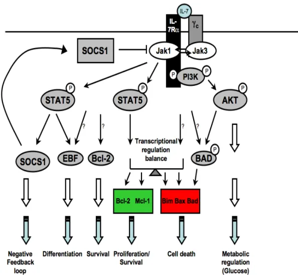
Figure 5. IL-7/IL-7R signaling cascade...30
Figure 6. Illustration of T-cell development in the thymus...43
Figure 7. Notch signaling pathway...51
Figure 8. Schematic representation of p53 activation and regulation of its downstream target genes. ...58
Figure 9. Schematic representation of c-Myc and Miz-1. ...64
Figure 10. Schematic representation of the two c-Myc-dependent regulatory pathways...67
Figure 11. Examples of positive and negative regulation by Miz-1 on p21CIP1 transcription. ...70
Figure 12. The BTB/POZ domain of Miz-1 influences the oligomerization capacity of the protein...279
Figure 13. Miz-1 is required for early B-cell development. ...283
Figure 14. Miz-1 is required for early T-cell development. ...287
Figure 15. Implication of Miz-1 during the β-selection checkpoint. ...292
Figure 16. A functional Miz-1 protein is required to regulate lymphoid precursors and early B- and T-cells functions. ...309
List of abbreviations
AID: Activation-induced cytidine deaminase ALP: All-lymphoid progenitor
Atm: Ataxia-telangiectasia-mutated Atr: Atm-Rad3-related kinase
B-ALL: B-acute lymphoblastic leukemia Bad: Bcl-2-antagonist of cell death Bax: Bcl-2-associated X protein Bcl-2: B-cell CLL/lymphoma-2 Bcl-XL : Bcl-2-like 1
BCR: B cell receptor
BLP: B cell-biased lymphoid progenitor
BTB: Broad-complex, Tramtrack and Bric-à-brac CCR9: CC-chemokine receptor 9
CDK: Cyclin-dependent kinase inhibitor CFU-S: Colony Forming Units-Spleen ChIP: Chromatin ImmunoPrecipitation Chk1: Checkpoint kinase 1
CLP: Common lymphoid progenitor CMP: Common myeloid progenitor CSR: Class switch recombination DC: Dendritic cell
DL: Delta-like ligand
DLBCL: Diffuse large B cell lymphoma DN: CD4-CD8- double negative
DP: CD4+CD8+ double positive E2A: Transcription factor 3 (Tcf3)
EBF: Early B cell factor
ELP: Early lymphoid progenitors
ERK: p44/42 Extracellular-Regulated Kinase ETP: Early T-lineage progenitors
Fl: Floxed allele carrying loxP sites Flt3: Fms-like tyrosine kinase 3 Flt3L: Flt3 ligand
Fo: Follicular
GM: Granulocyte/macrophage GMP: Granulo-monocytic precursors
HLH-LZ: Helix-loop-helix and leucine-zipper motif HSC: Hematopoietic stem cells
ICN: Intracellular domain of Notch IEL: Intraepithelial lymphocytes Ig: Immunoglobulin
IL-7 : Interleukin-7
IL-7Rα: Interleukin-7 receptor α chain JAK: Janus kinase
JNK: c-Jun NH2-terminal Kinase Lin: Lineage marker
LMPP: Lymphoid-primed multipotent progenitors LSK: Lin−Sca-1+c-Kit+
LT-HSC: Long-term hematopoietic stem cells MAML: Mastermind-like protein
MDM2: Double minute 2 protein or E3 ubiquitin-protein ligase MAPK: Mitogen-activated protein kinase
Mcl-1: Myeloid cell leukemia sequence 1, Bcl-2-related MegE: Megakaryocyte/erythroid
Miz-1: Myc-Interacting Zinc finger protein-1 (zbtb17) Miz-1ΔPOZ: Miz-1 lacking the BTB/POZ domain MPP: Multipotent precursors
Myc: Myelocytomatosis viral oncogene
MycV394D: c-Myc expressing an Aspartic Acid (D) instead of a Valine residue (V) at position 394 of its coding sequence
MZ: Marginal zone NK: Natural killer
p53: Transformation related protein 53
Pax-5: Paired-homeodomain transcription factor PI3K: Phosphatidylinositol 3-kinase
POZ: Pox virus Zinc finger domain pTα: Pre-TCRα chain
RAG: Recombination activating gene RSS: Recombination signal sequences SCF: Stem cell factor
SHM: Somatic hypermutation
SOCS: Suppressor of cytokine signaling 1 SP: CD4+ orCD8+ single positive
STAT: Signal transducer and activator of transcription ST-HSC: Long-term hematopoietic stem cells
T-ALL: T-cell Acute Lymphoblastic Leukemia TCR: T cell receptor
Tdt: Terminal deoxynucleotidyl transferase Tfh: T follicular helper cells
Tg: Transgenic
TGFβ: Transforming growth factor beta Th: CD4+ T helper cell
TSLP: Thymic-stromal-derived lymphopoietin receptor VCAM-1: Vascular cell adhesion molecule-1
V(D)J: Variable, diversity, joining recombination WT: Wild-type
«Le plus grand ressort c’est l’espoir; quand
il est cassé tout mouvement s’arrête en nous». Anonyme
Acknowledgments
Cette thèse est un accomplissement académique et personnel qui n’aurait jamais été possible sans l’aide et le soutien de plusieurs personnes à qui je ne dirais jamais assez MERCI! Je voudrais remercier en premier mon directeur de recherche, Tarik Möröy, qui m’a énormément appris scientifiquement et qui m’a permis d’entreprendre mes projets avec beaucoup de liberté, d’enthousiasme et de suivi. Vous avez été très généreux avec moi en plus d’être toujours disponible, compréhensif et encourageant. Je vous remercie de m’avoir conseillée et guidée durant ces cinq dernières années. Je remercie également mes collègues de laboratoire, particulièrement Christian, qui a été un grand frère, un partenaire et un mentor scientifique durant toute ma thèse. Ce fut un plaisir de travailler sur le projet Miz-1 que tu as amorcé et je crois qu’on peut être fier de ce que nous avons accompli ensemble. Je remercie aussi Mathieu pour tous les génotypages, les commandes et la Presse, mais pas le Devoir… Merci à Marissa, Marie-Claude, Cyrus, Lothar, Riyan, (Florian, Taro, Rachel, Khash, et Oliver), et à mes collègues d’étage, Astrid, Alex et Steve, avec qui j’ai eu autant de discussions scientifiques que non scientifiques. Nos manips jusqu’à trois heures du matin, nos pauses café, espresso ou thé et nos compétitions de ‘’êtes-vous observateur?’’ me manqueront. Je tiens à vous dire que vous avez été pour moi une deuxième famille à l’IRCM.
Je tiens aussi à remercier ma mère Sonia, mon frère Karl, ma famille et ma belle-famille pour leur amour et leurs éternels encouragements, mes amis Névine, Nathalie, Maryse, Fred, et Andrés pour leur soutien moral, leur aide (des fois scientifique) et pour leur disponibilité à m’écouter lorsque j’avais besoin de parler…
Enfin, Martin, mon complice et partenaire de vie, je te remercie du fond du cœur. Tu m’as toujours soutenue et encouragée, même dans mes moments les plus sombres. Tu as lu et critiqué mes demandes de bourses, articles et cette thèse même quand tu manquais de temps! Je ne pourrais énumérer tout ce que tu as fait, alors merci pour tout le reste...
Overview
The immune system has evolved over the years to arm the body with many strategies of defense against pathogens and infections. The innate immune system possesses sensors that initiate innate responses after promiscuous recognition. On the contrary, the adaptive immunity integrates signals from specific antigen receptors to transcriptional regulation events that govern the acquired response. The shape and size of the antigen receptor repertoire are also tightly regulated by immuno-surveillance mechanisms such as tolerance to avoid cross-reactivity, inflammatory pathologies and autoimmune diseases.
All mature blood cells originate from hematopoietic stem cells that mature in the bone marrow or in primary lymphoid organs through coordinated proliferation and differentiation events. By acquiring lineage restrictions, the progenitors loose stemness, but gradually gain proliferative capacity that needs to be tightly regulated by transcription factors. These transcription factors ensure the maintenance of the genetic programming that regulates cell growth, commitment and survival. Lymphoid malignancies arise from different stages of development and are often linked to a deregulated signaling pathway or transcriptional regulatory functions in B and T cells.
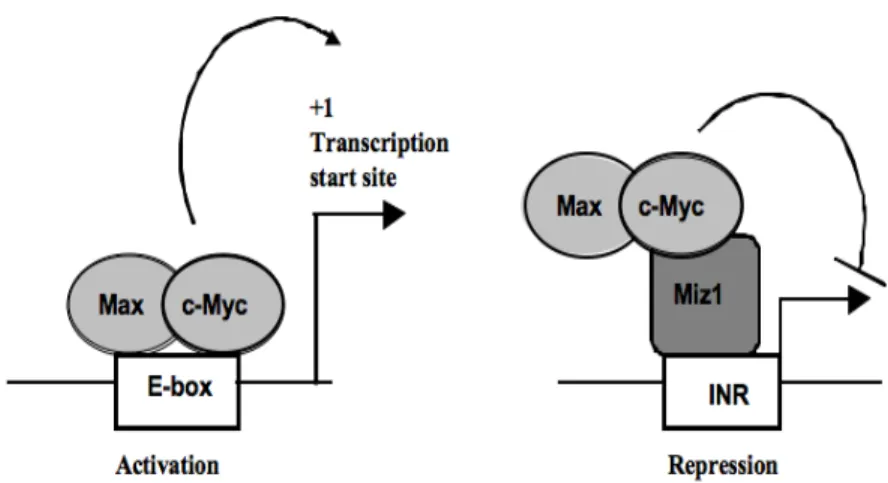
The transcription factor c-Myc plays important roles in hematopoietic differentiation and in the emergence of lymphoma and other blood cancers. It can regulate the transcription of its target genes through two pathways, one E-box-dependent and the other E-box-independent. The second E-box-independent pathway, in which c-Myc regulates gene expression, is dependent on the association of c-Myc with Miz-1. Miz-1 itself can control the expression of genes involved in proliferation. The identity of most of these genes and their function in c-Myc lymphocyte biology are unknown.
This work aimed to define the role of Miz-1 and the c-Myc/Miz-1 complex in the regulation of T- and B-cell survival, commitment and differentiation. As c-Myc has been
shown to play important roles in the development of progenitors and T cells, this project hypothesis was that its interacting partner Miz-1 also had implications in precursor commitment and lymphoid development processes. The characterization of these two transcription factors was thought to enable the identification of the interplay between c-Myc and Miz-1 functions during critical developmental checkpoint of B- and T-cell development.
The results obtained for this project will be presented in three chapters. The first chapter will contain the published article in ''Immunity'' regarding the requirement for a functional form of Miz-1 to regulate IL-7R signaling at early commitment stages of B-cell differentiation. The second chapter will expose data regarding the role of Miz-1 in IL-7R-dependent survival and differentiation of early T-lineage progenitors that were published in ''Blood''. The third chapter will present data from a manuscript recently accepted for publication in ''The Journal of Immunology'' on the implication of Miz-1 later in T-cell development, where it is important to coordinate the expression of TCRβ and p53 effector genes at the pre-TCR ‘’β-selection’’ checkpoint.
These studies revealed that Miz-1 is a new regulator of normal lymphoid development and that it exerts this role independently of c-Myc. Since it is known that both c-Myc and Miz-1 can affect the process of malignant transformation, their function in normal cells needs to be properly monitored in order to avoid the emergence of leukemia and lymphoma. Hence, this study not only presents a new insight on the mechanisms underlying normal lymphoid development, but it also provides new knowledge that is very likely important to design future therapeutic strategies against different types of blood cancers.
Introduction
Hematopoietic stem cells (HSC) are responsible for the generation of mature blood cells through a series of well coordinated proliferation and differentiation events. As cells progress through the early stages of hematopoiesis, they give rise to precursors that are more restricted because they gradually loose their multilineage and self-renewal potential which characterized the originating HSC [1, 2]. By acquiring a certain lineage restriction, the progenitors gradually loose their ability for self-renewal but gain proliferative capacity. These processes are tightly regulated by transcription factors that ensure the maintenance of the genetic programming that regulates cell growth, lineage commitment and cell survival [3].
Throughout the lifespan of a mouse, the hematopoietic progenitors generate mature lymphoid and myeloid cells that continue to replenish the innate and adaptive or acquired immune systems. The innate system constitutes the first line of defense and recognizes a limited number of evolutionary conserved molecules expressed by pathogen-associated molecules such as lipopolysaccharides and peptidogylcans [4, 5]. The major difference between these two branches of the immune system is that the adaptive immunity provides specificity to antigen recognition by the B cell receptor (BCR) or the T cell receptor (TCR) and secretes antibodies [6, 7]. Moreover, the innate immune system responds to antigen with a fast kinetic that lacks memory capacity whereas the adaptive immune system responds with a delayed kinetic, but possesses effective memory responses [4, 6]. The specificity of antigen recognition is achieved through several developmental pathways and
selection steps orchestrated by the interplay between the TCR or BCR signaling and transcription factor regulation [8]. Another particularity of the adaptive immune response is the clonal selection, a process that enables each cell that expresses a single receptor to expand based on the affinity of that receptor for its ligand. Hence, a small number of specific naïve cells will expand following the recognition of a particular antigenic epitope. Consequently, the repertoire of the adaptive immune system is unique to each individual, whereas the receptors of the innate immune system, that are retricted to specific motifs, are not clonally distributed and may be shared within individuals [8].
Hematopoietic cells bear characteristics of both the innate and the adaptive immune system. Yet, some subsets of cells do not classify within the criteria of the innate or adaptive response, as they possess properties of both branches. Examples of such cells include natural killer T (NKT) cells, γδ T cells, CD8αα T cells, B-1 B cells, marginal zone B cells and subsets of NK cells [9]. T and B cells are a lymphocyte lineage that is critical for the host defense system against many types of pathogens. Their functions are well characterized in the periphery, but major questions still remain regarding their origin and their developmental processes.
1. Hematopoiesis and lineage progenitors
Most hematopoietic lineages, including B cells, develop in the bone marrow, while T cells complete their development in the specific environment of the thymus. The migration of
progenitors out of the bone marrow allows the circulation of cells in the bloodstream through which they finally reach the homing organ [10]. Many markers have been proposed to accurately identify the correct progenitor that generates a specific lineage. For instance, lineage markers (Lin) are cell surface antigens that define specific populations of mature blood cells. The lack of lineage marker expression is found in an enriched population of cells with primitive hematopoietic stem cell or early progenitor cell characteristics. A bone marrow fraction that lacks Lin expression and expresses c-Kit (CD117) and Sca-1 is referred to as Lin−Sca-1+c-Kit+ or ''LSK'' cells that contain long-term and short-term hematopoietic stem cells (HSC) [11]. Long-term (LT)-HSC cells are the true hematopoietic stem cells that are capable of self-renewal, have a multi-lineage potential and can repopulate a transplanted host. LT-HSC can also be identified by the absence or low expression levels of Thy-1.1 (CD90) and Flt3 (Fms-like tyrosine kinase 3, Flk2, CD135), i.e. as Thy-1.1lo Flt3- cells or as CD34−Flt3− or CD150+CD244−CD48− cells [12, 13].
Self-renewing LT-HSC cells are the base of the hematopoietic system holding a very potent proliferative potential in order to produce differentiated mature blood cells. The differentiation steps required to generate blood cells are irreversible, as once a lineage potential is lost, it cannot be recovered [13, 14]. Many stages govern the differentiation and proliferation of hematopoietic precursors. LT-HSC give rise to short-term-HSC (ST-HSC) that can sustain hematopoiesis for only about 6 weeks in a mouse because of their restrictive self-renewal capacity [15]. ST-HSC can express Flt3 and are Thy-1.1loFlt3+ or CD34+Flt3- and CD34+Flt3+, functionally distinct subsets of short-term HSC [16], and
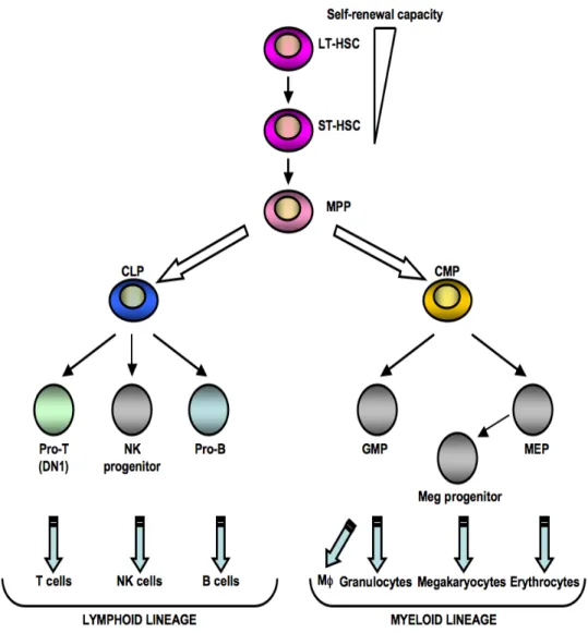
contribute to multi-lineage differentiation. The oldest technique for determining the capacity of progenitors to generate hematopoietic cell fates is an in vitro colony-forming cell assay where transplanted stem cells can form colonies in the spleen of irradiated mice [17-19]. ST-HSC are potent progenitors of the hematopoietic lineage and can generate colonies (CFU-S: Colony Forming Units-Spleen) that can be detected by this spleen colony technique after transplantation [15, 16]. ST-LSK will give rise to non-self renewing multipotent precursors (MPP) characterized by the expression of Thy1.1-Flt3+ or CD34+Flt3+. MPP have multilineage potential and mark the first step of lineage restriction during hematopoiesis [1, 2]. In addition, MPP have been identified as the progenitors of the common lymphoid progenitor (CLP) and common myeloid progenitor (CMP) [20, 21] (Figure 1).
Figure 1. The classical model of hematopoiesis. Hematopoietic stem cells lose their self-renewal activity and their first lineage fate decision is to engage in lymphoid or myeloid differentiation. In this model, the common lymphoid progenitor (CLP) and the common myeloid progenitor (CMP) are symmetrically derived from the same multipotent precursors (MPP). Each progenitor gives rise to the indicated mature blood cells. GMP, granulocyte/macrophage progenitor; MEP, megakaryocyte/erythroid progenitor; Meg, megakaryocyte; NK, natural killer; Mφ, macrophage (adapted from [22]).
1.1. Models for the hematopoietic cell differentiation
1.1.1. The classical model
Many groups have used the fractionation of hematopoietic precursors into subsets with different lineage specifications to further define hematopoietic differentiation. The first
dichotomy model was established in 1997 by the identification of the symmetrical division of MPP into CLP and CMP [20, 21]. CMP cells give rise to two sets of restricted bipotent progenitors: the megakaryocyte/erythroid (MegE) progenitors (CD34−FcγRIII−Thy1.1− IL-7Rα−Lin−Sca-1−c-Kit+) and the granulocyte/macrophage (GM) progenitors (CD34+FcγRIII+Thy-1.1−IL-7Rα−Lin−Sca-1−c-Kit+) [21]. CLP (Lin-IL-7Rα+Sca-1low c-KitlowThy-1.1−) are proposed to be the common lymphoid progenitor that gives rise to B and T cells. Refinements to this model were added by Adolfsson and his collaborators who reported that MPP could be divided into two groups based on Flt3 expression [23]. The highest Flt3 expressing subset had lost its MegE differentiation potential, but retained robust GM-, T- and B-cell differentiation potential [23]. Further analyses characterizing the expression of Flt3 expression allowed defining the branching point for most potent lymphoid progenitors to develop into the lymphoid lineage.
1.1.2. The revised classical model
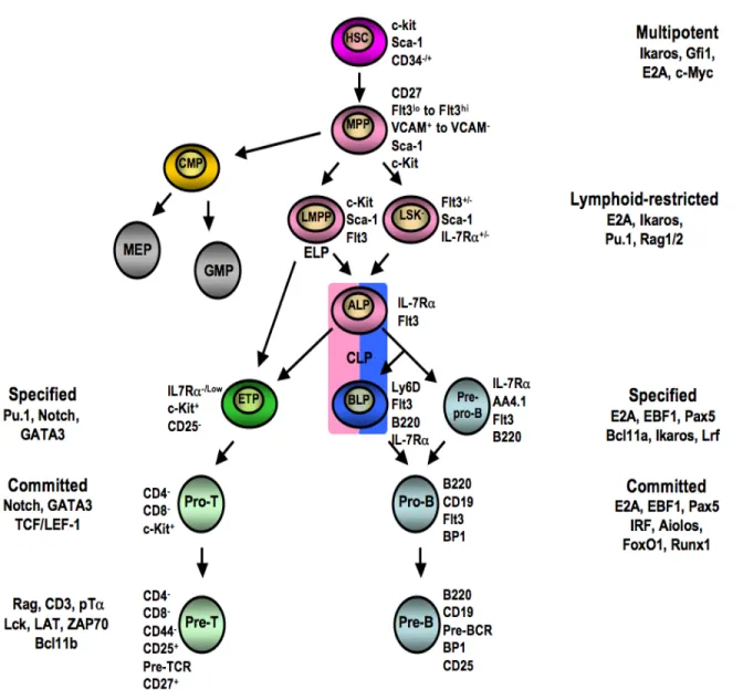
By using Flt3 as a marker in combination with vascular cell adhesion molecule-1 (VCAM-1), Kondo’s group could further subdivide the MPP population into three subsets: Flt3loVCAM-1+, Flt3hiVCAM-1+, and Flt3hiVCAM-1− MPP [24, 25]. The Flt3loVCAM-1+ MPP population seems to contain the true multi-lineage progenitor able to differentiate into MegE, GM and lymphoid precursors [25]. In agreement with the findings from Adolfsson and collaborators, the Flt3hiVCAM-1+ MPP can no longer differentiate into MegE lineage, but can give rise to GM and T and B cells [25]. Flt3hiVCAM-1− MPP cells still have GM differentiation potential, but preferentially give rise to lymphocytes [24, 25]. Only
Flt3loVCAM-1+ MPP can give rise to CMP, whether the more developmentally defined Flt3hiVCAM-1− MPP cell give rise to CLP [24]. Adolfsson and colleagues proposed an alternative lineage commitment step to the classical model where ST-HSC or MPP first diverge into the MegE lineage, and the loss of this MegE potential is a prerequisite for the differentiation into GM or lymphoid lineages [23]. This revised model suggests that CLP are not necessarily generated from the same MPP that commit to the CMP lineage (Figure 2). This refinement is particularly important to understand lymphoid lineage commitment and differentiation processes. These processes appear more complex compared to the myeloid-lineage commitment and differentiation program because of the multiple lineage restriction steps and the loss of the MegE and GM differentiating potentials prior to lymphoid lineage commitment.
1.1.2.1. Lymphoid versus myeloid lineage potential
One of the MPP subsets which expresses high levels of the Flt3 (Flt3+LSK) has been termed ‘lymphoid-primed multipotent progenitors’ (LMPP). LMPP are the precursors of the common lymphoid progenitor (Lin-IL-7Rα+Sca-1lowCD117lowThy-1.1−) and early lymphoid progenitors (ELP, Lin-IL-7Rα-CD44+Sca-1highCD117high) [20, 26-28] (Figure 2). ELP are lymphoid progenitors expressing the recombination activating gene-1 (RAG-1) [29, 30]. They are similar to Flt3hiVCAM-1- MPP which have been proposed to be lymphoid-biased progenitors that gradually down-modulate their myeloid potential. They also express CC-chemokine receptor 9 (CCR9), the chemokine receptor for CCL25 [31], which is only detected in the thymus [32], not in the bone marrow. Thus CCR9 enables
ELP to home to the thymus [20, 22, 29, 33]. CCR9 expression is not detected on CLP [34]. Consequently, their T cell potential is believed to occur prior to the silencing of the myeloid program and before the CLP stage [22]. Recently, the marker Ly6D has been used to identify the branch point where CLP divide into B cell-biased lymphoid progenitor (BLP) and all-lymphoid progenitor (ALP) that either give rise to the first stages of B-cell development or contribute to the T-cell development, respectively [35]. Ly6D- CLP possess B, T, natural killer (NK), dendritic cell (DC), and some degree of myeloid potential, whereas Ly6D+ CLP are B cell lineage restricted progenitors [35]. Ly6D- CLP are thought to enter the thymus, since after intravenous transfer, they can generate thymocytes [35, 36] (Figure 2). Both the CLP that express IL-7R gene and the ELP progenitors relay on IL-7/IL-7R signaling or priming in order to contribute to the B- and T-cell development [37-39]. The notion of priming was demonstrated by visualizing the history of IL-7R gene expression in a study by Schelnner and collaborators. The experiments performed in this study showed that, although the vast majority of ELP lacked IL-7R mRNA expression, they were marked by a prior IL-7R expression. This indicated that they are primed by the IL-7R gene signature and that they descend from a progenitor that is IL-7R-positive [39].
Figure 2. Schematic representation of the revised model of hematopoietic lineage differentiation, specification and commitment. Cells in pink can give rise to the myeloid lineage (gray) and the lymphoid lineages (blue-green). Common lymphoid progenitors (CLP) containing the ALP and BLP are in both pink and blue. ELP are also indicated under the LMPP cells as they represent a more immature stage compared to CLP. ELP and ALP can give rise to ETP, whereas BLP give rise to B cells. Specific surface markers for each cell types are indicated. Examples of key-player genes at the different differentiation stages are listed. HSC, hematopoietic stem cell; MPP, multipotent progenitor; CMP, common myeloid progenitor; MEP, megakaryocytic/erythrocyte progenitor; GMP, granulocyte/macrophage progenitor; LMPP, lymphoid-primed multipotent progenitor; LSK, Lin-Sca-1+c-Kit+; ALP, All lymphoid progenitor; ETP, early thymic progenitor; BLP, B cell-biased lymphoid progenitor; CLP, common lymphoid progenitor (adapted from [40]).
1.1.3. The myeloid-based model
The common lymphoid progenitors were described as the precursors that give rise to B, T and NK cells, but not to myeloid cells [20]. The prediction from this model was that CLP migrate to the thymus from the bone marrow to initiate T-cell development. However, some reports showed that the predominant thymus-seeding cells do not resemble CLP, but have characteristics of earlier hematopoietic progenitors [41]. Other evidence came from the characterization of Ikaros, member of the Ikaros family of transcription factors that contribute to multiple aspects of hematopoietic development and homeostasis [42, 43]. Ikaros-knockout mice lack B cells, but maintain their T cell pool despite the fact that they lack CLP because they retain the presence of an early T-lineage progenitor population in the thymus [44, 45]. These findings suggested that specific early T-lineage progenitors rather than CLP are the true T cell progenitors and that both can develop independently of each other. Therefore, an additional diversion at the CLP commitment step may be responsible for B- and T-cell development. Moreover, studies in mice showed differences in hematopoiesis between fetal and adult stages. In fetal mice, the myeloid potential persisted even after the lineage branch of hematopoiesis divided toward T and B cells [46-48]. Therefore, it was proposed that an additional 'myeloid-based' model of hematopoiesis exist, in which a hematopoietic stem cell initially generates common myelo-erythroid progenitors and common myelo-lymphoid progenitors. Recent studies validated this model for adult hematopoiesis, providing evidence that the early cell populations in the adult thymus contain progenitors that have lost the potential to generate B cells but retained substantial myeloid potential [49, 50]. These studies argue against the classical dichotomy
model in which T cells are derived from CLPs. It supports instead a myeloid-based model for both adult and fetal hematopoiesis.
Although compelling, the myeloid-based model of hematopoiesis is mainly supported by in vitro evidence. The proposed common progenitor for T and myeloid lineages has recently been challenged by an in vivo model able to map the fate of T- and myeloid cell development by visualizing the history of IL-7R expression. These elegant experiments provide evidence suggesting that lymphoid-restricted progenitors are the major source of T-cell differentiation and that the separation between lymphoid and myeloid progenitors is a fundamental hallmark of hematopoiesis [39]. In vitro and in vivo differentiation potentials must carefully be examined as both studies may generate artifactual results. On the one hand, non-physiological high concentrations of cytokines or growth factors may have been used in vitro that allow progenitors to differentiate into cell lineages that they are unable to generate under in vivo conditions. On the other hand, in vivo studies implicate isolation and manipulation of progenitors prior to their injection or transplantation which may force or alter the migration potential to distinct niches within the bone marrow or to the thymus [51] that only support the maturation of certain lineages.
Taken together, these studies show that the classical model of the CLP/CMP branching point of hematopoiesis is still valid, although some subdivisions in the subsets of MPP or ST-HSC that give rise to lymphoid or myeloid lineages must exist. Further analysis of bone marrow progenitors with T-cell differentiation potential will have to be done to clarify
whether only one or multiple subpopulations contribute to thymopoiesis. This clarification may have implications for improving bone marrow transplantation strategies to treat leukemia [52]. It takes months to fully reconstitute T cells after bone marrow transplantation [53]. During this period, patients are susceptible to infections. To improve the reconstitution of the peripheral T cell pool in a transplanted patient, it may become important to increase the number of true T-lineage committed progenitors in the transplant. In fact, while LMPP and CLP are able to generate CD4+CD8+ double positive (DP) cells in the thymus, HSC failed to do so after transfer. HSC, CLP and LMPP subsets can produce DP cells after intrathymic transfer, but HSC are unable to settle the thymus to generate DP cells. They can do so if the homing step is bypassed [54]. These experiments indicated that thymic settling is selective toward the circulating progenitors and that certain signals are required for thymic entry [54]. To improve stem cell transplantation efficiency, it was shown that the co-transplantation of CLP and hematopoietic stem cells in irradiated hosts can improve the recovery of the T-lineage [55]. Recently, Van den Brink’s group successfully transferred precursors generated in vitro on OP9DL1 bone marrow stromal cell cultures that improved peripheral T cell numbers in irradiated mice [56]. This in vitro technique has the advantage of generating a large number of T-committed precursors in order to improve the peripheral T cell pool that is suitable for transfer and that expresses a normal T cell receptor repertoire. These precursor-derived T cells also had normal cytokine production and proliferated in response to antigenic stimulation [56]. The increase in donor T cells after transplantation was shown to improve the resistance to Listeria infection and enhanced graft-versus-tumor responses [56]. Finally, the generation of T cell precursors
from human CD34+ cord blood cells using Notch signaling in vitro was also successful [57]. Therefore, whether T-lineage progenitors derived from bone marrow or from in vitro cultures are used, the co-transfer of true T cell progenitors and HSC may allow for an enhanced and accelerated T cell reconstitution after stem cell transfer.
2. Temporal and spatial regulation of transcription factors in
stem cells
Lineage specification and commitment of HSC is dependent, for example, on the stem cell niche, the profile of chemokine expression, cytokine signaling and transcription factors that mediate their survival and development [58-60]. The molecular programming of hematopoietic progenitor subsets depend on gene regulation, which occurs based on transcription factors, co-factors, signaling molecules present in the cell and on the context of the regulatory elements of each gene. Moreover, the function of a transcription factor can change during developmental transitions, but the regulatory elements of each gene do not vary with each transitional step [61]. Stem cells and multilineage precursors co-express lymphoid, erythroid and myeloid genes [62, 63]. Some genes can be qualified as differentiation genes that are true markers of lineage commitment [64]. For example, GATA, Pu.1, Id factors, SCL, Ikaros, Gfi1 and Gfi1b are transcription factors required to enable precursors to make specific lineage fate decisions [65, 66].
2.1. GATA and Pu.1 during lineage commitment
Sequence-specific DNA-binding proteins are important regulators of chromatin configuration that either increase or inhibit gene expression [67]. For example, the zinc finger transcription factor GATA-1 directly occupy looped enhancers and target gene promoters at the β-globin locus [68]. GATA-1 is essential for the erythroid lineage differentiation [69] and mutations in the GATA-1 gene are associated with megakaryoblastic leukemia and anemia [70]. GATA-1 is a transcriptional activator of many erythroid specific genes and can also function as a repressor of proto-oncogenes like Myc and Kit in order to terminate cell proliferation when erythroid maturation is achieved [71-73]. The zinc finger transcription factor GATA-2 is another GATA family member that is expressed in hematopoietic stem cells, multilineage progenitors and early committed erythroblasts. The erythroid differentiation is orchestrated by an exchange between the GATA factors as GATA-1 levels increase leading to the silencing of GATA-2 expression upon cell maturation [74, 75]. Furthermore, GATA2 exerts different functions regulating development, cell cycle and lineage commitment processes. It is expressed in hematopoietic stem cells during fetal and adult development [76, 77]. GATA2 deletion is lethal at mid-gestation due to severe defects in hematopoiesis [78]. GATA2 seems required to promote the proliferation and survival of early hematopoietic cells and mast cells, but it is not needed for terminal myeloid differentiation [79]. Mice deficient for one allele of GATA2 (GATA2+/-) show decreased numbers of HSC that are unable to compete with normal cells in transplantation assays [80]. It has been suggested that the decrease in GATA2 expression increases apoptotic cell rates and ultimately provokes cell death [80].
The GATA/Pu.1 axis is important for the separation between erythro-megakaryocytic and myeloid lineages [81]. GATA-1 drives erythro-megakaryocyte cell differentiation in opposition to the Ets (E26 transformation specific) family factor Pu.1, which directs differentiation towards the myeloid fate [82-84]. The transcription factor Pu.1 is expressed at the highest level in macrophages. It induces myeloid genes such as Mac-1 (CD11b), F4/80, GM-CSF receptor (CD116) and M-CSF receptor (CD115) [85, 86]. The disruption of Pu.1 results in many hematopoietic abnormalities [87-89]. Up-regulation of Pu.1 drives the cells into the myeloid lineage, while low Pu.1 expression levels are necessary for B-cell development [90, 91]. At low levels of Pu.1 expression, the IL-7Rα is induced to promote both the survival and proliferation of pro-B and pro-T cells [91, 92]. Moreover, Pu.1 must be downregulated as cells differentiate toward the T cell lineage to avoid activation of myeloid genes and cell death in T cell precursors [89, 93, 94].
2.2. Id1 to Id4 during lineage commitment
Id1-4 proteins (inhibitor of differentiation) are members of the helix-loop-helix protein family of transcription factor that play important roles during embryogenesis, cell fate determination and cell cycle progression [95]. During myeloid development, Id2 expression is up-regulated as the cells progress to terminally differentiated monocyte-macrophages, granulocytes and erythrocytes [96, 97]. Id proteins are also required for lymphopoiesis since Id2-/- mice display defects in NK cells that can emerge from the bone marrow or the thymus. Their development is regulated by the inhibition of basic helix-loop-helix E protein
functions which is mediated by high levels of Id protein expression [98, 99]. Id proteins lack the basic domain that allows helix-loop-helix proteins to bind to DNA. The heterodimerization of E- and Id-proteins abolishes the capacity of E-proteins, for example E2A (transcription factor 3), to bind to the DNA [100]. Id2 and Id3 are not expressed in early differentiating thymocytes as they interfere with their lineage commitment presumably by influencing the function of other factors such as E2A and HEB [101-103]. Id2-deficient precursors still give rise to T cells but not to NK cells, whereas forced expression of Id3 pushes thymocytes towards the NK fate [98, 103].
2.3. SCL/Tal-1 during lineage commitment
The helix-loop-helix stem cell leukemia (SCL/Tal-1) transcription factor is essential for the very early steps of hematopoiesis [104-107]. SCL/Tal-1 is also an erythroid differentiation cofactor along with the LIM-domain (for Lin11, Ist-1, Mec-3) protein LMO-2 [108, 109]. Maturation of T cells is associated with the downregulation of SCL/Tal-1 and LMO-1 and LMO-2, and a parallel up-regulation of E2A and HEB expressions. Enforced expression of SCL/Tal-1 and LMO-1 blocks T-cell development by inhibiting HEB target genes like the pre-TCRα chain (pTα) [110, 111]. Moreover, the deregulation of SCL/LMO-1 or LMO-2 complexes in T cells alters their normal growth pattern leading to the formation of leukemia [110, 112]. It is also involved in t(1;14) translocation observed in childhood T-cell acute lymphoblastic leukemia (T-ALL) [113, 114].
2.4. Ikaros during lineage commitment
Ikaros is the founding member of the Ikaros family of transcription factors that contributes to multiple aspects of hematopoietic development [115]. The Ikaros family members regulate, in particular, lymphocyte development and homeostasis [43, 44, 116, 117] and were originally described as transcription factors that recognize regulatory sequences of genes expressed in lymphoid cells [118, 119]. The Ikaros gene contains seven exons and can give rise to eight isoforms by alternative splicing [119]. The Ikaros proteins share a common C-terminal domain with two zinc fingers, whereas their N-terminal domains contain different combinations of one to four zinc finger motifs [119]. The N-terminal domains mediate DNA-binding and the C-terminal motif mediates self-dimerization or multimerization with other Ikaros family members such as Helios or Aiolos [120-122]. It takes three N-terminal zinc fingers for high affinity DNA interaction. Therefore only Ikaros-1, Ikaros-2 and Ikaros-3 are able to bind to sequences that share the core motif GGGA [119]. Ikaros-4 has two N-terminal zinc fingers and binds to tandem recognition sites that share the GGGA sequence [119]. On the contrary, Ikaros-5, Ikaros-6, Ikaros-7 and Ikaros-8, which have one or no N-terminal zinc fingers cannot bind to DNA [118]. The various isoforms of Ikaros act as activators or repressors of transcription [118]. For example, Ikaros-6 lacks a DNA-binding domain and acts as a dominant negative regulator of Ikaros function [118].
All Ikaros proteins are expressed in self-renewing populations of stem cells [115]. The role of the Ikaros gene was studied by deleting its last translated exon which is shared by all of
the Ikaros proteins. Ikaros-null mice lack B, NK and fetal T cells, but some T cell progenitors in the thymus and mature T cells in the periphery are found [44]. Although hematopoietic stem cell activity is defective in Ikaros-deficient mice, their myeloid differentiation is relatively normal [123, 124]. Mice expressing reduced amounts of Ikaros fail to undergo pro-B to pre-B cell differentiation and their bone marrow cells do not form colonies in response to IL-7 in vitro [125]. One explanation for these finding is provided by studies showing that Ikaros regulates the expression levels of several genes promoting lymphoid lineage differentiation such as terminal deoxynucleotidyl transferase (Tdt), Rag-1/2, Flt3 and IL-7R [124, 125].
2.5. Gfi1, Gfi1b and their functions
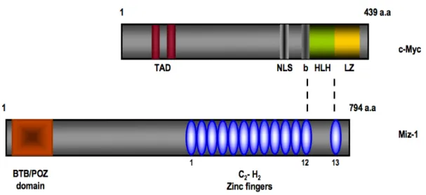
Growth factor independent-1 (Gfi1) and its closely related paralogue Gfi1b are transcription factors with a SNAG domain at the N-terminus and six zinc fingers at their c-terminal end [126-129]. The region between these two domains is smaller in Gfi1b than Gfi1 and is not conserved between the two proteins. Gfi1 is a transcriptional repressor that also plays essential roles outside of the hematopoietic system. For example, Gfi1 is important for the inner ear development [130, 131] and for the development of subsets of intestinal cells [132, 133]. Moreover, the SNAG domain repressor Gfi1 can regulate cell cycle progression through its interaction with the zinc finger protein Miz-1 (Myc-interacting zinc finger protein-1) [134]. Miz-1 is a transcriptional transactivator or transrepressor depending on its interacting partner and the best documented activation potential of Miz-1 is exemplified by its regulation of the expression of two genes encoding
for the cyclin-dependent kinase inhibitors Cdkn2b (p15INK4B) and Cdkn1a (p21CIP1) [135-138]. It has been shown that Gfi1 can bind to the cell cycle inhibitor Cdkn1a promoter through the zinc finger protein Miz-1, thereby influencing the outcome of its transcriptional regulation [134]. In this study, the association of Gfi1 with Miz-1 seems to allow Gfi1 to control cell proliferation in response to TGFβ stimulation [134]. Another study showed that the Miz-1/Gfi1 complex was important to repress the transcription of another cell cycle inhibitor, Cdkn2b [139]. This mutual recruitment of Gfi1 and Miz-1 to target gene promoter represents an novel transcriptional regulation that require more work in order to elucidate the full potential of this mechanism.
During hematopoiesis, Gfi1 and Gfi1b are differentially expressed [140]. Myeloid cells, in particular granulocytes, express high levels of Gfi1 whereas Gfi1b is completely absent. Gfi1 is also expressed in activated macrophages, granulo-monocytic precursors (GMP), stem cells, thymocytes and developing B and T lymphocytes, but absent or expressed at low levels in mature resting B and T cells, respectively [126, 130, 141-144]. Loss of Gfi1 in mice affects early B- and T-cell development [145] and neutrophil development [142, 146]. In addition, the deletion of Gfi1 in mice affects the frequency and function of HSC and greatly affects CLP numbers in the bone marrow. Moreover, Gfi1 is important to restrains the proliferation of HSC in order to control their self-renewal capacity and long-term engraftment abilities [147]. It also protects HSC from stress-induced apoptosis [148]. As for the implication of Gfi1 in B-cell development, it was demonstrated that
Gfi1-deficient B cell precursors can no longer integrate the signals initiated by the IL-7/IL-7R cascade [149].
Conversely, Gfi1b is absent from granulocytes, activated macrophages that reside in the bone marrow and GMP. Gfi1b is not expressed in almost all stages of T-cell differentiation, but it is present in early steps of B-cell development [144]. Moreover, Gfi1b is highly expressed in hematopoietic stem cells and in erythroid precursors, megakaryocytes and their progenitor cells (MEP), where Gfi1 is mostly undetectable [144, 150, 151]. The deletion of Gfi1b is lethal at mid-gestation probably because of the disrupted erythroid and megakaryocyte development [144, 150]. To circumvent this lethality, conditional deletion of Gfi1b in adult mice was achieved and showed that the deficient HSC significantly expanded in the bone marrow and blood. This expansion correlates with increased levels of reactive oxygen species and disturbed expression of cell surface receptors that mediate stem cells niche localization [148]. The co-expression of Gfi1 and Gfi1b was observed in early stages of B cell and in a subset of early T cells, suggesting a tight regulation of both transcriptional factors in a cell-specific manner [143, 144].
2.6. Transcriptional regulation of B- versus T-lineage choice
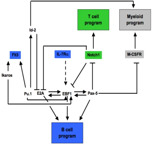
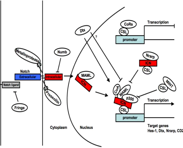
The dichotomy between Notch signals and Paired-homeodomain transcription factor Pax-5 dictates the T- versus B-lineage commitment respectively. Notch signals activate RBPJ (Recombining binding protein suppressor of hairless) transcription factor, also known as CSL (CBF-1, Suppressor of Hairless, Lag-1) which triggers and sustains a T lymphocyte
program while blocking any B-cell differentiation potential. Conversely, Pax-5 directs the B cell program while blocking the T cell fate, in part by inhibiting Notch1 expression [152-154]. These transcription factors are key-players in the cell-type specific regulation of lineage fate. They can repress or inhibit a cascade of other factors important for alternative pathways in order to activate the cell-specific fate (Figure 3) [155].
Figure 3. A simplified version of the transcriptional network governing B-, T- and myeloid lineage differentiation. Key surface receptors for B cells (blue), T cells (green) and myeloid cells (gray) are shown in rectangles. Transcription factors involved in different stages of hematopoiesis are in black text. Positive gene regulation is represented by arrows, while barred lines represent transcriptional repression. Indirect interactions are in dashed arrows (adapted from [40]).
3. B-cell development
B cells are typical representative members of the adaptive immune system because they provide both a specific response and a long-lasting protection against invading pathogens. B cell activation is initiated following the recognition of antigens by the BCR which results in cell proliferation and differentiation. Activated B cells can differentiate into either plasma cells that are responsible for antibody secretion or memory cells that provide protection during secondary responses against the same invading pathogen [156]. The proper functioning of B effector cells is coordinated during the immune response. Most importantly, it is tightly regulated earlier during development to assure that only functional mature B cells populate peripheral lymphoid organs to mount efficient responses against potential pathogens and infections.
3.1 Early stages of B-cell differentiation
Emergence of CLP from LMPP progenitors is marked by the up-regulation of IL-7Rα (CD127) expression that, together with the common γ chain (CD132), forms the IL-7R [157]. The IL-7Rα chain is also a component of the thymic-stromal-derived lymphopoietin (TSLP) receptor [158]. Given that IL-7Rα is required for two different receptors that function in B-cell development, it is to be noted that IL-7Rα-deficient mice have more pronounced B cell deficiencies compared to mice lacking the other chain of IL-7R, the common γ chain, since the γ-deficient mice have an intact TSLP-R [37, 159]. Mice lacking the IL-7 cytokine itself (IL-7-/-) have a similar phenotype to IL-7Rα-deficient mice, but their B-cell development arrest at a later stage [38]. This difference is probably caused by other
cytokines that use the IL-7Rα, such as TSLP. IL-7-/- mice also showed similar lymphocyte abnormalities to γ-deficient mice [160], even if this receptor unit is shared by other common γ chain user cytokines. These studies indicate that the most severe lymphocyte developmental abnormalities observed in γ-deficient mice are mainly due to IL-7 and not the other cytokines that bind to this receptor.
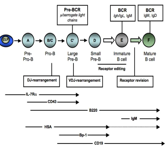
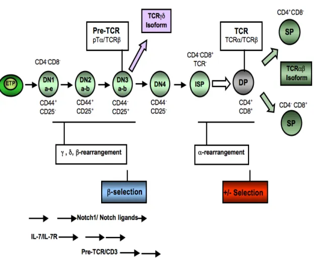
Although CLP express IL-7Rα, this signaling cascade is not necessary for the generation of these progenitors, but is crucial for their ability to differentiate into pre-pro-B lymphocytes and to undergo cytokine-induced expansion [161, 162]. B-cell commitment can be analyzed by tracking the surface markers B220 (CD45R), HSA (CD24), CD43 and CD19 which follow a precise ordered expression pattern. The most immature B cells express B220 and CD43, whereas more mature stages start expressing HSA and CD19 [163]. The B220+ subset in the CD19-negative cells is also referred to as fraction (Fr) A, which contain the uncommitted pre-pro-B cells (B220+CD19-) and are the source for committed CD19+ pro-B cells (B220+CD19+CD43+IgM-). These cells can be further subdivided into Fr B and Fr C or pro-B cells, that differentiate into Fr C-prime or early/large pre-B cells and ultimately give rise to Fr D or late/small pre-B cells [164, 165] (Figure 4).
The transition from pro-B cells to pre-B cells is the first stage of B-cell development that is accompanied by the expression of the pre-B cell receptor. This pre-BCR is composed of the surrogate light chain, VpreB or Vλ5, that pairs with the immunoglobulin µ heavy chain (Igµ) in large pre-B cells [166].The diversity of the BCR repertoire is generated by the
variable (V), diversity (D), joining (J) recombination events that are sequentially coordinated during B-cell development [166]. The immunoglobulin heavy chain (Igh) locus undergoes rearrangements in the pro-B cells, and successful rearrangement leads to the expression of the Igµ heavy chain protein. Subsequently, the recombination of the Ig-light chain genes Igk or Igl takes place later in the small pre-B cells, which replaces the surrogate VpreB and Vλ5 chains resulting in the expression of the BCR [166] (Figure 4).
Diversity is generated during these early B-cell differentiation steps by random rearrangement of the immunoglobulin genes. This process mainly generates auto-reactive immature B cells that are eliminated from the repertoire by clonal deletion [167] or by the induction of anergy, which renders auto-reactive B cells non-responsive to a BCR-mediated simulation [167, 168]. Another mechanism to eliminate auto-reactive B cells is receptor editing, where a new Ig gene rearrangement occurs to generate a new light chain to pair with the existing Ig heavy chain [169-171]. The new combination, if successful, will generate non-self reactive BCR which prevents the cells from deletion by apoptosis [172]. Some auto-reactive B cells can also be generated later in the development, after mature B cells leave the bone marrow and enter the spleen where they undergo somatic mutations of the Ig variable region genes. Hence, tolerance mechanisms operate in the periphery such as receptor revision [173, 174] or by blocking activated B cells from becoming antibody-secreting plasma cells [175] (Figure 4).
Figure 4. Illustration of B-cell development in the bone marrow. The B-cell lineage is initiated by a common lymphoid precursor (CLP) that give rise to cytokine-dependent pro-B cells. Pro-pro-B cells start V(D)J recombination and the µ heavy chain is produced in the large pre-B cell stage. Cells that fail to produce this chain are eliminated by apoptosis. In large pre-B cells, the µ heavy chain pairs with two surrogate light chains, λ5 and VpreB, and form the pre-B cell receptor (BCR). The pre-BCR signaling mediates proliferation and differentiation of small pre-B cells. Subsequently, rearrangement of the kappa and lambda light chains takes place and a fully assembled BCR, mainly IgM molecules at this stage, is expressed on the surface of immature B cells. These immature B cells emigrate from the bone marrow to further maturate in the spleen and lymph nodes where they encounter antigen and go through the process of receptor editing. The different B cell maturation steps are defined by the presence of specific markers, some of which are indicated (adapted from [176-178]).
3.2. B-cell differentiation and cytokines
Adult B-cell development takes place in the bone marrow through well-defined steps involving cytokine signaling, V(D)J recombination and gene regulation [179, 180]. The differentiation, proliferation and survival of early B-cell progenitors are dependent on cytokine signaling, in particular on Flt3 and IL-7R [27, 181, 182]. Most of the relevant cytokines for B-cell development are produced in the bone marrow by stromal cells that reside in the medullary cavity. Stromal cells that support pre-pro-B cells are CXCL12+IL-7 -, whereas those that support pro-B cell differentiation are CXCL12-IL-7+ [183].
IL-7 has been documented by many groups to be a necessary B lymphopoietic factor since B-cell differentiation does not occur in IL-7R-deficient mice [38]. The expression of the pre-BCR is dependent on stromal cell interactions and IL-7 receptor signaling. This signaling up-regulates the expressions of the IL-7Rα chain itself and the enzyme terminal deoxynucleotidyl transferase (Tdt), responsible for adding nucleotides at the joining region between V and D segments during recombination [184]. However, adult and fetal B lymphopoieses differ in their requirements for IL-7. For example, B-1 B cells, which are mainly produced during fetal development, may be independent of IL-7 since they are still produced in IL-7-deficient mice [185]. This may be attributable to the compensatory effect of thymic stromal lymphopoietin on fetal B cell progenitors [159, 186]. Contrary to IL-7-deficient mice, B-1 B cells are severely impaired in the absence of the IL-7 receptor. This indicates that the IL-7R is the key factor for adult B-cell development [187].
3.2.1. IL-7/IL-7R signaling in B cells
IL-7R signaling activates three major axes or pathways named after the Janus kinase- signal transducer and activator of transcription (JAK-STAT), the phosphatidylinositol 3-kinase (PI3K)-Akt and the RAS-mitogen-activated protein kinase (MAPK) [188]. The IL-7/IL-7R signaling cascade is induced when IL-7 binds to its receptor, which activates JAK3 that phosphorylates the IL-7Rα-associated JAK1 and the IL-7Rα chain itself. The phosphorylation of JAK proteins creates a docking site for STAT5, which is itself activated by phosphorylation following its recruitment to IL-7Rα chain [157]. The transcription factor STAT5 consists of two related isoforms, STAT5A and STAT5B, encoded by separate genes. In lymphocytes, STAT5A and STAT5B play redundant roles as the deletion of either one of them has only minor consequences on cellular functions [189]. Phosphorylated STAT5 proteins dimerize and translocate to the nucleus where they activate the transcription of IL-7-dependent target genes [157, 189] (Figure 5).
One outcome of IL-7R signaling is the maintenance of cell survival by promoting a positive balance of Bcl-2-family members. This is achieved by increasing the expression of anti-apoptotic Bcl-2 (B-cell CLL/lymphoma-2) and Mcl-1 proteins (Myeloid cell leukemia sequence 1, Bcl-2-related), and by redistributing the cell-death proteins Bax (Bcl-2-associated X protein) and Bad (Bcl-2-antagonist of cell death) [190]. On the one hand, pro-apoptotic Bax, Bad and Bak (Bcl-2-antagonist/killer) proteins form homo-oligomeric pores in the mitochondrial membrane and are critical for cytochrome c release. On the other hand, Bcl-2, Mcl-1 and Bcl-XL (Bcl-2-like 1) are anti-apoptotic proteins that maintain