1
VISUOTOPY & OPTIC FLOW
PROCESSING IN MONKEY’S
VISUAL CORTEX:
AN FMRI INVESTIGATION
Visuotopie et traitement du flux optique dans le cortex visuel du singe :
une investigation par IRMf
3
Acknowledgements
Starting acknowledgements is never an easy task, for the rules of language impose that you start somewhere, implicitly prioritizing what you are thankful for. I wish that all people being thanked here, know that you were pillars in the elaboration of this work, for each of you have brought exactly what was needed, at the right time and the right place. Here goes nothing.
Jean-Baptiste, you have been the older brother I have never had. You are a true role model and I know that all PhD students in this lab, wished to have you as a supervisor. You taught me patience and perseverance, you taught me how to love what I am doing despite the hard times, you taught me the true meaning of being a mentor. You trusted me when you sent me to Leuven, you trusted me when I came back. I think you knew I would come back. I shouldn’t make this longer than it needs to be. You gave me a lot, and I know my job is to transmit that to as many students as I can.
Asma et Chaoukat, mes parents, sans vous tous cela n’aurait pas été possible. Non pas parce que vous m’avez donné la vie, mais parce que vous m’avez donné l’envie, et le plaisir de la vivre. Ingrid, je te remercie parce que tu as persisté. Je t’aime parce que tu fais sortir le mieux en moi. Je te remercie parce que tu m’as aidé et soutenu comme personne ne sait le faire.
Youn, Benoît & Marcello. You have also been great teachers and friends, the interactions we had were of utmost significance, both scientifically and humanly. I wish we could continue working together. I love you guys.
Je remercie le Croissant Fertile et tous mes camarades, avec qui je partage ce projet de vie, de renaissance !
Je remercie l’équipe de l’animalerie, passée et présente. Camille et Emilie, je vous remercie aussi de votre aide, mais surtout de votre ténacité, qui m’a appris à être carré (pas autant que vous l’auriez souhaité peut-être, mais ça viendra, promis) dans mes interactions avec les animaux. Je remercie aussi Nathalie, Jean-Pierre, Yohann, Fred et Hélène. Vous avez été extrêmement aimables et surtout indispensables. C’était un plaisir de travailler avec vous, et surtout de prendre l’apéro à l’IRM lorsqu’on un animal a bien travaillé !
I thank also all of the people I met at the CerCo, past and present members. This network of exceptional people has been the birthplace of original ideas, solid friendships and unforgettable memories.
4
“The seeker after truth is not one who studies the writings of the ancients and, following his natural disposition, puts his trust in them," the first scientist wrote, "but rather the one who suspects his faith in them and questions what he gathers from them, the one who submits to argument and demonstration and not the sayings of human beings whose nature is fraught with all kinds of imperfection and deficiency. Thus, the duty of the man who investigates the writings of scientists, if learning the truth is his goal, is to make himself an enemy of all that he reads, and, applying his mind to the core and margins of its content, attack it from every side. he should also suspect himself as he performs his critical examination of it, so that he may avoid falling into either prejudice or leniency.” ― Ibn al-Haytham هناميإ قّلعُي نم وه لب ،اهيف هتقث عضيو اهتلاح ىلع ،ءامدقلا تاباتك سردي نم وه سيل ةقيقحلا نع ثحابلا نإ لاو ،ةجحلا نع ثحبي يذلا وه .مهنم هانج يذلا ام لءاستيو مهب عاونأ لك اهلأمي هتعيبط ناسنإ لاوقأ ىلع دمتعي ،هفده ةقيقحلا نع ثحبلا ناك اذإ ،ءاملعلا تاباتك يف ققحي نم ىلع بجاولا نم نإ ف يلاتلابو .روصقلاو صقنلا يف ككشتي نأ هيلعو بناج لك نم راكفلأا كلت ثحبل عاخنلا ىتح هل قع مدختسيو ،هأرقي ام عيمج ركنتسي نأ وه هتسارد جئاتن .لهاست وأ زيحت يأ يف عوقولا بنجتي ىتح ،اًضيأ -مثيهلا نبا
5
Preamble
My path towards academic research started when I conducted my first internship in the field of Neuroscience with Dr. Jean-Baptiste Durand during the first year of my Master’s degree. I had met J-B’s brother during one of my courses and we had become close acquaintances. During a conversation about our career goals, I had mentioned to him my will to work in Neuroscience. Promptly he told me that his brother was a researcher in the domain at the CNRS and that I should contact him. In addition to getting frisky with a male macaque’s teeth, my first in-depth research experience under the supervision of J-B was extremely motivating and enriching. He had all the right mentorship skills to convey his passion towards his area of expertise. It was during the time the CerCo had first moved to a new location and the MR platform had just entered service. J-B, along with some of his colleagues had the daunting task of putting together the awake monkey fMRI technique from scratch, with very limited monetary, technical, and human resources. At the end of my internship, and after having accumulated all the positive energy from that inspiring team, I asked J-B to help me find a lab in which I could familiarize myself better with monkey fMRI. Happy with my request, he enthusiastically recommended me to the Leuven lab in which he performed one of his postdocs, to conduct my master’s thesis under the supervision of Pr. Wim Vanduffel and Dr. John Arsenault.
The object of my master’s thesis was the study of the role of the primate ventral tegmental area in reinforcement and motivation. This internship was a true challenge, as it opposed me to my deepest fears and weaknesses. Extensive work and sleepless nights, during which I had to learn new complex skills, have profoundly changed me. Nevertheless, I am grateful to have endured them. During this internship, I have gained a lot knowledge about the neuroscience of reward. I also learned to manipulate macaques, I have assisted in many surgeries and learned to operate an MRI scanner and acquire and analyze fMRI data. I also learned a great deal about chronic implanted electrodes and electric microstimulation. Amid that tsunami of information and struggle to adapt
6 to a new society, I realized that I had surmounted a lot of obstacles and broken many barriers within my own psyche. I had learned to seek new experiences instead of avoiding them, to boldly defend my ideas and even to go meet my idols. Antonio Damasio was invited to Leuven to be awarded an honorary doctorate, and gave a talk in the university, which I attended. I went to meet him, and we had a long hour discussion about everything, from science to politics, and I had finally asked him to be a reviewer for my thesis, which he accepted.
Upon the end of my master’s thesis and having co-authored a paper published in Current Biology (Annex 1), which is an annex in the present manuscript, I was resolved to come back to Toulouse and conduct a PhD under the supervision of J-B. I applied to and won a grant from a Lebanese organization, which then allowed me to contact J-B and discuss with him that opportunity. I came back to the CerCo because I wanted to take part in developing the awake monkey fMRI technique to be able to implement elsewhere. But most importantly, I wanted to continue studying the primate visual cortex.
Knowing that the development a technique as heavy and complex as awake monkey fMRI, and publish with that technique within the 3 years of my PhD funding, was a risky bet. Which is why I developed a secondary axis of research which relates to the subject of my Master’s thesis. Fortunately, we were not only able to finish developing the technique and publishing the first awake monkey 3T fMRI paper in France, but we also managed to submit a paper describing a new visuotopic cluster in the monkey posterior parietal cortex. Furthermore, we submitted a study about the effects of reward on visual perception, which is included as an annex in this manuscript (Annex 2).
7
Summary
Functional magnetic resonance imaging (fMRI) allows addressing the functional organization of the human brain with minimal invasiveness and in healthy individuals. The implementation of that technique in non-human primates represents an important achievement in systems neuroscience. On the one hand, monkey fMRI contributes to the reduction and refinement of invasive approaches in non-human primates, by revealing the regions of interest in which focal electrophysiological and/or anatomical investigations should be carried out. On the other hand, the knowledge acquired with such invasive approaches can be more safely transposed to humans, once inter-species homologies and differences have been identified through the use of similar fMRI protocols in human and non-human primates.
The first part of this thesis reviews the most common approaches that have been used to study brain functions, either in humans or in non-human primates. It is shown that despite progresses in the human approaches, invasive studies in monkeys remain necessary for understanding the neuronal mechanisms underlying cognitive functions. Then follows a description of the evolution of the monkey fMRI techniques and some of its achievements in bridging the gap between non-invasive human studies and invasive animal studies, notably for deciphering the neural mechanisms supporting visually-guided grasping. The end of this first part is purely methodological. It undertakes the description of the monkey facilities and the MR platform in Toulouse, and details the necessary milestones for conducting fMRI research in macaque monkeys. The second part of the thesis presents the 4 studies we have conducted with monkey fMRI. The first study is a preparatory experiment for characterizing the monkey hemodynamic response function, which is a prerequisite for proper analysis of subsequent monkey fMRI data. The second study addresses the visuotopic organization of the primate dorsal visual cortex with a novel technique of wide-field (80°) phase-encoded visual stimulation, coupled with a state of the art surface-based analysis of population receptive fields. The results obtained in 2 animals uncover a new cluster of visuotopic areas in the posterior parietal cortex of the macaque monkey, bringing a fresh view to the functional organization of this piece of cortex and opening a promising avenue for inter-species comparisons. The third study unveils the cortical network involved in optic flow processing in non-human primates and it compares this network to that recently described in humans. To that end, we replicated in macaque monkeys an experiment previously conducted in human subjects with optic flow stimuli that are either consistent or inconsistent with egomotion. Besides confirming the involvement of areas previously identified through electrophysiological recordings, our results reveal new cortical areas involved in the processing of optic flow, drawing the picture of a network sharing many similarities, but also striking differences, with that documented in the human brain.
In summary, the ambition of this thesis is two-fold: (1) providing guidelines for setting-up monkey fMRI techniques, drawn from our own experience and (2) exposing a set of studies we have conducted with this approach, dealing with the visuotopic organization of the dorsal visual cortex and its involvement in the processing of visual motion. Besides bringing a fresh view to the functional organization of the dorsal visual pathway in non-human primates, these studies illustrate how monkey fMRI bridges the gap between electrophysiological studies in non-human primates and functional imaging studies in humans.
Keywords: Visuotopic organization, motion processing, visual cortex, non-human primates
8
Résumé en français
L'imagerie par résonance magnétique fonctionnelle (IRMf) permet d'examiner l’organisation fonctionnelle du cerveau humain de manière non-invasive et chez les sujets sains. L’implémentation de cette technique chez des primates non-humains représente un progrès important dans les neurosciences des systèmes. D’une part, l'IRMf singe permet la réduction et le raffinement des protocoles invasifs impliquant des primates non humains, en révélant les régions d’intérêts dans lesquelles les approches focales invasives, électrophysiologiques ou anatomiques, devraient être menées. D’un autre côté, les connaissances acquises avec ces approches invasives peuvent être transposées plus aisément à l’homme, une fois que les homologies et différences interspécifiques ont été identifiées au travers de protocoles d’IRMf menées en parallèle chez les primates humains et non-humains.
La 1ère partie de cette thèse présente les approches conventionnelles d’étude des fonctions cérébrales. Nous montrons que des études invasives chez l’animal demeurent nécessaires pour comprendre les mécanismes neuronaux qui sous-tendent nos fonctions cognitives, malgré le progrès des techniques d’investigation chez l’homme. Suit une revue sur l'évolution des techniques d’IRMf singe et certaines de ses réalisations majeures comme pont dressé entre les études non-invasives menées chez l’homme et les études invasives réalisées chez l’animal, notamment en ce qui concerne notre compréhension des mécanismes neuronaux permettant la saisie manuelle d’objets sous contrôle visuel. Purement méthodologique, la fin de cette 1ère partie décrit l’animalerie et la plate-forme d’IRM à Toulouse et expose les jalons de l’implémentation de l’IRMf chez le singe macaque vigile. La 2ème partie de la thèse présente les 4 études que nous avons menées en IRMf singe. La 1ère étude modélise la réponse hémodynamique chez le singe, un outil indispensable à l’analyses de données d’IRMf, acquises dans les études suivantes. La 2ème étude traite de l'organisation visuotopique du cortex visuel dorsal des primates, et y décrit un nouvel assemblage d’aires visuotopiques chez 2 animaux, grâce à l’usage de nouvelles techniques de stimulation visuelle et d’analyse de champ récepteurs. Ces résultats apportent un point de vue neuf sur l'organisation fonctionnelle de la voie visuelle dorsale et ouvrent de nombreuses perspectives pour les comparaisons entre espèces. La 3ème étude cartographie le réseau d’aires corticales impliqué dans le traitement du flux optique chez les primates non humains et le compare à celui décrit récemment chez l’homme. Grâce à la réplication d’une étude réalisée chez l’homme, nous avons confirmé chez 3 macaques l'implication de zones précédemment identifiées par des enregistrements électrophysiologiques. Nos résultats révèlent de nouvelles zones corticales impliquées dans le traitement du flux optique, dessinant l'image d'un réseau cortical partageant de nombreuses similitudes, mais ayant également des différences frappantes, avec celui documenté dans le cerveau humain.
En résumé, l'ambition de cette thèse est double : (1) fournir des recommandations pour la mise en place de techniques IRMf chez le singe, tirées de notre propre expérience et (2) exposer les résultats d’un ensemble d'études que nous avons menées avec cette approche, traitant de l'organisation visuotopique du cortex visuel dorsal et de son implication dans le traitement du mouvement visuel. En plus d'apporter une perspective nouvelle sur l'organisation fonctionnelle du cortex visuel chez les primates non humains, ces études illustrent la contribution de l’IRMf singe comme pont entre études électrophysiologiques chez les primates non humains et études d'imagerie fonctionnelle chez l'homme.
Mots clés : Organisation visutopique, traitement du mouvement visuel, cortex visuel, primates non-humains
9
Résumé long en français
Le développement de l’IRMf (Imagerie par Résonance Magnétique fonctionnelle) chez des primates non-humains (PNH) a instauré la possibilité d’établir le lien tant attendu entre les données issues d’études invasives chez le PNH et celles d’imagerie fonctionnelle obtenues chez l’humain. Cette technique représente un progrès important car elle permet la réduction et le raffinement des protocoles invasifs impliquant des primates non humains, en révélant les régions d’intérêts dans lesquelles les approches focales invasives, électrophysiologiques ou anatomiques, devraient être menées. Par ailleurs, les connaissances acquises avec ces approches invasives peuvent être transposées plus aisément à l’homme, une fois que les homologies et différences interspécifiques ont été identifiées au travers de protocoles d’IRMf menées en parallèle chez les primates humains et non-humains. Conscient des avantages que fourni le modèle PNH, l’objectif primordial de cette thèse était le développement de la technique IRMf chez le singe éveillé au CerCo (Centre de recherche sur le cerveau et la cognition) à Toulouse, afin d’étudier l’organisation fonctionnelle de la voie dorsale chez cette espèce et de la comparer avec celle de l’Homme.
En guise d’introduction, cette thèse présente les approches conventionnelles d’étude des fonctions cérébrales et montre que des études invasives chez l’animal demeurent nécessaires pour comprendre les mécanismes neuronaux qui sous-tendent nos fonctions cognitives, malgré le progrès des techniques d’investigation chez l’homme. Suit une revue sur l'évolution des techniques d’IRMf singe et certaines de ses réalisations majeures comme pont dressé entre les études non-invasives menées chez l’homme et les études non-invasives réalisées chez l’animal, notamment en ce qui concerne notre compréhension des mécanismes neuronaux permettant la saisie manuelle d’objets sous contrôle visuel. Cette revue bibliographique est suivie par un chapitre purement méthodologique, qui décrit l’animalerie et la plateforme IRM du pavillon Baudot qui accueille le CerCo. Cette partie expose les étapes qui ont permis l’implantation des techniques d’IRMf singe sur la plateforme. Elle détaille la mise en place des 2 postes de conditionnement qui, en mimant
10 l’environnement de l’IRM, permettent la familiarisation des animaux aux différentes contraintes de ce milieu et leur entrainement aux différentes tâches qu’ils doivent pratiquer pendant les sessions d’acquisition d’images fonctionnelles. Sont discutés aussi le développement d’une antenne 8 canaux dédiés à l’imagerie chez les macaques et la création d’outils d’analyses des données fonctionnelles et oculométriques.
Dans la continuité de la description de la mise en place de l’IRMf chez le singe éveillé à Toulouse, la partie suivante décrit la modélisation de la réponse hémodynamique du cortex visuel du singe macaque. L’IRM n’offre qu’une mesure indirecte de l’activité neuronale, qui repose sur la détection des perturbations du champ magnétique dans un volume de cerveau défini (voxel) induit par une modification du rapport d’hémoglobine oxygénée/hémoglobine désoxygénée et dont la dynamique est nommée réponse hémodynamique (HRF). L’analyse des données d’IRMf dépend d’une bonne estimation de la HRF, qui est non seulement propre à chaque espèce, mais peut aussi varier entre les différentes aires du cerveau. Dans cette étude préliminaire, nous avons estimé la HRF du cortex visuel sur 3 macaques, à partir des activations IRM induites par une impulsion visuelle (l’affichage très bref d’un damier scintillant couvrant une grande partie du champ visuel) et d’une fonction modèle de type double-gamma. Les paramètres de ce modèle sont discutés en termes de variabilités interindividuelles et interspécifiques.
Ce modèle de HRF est ensuite utilisé dans 2 études. Dans la première, qui constitue le cœur de ma thèse, je me suis intéressé à l’organisation visuotopique de la voie dorsale chez le singe macaque. Le cortex visuel du macaque a été extensivement étudié et de multiples modèles de son organisation fonctionnelle ont été proposés. Cependant, l’organisation visuotopique de certaines aires et notamment celles de la voie dorsale demeurent débattues. Plusieurs difficultés surgissent lors de l’investigation de cette région de cortex. D’abord, les aires visuelles de la voie dorsale présentent de grands champs récepteurs couvrant les zones les plus périphériques du champ visuel. De plus, beaucoup de ces zones ne présentent pas une organisation rétinotopique conventionnelle du fait
11 qu’elles traitent aussi d’autres modalités sensorielles que la vision. Enfin, l’attention semble jouer un rôle important dans l’activation de ces zones. Ainsi, l’activation de cette région requiert des stimuli capables de couvrir la quasi-totalité du champ visuel et qui présentent des attributs d’importance comportementale, pouvant attirer l’attention des sujets.
Grâce à l’emploi d’une technique de stimulation large-champ (80° d’excentricité) et l’utilisation d’un stimulus visuel représentant un panier de fruits en mouvement comme stimulus, nous avons été capable d’activer de façon robuste une grande partie de la voie visuelle dorsale chez 2 macaques. Suite à la projection des données volumétriques sur la surface corticale, nous avons utilisé un filtre fréquentiel avant d’effectuer une analyse des champs récepteurs de population. L’analyse des résultats issus de ces traitements nous a permis de détecter des inversions de gradients d’angle polaire et d’excentricité des champs récepteurs de population le long de la surface corticale, qui confirment et synthétisent les connaissances acquises au moyen d’approches invasives sur ce modèle. Ces inversions de gradients révèlent de plus les frontières d’un nouvel assemblage d’aires visuotopiques. Cet assemblage d’aires (« cluster »), nommé PIP (posterior intraparietal) est situé dans la partie postérieure du sillon intra pariétal et est constitué de 2 aires récemment décrites (CIP 1/2) et de 2 nouvelles aires (PIP 1/2). Ainsi, grâce à la combinaison de l’IRMf et de la cartographie rétinotopique large-champ, nous avons décrit des structures organisationnelles non caractérisée jusque-là, que ça soit par des méthodes invasives ou des méthodes de cartographie rétinotopique traditionnelles.
La seconde étude avait pour but de caractériser le réseau d’aires corticales impliquées dans le traitement du flux optique compatible avec le mouvement de soi. Une étude récente publiée par Andrew Smith du Royal Holloway à l’université de Londres, décrit l’implication d’un large réseau d’aires corticales dans cette fonction, bien plus étendu que ne pouvait suggérer les données invasives chez l’animal. Cette étude confirme certaines aires qui correspondent à des aires homologues trouvées chez le singe par électrophysiologie mais décrit surtout une aire qui semble
12 être particulièrement sélective au mouvement de soi. Cette aire dénommé CsV (Cingulate sulcus Visual) ne possède aucun homologue connu chez le singe. En collaboration avec Andrew Smith, nous avons répliqué l’étude menée chez l’homme afin de caractériser le réseau cortical impliqué dans le traitement de mouvement et d’évaluer un homologue de l’aire CsV chez le singe. En contrastant l’activité corticale résultante d’un stimulus de flux optique compatible avec le mouvement et celle du même stimulus répliqué 9 fois (3x3) sur l’écran (incompatible avec le mouvement de soi), nous avons révélé un réseau cortical qui soutient les données électrophysiologiques et anatomiques précédemment décrites chez le PNH. La comparaison de ces données avec les données humaines révèlent que malgré le fait que les réseaux corticaux chez les 2 espèces partagent des similitudes (MST/hMST, VIP/hVIP,VPS/PIC et pmCsV/CsV), ils demeurent différents. D’une part, certaines aires sont propres au singe et ne sont pas retrouvées chez l’humain, tel que les aires 7a, STPm, FEFsem et FEFsac. D’autre part, des activations robustes des aires V6, P2V et Pc ne sont pas retrouvées chez le singe.
Cette thèse poursuivait un double objectif :1) fournir des recommandations pour la mise en place de techniques IRMf chez le singe, tirées de notre propre expérience et (2) exposer les résultats d’un ensemble d'études que nous avons menées avec cette approche, traitant de l'organisation visuotopique du cortex visuel dorsal et de son implication dans le traitement du mouvement visuel. En plus d'apporter une perspective nouvelle sur l'organisation fonctionnelle du cortex visuel chez les primates non humains, ces études illustrent la contribution de l’IRMf singe comme pont entre études électrophysiologiques chez les primates non humains et études d'imagerie fonctionnelle chez l'homme.
Mots clés : Organisation visuotopique, traitement du mouvement visuel, cortex visuel, voie dorsale, primates non-humains
13
Table of content
A. General Introduction... 17
A.1 Human approach to cognitive neuroscience ... 19
A.1.1 Human lesions ... 19
A.1.2 Brain imaging ... 21
A.1.3 Causal approaches ... 24
A.1.4 Neural recordings in patients ... 24
A.2 Non-human primates: a necessary alternative ... 25
A.2.1 Anatomical exploration of NHP cortex ... 26
A.2.2 Monkey neurophysiology: the backbone of modern neuroscience... 26
A.2.3 Monkey lesion and interference ... 27
A.2.4 Complications in relating human and monkey studies ... 28
A.3 Conclusion ... 30
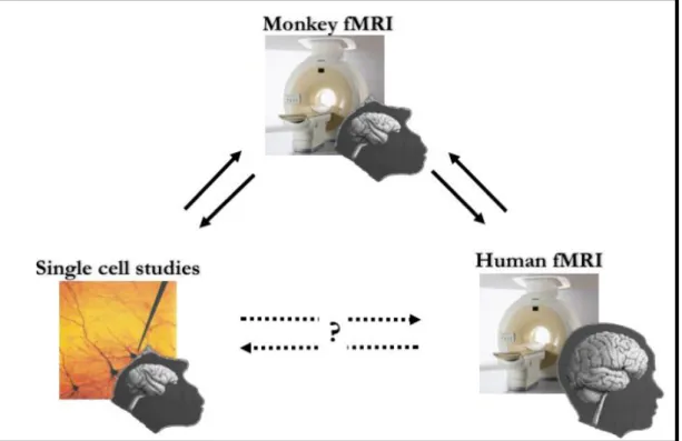
B. fMRI bridges the gap between single cell and human imaging studies ... 31
B.1 Closing the loop ... 33
B.2 Monkey fMRI: the journey ... 35
B.2.1 First attempts ... 35
B.2.2 The battle for SNR ... 36
B.2.3 Pedal to the metal: High-field vertical fMRI ... 37
B.2.4 On the home stretch: democratizing monkey fMRI ... 37
B.3 Feather in the cap: some achievements of monkey fMRI ... 39
B.3.1 Monkey fMRI sheds light on the nature of the BOLD signal ... 39
B.3.2 The grasping network ... 40
B.3.3 Comparing human and monkey neuroimaging ... 43
B.4 Conclusion ... 45
C. Methodology for Monkey fMRI ... 47
C.1 Monkey fMRI facilities in Toulouse ... 48
C.2 Creating the setups: an environment for fMRI conditioning ... 50
C.3 Animal preparation ... 52
C.3.1 Legal authorizations ... 52
C.3.2 Surgical procedures ... 52
C.3.3 Behavioral training ... 53
14
C.5 Magnetic resonance imaging ... 55
C.5.1 Set-up overview... 55
C.5.2 Acquisition sequences ... 56
C.5.3 Anatomical and functional templates ... 57
C.5.4 Cortical surface reconstruction ... 57
C.6 Functional data processing ... 59
C.6.1 Functional signal: BOLD vs. IRON ... 59
C.6.2 Basic pre-processing steps ... 61
C.6.3 Visual field mapping techniques ... 62
C.6.4 Basic statistical analyses ... 63
D. Estimating the Monkey hemodynamic response function (HRF) ... 67
D.1 The hemodynamic response ... 69
D.2 Motivation to measure the HRF ... 70
D.3 Estimating the HRF in macaque monkeys... 71
D.3.1 Stimuli ... 71
D.3.2 Preprocessing ... 72
D.3.3 Data analysis ... 73
D.4 Results ... 74
D.5 Discussion ... 76
E. Wide-field retinotopy of macaque visual cortex ... 79
E.1 Retinotopy is a solution to costly wiring ... 81
E.2 Retinotopic mapping using fMRI ... 82
E.2.1 The standard paradigm: Travelling wave ... 82
E.2.2 Population receptive field modelling ... 83
E.2.3 Homologies of retinotopic maps in humans and monkeys ... 85
E.3 Investigating the visuotopic organization of the posterior parietal cortex (PPC) ... 87
E.3.1 Introduction ... 87
E.3.2 Material and Methods ... 88
E.3.3 Results ... 98
E.3.4 Discussion ... 110
F. Processing of egomotion-consistent optic flow in the rhesus macaque cortex ... 113
F.1 Introduction ... 115
F.2 Material and methods ... 118
F.2.1 Animal model ... 118
15
F.2.3 MRI recordings ... 120
F.2.4 Data processing ... 122
F.3 Results ... 127
F.3.1 Cortical network involved in processing optic flow ... 127
F.3.2 Quantitative analysis of egomotion selectivity ... 133
F.4 Discussion ... 140
F.4.1 Overview ... 140
F.4.2 Activations in temporal cortex: MSTd and STPm ... 140
F.4.3 Activations in parietal cortex: VIP, 7a and LIPd ... 142
F.4.4 Activation in parieto-insular cortex: VPS ... 144
F.4.5 Activations in frontal cortex: FEFsem and FEFsac ... 145
F.4.6 Activation in cingulate cortex: pmCSv ... 146
F.4.7 Homologies with the human EC-selective areas V6, Pc and 2v? ... 146
F.5 Conclusion ... 147
G. General discussion & Conclusion ... 149
G.1 Summary of results ... 151
G.2 Considerations pertaining to the awake monkey fMRI technique ... 151
G.3 Considerations pertaining to the measurement of the HRF ... 154
G.4 Considerations pertaining to the visuotopic mapping of the dorsal visual pathway ... 154
G.5 Considerations pertaining to the motion sensitivity of the dorsal visual pathway. ... 156
G.6 Perspectives ... 157
H. Bibliography ... 159
I. Annex 1... 185
17
19
A.1 Human approach to cognitive neuroscience
With its billions of interconnected neurons, the human brain is the most complex system in the known universe and as such, a paramount challenge for scientific research. Our brain supports recognition, attention, memory, decision making and many other higher cognitive functions by which we can challenge daily basic struggles, navigate, use tools, cooperate and build societies. Although the anatomy and physiology of many other organs can be reasonably apprehended through studies in rodents, taking advantage of their high similarities with human organs, studying the human brain is another level of dilemma. Many of the higher cognitive functions it operates are specific to humans or shared only with its closest relatives, i.e. the non-human primates. Understanding those functions requires in-depth knowledge of the underlying neural mechanisms, knowledge that spans across the functional organization of the brain to the dynamics of single neurons. Accessing this knowledge constitutes an ethical quandary, since it requires invasive procedures that are difficult to conduct in humans. However, much progress has been made through approaches involving notably human brain lesions studies and more recently, non-invasive functional imaging studies. Those approaches are briefly described in the following sections.
A.1.1 Human lesions
In 1861, Paul Broca boldly suggested that the third convolution of the inferior frontal gyrus is involved in speech production. Support for this claim came from the brain of a patient (Figure 1) who had been able to produce only one syllable (tan), in the form of stereotyped recurrent utterances: tan-tan-tantan… Although Broca was not the first to relate language to the left hemisphere, his finding launched a new age of discovery. Specifically, Broca’s approach to localizing human brain function by studying the correlation between the location of a brain lesion
20 and a behavioral affliction founded a long tradition of neuropsychological research, which has greatly advanced our understanding of brain function.
Figure 1. Leborgne’s brain (source: Dronkers NF, et al., Paul Broca’s historic cases: high resolution MR imaging of the brains of Leborgne and Lelong. Brain 2007;130:1432-1441)
Although in historical terms, lesions caused by accidents, war injuries, strokes or even neurosurgical excisions were the pillar of neurophysiology for a long time (Cowan et al., 2000), there are several fundamental problems with the lesion method that have caused neuroscientists to become critical about the technique.
Firstly, in the case a lesion could be detected and localized, highly sophisticated psychological and physiological assessments are needed to be able to pinpoint the nature of the impairment caused by the lesion. Furthermore, many brain functions might be carried out in a distributed manner, with large portions of the brain working in a flexible fashion rather than each region having a fixed function. Consequently, it is very difficult to exclusively link a lesion to a cognitive function. Indeed, accidental lesions or lesions caused by trauma, almost always span across many cytoarchitectonic areas. Additionally, almost constantly concomitant white matter insults disengage the connection between different areas, which implies that it cannot be excluded that the diagnosed impairment is not due to an interruption of functional connectivity instead of a focalized lesion per se. Moreover, since a certain amount of time would have passed between the lesion and the beginning of its investigation, functional reorganization of the cortex would have most probably occurred, thus misleading our interpretation about the function of the area in the normal brain.
21 The human ‘lesion method’ was seminal for our understanding of functions as diverse as language, memory, hemispheric specialization, emotion, vision and motor control, but to relate behavioral functions to anatomy using the lesion approach, it is necessary to identify the location and extent of a brain injury. At the time when Broca conducted his work (in the 1860s), researchers had to wait for a patient to die before they could examine the brain, a new method was thus needed.
A.1.2 Brain imaging
Fortunately, William H. Olendorf got unsatisfied with the traumatic, tedious, invasive studies on his patients at the University of Minnesota Hospital. Indeed, these studies provided only limited and indirect information about the brain. So, he strived for something better. William Henry Oldendorf (1925–1992) was an American neurologist, physician, researcher, medical pioneer and founding member of the American Society for Neuroimaging (ASN). From his basement, he modeled a new instrument incorporating principles and hardware used in modern computed tomography (CT) scanners. The description of the model was published in 1961. Standard X-Ray manufacturers were not thrilled with Olendorf’s idea and dismissed it as impractical. A letter from one company ended: “Even if it could be made to work as you suggest, we cannot imagine a significant market for such an expensive apparatus which would do nothing but make a radiographic cross-section of a head”. Despite his failure to interest manufacturers with his invention, Oldendorf’s description was patented in 1963 and this has been of invaluable importance to researchers in the field of neuroimaging.
Soon after the invention of CAT (Computer Automated Tomography), the development of radioligands started the functional imaging revolution. Thanks to the work of Marcus Raichle and coworkers, functional imaging took a large step forward with the development of oxygen-15 labelled water (H2O15) imaging (Herscovitch et al., 1983). H2O15 emits positrons and creates images
22 H2O15 positron emission tomography (PET) allowed investigators to make regional maps of brain
activity during various cognitive tasks. Concurrently, magnetic resonance imaging (MRI or MR scanning) was developed. Scientists soon learned that the large blood flow changes measured by H2O15PET were also imaged by MRI. Functional magnetic resonance imaging (fMRI) was
born(Ogawa et al., 1990). Since the 1990s, fMRI has come to dominate the brain mapping field due to its low invasiveness, lack of radiation exposure, and relatively wide availability.
Studying brain functions in healthy subjects using very precise cognitive tasks performed inside the scanner provides access to peak activity that is restricted to cytoarchitectonic areas and that lie in grey and not white matter, without the worry of possible cortical reorganization such as can be found in patients. Whether PET or fMRI is used, the functions of an area can be inferred by contrasting tasks that differ in only one aspect. Nevertheless, showing that an area is active during a task does not necessarily means that it is mandatory for performing the task accurately, nor does it indicate its precise functional role. For example, many studies have reported activity in the anterior cingulate cortex when subjects are engaged in judgement tasks about the thoughts of others (theory of mind) (Gallagher 2003). However, a patient with a large bilateral lesion of this area had no problem in succeeding on similar tasks (Bird et al., 2004), questioning the central role of this area in such tasks. Recent developments in fMRI may also allow measuring the covariance in activity between various areas (Rogers et al., 2007), introducing the possibility to study functional interactivity between distant portions of the cortical surface. Yet these methods still rely on measures of correlation, a limitation which can only be resolved through interference with the activity of one of the areas. There are several other limitations to brain imaging techniques such as PET or fMRI: neither provides a direct measure of neural activity. The signal measured in fMRI is blood oxygen level dependent (BOLD), which is an indirect measure of blood flow. Granting that PET measures direct fluctuations of blood flow, both techniques indirectly measure the activity of a whole population of cells, with no information on specific cellular dynamics. Admitting that these
23 techniques are suitable for functional studies, the same cannot be said for the study of how functions are implemented at the neuronal level. The cellular machinery has a temporal resolution on the order of milliseconds, while PET has a temporal resolution of around 60 seconds and fMRI has a rough temporal resolution of 1-3 seconds. Consequently, we cannot fathom the order of events in the brain. The temporal caveats of fMRI and PET can be resolved with techniques such as the electroencephalogram (EEG) or the magnetoencephalogram (MEG). EEG measures voltage fluctuations resulting from ionic current within the neurons of the brain at a distance. The first human EEG was recorded in 1924 by German physiologist and psychiatrist Hans Berger (1873–1941) (Swartz, 2017). It is a non-invasive method that detects local variations of the electric field with electrodes placed along the scalp. EEG has a temporal resolution on the order of the millisecond and even the microsecond, which makes it ideal to study signal propagation. EEG is less subject to motion artifacts and it is a portable technique that can even be used on infants and claustrophobic subjects. One serious drawback is its poor spatial resolution. Indeed, because of the bad conductibility of the scalp, the measured electrical field fluctuations tend to disperse, making it difficult to localize their sources. The other technique with a high temporal resolution, is MEG. MEG signals were first measured by University of Illinois physicist David Cohen in 1968 (Cohen, 1968), It measures changes in magnetic fields on the surface of the scalp. The additional advantage over EEG is that it possesses a better spatial resolution, because magnetic field fluctuations are not distorted by the scalp. MEG is an expensive technique though, it is not portable and requires pairing with MRI for proper areal localization.
Until today, no single novel technique could fuse the advantages of fMRI and EEG/MEG. Although combined studies of fMRI and EEG have achieved noteworthy results(Mullinger and Bowtell, 2011; Huster et al., 2012), despite the inherent methodological problems that arise from the combination of these techniques, a tool with the ability to acquire both high temporal and spatial resolution data would be the holy grail of brain imaging.
24
A.1.3 Causal approaches
The aforementioned imaging techniques, no matter their resolution remain correlational approaches, they inform about the relationship between indirect measures of brain activity and behavioral performances in sensory, motor or cognitive tasks. However, knowing whether the recorded activity in a particular brain location is necessary for executing the task at hand or simply an epiphenomenon requires tampering with its activity. Application of electrical currents to modify/investigate brain functions is a technique that dates back to the 19th century (Selimbeyoglu,
2010). Methods such as trans-cranial magnetic stimulation (TMS) (Sparing and Mottaghy, 2008), that deliver localized magnetic perturbations inducing increased or debilitated activity depending on the adopted mode, can inform us on the causal role of a cortical area. This technique is nevertheless restricted to the first 3 cm of cortical depth, as it cannot reach deep brain structures such as the thalamus or the basal ganglia. Additionally, it is only as precise as the experimenter’s localization and positioning abilities, although precision can be increased using online neuro-navigation. In addition, the effects of TMS are quite brief.
Other causal approaches include transcranial direct current stimulation (TDCS). This technique is inexpensive, easy to administer, non-invasive and virtually painless. TDCS has been shown to modulate human brain functions by inducing focal, prolonged, but reversible shifts of cortical excitability. Studies combining TDCS with other brain imaging and neurophysiological mapping promise to provide invaluable insights on the causation between modification of behavior and its underlying neurophysiologic underpinnings (Sparing and Mottaghy, 2008).
A.1.4 Neural recordings in patients
Measuring directly the neural activity of single cells or cell populations during cognitive tasks represents an essential step for gaining a better knowledge of the neural codes underlying those cognitive functions. Electrophysiological recordings in humans, while feasible, are restricted to
25 patients suffering from epilepsy and that are about to undergo ablation surgery of the epileptic foci (Nobre and Mccarthy, 1995), or patients being outfitted with brain-machine interfaces for the control of prosthetic limbs or for interaction with computers (Andersen et al., 2004). Despite the limitations, this technique is a promising one, as it already brought decisive findings, such as the “Jennifer Anniston” neurons in the hippocampus (Quiroga et al., 2005).
A.2 Non-human primates: a necessary alternative
Non-human primate (NHP) research has helped resolve a great deal of the limitations inherent to the study of human brain functions. The brain of the macaque monkey closely resembles that of humans in both structural and functional terms (Orban et al., 2004; Sereno and Tootell, 2005), furthermore they share with us similar cognitive and perceptive abilities, and they can be taught to perform complex tasks. Thus, the macaque model is fundamental to our understanding of our mental world.
Monkey research has provided the basis for ground-breaking discoveries despite being the focus of very vocal and sometimes violent opposition from well-funded groups waging war against animal-based biomedical research. Opposing groups often argue that the palette of techniques available in humans, combined to modeling approaches, is sufficient for studying brain functions (Bailey and Taylor, 2016). Yet this argument fails to distinguish between methods that record or disrupt large cell populations in cortical regions dictated by clinical considerations, and methods that disrupt small populations or even single cells in chosen regions of interest. As mentioned previously, the spatial resolution of techniques such as fMRI, EEG and MEG is adequate for linking anatomy to behavior, but much finer spatial and temporal resolution is needed to understand the underlying neuronal computations. Invasive studies are necessary, notably because the only way to understand the function of brain areas and to understand the language of the brain,
26 is to directly and selectively interfere with the activity of these regions and record each set of neuronal types.
A.2.1 Anatomical exploration of NHP cortex
Anatomically speaking, the brain of the rhesus macaque has been explored in detail. The cortical circuitry of the monkey cortex has been thoroughly studied using tractography (Felleman and Van Essen, 1991; Young, 1992, 1993). By injecting tracers in localized areas and following their migrations along axons, one can establish the connectivity pattern between cortical areas. A database that collates information from 391 papers, including data on 7007 sites and around 37000 connection details, is available online (www.cocomac.org). This database also includes information on the specific interconnectivity of cortical layers. Feedforward and feedback connections do not project to the same cortical layers. Thus, knowing how cortical layers connect to each other is crucial for understanding the flow of information in the brain. Diffusion tractography imaging (Soares et al., 2013), a variant of fMRI can provide information about the patterns of wiring, yet the level of detail that we have in macaque monkeys is far from being reached in the human brain. Although there has been criticism about probable differences between macaque and human connectomics (Miranda-Dominguez et al., 2014), no major differences have been observed so far.
A.2.2 Monkey neurophysiology: the backbone of modern neuroscience
The pioneering work on monkeys of Hubel and Wiesel (1968) propelled electrophysiological recordings in monkeys to the stature of a pillar in systems neuroscience. By using metal, glass or silicon electrodes to record electrical signals associated with ion fluxes across neuronal membranes, electrophysiology allows listenning directly to the language of neurons at an extremely high signal-to-noise ratio. This is the main strength of the method, because electrical activity is recorded directly, without the need for a 'translator', that is, a probe that transforms electrical activity into a different signal. However, this is also the main weakness of electrophysiology, because access to
27 the electrical activity of neurons necessitates physical contact with the tissue under investigation. The advantages this technique presents over its equivalent in humans are unquestionable. Firstly, recordings are done on normal brains in monkeys, whereas recordings in humans are normally done on patients. Second, the time limit to record single cells in monkeys is enough to test each under a variety of conditions (Rainer et al., 1999), which is not the case for patients given the requirements of surgery. Furthermore, electrode arrays can be used to acquire data from multiple cells concomitantly, and from several cortical areas (Takeda and Funahashi, 2004). Furthermore, computers models of signal integration can then be constructed through the combination of information from the different sources (Carmena et al., 2003).
The advancement of neurophysiology in NHPs has been and will always be the necessary precursor to human neurophysiological recording and stimulation. It is not imaginable to conduct such procedures in humans without validating them in NHPs beforehand. The development of direct brain computer interfaces in NHP will undoubtedly introduce new major technologies not only for the palliation of motor deficits in patients, but also for the enhancement of normal subjects (Nicolas-alonso and Gomez-gil, 2012).
A.2.3 Monkey lesion and interference
Electrophysiological recordings can also be combined with causal techniques. Lesion studies in NHPs are more precise than their human counterparts, considering that the lesions are experimentally controlled and can be restricted to the grey matter of a single cytoarchitectonic area. Furthermore, one does not depend on meta-studies encompassing dozens of patients, since the same lesion can be replicated across multiple animals. To avoid induced cortical reorganization, one can replace physical insults with temporary pharmacological perturbations (Tanji and Shima, 1994; Shima and Tanji, 2011) or use cooling for focal and transient cortical inactivation (Lomber, 1999). Perturbation experiments include also electrical stimulation of both cortical matter and deep
28 nuclei. A recently developed tool, optogenetic stimulation (OS) (Gerits et al., 2012) allows controlling of cell-type-specific neuronal activity on a millisecond timescale, which has been shown to induce changes in behavior. Furthermore, the combination of OS and electrophysiological recording established a selective koniocellular LGN influence on V1 supra-granular layers (Klein et al., 2016). Interference techniques can be used in combination with cell recordings to elucidate the link between 2 cortical areas (Mushiake et al., 1991; Chafee and Goldman-Rakic, 2000; Wallis and Miller, 2003).
In the wake of my master’s thesis, I have been involved in a study that exemplifies many of the advantages offered by the NHP model for understanding the link between behavior and its neuronal underpinnings (Arsenault et al., 2014). This work had a twofold purpose: 1) investigating the role the ventral tegmental area (VTA) on reinforcement learning, and 2) characterizing the network of areas activated by VTA electrical stimulation. This required us to implant 2 monkeys with chronic electrodes. To make sure we properly targeted this deep structure, we used anatomical scans to monitor the position of the electrode during surgery, which itself required precisely controlled anesthesia and state of the art imaging for trajectory correction. Electrical stimulation of VTA was subsequently used to demonstrate its causal involvement in establishing instrumental associations. Furthermore, combining such stimulation with fMRI allowed us to reveal the areas functionally connected to VTA. Of course, such a study has little chance of being conducted in humans. Although some patients are fitted with electrodes for deep brain stimulation, the big majority are not compatible with fMRI. Furthermore, it would be ethically unfathomable to use such technique to manipulate instrumental preferences for stimuli.
A.2.4 Complications in relating human and monkey studies
Since modern macaque monkeys and humans have been separated for 30 million years (Kay 1997), one would expect to find significant differences between the brains of monkeys and humans.
29 Anatomical differences are not the only thing differentiating these 2 species (Kaas, 2006). There are marked behavioral differences, and these must depend partly on differences in the brain and its selective adaptation to distinct environments. Humans can speak and use grammar, reflect on their own mental states as well as those of others and can mold the environment through explicit understanding of causes in the physical and mental world.
A hypothetical monkey of the same body weight as human would have a brain that is 4.8 times smaller. Furthermore, the proportions of the human brain are not those that would be predicted by a plot of the changes in proportions in other primates as brain size increases. For example, the human neocortex is 35% larger than predicted for a primate with a similarly sized brain (Passingham, 2009). An increase in brain size could mean an increase in the number of specialized regions. In addition, the amount of cortical tissue devoted to a body part relates to the sophistication of the analysis or control, rather than the size of that part: the amount of information received by the eye of a monkey and that of a human does not greatly differ, yet the inferior temporal cortex, devoted to the identification of visual objects in both species (Denys, 2004), is 12 times larger in the human brain (Kaas, 2006). The absence of one-to-one correspondence indicates that homologies but also differences are likely to exist in the functional organization of the cortex, which prevents direct transposition of NHP research to humans. Then, one might argue that there is a problem interpreting functional imaging data in humans with data obtained from cells in NHPs. Undoubtedly, how can one know that the area activated in human brains is the homolog area from which recording has been undertaken in the monkey brain? Indeed, establishing a relationship between human and monkey studies is far from straightforward. Signals measured with non-invasive techniques in humans are not direct estimations of neural activity. In contrast, non-invasive electrophysiology in macaques measures the direct electrical activity of single cells or a small population of neighboring cells. Differences in the nature of measured signals renders it difficult
30 to disentangle dissimilarities caused by parallel brain evolution from those caused by a difference of recorded signals.
A.3 Conclusion
The brain is a highly complex system of which the workings occur on multiple spatial and temporal scales. Thus, understanding such a machinery requires the combination of multiple techniques, some of which can be implemented in humans (non-invasive) and others not (invasive). For such, the NHP model imposes itself as a necessity for what can only be resolved through invasive techniques. The major challenge then is to integrate the knowledge gathered from the invasive approaches in NHPs with the data acquired from non-invasive approaches in humans.
31
B. fMRI bridges the gap
between single cell and human
imaging studies
33
B.1 Closing the loop
FMRI is an important technique that helps identify the neuronal structures underlying cognitive functions in healthy human volunteers and patients. But direct comparison between monkey electrophysiology and human fMRI is difficult because those approaches differ in species and technique. Indeed, properly establishing a link between human imaging and single cell studies must begin with the comparison between single cells with fMRI in monkeys and addressing the effect of the technique, and then fMRI in humans and monkeys, to investigate species differences. Monkey fMRI is an important advancement in systems neuroscience. On one hand, it supplies a bird’s eye view of the cortical distribution of cognitive functions, which constitutes a precious guide for electrophysiological recordings and reversible perturbations. Consequently, monkey fMRI serves both the reduction and refinement of invasive protocols involving non-human primates. Combining fMRI with invasive techniques can also help us better understand and interpret (BOLD) signal. fMRI has been in use since the early 1990s, and while the BOLD signal is obviously a marker of brain function, it is still not clear what exactly it represents. For example, it is not known whether all neural processes elicit a BOLD response (e.g. synaptic input vs. spiking activity, stimulus-driven and neuromodulatory activity, feedforward and feedback processes, inhibitory and excitatory potentials) or whether these processes are all equally represented in the BOLD signal (Goense and Logothetis, 2008).
On the other hand, knowledge acquired with monkey fMRI can be directly related to that acquired in humans with the same approach, allowing the identification of homologies and differences in brain functions, a necessary step for integrating the wealth of results from invasive research in animals with the ever-expanding human imaging data sets. NHP research helps with the interpretation of findings obtained with neuroimaging techniques in humans, and, vice versa, findings in humans aid in the interpretation of the results obtained in NHPs.
34
35
B.2 Monkey fMRI: the journey
B.2.1 First attempts
Functional imaging in monkeys did not await the advent of fMRI. While some have attempted using PET imaging in monkeys (Takechi et al., 1997), this technique never really gained wide use because of multiple methodological problems and inherent technical limitations. Indeed, PET requires the use of radioligands that expose the subject and experimenters to radioactivity, which limits the repeatability of experiments, thus restraining the robustness of data, especially when this data is issued from complex cognitive tasks. Furthermore, PET has very low temporal and spatial resolutions. A transition towards fMRI was thus inevitable and many obstacles were to overcome for the proper transfer of this technology from humans to monkeys.
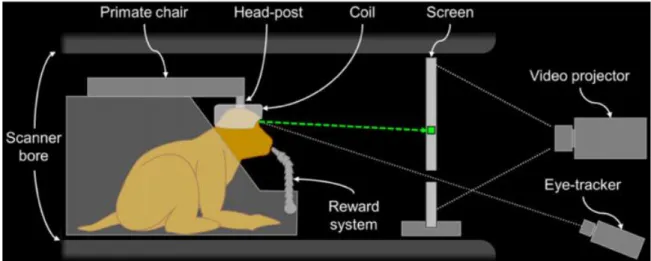
The first studies (Dubowitz et al., 1998; Stefanacci et al., 1998) to report stimulus-induced neuronal activity-related signals in the visual cortex of an awake monkey using fMRI, employed a low-field (1.5 T) clinical horizontal MR scanner. They trained rhesus macaques in a mock MR environment to remain immobile in a prone position, with body restrained and head fixed to the primate chair via a custom-designed, nonmetallic head-post surgically attached to their skull. With the purpose of activating the visual cortex, the experimenters subjected the monkey to visual stimuli rich in movement, color and texture. In one of the studies (Dubowitz et al., 1998), the images that figured in the study show activation of a small region at the pole of the occipital lobe mixed with some ghosting artifacts. This activation was problematic, as it was located bilaterally about 8 degrees peripheral to the cortical representation of the monkey’s fovea. No response was evident in the animal’s cortical representation of the macula. In the other study (Stefanacci et al., 1998), stimulus-induced activation was impossible to localize because signal variation was dominated by motion artifacts. These first attempts at monkey fMRI provided rich insight on necessary improvements to make such procedure worthwhile, all while raising important issues.
36
B.2.2 The battle for SNR
FMRI eliminated the risks related to exposure to radioactivity, while providing better spatial and temporal resolutions compared to PET. Yet, many obstacles are still to overcome for the proper use of monkeys in an MR environment, especially if the purpose is to test humans and monkeys in similar conditions with relatable cognitive tasks. Generally speaking, scanning strategies and methodologies cannot be simply transferred from human fMRI to awake monkey fMRI. There are several significant technical challenges involved in carrying out fMRI experiments in monkeys, arising from the need to use alert animals.
Monkeys have relatively small brains and smaller functional structures compared to humans. The average area of the cerebral cortex in macaque monkeys is 100 cm2 (Felleman and Van Essen,
1991), compared to 2500 cm2 in humans. Functional structures are also smaller, for example the
ocular dominance columns in macaque monkeys (Ts’o et al., 1990) are half the size of those in humans (Cheng et al., 2001; Yacoub et al., 2007). The higher spatial resolution that is necessary for the smaller monkey brain causes a loss of signal-to-noise ratio (SNR). For human fMRI, resolution is typically about 3 × 3 × 3 mm3, which leads to a voxel size of almost 30 μl before any spatial
smoothing is applied. For awake monkeys, a typical spatial resolution is on the order of 1.5 × 1.5 × 2 mm3 (4.5 μl voxels), while for anesthetized monkeys the resolution is even higher.
Depending on acquisition and pre-processing parameters, the SNR of monkey fMRI can easily be 5–30 times lower than that of human fMRI. This is a large SNR loss that needs to be taken into account when the experiment is designed, for instance by offsetting the SNR loss with smaller RF-coils (Logothetis et al., 2002; Janssens et al., 2012), using iron-based contrast agents to enhance the functional signal (Vanduffel et al., 2001; Leite et al., 2002; Fize et al., 2003), or using high-field scanners.
Another significant problem is animal motion. While human subjects can be selected based on their ability not to move, the task is harder for animals that are naturally uncooperative. Any
37 movement of the animal that causes the brain to change position inside the magnetic field during scanning produces inconsistencies in phase and amplitude, which can generate blurring and ghosting motion artifacts larger even than the activation signals (Pfeuffer et al., 2007).
B.2.3 Pedal to the metal: High-field vertical fMRI
Another adopted course for monkey fMRI was through the use of custom built high-field (4.7 T) vertical bores. The pioneers of this technique (Logothetis et al., 1999) showed that BOLD imaging at very high spatiotemporal resolution is possible in both the anesthetized and the alert monkey. In the initial study, fifteen animals in total were scanned, distributed between behaving and anesthetized configurations. In the latter configuration, the level of anesthesia, the precise control of respiration and the correct positioning of the visual stimulus, virtually eliminated motion artifacts whether they were voluntary or simply related to physiological function. In contrast, experiments on anesthetized monkeys were hampered by the upright position of the animal, as it became substantially more difficult to maintain normovolemia and constant blood pressure. On the other hand, the behaving animal configuration, benefitted greatly from the upright position. Being more natural, this position permitted easier training of the animals and reduced unwanted body movements. In both configurations, stimulus-specific activation was observed in the LGN, striate and early extrastriate cortices, and in the temporal and mediotemporal structures that are involved in the processing of facial identity and expression. In the anesthetized monkey, activation was highly dependent on the depth of anesthesia.
This high complexity and cost of setting up this technique and maintaining it proved to be to prohibitive to allow its widespread adoption.
B.2.4 On the home stretch: democratizing monkey fMRI
The beginning of the 21st century witnessed a steep increase of interest in awake monkey fMRI. Thus, striving to render this technique more accessible, without the need to build expensive custom scanners, one lab greatly contributed to the simplification and refinement of this technique.
38 Amongst the flagship studies that set the tone for subsequent monkey fMRI, was one published in 2001 by Vanduffel and colleagues. They demonstrated that robust visually driven activations could be detected using the more available horizontal, low field, clinical scanners. To accomplish this feat, they used iron oxide contrast agents to significantly enhance the functional brain imaging in awake behaving macaques, compared to the BOLD signal. They showed that the use of such contrast agents yielded approximately a 10-fold increase in the percent signal change relative to comparable BOLD measurements at 1.5T, thus becoming an excellent alternative to BOLD imaging with high field scanners. This enhancement allowed them to link between the motion sensitive areas found in fMRI with and those that have been described in single cell studies. The advancements provided by this team over the years made their approach to monkey fMRI a reference for those who wanted to try their hand at scanning monkey brains. Significant advancements in monkey fMRI techniques were introduced in the following years, with the development of implanted focal single loop coils (Logothetis et al., 2002), combined EEG/fMRI at 4.7T (Schmid et al., 2006) external phased array coils (Ekstrom et al., 2008), spin-echo imaging (Ku et al., 2011), and implanted phased array coils (Janssens et al., 2012).
39
B.3 Feather in the cap: some achievements of monkey fMRI
B.3.1 Monkey fMRI sheds light on the nature of the BOLD signal
Since the introduction of fMRI in humans as a method for measuring behavior-related neural activity in the human brain, the relation between the BOLD signal and underlying neural activity has been an open and actively researched question. BOLD measures a combination of cerebral blood flow and oxygen metabolism. Theoretical predictions (Attwell and Laughlin, 2001; Attwell and Iadecola, 2002) suggest glucose utilization primarily reflects in the work involved in synaptic signalling and that metabolic measures of brain activity correlate most strongly with measures that reflect synaptic processing rather than spike rate alone.
Local field potential (LFP) are thought to represent post-synaptic and pre-synatpic activity at multiple neurons, and thus is most accurately described as “peri” synaptic activity. A landmark study (Logothetis et al., 2001) that attempted to empirically determine whether spike rate or LFPs correlated with the BOLD signal, constructed a recording device that allowed simultaneous acquisition of fMRI, LFPs, and the spiking activity of neurons in monkey visual cortex. Anesthetized monkeys viewed contrast gratings while BOLD and electrophysiological activity was recorded in primary visual cortex. The authors reported a strong correlation between BOLD and LFPs and robust but slightly weaker correlation between BOLD and multi-unit activity (MUA). The fact that the LFP accounted for significantly greater amounts of variance across recording sites suggested that the LFP correlated better with the BOLD signal than spike rate (Logothetis and Wandell, 2004). The subsequent investigation of individual recording site indicated that dissociations between spiking and the LFP always resulted in a strong correlation between the BOLD signal and the LFP and not spike rate. These data suggested that correlations between spike rate and LFPs contributed to the correlation between BOLD and spiking activity. Later studies also suggested that significant (but weaker) correlations between BOLD and LFPs could be obtained in the visual cortex of awake behaving monkeys as well (Goense and Logothetis, 2008).
40
B.3.2 The grasping network
The brain is remarkably adept at orienting the wrist and shaping the span of the fingers to match an object. The prehensile hand is a major characteristic that distinguishes primates from other mammal species. All primates can grasp an object and hold it in part or entirely using a single hand. Comparative kinematic studies on grasping behavior in humans and macaques have been carried out to investigate the similarities and differences existing across the two species (Fogassi et al., 1991; Roy et al., 2000, 2006; Christel and Billard, 2001; Sacrey et al., 2009; Jindrich et al., 2011; Pouydebat et al., 2014). These studies mostly indicate similarities in hand shaping across species. For example, the fact that the hand aperture seems to be scaled relatively to the size of the object. Although some differences emerge, the similarities still place the macaque monkey as an excellent model for the study of the neuroscience of grasping.
B.3.2.1 From single cells to whole brains
A particular brain region in the non-human primate parietal cortex is strongly associated with this ability. This region, termed AIP, is found in the anterior-lateral intraparietal sulcus (area 7b). The functional properties of area AIP have been extensively investigated at the single unit level (Taira et al., 1990; Jeannerod et al., 1995; Sakata et al., 1995; Murata et al., 2000; Gardner et al., 2007) while macaque monkeys performed visually guided grasps of differently shaped 3D objects. The visual and motor responses of AIP neurons were tested in three experimental conditions: grasping in light, grasping in dark, and object observation. The results showed that while some AIP neurons respond during grasping execution in light and dark, others respond only during grasping in light and finally, some neurons discharge when the monkey fixates an object even when no grasping of the object is required. There is congruence between the visual and motor responses of AIP neurons: a neuron is active when the monkey observes one object selectively and discharges for grasping of the same object. Indeed, grasping related movements require processing of the visual properties of the object and those that control hand movement. It is thus primordial to perform a
41 rigorous transformation of an object’s properties such as size, orientation and shape into a fitting motor scheme, shaping the hand for proper grasping.
The observation that single neurons in AIP display a combination of visual and motor properties suggests that these neurons code the visual features of the observed objects and that together with F5 (premotor cortex) neurons they transform them into the appropriate hand configuration for grasping (for review Rozzi and Coudé, 2015). The visuomotor properties of area AIP also include the processing of the 3D shape of objects. The early studies in area AIP had reported object-selective responses in this area (Murata et al., 2000) but it was unclear whether these neurons encoded differences in 3D structure, 2D contour, orientation or any other feature that differed between the objects used in those experiments. A recent study (Srivastava et al., 2009) recorded single-cell activity in the AIP of awake fixating rhesus monkeys using disparity-defined curved surfaces. They report robust selectivity for disparity-defined slanted and curved surfaces in a high proportion of AIP neurons, thus confirming its involvement in the processing of the 3D shape of objects.
Reversible pharmacological lesions of area AIP, have been reported to affect hand preshaping (i.e., grasping) (Gallese et al., 1994), leaving the reach component unaffected (Murata et al., 2000). The deficit was evident only, or mainly, when a precision grip was required. The fact that this impairment is more pronounced in the preshaping phase of grasping rather than during object manipulation emphasizes the crucial role of AIP in visuomotor transformation. The implication of AIP as part of the network responsible for 3D-shape perception was further stressed in a recent experiment of focal perturbation (Verhoef et al., 2016). Through a task of categorization of disparity-defined 3D shapes during concomitant microstimulation of 3D-shape selective AIP neurons, Verhoef and colleagues (2015) found that microstimulation effects on preferences and latency depended on the 3D-shape preference of the stimulated site. Additionally, they show that