ANALYSES ISOTOPIQUES ET GÉOCHIMIQUES MENÉES SUR LES POISSONS FOSSILES ET LES SÉDIMENTS DE LA FORMATION D'ESCUMINAC (DÉVONIEN
SUPÉRIEUR, MIGUASHA, QUÉBEC) ET IMPLICATIONS PALÉOENVIRONNEMENTALES
MÉMOIRE PRÉSENTÉ
COMME EXIGENCE PARTIELLE
DE LA MAÎTRISE EN SCIENCES DE LA TERRE
PAR
OLIVIER MATION
UI\JIVERSITÉ DU QUÉBEC À MOI\JTRÉAL Service des bibliothèques
Avertissement
La diffusion de ce mémoire se fait dans le respect des droits de son auteur, qui a signé le formulaire Autorisation de reproduire et de diffuser un travail de recherche de cycles supérieurs (SDU-522 - Rév.01-2006). Cette autorisation stipule que «conformément
à
l'article 11 du Règlement no 8 des études de cycles supérieurs, [l'auteur] concèdeà
l'Université du Québecà
Montréal une licence non exclusive d'utilisation et de publication de la totalité ou d'une partie importante de [son] travail de recherche pour des fins pédagogiques et non commerciales. Plus précisément, [l'auteur] autorise l'Université du Québec à Montréal à reproduire, diffuser, prêter, distribuer ou vendre des copies de [son] travail de rechercheà
des fins non commerciales sur quelque support que ce soit, y compris l'Internet. Cette licence et cette autorisation n'entraînent pas une renonciation de [la] part [de l'auteur]à
[ses] droits moraux nià
[ses] droits de propriété intellectuelle. Sauf entente contraire, [l'auteur] conserve la liberté de diffuser et de commercialiser ou non ce travail dont [il] possède un exemplaire.»commencement de cette maîtrise, fut la personne qui m'introduisit avec passion à la paléontologie et dont les connaissances à propos du site de Miguasha sont si précieuses, et Ross Stevenson de l'UQAM qui a si bien su me guider avec patience, gentillesse et rigueur dans les voies parfois mystérieuses de la géochimie.
J'aimerais également rendre hommage à l'ensemble de l'équipe, présente et passée, du parc national de Miguasha. L'intérêt que vous portez à vos responsabilités, aux trésors de la Formation d'Escuminac et aux projets de recherche qui s'y penchent n'a pu que renforcer le respect que je porte à ce site exceptionnel. Je pense spécialement au Dr Sylvain Desbiens pour ses commentaires constructifs, à Johanne Kerr pour m'avoir donné accès à la salle de collection du Musée d'histoire naturelle de Miguasha ainsi qu'à Norman Parenti, Jason Willetti et à mes compagnons fouilleurs pour vos connaissances de terrain et le plaisir partagé avec vous dans la falaise.
Tout au long de ma maîtrise j'ai également eu la chance de pouvoir compter sur la disponibilité et le professionnalisme du personnel du département des Sciences de la Terre et de l'Atmosphère et du GEOTOP-UQAM-McGILL. Je remercie donc le Dr Michel Lamothe pour m'avoir permis de venir étudier au département, Micheline Lacroix pour toutes les questions administratives, Raymond Mineau au MEB, Brieuc Lefevbre (UQAM) et Bill Minarik (Université McGill) pour leur pilotage du La-ICP-MS, Chantal Gosselin lors de la préparation de présentations orales et finalement Raynald Lapointe pour son aide technique régulière.
Pour m'avoir diverti et encouragé lorsque je faiblissais, je dois souligner le soutien indéfectible de la musique rock, de ma famille et de mes amis, notamment Émilie, France, Isabelle et Philippe avec qui je pouvais échanger sur notre parcours commun. Des
111
remerciements tout particuliers à Mélanie qui fut la toute dernière à m'aider, lors de la mise en page finale de mon mémoire, à quelques heures de mon dépôt.
À la base de tout, mes parents ont toujours eu à cœur mon bien-être et ont stimulé ma curiosité. Merci pour tout, de mon premier livre d'enfant portant sur la paléontologie jusqu'à vos encouragements au cours de mes nombreuses années universitaires. J'ai toujours senti que vous étiez avec moi.
Finalement, des pensées particulières vont à la personne qui a côtoyé ce projet au quotidien. Merci Geneviève, toi qui sais si bien me faire sourire et oublier mes soucis. Pour m'avoir précédé dans les études graduées, tu auras été la meilleure pour m'inspirer et me rassurer tout au long de ma maîtrise.
LISTE DES TABLEAUX viii
RÉSUMÉ ix
INTRODUCTION 1
CHAPITRE 1
ISOTOPIC AND GEOCHEMICAL ANALYSES OF FOSSIL FISH REMAINS AND SHALES FROM THE UPPER DEVONIAN ESCUMINAC FORMATION (MIGUASHA,
QUÉBEC): PALEOENVIRONMENTAL IMPLICATIONS 3
O. Abstract 3
1. Introduction 4
1.1. Background 4
1.2. Preservation of pristine Sr isotope signatures in bioapatites 5 1.3. Preservation ofpristine rare earth elements abundances ofbioapatites 7
2. Geological setting 9
3. Material and methods 11
3.1. Sampling 11
3.2. Scanning electron microscopy (SEM) and Laser ablation-inductively
coupled plasma-mass spectrometry (LA-ICP-MS) 11
3.3. Strontium, neodymium and samarium isotope analyses 12
4. Results 14
4.1 X-ray maps 14
4.2 Sr in bioapatites 16
4.3 REE in bioapatites and diagenetic calcite 21
4.4 Sm-Nd in bioapatites and shales 23
5. Discussion 25
v
5.2 Diagenetic considerations for REE in ichthyoliths 26
5.3 Do the Nd isotopes reflect seawater? Comparisons with conodonts 26
6. Conclusions 31
7. References 32
CONCLUSION 39
2005) 9 2. X-ray maps of ichthyoliths from the Escuminac Formation. Calcium and
phosphorus distribution in the cancellous bone layer of Bothriolepis canadensis (a and b) and in tooth of Eusthenopteron foordi (c and d). bt, bone
trabeculae; d, dentine; e, enamel; pc, pulp cavity; s, space; c, calcite 15 3. 87S r/86S r variation for fish material and diagenetic calcite from
the Escuminac Formation. The grey area shows the amplitude of variation of seawater 87Sr/86S r for the Middle Frasnian (Veizer et al., 1999) and the arrow is pointing toward freshwater values. The scale bar represents
average 2cr 19
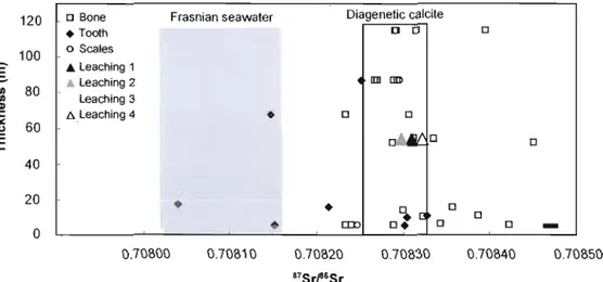
4. 87S r/86S r variation by tissue type and calcite leaching experiment. The
grey area shows the amplitude of variation of seawater 87Sr/86S r for the Middle Frasnian (Veizer et al., 1999) while the white area encompasses the values obtained for diagenetic calcite used in this study. "Leaching 1-4" refers to progressive leaching of diagenetic calcite, from initial immersion in 0.1 M HNO) ("Leaching 1") to complete dissolution in HCI and HF
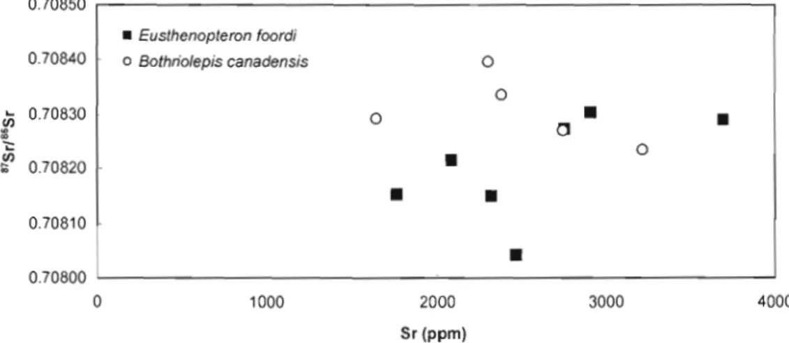
("Leaching 4"). The scale bar represents average 2cr 19 5. Covariation between 87Sr/86Sr and Sr concentration for Bothriolepis canadensis
and Eusthenopteronfoordi 20
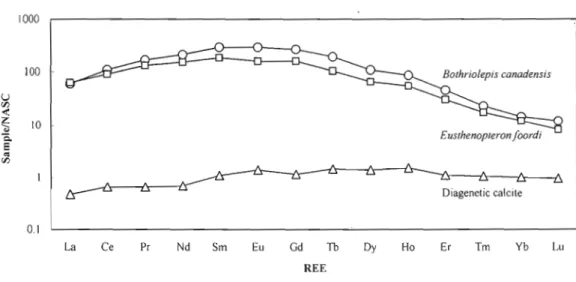
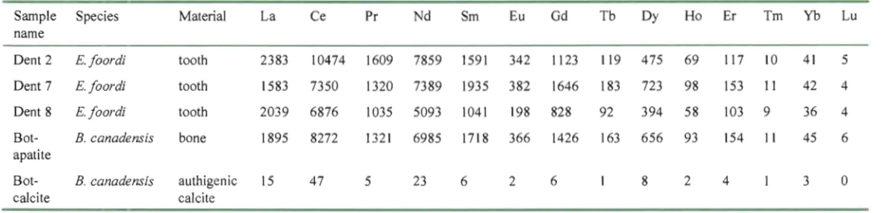
6. Comparison of shale-normalized (NASC) REE concentrations between ichthyoliths and authigenic
calcite from the Escuminac Formation 21
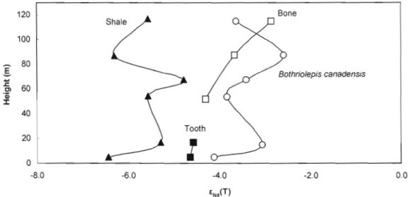
7. ENd(t) variation along the Escuminac Formation for Bothriolepis canadensis, Eusthenopteronfoordi and shales. "Tooth" and "Bone"
series belong to Eusthenopteronfoordi 27
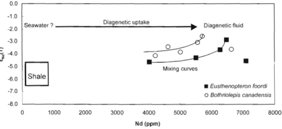
8. Diagram showing ENd(T) values of ichthyoliths plotted against their Nd concentration. The samp1es differ from the shale from the Escuminac Formation. An hypothetical seawater value is presented as a possible source
ofREE uptake during diagenesis 28
9. Sr isotope composition and ENd(T) oftooth and bone from the Escuminac Formation. Sampies exhibit relatively homogeneous ENd(T) in spite of
Vll
10. ENd(T) comparison between the material used in this study and sediments and fossils coming form other localities and periods. Black arrows represent the general trend seen in North American shales (see Hurowitz and McLennan, 2005). Data for conodonts are taken in Wright (1995; for North America) and Dopieralka et al. (2006; for Poland). Figure
1. Major element composition of fish tooth enamel, dentine and conodont crown
and basal body 15
2. Sr isotope measurements of Miguasha bioapatites and diagenetic calcite 17 3. REE abundances (ppm) for Miguasha fossil material and authigenic calcite 22 4.
Sm-Nd isotope data
forMiguasha shales and fossil material
24RÉSUMÉ
Le cadre paléoenvironnemental de la Formation d'Escuminac a été investigué par le biais d'analyses géochimiques (terres rares) et isotopiques (Rb-Sr, Nd-Sm) effectuées sur du matériel fossile et des sédiments de la section type de cette formation. Pour ce faire, des échantillons d'argilite et des ichthyolithes appartenant à cinq espèces de poissons fossiles différentes (le placoderme Bothriolepis canadensis, l'acanthodien Homalacanthus concinnus, l'actinoptérygien Cheirolepis canadensis, le dipneuste Scaumenacia curta et l'ostéolépiforrne Eusthenopteron foordi) et pour lesquels la position stratigraphique est connue ont été étudiés. Les analyses isotopiques furent réal isées en spectrométrie de masse par ionisation thermique et les abondances de terres rares ont été mesurées par ablation laser ICP-MS.
Les résultats obtenus dans cette étude suggèrent que les bioapatites ont subi un certain grade de diagénèse mais qu'il est malgré tout possible d'y récupérer des signatures paléoenvironnementales pertinentes. Le matériel dentaire apparaît plus résistant à l'altération que les os ou les écailles. Les ratios 87Sr/86Sr des bioapatites varient de 0,70804 à 0,70845.
Les signatures les plus sûres sont similaires à la composition isotopique en Sr de l'eau de mer au Frasnien, suggérant un environnement marin ou saumâtre pour la Formation d'Escuminac, sans variation interspécifique ou stratigraphique claire. La majorité des échantillons analysés présentent cependant une déviation significative des valeurs marines vers un signal plus radiogénique. Cette déviation serait le résultat d'échanges post-mortem de Sr entre les fossiles et un fluide diagénétique isotopiquement distinct des eaux dans lesquelles ont vécu les poissons.
Tous les ichthyolithes sont fortement enrichis en terres rares et montrent un patron normalisé au NASC similaire à ceux décrits dans les bioapatites paléozoïques d'origine marine. Les ENd(T) des argilites varient de -4,8 à -6,4, suggerant que de jeunes sources appalachiennes ont dominé la sédimentation détritique tout au long de la mise en place de la Formation d'Escuminac. Une composition isotopique en Nd plus radiogénique (de -2,6 à 4,6) est obtenue pour les bioapatites. La divergence entre le ENd(T) des sédiments et des fossiles implique la présence d'un autre réservoir de Nd pour les ichthyolithes de la Formation d'Escuminac. Les fossiles de la Formation d'Escuminac portent une composition isotopique en Nd qui les rapproche plus de conodontes de la région baltique que des conodontes nord-américains. Un contact entre le bassin collecteur de la Formation d'Escuminac et l'océan Rhéique est ainsi proposé.
vel1ébrés inférieurs, notamment au sujet de la transition cruciale entre les poissons et les tétrapodes. Cependant, en dépit de cet intérêt scientifique soutenu, aucun cadre paléoenvironnemental ne fait consensus pour la Formation d'Escuminac. Des données parfois contradictoires, provenant de la paléontologie, de la sédimentologie et de la géochimie ont conduit à une gamme d'interprétations paléoenvironnementales allant du milieu lacustre à l'environnement marin. Jusqu'à maintenant, les études géochimiques se sont concentrées sur les sédiments de la Formation d'Escuminac. Les signatures géochimiques obtenues par l'étude de roches sédimentaires étant plutôt tributaires de l'histoire géologique de ces sédiments, il est préférable d'analyser le matériel fossile si l'on souhaite obtenir des informations pertinentes sur le milieu de vie des organismes anciens. Ce travail représente la première investigation géochimique à grande échelle du matériel fossile provenant de la Formation d'Escuminac.
Ce travail vise dans un premier temps l'étude de la structure interne et de la composition en éléments majeurs des fragments de poissons fossiles utilisés dans ce projet, afin de vérifier si la minéralogie originelle est préservée. Une fois cette étape préliminaire réalisée, les analyses géochimiques et isotopiques peuvent être effectuées sur les fossiles et les sédiments de la Formation d'Escuminac. Les éléments étudiés sont le strontium (Sr), le rubidium (Rb), le samarium (Sm), le néodyme (Nd) ainsi que les autres terres rares. Ces analyses visent à renseigner sur (1) le degré de diagénèse subit par les fossiles de la Formation d'Escuminac, (2) la paléosalinité du milieu aquatique dans lequel vivait les poissons étudiés, (3) les sources sédimentaires ayant alimenté le bassin collecteur de la Formation d'Escuminac et (4) le degré de similitude entre le milieu de vie des poissons de Miguasha et d'autres masses d'eau contemporaines.
Pour réaliser ces objectifs, des échantillons d'argilite et de diverses espèces de poissons fossiles provenant de la Formation d'Escuminac ont été sélectionnés. Le choix s'est porté sur
2
le matériel bien conservé et pour lequel une position stratigraphique précise est connue. Une fois nettoyés des sédiments encaissants, les échantillons ont été attaqués chimiquement jusqu'à dissolution puis l'on a isolé les isotopes à l'étude (i.e., Rb, Sr, Sm, Nd) à l'aide de résines spécifiques. Ces isotopes furent analysés à l'aide d'un TIMS. Finalement, d'autres fossiles ont été étudiés au microscope électronique à balayage et au La-1CP-MS afin d'en étudier la structure et la composition en éléments majeurs et en terres rares.
La première section de ce travail présente les notions théoriques impliquées dans cette étude à propos du comportement du strontium et des terres rares. Suit une description du cadre géologique de la Formation d'Escuminac puis une présentation de la méthodologie utilisée. La quatrième section de ce mémoire exposera les résultats obtenus lors des analyses. Enfin, une interprétation paléoenvironnementale des divers résultats sera proposée.
IGEOTOP UQAM-McGill and Département des Sciences de la Terre, Université du Québec à Montréal, c.P. 8888, Succ. Centre-Ville, Montréal, Qc, Canada, H3C 3P8
2Parc national de Miguasha, 231 Miguasha Ouest, c.P. 183, Nouvelle, Qc, Canada, GOC 2EO
3Chaire de paléontologie et biologie évolutive, Université du Québec à Rimouski, 300 allée
des Ursulines, Rimouski, Qc, Canada, G5L 3AI
*Author to whom correspondance should be addressed (richard_cloutier@uqar.qc.ca)
Abstract- The paleoenvironmental context of the Middle Frasnian Escuminac Formation was investigated through direct geochemical (rare earth elements) and isotopic (Rb-Sr, Nd-Sm) analyses of fossil material and shales. Shale samples as weil as ichthyoliths of different fish species (placoderm Bothriolepis canadensis, acanthodian Homalacanthus concinnus,
actinopterygian Cheirolepis canadensis, dipnoan Scaumenacia curta and osteolepiform
Eusthenopteron foordi) coming from the base to the top of the Escuminac Formation were analysed. The isotopic analyses were performed by thermal ionisation mass spectrometry (TlMS) and the rare earth element abundances were measured by laser ablation lCP-MS.
87 Sr
l6
Sr ratios for ichthyoliths vary from 0.70804 to 0.70845 and overlap with Middle Frasnian seawater Sr isotope composition, though most of the biogenic apatites analyzed show a variable drift from marine values toward a more radiogenic continental signature. This trend is possibly the result of post-mortem Sr exchange between fossils and a fluid isotopicaJly distinct from the waters in which they developed.AIl ichthyoliths are heavily enriched in rare earth elements with bell-shaped shale normalized patterns. The ENd(T) of the shales range from -4.8 to -6.4, suggesting that a young Appalachian source dominated the detrital sedimentation for the Escuminac Formation. In contrast, more radiogenic ENd(T) values (ranging from -2.6 to -4.6) were obtained for the ichthyoliths. The discrepancy between shales and ichthyoliths ENiT) implies the presence of a second Nd reservoir for the Escuminac bioapatites. The similarity between Nd isotope compositions of Escuminac fossils and Baltic conodonts versus North American conodonts argues for a close connection between waters of the Rheic Ocean and the Escuminac Formation collector basin.
1. INTRODUCTION
1.1. Background
The Middle Frasnian Escuminac Formation (Miguasha, Québec, Canada) is renowned for (1) the abundance, (2) the quality of preservation and (3) the phylogenetic and evolutionary importance of its fossils. Since its discovery in 1842, over 19 000 specimens have been collected from the Miguasha cliffs. The biota includes terrestrial plants, invertebrates (i.e., polychaetes, eurypterids, crustaceans, terrestrial scorpions and millipedes) as weil as vertebrates. Vertebrates are represented by 20 fish species that encompass most of the Late Devonian vertebrate diversity, with the exception of chondrichthyans or cartilaginous fishes and tetrapods. The diverse ichthyofauna of the Escuminac Formation has been under scrutinity for decades, providing invaluable information on the systematic, fine anatomy and paleoecology of extinct vertebrates (Schultze and Cloutier, 1996; Arsenault et al., 2004; Burrow, 2005; Janvier et al., 2006).
Reconstructing past habitats is intricately linked to our understanding of life evolution. For instance, the environment in which the first tetrapods evolved is still disputed (Clack and Neininger, 2000) and an improved understanding of the early tetrapods habitat would allow a better conceptualization of the abiotic and biotic framework that favoured the origin of tetrapods. The Escuminac assemblage includes two taxa sharing anatomical features that 1ink them to tetrapods, the osteoJepiform Eusthenopteron foordi and the elpistostegalian
Elpistostege watsoni (Cloutier and Ahlberg, 1996), with the latter being considered as the sister group of tetrapods (Schultze and Arsenault, 1985; Cloutier and Ahlberg, 1996; Schultze, 1996; Daeschler et al., 2006). Furthermore, resolving the paleoenvironmental setting of the Escuminac Formation would provide insight into factors that play a role in the formation of a Konzentrat and Konservat LagersUitte (Parent and Cloutier, 1996) like the Escuminac Formation.
Devonian üld Red Sandstone fish assemblages have been long considered to have a freshwater origin and, by extension, so was the Escuminac assemblage (Dineley and
Williams, 1968; Brideaux and Radforth, 1970; Gray, 1988; Rust et aL, 1989). However, this paradigm is disputed (Ah1berg, 1989; Schmitz et aL, 1991; Schultze, 1995) and other interpretations of the depositional environment of the Escuminac Formation have been proposed: marine (Schmitz et aL, 1991), coastal marine (Schultze and Arsenault, 1985; Vézina, 1991; Schultze, 1995), transitional (Chidiac, 1989; 1996), marine to brackish (Schultze and Cloutier, 1996; El Albani et al., 2002) and estuarine (Hesse and Sawh, 1992; Schultze, 1995; Cloutier et aL, 1996; Maples, 1996). Among these workers, few have used geochemical parameters in their studies. Studies of the stable isotopes of C, 0, and Band B concentrations (Chidiac, 1996); Na, F, Sr and La concentrations and Sr isotopes (Schmitz et al., 1991) and Mg, Ca and Fe concentrations (Vézina, 1991) are ail suggestive of a marine influence for the Escuminac Formation (El Albani et aL, 2002). With the exception of Schmitz et al. (1991) who used a single bony fragment, ail the remaining geochemical studies were performed on the sediments rather than on bioapatites. The goal of this paper is to address this paleoenvironmental debate through a broader geochemical (rare earth elements) and isotopie (Sr and Nd) study of fossi1 fish remains and the enclosing sediments of the Escuminac Formation.
1.2. Preservation of pristine Sr isotope signatures in bioapatites
87S r is produced by the radioactive decay of 87Rb, which is generally highly abundant in continental rocks. This means that water from lakes and rivers will normally have higher, more radiogenic, 87Sr/86S r ratios that reflect the age and Rb/Sr ratio of the bedrock in the drainage basin, than seawater (Schmitz et aL, 1991). In addition, because the residence time of Sr in seawater (4-5 Ma) exceeds the mixing time of ocean water (-J03 years), oceans have
a uniform Sr isotopie composition at a given time (Dasch and Campbell, 1970). For instance, the present-day world's oceans possess a homogenous 87Sr/86Sr ratio of 0.7092 (De Paolo and Ingram, 1985), whereas continental waters have higher and more variable 87S r/86S r ratios, ranging from -0.710 to ~0.730 (Veizer and Compston, 1974; Palmer and Edmond, 1989).
6
The Sr concentration is also different between seawater and freshwater: Sr in seawater is generally two orders of magnitude greater than in freshwater (Bryant et al., 1995; Holmden et al., J997). This large difference leads to difficulties in identifying between mixtures of seawater and freshwater because even in brackish environments, the freshwater 87S r/86S r signature is often completely overwhelmed by the saltwater signal (Veizer and Compston,
1974; Holmden et al., 1997; Kohn and Cerling, 2002; Holmden and Hudson, 2003).
In paleoenvironmental investigations, the 87Sr/86Sr ratio of calcitic and phosphatic biomineralization can be used as a proxy for the seawater composition because Sr2+ easily substitutes for Ca2+ in biogenic calcite and apatite owing to their similar ionic radii. Moreover, it is weil established that Sr is incorporated in vivo in biomineralization without significant fractionation (Dasch and Campbell, J970; Schmitz et al., J99 J; Koch et al., 1992; Kaufman et al., 1993), thus recording the Sr-isotopie composition of the environment in which the animal is living (Shaw and Wasserburg, 1985; Staudigel et al., 1985). A crucial issue, however, is whether the original signal is preserved after the death of the animal (Schmitz et al., 1997). The case of bioapatites is relevant. Ichthyoliths (i.e., small fish remains such as isolated bon es, teeth, scaJes) and conodont elements (i.e., small feeding apparatus of an extinct group of jawless chordates) share a common general mineralogy, a carbonated form of hydroxyapatite [CalO(C03,P04MOH)2; Trueman and Tuross, 2002]. This calcium phosphate is thermodynamically unstable and during buriaJ and diagenesis, it may be wholly or partially altered. During the process of fossilization, the organic components are lost, increasing the porosity within bioapatites and simultaneously, authigenic mineraIs precipitate, either in intercrystalline spaces or in larger vascuJar spaces (permineralization), thus increasing bioapatite density (Trueman and Tuross, 2002). It is this increase in density that gives stability and stops further exchange between biominerals and their environment (Trueman and Tuross, 2002). During these processes, trace elements are either removed or added (e.g., Sr) in the bioapatite (Nelson et al. 1986; Barrat et al., 2000). These processes lead to the introduction of secondary Sr and a rapid increase in Sr content during early diagenesis (Schmitz et al., 1991) by up to several thousand ppm in fossil apatite (Staudigel et al., 1985; Grandjean et al., 1987; Schmitz et al., 1991). Several authors have proposed that these exchanges can drastically alter biological and environmental signais recorded in vivo
(Nelson et al., 1986; Bertram et aL, 1992; Holmden et al., 1996, Dufour et aL, 2007). On the other hand, other workers (Shaw and Wasserburg, 1985; StaudigeJ et al., 1985, Holmden et al., 1996) suggest that preservation of the original paleoenvironmental signature is possible; if the bioapatite exchanged little Sr with its burial environment or if it happened when pore fluids were still in contact with waters in which the organism was living.
In their broad study, Schmitz et al. (1991) analysed the 87S r/86Sr ratio of placoderm material (Bothriolepis canadensis) from the Escuminac Formation. However, their analyses were conducted on a single bony fragment and they bel ieved that the specimen may have been diagenetically altered. For these reasons they recommended further geochemical investigations of the Escuminac Formation fauna. Other studies focusing on the sediments of the Escuminac Formation have also indicated that the Escuminac Formation is diagenetically altered (Chidiac, 1989; Hesse and Sawh, 1992; El Albani et al., 2002). However, there is no unequivocal consensus on the degree of aIteration, with El Albani et al. (2002) suggesting a lower level than Chidiac (1989) and Hesse and Sawh (1992).
1.3. Preservation of pristine rare earth elements abundances in bioapatites
No vital raie is known for the rare earth elements (REE) and they are thus found in very low concentrations in vivo (Trueman, 1999). However, studies have shown that there is a spectacular post mortem enrichment of these elements in both recent and ancient bioapatites. Like Sr, the substitution occurs in the Ca site of apatite. This drastic diagenetic enrichment, up to 103_106 time their initial concentration in ichthyol iths and conodonts (Wright et al., 1984; Shaw and Wasserburg, 1985; Holmden et al., 1996), will normally swamp any signal recorded during the life of the animal. However, it has been noted that the incorporation happens very quickly, near the water-sediment interface, in modem fish apatite (StaudigeJ et aL, 1985; E1derfield and Pagett, 1986; Wright et a1., 1987; Martin and Haley, 2000). If the bioapatites do not reach equilibrium with pore waters during their burial history (Martin and Haley, 2000), the REE characteristics of bottom water or early diagenetic environment cao then be recovered (Wright et al., 1984; Staudigel et a1., 1985; Keto and Jacobsen, 1987;
8
Wright et aL, 1987; Trueman and Tuross, 2002) giving insights on the evoJution of marine REE patterns over geological time (Grandjean et aL, 1987; Wright et aL, 1987; Grandjean Lécuyer et al., 1993), the redox state of ancient seawater (Wright et aL, 1984; Elderfield and Pagett, 1986; Hoiser, 1997; Picard et al., 2002; Kemp and Trueman, 2003) and the provenance of the sediments (Wright, 1995). On the other hand, sorne studies tend to demonstrate that late diagenetic processes could alter the original signature (Elderfield and Pagett, 1986; ShoJkovitz et al., 1989; Toyoda and Tokonami, 1990; Holmden et al., 1996).
- - - -
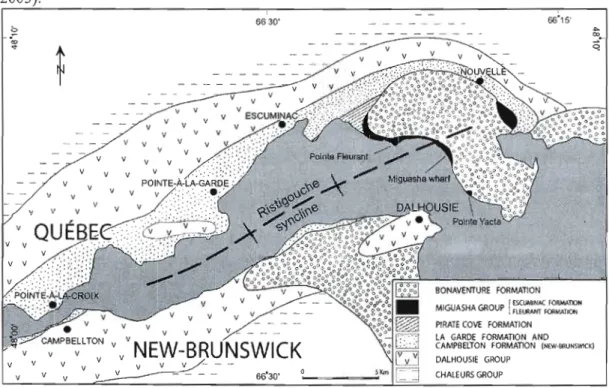
-River estuary but, recently, new outcrops have been discovered as much as 40 km inland from the type section in Miguasha. The type section of the Escuminac Formation is 119 m thick (Sawh, 1982) and is exposed aJong cliffs that range from 3 to 30 m in height. The Escuminac Formation conformably overlies the Fleurant Formation, a conglomerate with sandstone Jenses interpreted as proximal alluvial deposits of early Late Devonian age (Rust et al., 1989). Together, these two formations form the Miguasha Group (Dineley and Williams,
1968).
Figure 1. Simplified geologicaJ map of the Miguasha area (modified from Desbiens et al., 2005). 6630'
...
1:> .;;; ~•.. .,. v v Q v l' BONAVENTURE FORMATION MIGUASHA GROUP [;~::;~=~N PIRATE COYE FORMATIONv V V v
LA GARDE FORMATION AND CAMPBELTON FORMATION (NEW·&1lUNIWlCK) v V V V v
NEW-BRUNSWICK
DALHOUSIE GROUP
v v v
10
The Escuminac Formation is overlain along an angular unconformity by the Mississippian Bonaventure Formation that is composed of alternating conglomerate and coarse sandstone (Desbiens et al., 2005). A middle Frasnian (Upper Devonian) age has been assigned to the Escuminac Formation, based on its miosporal content and fish assemblage (Cloutier et al., 1996). The main lithologies of the Escuminac Formation are, in decreasing order of abundance: shale, sand stone, siltstone, laminite and conglomerate. The laminite lithofacies consists of altemating dark (clay and organic matter) and light (silt-size calcite and quartz) laminae. This siliciclastic lithofacies has been interpreted as tidal-controlled deposits (Cloutier et al., in prep.). Leduc and Belles-Isles (1989) divided the formation into 394 beds of variable thickness, corresponding to major successive lithologica1 changes. The base of the formation (beds 1-7), corresponds to a shallow water littoral environment subjected to episodic aerial exposures (Desbiens et al., 2005). The fish remains first appear in bed 8 and continue to the upper limit of the formation, although their distribution is uneven throughout the stratigraphic sequence (Cloutier et al., 1996). Fibrous calcite and carbonate concretions are also found at different levels and are interpreted to be of early diagenetic origin (El Albani et al., 2002). This is supported by the fact that concretions frequently contain well preserved, articulated remains, indicating that carbonate cementation began early and was completed before decay of the organic parts could occur (Canfield and Raiswell, 1991 a). Numerous lines of evidence suggest that anoxic/hypoxic conditions prevailed at the water sediment interface and in the sediment at different periods of .deposition of the Escuminac Formation, mainly when laminated lithofacies were formed (Cloutier et al., in prep). The evidence includes; (1) the abundance of concretions and pyrite (Canfield and Raiswell, 1991a, 1991 b), (2) the absence of bioturbation, (3) the excellent state of preservation of the fish remains (Canfield and Ra:iswell, 1991 b), (4) the poor diversity of the benthic assemblage, (5) lack of evidence of macrophagous or scavenger activity, and (6) the repetitiveness of acanthodian mass mortality events.
Ali specimens were collected in the type section of the Escuminac Formation during the summer 2006. Emphasis was given on material for which a precise stratigraphic position was known in order to investigate potential temporal variations of different geochemical signatures during the sedimentation in the collector basin of the Escuminac Formation. When available, complete tooth material was used preferentially; if not, tooth fragments, bones or scales were picked. Bones and dentine have smaller crystal size (greater surface area for exchange) and higher organic content than enamel (Trueman and Tuross, 2002). For these reasons, enamel is considered more resistant to diagenetic overprinting than bone and dentine (Elliott, 2002). However, tooth enamel from the Miguasha teeth was frequently too thin to provide sufficient sample for analyses, thus whole teeth were analysed. Ichthyoliths were mechanically isolated and cleaned of detrital sediments and diagenetic calcite under binocular microscope using stainless steel instruments and 5% acetic acid. Samples were subsequently boiled in methanol in order to remove any traces of recent organic contaminants.
3.2. Scanning electron microscopy (SEM) and Laser ablation - inductively coupled plasma - mass spectrometry (LA-lep-MS)
As a preliminary characterization, three teeth from the osteolepifOlm Eusthenopteron foordi and one dermal bone plate from the placoderm Bothriolepis canadensis were mounted in epoxy resin in preparation for scanning electron microscopy, X-ray mapping and REE analysis by laser ablation ICP-MS. X-ray mapping and electron microscope photographs were obtained using energy dispersive X-ray (EDX) analyses coupled to a Hitachi S 4300SE/N variable pressure - scanning electron microscope at Université du Québec à Montréal. The REE analyses were conducted at McGi11 University with a New-Wave UP·213 Laser Ablation System coupled to a PerkinElmerlSCIEX ELAN 6100 DRCpius ICP-MS. REE data in the fossi 1material were normalized against the Ca content of hydroxyapatite that
12
forms the bone and tooth structures and repeated measurements of REE in the NIST-61 0 glass standard yielded average errors that were less than 10% relative standard deviation.
3.3. Strontium, neodymium and samarium isotope analyses
The rest of the fossil material (n
=
34), along with the shale samples (n=
6), was used for Sr, Nd and Sm measurements by thermal ionization mass spectrometry. (TIMS). The preparation of these sampI es was performed under clean room conditions. Specimens of the two main species involved in this study (Bothriolepis canadensis and Eusthenopteron foordi) were selected to represent seven stratigraphic levels: (1) Beds 8-10; (2) 23-28; (3) 39-46; (4) 214-219; (5) 272; (6) 351; (7) 390. In addition, fragments of three additional fish species (the acanthodian Homalacanthus concinnus, the actinopterygian Cheirolepis canadensis and the dipnoan Scaumenacia curta), were collected either from level (1) or (6) to investigate possible interspecific variation in the Sr isotopic ratio.After cleaning, approximately 20 mg of each sample was weighed, sorne of which were spiked with a 87 Rb·84Sr tracer solution (spike) to determine Rb and Sr concentrations by isotope dilution and ail samples were spiked with a '49Sm_150Nd solution to determine Nd and Sm concentrations and correction for production of '43Nd since the time of deposition. Samples were subsequently dissolved in Savillex™ beakers using 3 M HN03. After evaporation at about l30°C, dry residues were taken up with 1 ml of 3 M HN03 and then centrifuged. The resulting solution was passed through a Sr specific resin (Eichrom© Sr-spec resin) to isolate the Sr fraction. The rinse from this column was collected and evaporated in preparation for isolating the REE fraction. The REE were concentrated by passing the remaining samples through Eichrom© TRU-spec-resin followed by separation of Nd and Sm using Eichrom© Ln-spec-resin. The 87S r/86Sr ratio of diagenetic calcite was also investigated using a bony plate from Bothriolepis canadensis in which the median cancellous bone layer was filled with calcite crystals. The bony plate was immersed in 0.1 M HN03 for 30 minutes, allowing the calcite to partially dissolve, while leaving the bone structure intact. The solution was then' removed, centrifuged and the Sr fraction was isolated as described above. For
comparison, three other samples from the same specimen were collected and leached for 60 minutes (0.1 M RN03), 180 minutes and finally until complete dissolution in HCI and HF. Again, the Sr fraction was separated by the method described above. Finally, Nd and Sm isotope compositions were determined on shale samples from the same stratigraphic levels used for the fossils, with the exception of level (2). After being crushed, approximately 0.1 g of the powdered shale was spiked with a mixed Sm-Nd tracer and dissolved in a Teflon bomb with 14 M HN03 and concentrated HF over a period of 5-7 days at 150°C. The resulting solution was evaporated, then redissolved in a small amount of perchloric acid (HCI04) and evaporated once more in order to convert fluoride salts to chloride salts. The samples were then taken up in 6N HCl and fluxed ovemight to ènsure dissolution. The solution was then loaded on a column with anion exchange resin (AG I-X8) in order to remove Fe and then the REE and Sm and Nd were concentrated using the procedures described above.
Ali isotopic measurements were conducted on a VG SECTOR 54 mass spectrometer at the Université du Québec à Montréal. The Sr fractions were loaded onto a single Re filament in a phosphoric acid - tantalum oxide solution and measured in static mode. Replicate analyses of the Sr standard NBS987 yielded a value of 0.710241 ± 18 (2a, n = 6). The Sm and Nd fractions were loaded onto Ta side filaments and measured as a triple filament array with a Re center filament. Sm was analysed in static mode and Nd was measured in dynamic mode. Replicate analyses of the Nd standard JNdi-1 yielded a value of 0.512153 ± II (2o, n
= 10) during the period ofthis study. This value is higher than the accepted value of 0.512115 ± 7 for JNdi-1 (Tanaka et aL, 2000) and thus ail Nd analyses were normalized to this accepted value.
4. RESULTS
4.1. X-ray maps
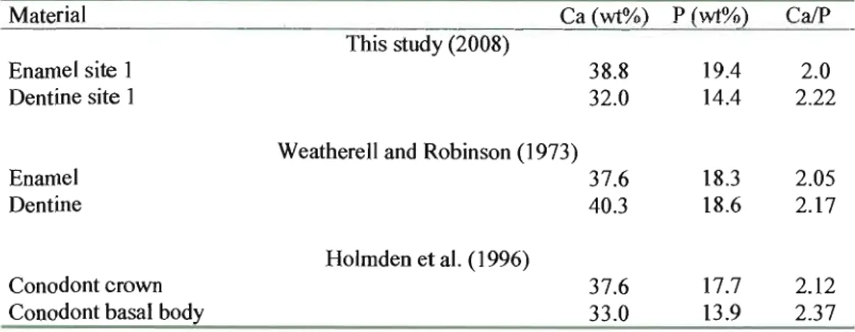
The results of scanning electron microscopy of representative Bothriolepis canadensis and Eusthenopteron foordi biomineralizations used in this study are given in Figure 2 and Table 1. Figure 2 shows X-ray maps for Ca and P for each specimen produced by energy dispersive (EDX) analyses with the percent abundances for the elements given in Table 1 along with the CaiP ratios. The abundance of Ca and P in both specimens is consistent with the calcium phosphate/apatite composition of fossil bone and tooth material. The X-ray mapping reveals the bony structure of the cancellous bone layer of Bothriolepis plate (Fig. 2a, b) in which calcium is found in both the bony wall (the bone trabecuJae; Burrow, 2005) and the spaces and phosphorus is concentrated in bone trabeculae. This suggests that the bone trabeculae are composed of apatite while the spaces are filled with calcite.
The pulp cavity of the tooth of Eusthenopteron foordi is also filled with secondary calcite (Fig. 2c). Both calcium and phosphorus are found in the dentine and enamel of the tooth (Fig. 2c, d) with CaiP ratios (~ 2) indicative of stôichiometric apatite and comparable to CaiP values of Ordovician conodonts (Holmden et al., 1996) and teeth (Weatherell and Robinson,
Figure 2. X-ray maps of ichthyoliths from the Escuminac Formation. Calcium and phosphorus distribution in the cancellous bone layer of Bothriolepis canadensis (a and b) and in tooth of Eusthenopteron foordi (c and d). bt, bone trabeculae; d, dentine; e, enamel; pc, pulp cavity; s, space; c, calcite.
Table 1. Major element composition of fish tooth enamel, dentine and conadont crown and basal body.
Material Ca (wt%) P (wt%) Ca/P
This study (2008)
Enamel site 1 38.8 19.4 2.0
Dentine site 1 32.0 14.4 2.22
Weatherell and Robinson (1973)
Ename1 37.6 18.3 2.05
Dentine 40.3 18.6 2.17
Holmden et al. (1996)
Conodont crown 37.6 17.7 2.12
16
Studies of hydroxyapatite in fossil bone and fish teeth indicate that CaiP ratios vary between 1.67 and 2.2 and that ratios above or below these values are either altered or conta in admixtures of secondary minerais (quartz, calcite; Nemliher et al., 2004). These values argue in favour of preservation of the apatitic composition of both bone and tooth from the Escuminac Formation with addition of diagenetic calcite in pore spaces. The banded pattern evident in the tooth (Fig. 2c, d) likely reflects the growth of dental tissue.
4.2. Sr in bioapatites
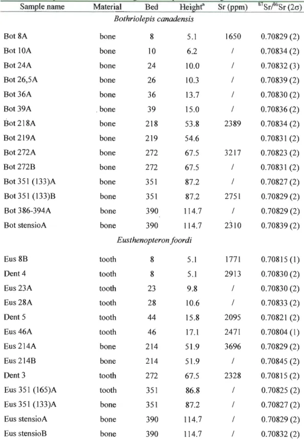
The 87S r/86S r ratio along with selected Sr concentrations for Bothriolepis canadensis, Homalacanthus concinnus, Cheirolepis canadensis, Scaumenacia curta, Eusthenopteron foordi and diagenetic calcite are given in Table 2. Stratigraphie variation of 87Sr/86Sr for five fish species and for different types of material are shown respectively in Figure 3 and Figure 4. Individual samples are shown in Figure 4, whereas Figure 3 depicts, where possible, the average of values obtained for the same species in a single bed. This explains the greater dispersion of values seen in Fig. 4.
Table 2. Sr isotope measurements of Miguasha bioapatites and diagenetic calcite.
Sam le name Material Bed Hei he Sr( m) 87Sr/86Sr (20) Bothriolepis canadensis
Bot 8A bone 8 5.1 1650 0.70829 (2)
Bot 10A bone 10 6.2 / 0.70834 (2)
Bot 24A bone 24 10.0 / 0.70832 (3)
Bot 26,5A bone 26 10.3 / 0.70839 (2)
Bot 36A bone 36 13.7 / 0.70830 (2)
Bot 39A . bone 39 15.0 / 0.70836 (2)
Bot 218A bone 218 53.8 2389 0.70834 (2)
Bot 219A bone 219 54.6 0.70831 (2)
Bot272A bone 272 67.5 3217 0.70823 (2)
Bot 272B bone 272 67.5 / 0.70831 (2)
Bot351 (l33)A bone 351 87.2 / 0.70827 (2)
Bot351 (133)B bone 351 87.2 2751 0.70829 (2)
Bot 386-394A bone 390 114.7 / 0.70829 (2)
Bot stensioA bone 390 114.7 2310 0.70839 (2)
Eusthenopteron foordi
Eus 8B tooth 8 5.1 1771 0.70815 (1)
Dent 4 tooth 8 5.1 2913 0.70830 (2)
Eus 23A tooth 23 9.8 / 0.70830 (2)
Eus 28A tooth 28 10.6 / 0.70833 (2)
Dent 5 tooth 44 15.8 2095 0.70821 (2)
Eus 46A tooth 46 17.1 2471 0.70804 (1)
Eus 214A bone 214 51.9 3696 0.70829 (2)
Eus 214B bone 214 51.9 / 0.70845 (2)
Dent 3 tooth 272 67.5 2328 0.70815 (2)
Eus 351 (165)A tooth 351 86.8 / 0.70825 (2)
Eus 351 (l33)A bone 351 87.2 / 0.70827 (2)
Eus stensioA bone 390 114.7 / 0.70829 (2)
18
Dent 6 tooth unknown unknown 2760 0.70827 (1)
Cheirolepis canadensis Che8A bone 8 5.1 / 0.70823 (1) Che 8B bone 8 5.1 / 0.70824 (2) Homalacanthus concinnus Hom8A scales 8 5.1 / 0.70825 (2) Hom8B bone 8 5.1 / 0.70842 (2) Scaumenacia curta
Seau 351 (165)A bone 351 86.8 / 0.70830 (2)
Seau 351B bone 351 86.8 / 0.70829 (2)
Authigenic calcite
Bot218 calc1 authigenic 218 53.8 / 0.70831 (2)
calcite
Bot218 calc2 authigenic 218 53.8 / 0.70830 (2)
calcite
Bot218 calc3 authigenic 218 53.8 / 0.70826 (2)
calcite
Bot218 calc4 authigenic 218 53.8 / 0.70832 (2)
calcite
(a) Heigbt (in meters) based on the stratigraphie column of Sawh (1982).
--Figure 3. 87Sr/86S r variation for fish material and diagenetic calcite from the Escuminac Formation. The grey area shows the amplitude of variation of seawater 87Sr/86Sr for the Middle Frasnian (Veizer et al., 1999) and the arrow is pointing toward freshwater values. The scale bar represents average 2u. Frasnian seawater 120 --0-B. canadensis - 0 -E. (oordi ____ H. concinnus 100
§:
--.- C. canadensis III 80 -+-S. cuita III G) _ _ Diagenetic calcite .>t: 0 60 :E c:....
1 40 20 0 0.70800 0.70810 0.70820 0.70830 0.70840 0.70850 87SrrS rFigure 4. 87Sr/86Sr variation by tissue type and calcite leaching experiment. The grey area shows the amplitude of variation of seawater 87Sr/86Sr for the Middle Frasnian (Veizer et al., 1999) while the white area encompasses the values obtained for diagenetic calcite used in this study. "Leaching 1-4" refers to progressive leaching of diagenetic calcite, from initial immersion in 0.1 M HN03 ("Leaching 1") to complete dissolution in Hel and HF ("Leaching 4"). The scale bar represents average 2u.
Diagenetic calcite D Bone Frasnian seawater
120 D D 0 • Tooth o Scales 100 ... Leaching 1 IID [][Il
l
Leaching 2 4 CIl 80 CIl Leaching 3 CIl l: 6.Leaching 4 0 0 .li: 60•
.!::! ~ D~~D D 1 40 20•
•
D DDt []
lJ:D D D 0•
0.70800 0.70810 0.70820 0.70830 0.70840 0.7085020
The overall 87Sr/86Sr variation ranges from 0.70804 to 0.70845. Eusthenopteron teeth are the only fossil material to fall within or near Frasnian seawater values (Fig. 3, 4). Ali other species and materials fall above seawater values. The 87Sr/86Sr of Bothriolepis canadensis bone is similar to the ratio measured by Schmitz et al. (1991) for a single plate from the Escuminac Formation (0.708382). The diagenetic calcite values indicate that the first leach of the calcite is radiogenic (0.70831), whereas subsequent leaches are less radiogenic (0.70825 0.70829). The final dissolution ("Leaching 4") of the calcite returns to a radiogenic value similar to the initial leach (0.70832). Figure 5 shows 87Sr/86Sr ratios compared with Sr concentrations in representative samples of Bothriolepis canadensis and Eusthenopteron foordi. There is no consistent trend between 87Sr/86Sr and Sr concentration. This likely
reflects disturbance of the Sr isotope system owing to addition of Sr from diagenetic fluids.
Figure 5. Covariation between 87Sr/86Sr and Sr concentration for Bothriolepis canadensis and Eusthenopteron foordi.
0 . 7 0 8 5 0 · r - - - , • Eusthenopteron foordi 0.70840 o Bothriofepis canadensis 0
...
CI) ~ 0.70830 0 0 CI•
•
-.: ~ 0.70820•
o•
•
0.70810 0.70800•
0 1000 2000 3000 4000 Sr (ppm)4.3. REE in bioapatites and diagenetic calcite
The REE concentrations in ichthyoliths and diagenetic calcite are given in Table 3. REE profiles for Eusthenopteron teeth, Bothrio/epis plates and diagenetic calcite are shown in Figure 6. The REE abundances are normalized against the North American Shale Composite (NASe). The profiles for the fossil material form a distinctive bell-shaped pattern found in many marine bioapatites (Wright et al., 1984; Grandjean et al., 1987; Wright et al., 1987; Bertram et al., 1992; Reynard et al., 1999; Kemp and Trueman, 2003). The REE profile of the diagenetic calcite is reJatively fiat and 1-2 orders of magnitude 1ess enriched than the bioapatites. There is no correlation between REE content and stratigraphie position within the Escuminac Formation.
Figure 6. Comparison of shale-normalized (NASC) REE concentrations between ichthyoliths and authigenic calcite from the Escuminac Formation.
1000 100 u rf) <: ~ 10 C E rf)
'"
Diagenetic calcite 0.1 La Ce Pr Nd Sm Eu Gd lb Dy Ho Er Tm Yb Lu REETable 3. REE abundances (ppm) for Miguasha fossi! material and authigenic calcite. Sample
name
Species Material La Ce Pr Nd Sm Eu Gd Tb Dy Ho Er Tm Yb Lu
Dent 2 E.foordi tooth 2383 10474 1609 7859 1591 342 1123 119 475 69 117 10 41 5
Dent 7 E.foordi tooth 1583 7350 1320 7389 1935 382 1646 183 723 98 153 Il 42 4
Dent 8 E.foordi tooth 2039 6876 1035 5093 1041 198 828 92 394 58 103 9 36 4
Bot apatite B. canadensis bane 1895 8272 1321 6985 1718 366 1426 163 656 93 154 11 45 6 Bot-calcite E. canadensis authigenic calcite 15 47 5 23 6 2 6 1 8 2 4 1 3 0 N IV
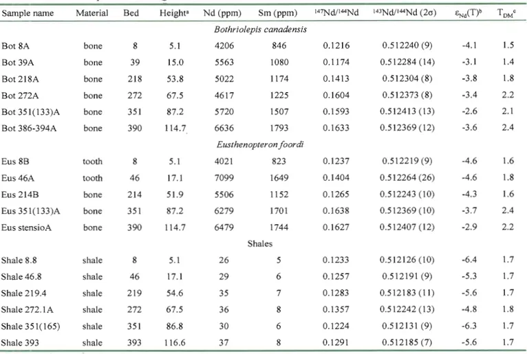
4.4. Sm-Nd in bioapatites and shales
The results for the Sm-Nd analyses of representative Eusthenopteron teeth and bone and Bothriolepis plates from different stratigraphie levels are given in Table 4 along with analyses of sediments (shales/siltstones) from the same level. The Sm and Nd concentrations in the fossil material range from 800 to 1800 ppm and 4000 to 7100 ppm, respectively. These concentrations are consistent with those determined by LA-lep-MS (Table 3). The 147 Sm/J44Nd ratios range from 0.117 to 0.164 and initial eNd values (at 380 Ma; Gradstein et
al., 2004) range between -2.6 and -4.6. The host sediments have Sm (5-8 ppm) and Nd (26 37 ppm) concentrations and '47Sm;I44Nd ratios (0.123-0.136) typical of crustal rocks (Taylor and McLennan, 1985). The initial eNd values of the sediments range between -4.8 to -6.4 and
Table 4. Sm-Nd isotope data for Miguasha shales and fossil material.
Sample name MateriaJ Bed Height8 Nd (ppm) Sm (ppm) 147Nd/I44Nd 143Nd/I44Nd (2a) ~iT)b TOMe
Bothriolepis canadensis
Bot 8A bone 8 5.1 4206 846 0.1216 0.512240 (9) -4.1 1.5
Bot 39A bone 39 15.0 5563 1080 0.1174 0.512284 (14) -3.1 1.4
Bot 218A bone 218 53.8 5022 1174 0.1413 0.512304 (8) -3.8 1.8
Bot 272A bone 272 67.5 4617 1225 0.1604 0.512373 (8) -3.4 2.2
Bot351(133)A bone 351 87.2 5720 1507 0.1593 0.512413 (13) -2.6 2.1
Bot 386-394A bone 390 114.7 6636 1793 0.1633 0.512369 (12) -3.6 2.4
Eusthenopteron foordi
Eus8B tooth 8 5.1 4021 823 0.1237 0.512219 (9) -4.6 1.6
Eus46A tooth 46 17.1 7099 1649 0.1404 0.512264 (26) -4.6 1.8
Eus 214B bone 214 51.9 5506 1152 0.1265 0.512243 (10) -4.3 1.6
Eus 351(133)A bone 351 87.2 6279 1701 0.1638 0.512369 (10) -3.7 2.4
Eus stensioA bone 390 114.7 6479 1744 0.1627 0.512407 (12) -2.9 2.2
Shales
Shale 8.8 shale 8 5.1 26 5 0.1233 0.512126 (10) -6.4 1.7
ShaJe 46.8 sha1e 46 17.1 29 6 0.1257 0.512191 (9) -5.3 1.7
Shale 219.4 shale 219 54.6 35 7 0.1283 0.512183 (Il) -5.6 1.7
Shale 272.1 A shaJe 272 67.5 36 8 0.1357 0.512242 (13) -4.8 1.8
Shale 351(165) shale 351 86.8 30 6 0.1224 0.512131 (9) -6.3 1.7
Shale 393 shaJe 393 116.6 37 8 0.1291 0.512185 (7) -5.6 1.7
(a) Height (in meters) based on the stratigraphie eolumn of Sawh (1982). (b) Age for ENd(T) ealeulation is 380 Ma.
(e) Depleted mantle model ages (TDM) ealeulated assuming a linear depleted mantle after Jacobsen (1988).
N
The high Sr content measured in the bioapatites from the Escuminac Formation indicate that there has been significant post-mortem uptake of this element (Nelson et al. 1986). The question is whether this uptake has altered the original isotopic signatures of the fossil material. For the same stratigraphic level, different ichthyoliths carry different Sr isotope compositions (Fig. 3). There are two potential explanations: different ratios reflect (1) different signatures recorded originally in vivo (and thus different environments exploited by the fish species), or either (2) different degrees of diagenetic alteration of the pristine signature. For many reasons, the second hypothesis is more plausible. Firstly, the lowest ratios are found in teeth (Fig. 4), a material considered less prone to post-mortem alteration (Trueman and Tuross, 2002). Secondly, many other biogenic samples used in the present study have 87Sr/86Sr ratios equivalent to, or even more radiogenic than calcite formed under diagenetic conditions. Thirdly, biogenic apatites that belong to the same species and coming from the same bed sometimes carry strongly different 87Sr/86Sr (e.g., Eusthenopteronfoordi in bed 8). These results suggest that the majority of specimens used in this study have experienced diagenesis to a certain extent. This alteration is likely owing to post-mortem interaction between ichthyoliths and a fluid isotopically distinct from the waters in which they developed. As noted by Martin and Scher (2004), the deviation is normally toward more radiogenic values when the biogenic apatites are hosted by silicate rocks. The Miguasha samples are consistent with this model. The lowest 87Sr/86Sr are found in the Eusthenopteron teeth. This likely reflects the fact that the teeth are overall less porous than bone (Trueman and Tuross, 2002) and infilling of the pore spaces by sediments and diagenetic calcite would lead to higher 87Sr/86Sr ratios. Slightly higher 87Sr/86Sr ratios in the other bioapatites might be due to the presence of trace amounts of sediments and calcite in the pore spaces or to a greater interaction with pore waters. Given that the lowest values, considered more reliable, overlap the Frasnian seawater Sr composition and that most of the Sr isotopic variation is close to seawater values, a marine influence cannot be discounted and may be more likely, suggesting a brackish to marine environment for the Escuminac Formation. In comparing the
26
Sr concentrations and 87S r/86S r ratios, there is no clearly defined trend that would indicate mixing of fresh and saltwater (Fig. 5). Owing to the variable degree of diagenesis experienced by the ichthyoliths used in this study, it is risky to concJude on any interspecific or stratigraphic variation of the Sr composition.
5.2. Diagenetic considerations for REE in ichthyolitbs
The high REE contents of the bone and teeth material refiect the uptake of REE during diagenesis (Wright et al., 1984; Shaw and Wasserburg, 1985). Again, the question is whether this uptake has altered the original ENd value. The bell-shaped profiles of ichthyoliths (Fig. 6; NASe normalized) are consistent with the profiles found in a number of Paleozoic marine bioapatites (Wright et al., 1984; Grandjean et al., 1987; Wright et al., 1987; Bertram et al., 1992; Reynard et al., 1999; Kemp and Trueman, 2003) and are distinguished from the REE profile of diagenetic calcite that is fiat and shows lower total REE abundances. The fiat pattern is typical of shale and suggests that the enclosing sediments are the major source of REE during calcite growth. The similar shapes of the REE profiles for the bone and teeth suggest that the REE enrichment is taxon-independent and dependent only on the diagenetic environment.
5.3. Do the Nd isotopes reflect seawater? Comparison with conodonts
The GNd values of the Escuminac sediments yield values comparable to other sediments shed as a result of the Taconian and Acadian orogenies (Eriksson et al., 2004; Hurowitz et al., 2005). Although there are small Nd isotopic variations within the sequence, no drastic GNd(T) shift is seen in the shales along the stratigraphic sequence (Fig. 7).
Figure 7. êNd(t) vanatlon along the Escuminac Formation for Bothriolepis canadensis, Eusthenopteron foordi and shales. "Tooth" and "Bone" series belong to Eusthenopteronfoordi. 120 Bone Shale 100 80
l
Bothriolepis canadensis 1: Cl 60 'Cii J: 40 20 0 -8.0 -6.0 -4.0 -2.0 0.0This suggests that sediment sources were relatively stable throughout deposition of the Escuminac Formation and were dominated by the erosion of the young Acadian Appalachians (Hesse and Sawh, ] 992; Prichonnet et al., 1996). However, the êNd(T) are more radiogenic for the Escuminac fossils (-2.6 to -4.6) than for the sediments. The difference in êNd(T) of shale and fossil material suggest the presence of at least two Nd reservoirs, one of which is Appalachian sediments and the other bearing a more radiogenic signature. Plots of êNd(T) and Nd concentration in fossil material (Fig. 8) also appear to suggest a trend that is consistent with mixing between two reservoirs; one reservoir that is 1ess radiogenic with lower êNiT) and low Nd concentration, and a second reservoir with higher êNiT) and much higher Nd concentration. The high Nd content suggests that the second reservoir is the
,
diagenetic fluid, but to what does it owe its Nd isotope composition? The fact that it is more radiogenic than the sediments suggests that the isotopie composition of the fossils reflects, at least in part, the isotopie composition of the seawater. Figure 8 illustrates that if the post mortem REE uptake is done in equilibrium with seawater, then, the isotopie composition may be preserved.
28
Figure 8. Diagram showing ëNd(T) values of ichthyoliths plotted against their Nd concentration. The samples differ from the shale from the Escuminac Formation. An hypotheticaJ seawater value is presented as a possible source of REE uptake during diagenesis.
0.0 -1.0 Diagenetic uptake _ _ _ _ _ _...;.;;:.;."",,;.~...:;.,;,,;...;.. ... Diagenetic fluid Seawater? -20 -30
~
ç -", -4.0 z w -5.0o
Mixing curves 0o
•
-601
5hale 1 -7.0 • Eusthenopteron (oordi o Bothriolepis canadensis -8.0o
1000 2000 3000 4000 5000 6000 7000 8000 Nd (ppm)The sinusoidal variation in ëNd(T) of the placoderm remains with respect to their stratigraph ic position (Fig. 7) mimics that of the sediments and likely reflects the presence of sedimentary (silicate?) material that was not completely removed during cleaning. The absence of a sinusoidal pattern among the osteolepiform teeth is likely because teeth are more easily cleaned. The difference in ëNd(T) between the bone and teeth is intriguing. The difference is unlikely to be due to incomplete cleaning because the placoderm fragments are more radiogenic. Could the difference in composition reflects different initial Nd isotope compositions for different species living in different water masses (e.g., pelagic vs. benthic)? Further tooth analyses are required to investigate this phenomenon.
A plot of the ëNd(T) values vs 87Sr/86Sr ratios of the Miguasha fossil material (Fig. 9) also potentially shows mixing between two reservoirs, one sedimentary and the other possibly marine. The bone material of both fossil species show a broad negative correlation from high 143Nd/144Nd, low 87Sr/86Sr to low 143Nd/144Nd, high 87Sr/86Sr. This latter component is the Appalachian sediment end of the mixing curve, whereas the high 143Nd/144Nd, low 87Sr/86Sr component trends towards what may have been seawater values. The sediment contamination
may be largely owing to the presence of sediment still trapped within pores and fractures in the bones and teeth. There are only two tooth samples for which both Nd and Sr isotope data are available so there is no clear correlation on this diagram. However, the displacement of the Eusthenopteron teeth compared to the bony material of the same species is significant. Note that the Nd isotope compositions of the more porous bone material of Eusthenopteron overlap with the bone material of Bothriolepis canadensis.
Figure 9. Sr isotope composition and êNd(T) of tooth and bone from the Escuminac Formation. Samples exhibit relatively homogeneous êNd(T) in spite of a wide range of 87Sr/86Sr.
0.0 , - - - , -1.0 -2.0 Seawater? Bone -3.0 o ~ ~ z w -4.0 -50
•
Tooth•
Appalachian -6.0 sediments -7.0 -8.0 0.70800 0.70810 0.70820 0.70830 0.70840 0.70850 87Sr/8·SrIt is relevant (Fig. 10) that êNd(T) values of bioapatites from the Escuminac Formation are similar (-2.6 to -4.6) to those recorded in Late Devonian conodonts from Variscan oceanic water (-1.1 to -6.5; Dopieralska et al., 2006). This oceanic realm was situated during the Devonian between Euramerica and Gondwana and included in its western portion the Rheic Ocean, which was bordering the eastern coast of Euramerica (Dopieralska et al., 2006). The radiogenic compositions of the Miguasha ichthyoliths may, thus, represent the diagenetic fluids in isotopie equilibrium with the Rheic Ocean water. These radiogenic signatures are also consistent with the erosion of a tectonically active region such as the eastern margin of Euramerica during the Devonian. The êNd(T) values of North American conodonts are similar to those of the Miguasha sediments and post-Taconic Appalachian sediments in
30
general (Fig. 10). The contrastingly more radiogenic cNd(T) values of the Miguasha fossils likely reflect the difference between a restricted epieric sea that received Nd (detrital and dissolved) from both the emerging Appalachian and older exposed continental rocks, and a more open marine environment on the Rheic Ocean as represented by the Miguasha fossils that received Nd largely from orogenic terranes. Decoupling of Nd isotope compositions between epicontinental seas and bordering oceans owing to restricted water circulation has been suggested in geochemical investigations of Paleozoic bioapatites (Holmden et al., 1998; Dopieralska et al., 2006). Considering the Nd isotope compositions, fossi 1 fishes from Miguasha have more in common with Baltic conodonts than North American conodonts. This connection is also supported by paleontological data. The Escuminac Formation ichthyofauna is considered to be closer to fish assemblages from Scottish and Baltic sequences rather than to other North American ichthyofaunas (Schultze and Cloutier, 1996) supporting the same aquatic connectivity suggested by the present study.
Figure 10. cNd(T) comparison between the material used in this study and sediments and fossils coming form other localities and periods. Black arrows represent the general trend seen in North American shales (see Hurowitz and McLennan, 2005). Data for conodonts are taken in Wright (1995; for North America) and Dopieralka et al. (2006; for Poland). Figure modified from Hurowitz and McLennan (2005).
0,0 . - - - . - . . . - - - , o Escuminac Formation (fossils)
f:,. Escuminac Formation (shale)
-4,0
Il
• North American conodonts•
-8,0 • ï=' ..~Oland C~OOi
·
'1
-12,0 w -16,0 20,0 --24,0 '-- ~ ~ " " - - -•
•
•
••
'__ __'__ ---J 350 400 450 500 550 600 TSTRA~Ma)of bioapatites and sediments from the Escuminac Formation. The main conclusions of this studyare:
1. The freshwater hypothesis is not supported by Sr isotope composition of bioapatites. Although most samples show a deviation toward more freshwater ratios, sorne tooth material is comparable to typical Frasnian seawater 87Sr/86Sr compositions. The signatures recorded in teeth are considered more reliable than those found in more easily altered structures such as porous bones and scales. The 87Sr/86Sr signature is more dependent on the nature of the fossil material (i.e., teeth, bone, scales) than by its stratigraphie position or its taxonomie affinity. A brackish to marine environment is suggested by 87 Sr/86Sr ratios of the ichthyoliths.
2. A significant post-mortem REE uptake occured in the Miguasha fossils. This diagenetic enrichment is partially derived from the enclosing sediments, as suggested by REE patterns in diagenetic calcite and similar cNd(T) fluctuation along the Escuminac Formation for shales and Bothriolepis bony plates.
3. As suggested by shale cNd(T) values, the detrital sedimentation in the Escuminac Formation was dominated by sources typical of post-Taconian Appalachians.
4. Differences between shale and fossil material cNd(T) values implies the presence of at least two Nd sources for ichthyoliths, the first of them being the sediments. We suggest that a second reservoir is seawater. During diagenesis, a significant Nd uptake from seawater oeeurs. If this uptake is done in equilibrium with seawater, the Nd isotope composition of seawater will be preserved in ichthyoliths. This might explain the similarity observed between cNd(T) for Miguasha fossils and other Upper Devonian marine bioapatites belonging to the Variscan oceanic realm. This homogeneity between bioapatites cNctCT) values from Rheic Ocean and Miguasha implies a Rheic Ocean component within the Escuminae Formation.
REFERENCES Ahlberg P. E. (1989) .. .for Devonian vertebrates. Nature 342, 738.
Arsenault M., Desbiens S., Janvier P. and Kerr J. (2004) New data on the soft tissues and external morphology of the antiarch Bothriolepis canadensis (Whiteaves, 1880), from the Upper Devonian of Miguasha, Quebec. In Recent Advances in the Origin and Early Radiation of Vertebrates (eds. G. Arratia, M.V.H. Wilson and R. Cloutier). Verlag Dr. Friedrich Pfeil, München, Gennany. pp. 439-454.
Barrat J. A., Taylor R. N., Andre J. P., Nesbitt R. W. and Lécuyer C. (2000) Strontium isotopes in biogenic phosphates from a Neogene marine formation: Implications for palaeoseawater studies. Chem. Geol. 168, 325-332.
Bertram C. J., Elderfield H., Aldridge R. J. and Conway Morris S. (1992) 87S r/86S r, 143Nd/144Nd and REEs in Silurian phosphatic fossils. Earth Planet. Sei. Let!. 113,239-249. Brideaux G. W. and Radforth, N.W. (1970) Upper Devonian miospores from the Escuminac Fonnation, eastern Québec. Cano J Earth Sei. 7, 29-45.
Bryant J. D., Jones D. S. and Mueller P. A. (1995) Influence of freshwater flux on 87S r/86S r chronostratigraphy in marginal marine environments and dating of vertebrate and invertebrates faunas. J. Paleont. 69, 1-6.
Burrow C. J. (2005) Histological structure of the cancellous bone layer in Bothriolepis canadensis (Antiarchi, Placodermi). Lethaia 38,205-210.
Canfield D. E. and Raiswell R. (1991 a) Carbonate precipitation and dissolution: its revelance to fossil preservation. In Taphonomy, releasing the data locked in thefossil record (eds. P. A. Ailison and D. E. G. Briggs). Topics in Geobiology, Volume 9, Plenum Press, New York. pp. 412-455.
Canfield D. E. and Raiswell R. (1991 b). Pyrite formation and fossi) preservation. In Taphonomy, releasing the data locked in the fossil record (eds. P. A. Allison and D. E. G. Briggs). Topics in Geobiology, Volume 9, Plenum Press, New York. pp. 338-388.
Chidiac Y. (1989) Analyse du paléoenvironnement de la Fonnation d'Escuminac (Dévonien supérieur), Miguasha, Québec, dans le contexte des données sédimentologiques, paléontologiques et géochimiques. Master's thesis (unpublished), Université du Québec à Montréal, Montréal.
Chidiac y. (1996) Paleoenvironmental interpretation of the Escuminac Formation based on geochemical evidence. In Devonian Fishes and Plants of Miguasha, Québec, Canada (eds. H.-P. Schultze and R. Cloutier). Verlag Dr. Friedrich Pfeil, München, Germany. pp. 47-53. Clack J. A. and Neininger S. L. (2000) Fossils from the Celcius Bjerg Group, Upper Devonian sequence, East Greenland: significance and sedimentological distribution. In New