O
pen
A
rchive
T
oulouse
A
rchive
O
uverte (
OATAO
)
OATAO is an open access repository that collects the work of Toulouse researchers and
makes it freely available over the web where possible.
This is an author-deposited version published in:
http://oatao.univ-toulouse.fr/
Eprints ID : 2822
To link to this article :
URL :
http://dx.doi.org/10.1016/j.powtec.2008.04.041
To cite this version :
Drouet, Christophe and Bosc, Françoise and Banu, Mihai
and Largeot, C. and Combes, Christèle and Dechambre, Gérard and Estournès,
Claude and Raimbeaux, G. and Rey, Christian ( 2009) Nanocrystalline apatites:
From powders to biomaterials. Powder Technology, vol. 190 (n° 1 - 2). pp.
118-122. ISSN 0032-5910
Any correspondence concerning this service should be sent to the repository
administrator:
staff-oatao@inp-toulouse.fr
Nanocrystalline apatites: From powders to biomaterials
Christophe Drouet
a,⁎
, Françoise Bosc
a, Mihai Banu
a, Céline Largeot
a, Christèle Combes
a,
Gérard Dechambre
a, Claude Estournès
b, Gwenaëlle Raimbeaux
b, Christian Rey
aa
CIRIMAT - Institut Carnot, ENSIACET, 118 route de Narbonne, 31077 Toulouse Cedex 4, France
b
Plateforme Nationale de Frittage Flash (PNF2)/CIRIMAT, Université Paul Sabatier, MHT, 118 route de Narbonne, 31062 TOULOUSE Cedex 9, France
A B S T R A C T
Keywords: Apatites
Resorbable bioceramics Low-temperature sintering
Non-stoichiometric nanocrystalline apatite powders are used to elaborate highly-bioactive biomaterials. Their exceptional surface reactivity arises from a structured but rather unstable hydrated layer involving ions in non-apatitic chemical environments, like in bone mineral. The initial powder characteristics can be tailored through precipitation parameters (pH, temperature, maturation time in solution). The drying of nanocrystalline apatite suspensions at very low temperature (4 °C) leads to ceramic-like materials exhibiting average mechanical properties (compressive strength 54 MPa) and a high porosity which could be exploited to entrap active organic compounds (e.g. growth factors). The consolidation at 150–200 °C of nanocrystalline apatite powders has also been studied using uni-axial pressing and spark plasma sintering (SPS). The results indicate only a limited alteration of the initial nanocrystals, and the bioceramics obtained show mechanical properties close to those reached with sintered stoichiometric HA. The high ion mobility in the hydrated layer of the nanocrystals can lead to“crystal fusion” processes. This capability to favor crystal–crystal interactions at low temperature, while preserving the non-stoichiometry and nanometer dimensions of apatite crystals, opens interesting perspectives for the elaboration of new resorbable and highly-bioactive bioceramics.
1. Introduction
Calcium phosphates (CaP) exhibit excellent biocompatibility and show osteoconductive properties. They are widely used as bone substitute materials and are often proposed for replacing autologous grafts or allografts. Among calcium phosphates, non-stoichiometric nanocrystalline apatites have appeared in the last decades as exceptionally promising biomimetic compounds. They are the main constituent of bone, and nature has learned to use all peculiarities of their surface and bulk reactivity[1]. They are also considered to be at the origin of the biological activity of orthopedic materials (bioglasses, polymers, ceramics, or cements)[2].
Apatite nanocrystals obtained by precipitation methods in solution were shown to exhibit physico-chemical characteristics similar to those of bone nanocrystals[3]. In particular, their chemical composi-tion departs from stoichiometry by calcium and hydroxide ions deficiency, leading to an increased solubility, and in turn bioresorption rate in vivo[4]. The nanocrystals have also the property to evolve in solution (maturation) like bone crystals. Thus, freshly-precipitated apatites have been shown to be analogous to embryonic bone mineral crystals whereas aged precipitates resemble bone crystals of old vertebrates [3]. This relatively fast maturation process has been
assigned to the very specific structure of apatite nanocrystals, with an apatitic core and a structured hydrated surface layer. Upon aging in solution, the apatitic domain develops progressively at the expense of the hydrated layer and the surface reactivity is altered.
CaP bioceramics are generally obtained by sintering techniques at high temperature, which lead to crystal alterations (grain growth, loss in surface area, dehydration, evolution toward stoichiometry), and the setup of new powder processing techniques capable of preserving the nanocrystals of biomimetic apatites appears rather delicate. Also, since the resorption rate of bioceramics is directly linked to the apatite non-stoichiometry [4], the conservation of a highly-non-stoichio-metric composition appears as an interesting, but still unsolved, challenge in view of the elaboration of bioactive and resorbable bioceramics.
To date, some prospective methods have been tentatively tested such as associations with macromolecules[5]or cements setting[6]. Apatite nanocrystals have also been recently proposed in coatings[7]
and they have generally shown superior biological activity and even in some cases osteoinductive properties[8]. The use of an unconventional sintering technique such as spark plasma sintering (SPS) has also been investigated for sintering hydroxyapatite (HA). SPS sintering was then shown to be more efficient when HA powders with small particle size were used, and the compacts exhibited excellent mechanical proper-ties[9]. The formation of transparent apatite ceramics with interesting surface properties has also been reported[10]and SPS-sintered HA generally showed superior surface reactivity in simulated-body-fluid
⁎ Corresponding author.
E-mail address:christophe.drouet@ensiacet.fr(C. Drouet).
(SBF) tests often considered to represent the biological activity[11]. However, these SPS studies were performed at high temperature (900– 1100 °C), although lower than those observed for natural sintering, and results cannot be directly transposed to nanocrystalline apatites exhibiting a relatively unstable hydrated surface layer.
The aim of this contribution is to illustrate the effect of synthesis experimental parameters on apatite powder characteristics, and to discuss methods for the processing of highly-reactive non-stoichio-metric nanocrystalline apatite bioceramics, based on low-temperature methods: gel hardening, uni-axial pressing at low temperature and spark plasma sintering (SPS).
2. Materials and methods
2.1. Synthesis and characterization procedures
The nanocrystalline apatites discussed in this paper (referred to as “hap”) have been synthesized at room temperature and at physiolo-gical pH by double decomposition between a solution of ammonium hydrogenphosphate (120 g (NH4)2HPO4in 1500 ml) and a solution of
calcium nitrate (52.2 g Ca(NO3)24H2O in 750 ml). The large excess of
phosphate provides pH buffering at 7.4. The calcium solution is rapidly poured into the phosphate solution at room temperature and stirred for 2 min. It is then possible to leave the precipitate in the mother solution for maturation (aging in solution) for a determined and variable amount of time. In the text, samples matured for“X” days will be referred to as“hap Xd”.
For investigations on freshly-precipitated nanocrystalline apatites, the precipitate is quickly vacuum-filtered after stirring for 2 min, and then washed with deionized water (2 l). The filtered cake of precipitate has then the consistency of a gel. In order to prevent further maturation of the apatite nanocrystals, the gel is freeze-dried and stored in a freezer. Experiments aiming at following the effect of drying on such gels were carried out in air at low temperature (4 °C). Calcium concentration was determined by complexometry with EDTA and phosphorus concentration by visible absorption spectro-photometry of a phosphovanadomolybdenum complex at 460 nm.
X-ray diffraction (XRD) was performed for crystal structure identifications, using a curved counter diffractometer (INEL CPS 120) and a monochromatic CoKα radiation (kCo= 1.78892 Å).
Fourier-transformed infrared (FTIR) analysis was used for complementary phase identification. The experiments were carried out on a Perkin Elmer 1600 spectrometer at 4 cm− 1resolution.
2.2. Powder processing techniques and mechanical tests
Three processing techniques were investigated in this work, at low temperature: gel hardening in air, uni-axial pressing and spark plasma sintering (SPS).
Gel hardening was carried out by drying apatite gels in air, at temperatures down to 4 °C.
The uni-axial pressing (50 MPa) of powders was performed by associating a Hounsfield press (model H25K-S) to the custom-built setup depicted inFig. 1. An 8-mm diameter mold was used. Heating was provided simultaneously by a custom-built low-temperature regulated oven (maximum operational temperature: 300 °C), and the actual temperature of treatment was determined by a thermocouple located near the sample.
Spark plasma sintering (SPS) experiments were performed on an SPS 2080 Sumitomo Coal Mining equipment. Powder samples without any sintering aids or additives were placed in an 8-mm graphite mold and introduced in the treatment chamber under low mechanical pressure (aimed at insuring the mold stability). DC pulsed current with a sequence of 12⁎2 (12 pulses and 2 dead times) was applied to heat the graphite mold. The temperature was measured by means of a thermocouple located in a narrow hole at the surface of the mold. In
this work, the samples were sintered under vacuum (several Pa) and heated in these conditions until spontaneous densification of the powder. A uni-axial pressure of 50 MPa was applied on the sample and the temperature was kept constant until completion of the sintering process. Fast cooling (~ 100 °C/min) and a release of the mechanical force were then operated.
Unless otherwise specified, the mechanical properties investigated were obtained by compression tests on cylindrical samples using the above-cited Hounsfield press.
Due to the size and shape of the SPS mold, the materials con-solidated by this technique exhibited a disk shape. Therefore, usual compression tests could not be performed and mechanical testing was carried out here by using diametral compression tests. In such experiments, an increasing force (F) is applied on the disks perpendi-cular to the cylinder axis until rupture. This leads to tensile strain perpendicular to F, and the corresponding tensile stress (σ), that is characteristic of the mechanical strength of the disk, is related to F by Eq. (1):
r ¼kDH2F ð1Þ
where D and H are respectively the diameter and height of the disk. 3. Results and discussion
3.1. Effects of synthesis parameters on apatite powder characteristics Several nanocrystalline apatite powders were synthesized at room temperature with varying maturation times in solution, between 0 and 20 days. X-ray diffraction patterns (Fig. 2) exhibit in all cases broad bands and an elevated background illustrating the nanocrystalline feature of the apatitic phases prepared. These patterns remind those of newly formed bone tissues (“young bones”)[3], therefore pointing to the biomimetic property of apatites prepared through the double decomposition synthetic route. According to the XRD patterns, maturation leads to an improvement of crystallinity.
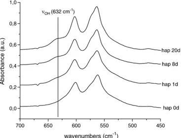
In parallel, FTIR data recorded for increasing maturation states (Fig. 3) clearly show the progressive increase of the OH−content, as observed from the increase of the band intensity at 632 cm− 1.
These variations can be related to structural modifications leading to apatite compositions moving toward stoichiometry. This can be linked to the growth of apatitic domains at the expense of non-apatitic ionic chemical environments, present within the surface hydrated layer on the nanocrystals. These results show that the extent of surface hydrated layer can be controlled through the maturation process. Elemental analysis (determination of Ca and P contents) carried out on such samples shows indeed an increase of the Ca/P molar ratio upon maturation, which is in agreement with the above results (Table 1).
The mean crystallite dimensions (platelets) were also derived from Scherrer's formula applied to diffraction lines (002) and (310) (Table 1), giving respectively estimates of the crystallite length and an average of their width/thickness. These data confirm in all cases the nanometer dimensions of the crystals constitutive of such apatites, and they point out an increase of crystal size upon maturation, especially of the crystallite length (along the c-axis of the unit cell).
The HPO42−content of such apatites can be determined by chemical
analysis after calcination at 600 °C, following the method proposed by Gee and Dietz [12]. This treatment converts the HPO42− ions into
pyrophosphate ions P2O4−7 which are analyzed: phosphorus is titrated
as orthophosphate at 460 nm by absorption spectrophotometry, before (PO43−only) and after (PO43−and HPO42−) acid hydrolysis of the P–
O–P bond of pyrophosphate ions at 100 °C for 1 h. Thus, the level of condensed phosphate (therefore that of HPO42−) is calculated from the
difference of the results of these two analyses. The results obtained showed a progressive decrease from 1.46 to 0.75 mol HPO42−per unit
formula, respectively for a non-matured sample and after 15 days of
maturation in solution. Thesefindings illustrate again the progressive evolution toward stoichiometry.
The effects of precipitation (and maturation) temperature and of pH on the apatite nanocrystals features were also investigated in this study. Tests were performed here on a nanocrystalline apatite matured for 3 days either at 50 °C or at room temperature but with an initial alkaline pH of 9.5. FTIR analysis of the samples obtained (Fig. 4) indicates in both cases an increase of the crystallinity level of the apatite, especially noticeable by the better resolution of theν4
(PO4) band, as well as an increase in hydroxylation, as shown by the
growth of the apatitic OH band at 632 cm− 1 (Fig. 4). These data indicate that an increased synthesis temperature and/or an increase in pH value lead to apatite phases closer from stoichiometry and better crystallized.
All the above results illustrate the effects of synthesis parameters (maturation time, temperature and pH) on the physico-chemical characteristics of the starting apatite powders. These features (crystallinity, non-stoichiometry, crystal size, presence of non-apatitic chemical environments, extent of hydrated layer, chemical composi-tion) can then be tailored in view of specific applications. Indeed, i) the bioresorbability rate of apatite materials is linked to the size and stoichiometry of the constitutive nanocrystals (larger and/or stoichio-metric crystals showing a much lower solubility), and ii) nanocrystal-line apatite reactivity (in particular through ion exchange [13] or adsorption [14] phenomena) involves the surface hydrated layer present on the nanocrystals[15,16] and therefore depends on the extent of this layer.
3.2. Low-temperature consolidation
The presence of a structured hydrated layer on the surface of apatite nanocrystals opens the possibility for strong crystal–crystal interactions upon drying. Such interactions have, in a similar way, been observed in vivo for bone mineral, and were described as a
Fig. 3. FTIR analysis of the effect of maturation on nanocrystalline apatites (450–700 cm− 1
region).
Table 1
Evolution of Ca/P ratio and crystallite dimensions for nanocrystalline apatites with varying maturation states
Maturation time (days) Ca/P (mol) Length (nm)a Width (nm)b
0 1.38 15 5
1 1.43 22 6
3 1.45 25 6.5 15 1.49 28 7
a
Assessed from (002) diffraction line analysis.
b
Assessed from (310) diffraction line analysis.
Fig. 4. FTIR analysis of the effect of temperature and pH on nanocrystalline apatites. Fig. 2. XRD analysis of the effect of maturation on nanocrystalline apatites.
“crystal fusion” process[17]. These observations led us to investigate here the possibility to consolidate nanocrystalline apatites, at low temperature in order to preserve as far as possible this hydrated structure. The results presented in the previous section indicated that apatite synthesis at room temperature (rather than heating) and with limited durations of maturation led to highly non-stoichiometric apatite nanocrystals and well-developed hydrated layers. It thus enabled us to select these powder preparation conditions as the most appropriate for consolidation. The following tests have thus been performed on apatites matured for durations lower or equal to 1 day.
Fig. 5shows the typical plate-like crystallite morphology obtained for such initial powders, with an average specific surface area of ca. 120 m2/g.
3.2.1. Apatite gel hardening
Apatite gels obtained just after precipitation (non-matured) and filtration were subjected to drying in air (studied here at temperatures down to 4 °C) and led, in an irreversible way, to ceramic-like materials, as had already been observed by Sarda et al.[5]. The process was found to be associated with an important shrinkage. Average mechanical properties were measured, indicating a compressive strength of about 54 MPa. A high porosity was also observed on the solid bodies obtained, with pore sizes in the range 5–25 nm, and an
increase of total porosity (from 30 to 50%) was seen upon maturation. Despite the rather limited mechanical resistance of such materials, the existence of this porosity might be considered as an interesting feature for the elaboration of drug carriers, as it could be used for example for trapping of organic molecules (e.g. growth factors) in view of activating bone repair after implantation in vivo. Also, the possibility to perform this gel consolidation at very low temperature makes it possible to manipulate rather unstable organic compounds, and also limits the alteration of the apatite nanocrystals.
The formation of solid bodies upon such gel drying is probably related to the presence of the hydrated layer, which is likely to be involved in the cohesiveness and adhesion processes between two adjacent apatite nanocrystals (and this property should therefore even subsist for low-temperature processing). The progressive drying is thought here to increase inter-crystal contacts, by the steady elimination of excess water molecules present in the gel, thus bringing two crystals in close contact and enabling the constitutive ions to interact through strong electrostatic interactions.
This mechanism is however probably not limited to gel drying, and should also apply to other low-temperature sintering methods, for which ion diffusion processes show limited kinetics and where the high ionic mobility of the hydrated layer might present a potential advantage. The notion of“low temperature” is intended here for heat treatments enabling the apatite crystals to remain nanosized and hydrated, therefore typically below 300 °C. In these conditions, the term“consolidation” is then probably more suited than “sintering” to describe the physical phenomena undergone by the initial powder samples during such heat treatments.
3.2.2. Uni-axial pressing
Uni-axial pressing of apatite powders was studied (see Fig. 1) under simultaneous heating, using a two-step process: afirst heating was performed at 80 °C for 20 min for evacuation of adsorbed water and a second heating step was then carried out at 150 °C for 30 min after applying a mechanical pressure of 75 MPa. In these experiments, the heating rates were set to 3 °C/min. After heating, the mold was allowed to cool down at 0.5 °C/min. This method led to solid ceramic-like materials. At this low temperature some crystal growth was observed from XRD data analysis as compared to starting powders, especially in the c-axis direction, and this was accompanied by a partial decomposition into dicalcium phosphate anhydrous (DCPA or
Fig. 5. TEM micrograph for typical nanocrystalline apatite morphology.
monetite), observable as a trace secondary phase by XRD (additional peaks at 2θ=30.97° and 35.26°, with kCo= 0.178892 nm), as well as a
further hydroxylation of the initial powder (increase ofνOHband seen
by FTIR) probably due to the internal hydrolysis of phosphate groups. Fully-dense ceramics could not be obtained, as apparent densi fica-tion rates of about 65% were measured. However, the compacts obtained by this method, starting with non-matured apatites, exhibit a high compression strength of 100–150 MPa and a Young modulus of ca. 10 GPa. Very interestingly, despite a lower densification, these values compare well with those obtained after sintering of stoichio-metric HA at much higher temperatures (1100–1250 °C).
Additional mechanical tests were performed on pre-humidified apatite powders, in order to unveil a possible positive effect of an increase in initial water content. For these tests, 1 g of powder was intimately mixed with 4.3 g water prior to uni-axial compression. However, the compacts obtained this way only showed poor mechanical resistance, about 6 times lower than for dry powders.
The above findings indicate that low-temperature pressing at 150 °C leads to consolidated materials which could be used for load bearing bone-related applications. However, the presence of traces of a secondary phase and the observed growth of the apatite nanocrys-tals led us to investigate in parallel the consolidation of apatites at low temperature by SPS.
3.2.3. Spark plasma sintering (SPS) at low temperature
Taking into account its unconventional heating source enabling fast heating and cooling, the SPS technique appeared as particularly interesting to apply to such rather unstable hydrated nanocrystalline apatites. Samples matured for 1 day were used in these tests. Under SPS conditions, the spontaneous densification of the powder was observed in the temperature range 150–190 °C.
The tests performed by SPS under vacuum, at 200 °C, led to compacts exhibiting a disk shape (due to the SPS mold configuration) and mechanical testing was thus carried out here using diametral compression tests. In this case, a tensile stress ranging from 18 to 25 MPa was found for a heating time of ca. 3 min from room temperature to 200 °C followed by a plateau at 200 °C for 2 min. Interestingly, these values are close to those obtained with stoichio-metric HA despite extremely short heating times. A densification rate of 70% was measured.
XRD analysis of the consolidated disks obtained by SPS treatment for 2 min at 200 °C corresponded to nanocrystalline apatite and no other crystalline phase was observed, unlike for uni-axial pressing. Compar-ison with the pattern of the initial powder indicated however an increase of the degree of crystallinity after SPS treatment, and the average crystallite dimensions were found to increase slightly, especially in length along the c-axis, after SPS treatment: length estimated to 26 nm (to be compared to 22 nm for the starting powder). However, this increase in crystal length was found to be noticeably lower than the one observed after uni-axial pressing, even at low temperature: 29.5 nm for a treatment at 200 °C for 15 min. Therefore, SPS consolidation for 2 min at 200 °C led to a lower alteration of the initial nanocrystalline powder than uni-axial pressing at 200 °C for 15 min.
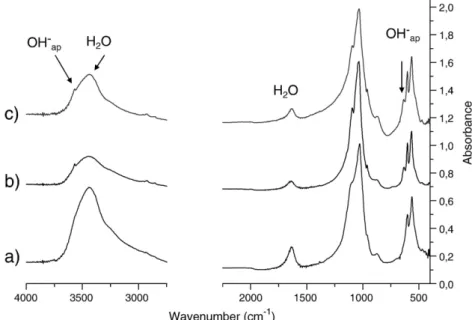
Thesefindings were confirmed by FTIR analysis showing that the partial loss of water and the increase in intensity of the OH−bands was smaller after SPS treatment than after uni-axial pressing, even at low temperature (200 °C for 15 min), as shown inFig. 6.
Although the mechanism involved in SPS sintering is not completely understood at this point, the consolidation of nanocrystal-line apatite powders at low temperatures most likely involves the high ion mobility on the surface of the nanocrystals for achieving crystal– crystal interactions, accompanied by a progressive release of water molecules from the hydrated layers of adjacent crystals.
SPS consolidation appears thus as a promising technique for obtaining compacts for use in bone repair applications, thanks to the limited alterations of apatite nanocrystals, preserving part of their
surface reactivity. In addition this technique allows the formation of ceramic-like materials preserving the non-stoichiometry of apatites and therefore their biological resorbability.
4. Conclusions
The physico-chemical characteristics of synthetic nanocrystalline powders analogous to bone mineral were investigated and the effect of synthesis parameters such as crystal aging in solution (maturation), temperature and pH were pointed out and discussed. These parameters could be used to tailor the apatite crystal size, chemical composition and non-stoichiometry in view of specific applications.
Low-temperature consolidation techniques were studied. Gel hardening leads to ceramic-like materials with average mechanical strength but exhibiting an array of pores possibly usable for the trapping and release of active agents such as drugs or growth factors. Low-temperature uni-axial pressing and spark plasma sintering (SPS) of nanocrystalline apatites matured for 1 day led, in contrast, to compacts exhibiting high mechanical strength while preserving the non-stoichiometry and nanometer dimensions of the crystals and limiting the physico-chemical alterations of the initial powder. Among these two techniques, SPS was found to lead to smaller alterations of the original nanocrystals, and no secondary phase was detected, making it a very promising technique in view of apatite consolidation at low temperature.
Thesefindings open thus a new field for the fabrication of new bioresorbable and highly-bioactive bioceramics using the unique surface properties, in particular in terms of ion mobility, of hydrated apatite nanocrystals.
References
[1] S. Cazalbou, C. Combes, D. Eichert, C. Rey, Adaptative physico-chemistry of bio-related calcium phosphates, J. Mater. Chem. 14 (2004) 2148–2153.
[2] C. Combes, C. Rey, Bioceramics, in: Ph Boch, J.C. Nièpce (Eds.), Ceramics Materials— Processes, Properties and Applications, ISTE, London, 2007, pp. 493–521. [3] C. Rey, A. Hina, A. Tofighi, M.J. Glimcher, Maturation of poorly crystalline apatites:
chemical and structural aspects in vivo and in vitro, Cells Mater. 5–4 (1995) 345–356.
[4] R.Z. Legeros, Biodegradation and bioresorption of calcium phosphate ceramics, Clin. Mater. 14 (1993) 65–88.
[5] S. Sarda, A. Tofighi, M.C. Hobatho, D. Lee, C. Rey, Associations of low temperature apatites ceramics and proteins, Phosphorus Res. Bull. 10 (1999) 208–213. [6] D. Knaack, M.E.P. Goad, M. Ailova, C. Rey, A. Tofighi, P. Chakravarthy, D. Lee, Resorbable
calcium phosphate bone substitute, J. Biomed. Mater. Res. 43 (1998) 399–409. [7] P. Habibovic, F. Barrere, C.A. van Blitterswijk, K. de Groot, P. Layrolle, Biomimetic
hydroxyapatite coating on metal implants, J. Am. Ceram. Soc. 85 (2002) 517–522. [8] J.D. deBruijn, H. Yuan, R. Dekker, P. Layrolle, K. de Groot, C.A. van Blitterswijk, Osteoinductive biomimetic calcium phosphate coatings and their potential use as tissue-engineering scaffolds, in: J.E. Davies (Ed.), Bone Engineering, EM Squared Inc., Toronto, 2000, pp. 421–431.
[9] Y.W. Gu, N.H. loh, K.A. Khor, S.B. Tor, P. Cheang, Spark plasma sintering of hydroxyapatite powders, Biomaterials 23 (2002) 37–43.
[10] D. Kawagoe, K. Ioku, H. Fujimori, S. Goto, In vitro estimation with simulated body fluid of OH-designed transparent apatite ceramics prepared by spark plasma sintering, Trans. Mater. Res. Soc. Jpn. 28 (2003) 841–844.
[11] A. Nakahira, M. Tamai, H. Aritani, S. Nakamura, K. Yamashita, Biocompatibility of dense hydroxyapatite prepared using a SPS Process, J. Biomed. Mater. Res. 62 (2002) 550–557.
[12] A. Gee, V.R. Dietz, Determination of phosphate by differential spectrophotometry, Ann. Chem. 25 (1953) 1320–1324.
[13] S. Cazalbou, D. Eichert, X. Ranz, C. Drouet, C. Combes, M.F. Harmand, C. Rey, Ion exchanges in apatites for biomedical applications, J. Mater. Sci., Mater. Med. 16 (2005) 405–409.
[14] C. Rey, E. Strawich, M.J. Glimcher, Non-apatitic environments in Ca–P biominerals; implications in reactivity of the mineral phase and its interactions with organic matrix constituents, in: D. Allemand, J.P. Cuif (Eds.), Biomineralizations, Bull. Océanogr., vol. 14 1, Monaco, 1994, pp. 55–64, n° special.
[15] D. Eichert, C. Combes, C. Drouet, C. Rey, Formation and evolution of hydrated surface layers of apatites, Key Eng. Mater. 284–286 (2005) 3–6 (Bioceramics 17). [16] C. Rey, C. Combes, C. Drouet, H. Sfihi, A. Barroug, Physico-chemical properties of
nanocrystalline apatites: implications for biominerals and biomaterials, Mater. Sci. Eng., C 27 (2007) 198–205.
[17] G. Daculsi, J. Menanteau, L.M. Kerebel, D. Mitre, Length and shape of enamel crystals, Calcif. Tissue Int. 36 (1984) 550–555.