HAL Id: inserm-02490517
https://www.hal.inserm.fr/inserm-02490517
Submitted on 25 Feb 2020
HAL is a multi-disciplinary open access
archive for the deposit and dissemination of
sci-entific research documents, whether they are
pub-lished or not. The documents may come from
teaching and research institutions in France or
abroad, or from public or private research centers.
L’archive ouverte pluridisciplinaire HAL, est
destinée au dépôt et à la diffusion de documents
scientifiques de niveau recherche, publiés ou non,
émanant des établissements d’enseignement et de
recherche français ou étrangers, des laboratoires
publics ou privés.
Distribution of Bacterial α1,3-Galactosyltransferase
Genes in the Human Gut Microbiome
Emmanuel Montassier, Gabriel Al-Ghalith, Camille Mathé, Quentin Le
Bastard, Venceslas Douillard, Abel Garnier, Rémi Guimon, Bastien
Raimondeau, Yann Touchefeu, Emilie Duchalais, et al.
To cite this version:
Emmanuel Montassier, Gabriel Al-Ghalith, Camille Mathé, Quentin Le Bastard, Venceslas Douillard,
et al.. Distribution of Bacterial α1,3-Galactosyltransferase Genes in the Human Gut Microbiome.
Frontiers in Immunology, Frontiers, 2020, 10, pp.3000. �10.3389/fimmu.2019.03000�. �inserm-02490517�
Edited by: Jia Sun, Jiangnan University, China Reviewed by: Runmin Wei, University of Texas MD Anderson Cancer Center, United States Xi Ma, China Agricultural University (CAU), China *Correspondence: Jean-Paul Soulillou [email protected] Laureline Berthelot [email protected]
†These authors have contributed
equally to this work
Specialty section: This article was submitted to Nutritional Immunology, a section of the journal Frontiers in Immunology Received: 25 June 2019 Accepted: 06 December 2019 Published: 13 January 2020 Citation: Montassier E, Al-Ghalith GA, Mathé C, Le Bastard Q, Douillard V, Garnier A, Guimon R, Raimondeau B, Touchefeu Y, Duchalais E, Vince N, Limou S, Gourraud P-A, Laplaud DA, Nicot AB, Soulillou J-P and Berthelot L (2020) Distribution of Bacterial α1,3-Galactosyltransferase Genes in the Human Gut Microbiome. Front. Immunol. 10:3000. doi: 10.3389/fimmu.2019.03000
Distribution of Bacterial
α
1,3-Galactosyltransferase Genes
in the Human Gut Microbiome
Emmanuel Montassier1,2,3, Gabriel A. Al-Ghalith4, Camille Mathé5,6, Quentin Le Bastard1,3,
Venceslas Douillard5,6,7, Abel Garnier5,6,7, Rémi Guimon5,6,7, Bastien Raimondeau5,6,
Yann Touchefeu8,9, Emilie Duchalais8,9, Nicolas Vince5,6, Sophie Limou5,6,
Pierre-Antoine Gourraud5,6,7, David A. Laplaud5,10, Arnaud B. Nicot5,6,
Jean-Paul Soulillou5,6*†and Laureline Berthelot5,6*†
1Microbiota Hosts Antibiotics and bacterial Resistances (MiHAR), Université de Nantes, Nantes, France,2Laboratoire
EA3826 Thérapeutiques cliniques et expérimentales des infections IRS2 Nantes Biotech, Université de Nantes, Nantes, France,3Department of Emergency Medicine, CHU de Nantes, Nantes, France,4Bioinformatics and Computational Biology,
University of Minnesota, Minneapolis, MN, United States,5Centre de Recherche en Transplantation et Immunologie UMR
1064, INSERM, Université de Nantes, Nantes, France,6Institut de Transplantation Urologie Néphrologie (ITUN), CHU de
Nantes, Nantes, France,7CHU de Nantes, CIC 1413, Pôle Hospitalo-Universitaire 11 Santé Publique, Clinique des données,
Nantes, France,8Institut des Maladies de l’Appareil Digestif, CHU Nantes, Nantes, France,9INSERM U1235, Nantes,
France,10Neurology department, CIC Neurology, CHU de Nantes, Nantes, France
Because of a loss-of-function mutation in the GGTA1 gene, humans are unable to synthetize α1,3-Galactose (Gal) decorated glycans and develop high levels of circulating anti-α1,3-Galactose antibodies (anti-Gal Abs). Anti-Gal Abs have been identified as a major obstacle of organ xenotransplantation and play a role in several host-pathogen relationships including potential susceptibility to infection. Anti-Gal Abs are supposed to stem from immunization against the gut microbiota, an assumption derived from the observation that some pathogens display α1,3-Gal and that antibiotic treatment decreases the level of anti-Gal. However, there is little information to date concerning the microorganisms producing α1,3-Gal in the human gut microbiome. Here, available α1,3-Galactosyltransferase (GT) gene sequences from gut bacteria were selectively quantified for the first time in the gut microbiome shotgun sequences of 163 adult individuals from three published population-based metagenomics analyses. We showed that most of the gut microbiome of adult individuals contained a small set of bacteria bearing α1,3-GT genes. These bacteria belong mainly to the Enterobacteriaceae family, including Escherichia coli, but also to Pasteurellaceae genera, Haemophilus influenza and
Lactobacillusspecies. α1,3-Gal antigens and α1,3-GT activity were detected in healthy
stools of individuals exhibiting α1,3-GT bacterial gene sequences in their shotgun data. Keywords: intestinal microbiome, α1,3-Galactosyltransferase, α1,3-Gal antigen, shotgun data, enterobacteriaceae
INTRODUCTION
Human beings and old-world primates are unable to express the α1,3-Galactose epitope because of a loss-of-function mutation in the GGTA1 gene (Glycoprotein α1,3-Galactosyltransferase 1) coding
α1,3GalactosylTransferase [see (1) for review]. As such, humans develop anti-α1,3-Galactose
(anti-Gal) antibodies (Abs) supposed to result from an immune response to gut microbiota within the first year of life (2,3).
Montassier et al. α1,3-Galactosyltransferase in Gut Microbiome
Humans have high levels of circulating Abs directed against the α1,3-Gal epitope, with more than 1% of B-cells exhibiting a
membrane receptor devoted to α1,3-Gal recognition (4).
Anti-Gal Abs have been identified in the 1980s as the main barrier to vascularized organ xeno-transplantation, where they act as the major etiological component of hyper-acute vascular xenograft
rejection (5), which does not occur if the transplant comes
from GGTA1 KO pigs (6). Anti-Gal Abs in humans have also
been proposed as protective against a variety of Gal-positive
microorganisms (7, 8) or viruses budding from non-human
host cells (9) and likely contribute to several host-pathogen
relationships, also including potential infection susceptibility. Anti-Gal may also play a role in some auto-immune processes. Patients with multiple sclerosis, with lower circulating titers of anti-Gal Abs (10) and with dysbiosis of the gut microbiota, may lack bacteria able to synthetize the α1,3-Gal epitope in their microbiota (11).
Importantly, presence of the α1,3-Gal epitope has been detected in engineered animal devices such as porcine or bovine biological heart valves (BHV) (12), and anti-Gal Abs may play a role in late valve deterioration, a major limitation of the success of BHV implantation in young adults. Thus, the level and type of microbiota able to synthetize the α1,3-Gal epitope may represent a key environmental factor controlling several pathological processes. The knowledge of the micro-organisms producing the α1,3-Gal antigens eliciting and maintaining blood anti-Gal levels may allow for the design of strategies to accordingly modulate antibody titers in some pathological states.
It is known that several bacteria are able to synthetize α1,3-Gal sugars (13) such as Klebsiella pneumonia (14), Escherichia coli (15), Salmonella (16), or Serratia marcescens (17) and that antibiotics can decrease the levels of blood anti-Gal in an experimental model (18). Yet, there is little information on which
α1,3-Gal positive bacteria in the human gut microbiome initiate
the primary response against the α1,3-Gal epitope in the first year of life, or which bacteria likely sustain the high level of anti-Gal antibodies in adults (19). In this paper, we analyzed for the first time the spectrum of α1,3GalactosylTransferase (α1,3GT) gene sequences in bacteria from human gut microbiome samples of 163 healthy adults using shotgun metagenomic analysis.
MATERIALS AND METHODS
Metagenomic Shotgun Sequences
We meta-analyzed two published population-based
metagenomics analyses to assess the presence of
α1,3GalactosylTransferase (α1,3GT) sequences in the gut microbiomes of human subjects. First, we analyzed metagenomic shotgun sequences from the Human Microbiome Project (HMP), including 239 adult subjects (20). Raw data are available at http://hmpdacc.org/. Stool sequences (n = 106 individuals) of this first cohort were randomly selected using the sample function in R programming language, and ultimately submitted to the meta-analysis. Full metadata and annotation protocols are available on the HMP DACC website (http://hmpdacc. org/HMMCP). We used the raw sequences downloaded from the website.
We also analyzed data of a second and more recent shotgun-sequencing project from the LifeLines-DEEP cohort. The raw sequence data from this Dutch population-based cohort are available from the European genome-phenome archive (https:// www.ebi.ac.uk/ega) at accession number EGAS00001001704. We randomly selected 57 individuals in the database and analyzed raw sequences downloaded from the EGA. We then analyzed a shotgun metagenomic DNA sequencing dataset of 20 samples from Hmong in Thailand (n = 15), Karen in Thailand (n = 5) from a multi-generational Asian American cohort (21).
α
1,3-GalactosylTransferase Gene
Sequences
The gene sequences encoding α1,3-GalactosylTransferase (α1,3GT) enzymes were collected from the Bioinformatics Resource Portal ExPASy (https://enzyme.expasy.org/EC/2.4. 1.87). We also collected all gene sequences corresponding to α1,3GT among the 4,800 described genomes of bacteria in the Kyoto Encyclopedia of Genes and Genomes (KEGG) (https:// www.genome.jp/kegg/) and the gene sequences coding α1,3GT proteins in UniProt (https://www.uniprot.org/). Evolution has provided a high diversity of enzymes able to create the branched α1,3 position in galactose, which can stimulate an immune response in humans. Supplementary Table 1 (also commented in the result section) provides the final list of sequences submitted for bioinformatics analysis.
α
1,3GalactosylTransferase Phylogenic Tree
Building
We retrieved 193 bacterial gene sequences of the α1,3GT from the KEGG and UniProt databases by using α1,3GT-related keywords and manually curated the obtained list (Supplementary Table 1). We found 55 duplicates of nucleotide sequences, 45 of which coming from different strains of E. Coli; the 10 remaining sequences were duplicates from the same genus except one, Acidaminococcus intestinis and Campylobacter lari. We removed the duplicates to conduct the alignment and the tree building as advised for these algorithms; the alignment was performed
using Clustal Omega from the EMBL-EBI website (22, 23).
Results in the FASTA format were then given as input for the
tree building. We chose RaxML (v. 8.2.12) (24) to compute
phylogenetic trees. It relies on Maximum Likelihood and bootstrapping to generate multiple trees and rank them from the most to the least likely. It allows for fast and reliable tree building, adapted to calculation on a cluster. Standard GTRCAT and rapid bootstrap analysis and search for best-scoring ML tree were the options provided to the algorithm. The RaxML output trees were handled and visualized with R (v. 3.5.1) with the help of the ggtree package (v. 1.14.6) (25,
26). Coloration was done with the order group rather than
genus or family in order to highlight the similarities between species and facilitate the interpretation. Orders of the bacteria were retrieved using BacDive (27), a database containing diverse phenotypic information.
Identification in Shotgun Sequences of
α
1,3-GalactosylTransferase Gene
Sequences
All of available α1,3GT gene sequences from bacteria were used to quantify their abundance in the shotgun sequences from the fecal samples from the publicly available population-based metagenomics analyses of the two cohorts. A minimal sequence identity cutoff of 85 or 93.5% between the reference
α1,3GT sequences and the shotgun reads sequences from
the HMP and lifelines-DEEP datasets were both used in our assignment procedure. A 90%-confidence lowest common ancestor taxonomy assignment strategy was used to call taxonomy for each read whenever such a read mapped to the α1,3GT genes of multiple reference organisms.
Bioinformatic Analysis
We processed and trimmed the raw sequences from the two
metagenomic datasets for quality using SHI7 (28). We then
used the BURST DNA aligner (https://github.com/knights-lab/ BURST) in CAPITALIST mode at 85 and 93.5% identity against the representative set (i.e., α1,3GT bacteria gene sequences). BURST is an optimal, high-speed pairwise sequence aligner specialized in aligning many short reads against reference databases under a range of sequence similarity assumptions
(28). The reason BURST was chosen is because it does not
use any type of heuristic approximation in its alignment calculations, unlike other popular short read aligners designed for high-similarity matches only. Instead, it performs exhaustive pairwise alignment on all sequences using dynamic programming without statistical approximations, and hence avoids the need for ensemble analysis that may be required when using other approximate tools that may have sensitivity/specificity tradeoffs in pursuing or calculating alignments. Especially for lower alignment identities, general-purpose techniques designed for speedy approximate alignment (read mappers, classifiers) are not applicable, as they use approximations that are reasonable for short-read mapping but not lower-identity alignment (under some metrics of precision/accuracy/sensitivity/recall) (29–31). Because we are looking for potentially divergent sequences, we selected BURST over high-speed metagenomic classifiers
such as MEGAN, MEGAN-LR (31) or Kraken (29) despite the
increased computational cost. Here, our reference database was the collection of α1,3GT gene sequences collected from the literature, ExPASy, KEGG, and UniProt as described above.
Detection of α1,3-Gal Antigen
For flow cytometry, cells were labeled with IB4 lectin coupled with FITC (Sigma Aldrich) at dilution 1:50 in PBS-BSA 1% at 4◦
C for 20 min. After washing, cells were analyzed using Celesta flow cytometer (BD Biosciences) and Flow Jo software.
Stool samples from 6 healthy volunteers were used. A written consent was obtained for all individuals and the study was approved by the local ethical committee. For ELISA, maxisorp plates (Nunc, Thermo Fisher Scientific) were coated with proteins at 20 µg/ml from the different stool lysates in PBS 2 h at 37◦
C. After washing with PBS, Tween 0.05%, blocking was
performed using PBS-BSA 1% for 1 h. An IgM anti-α1,3-Gal (clone M86, Enzo) was incubated at dilution 1:50 in PBS-BSA
1% for 1 h at 37◦
C. For the revelation, an anti-IgM coupled with horse radish peroxidase 1/500 was added. The substrate TMB (BD biosciences) was then incubated and coloration was stopped with sulfuric acid (0.18 M, Sigma Aldrich). Plates were read at 450 nm using a Spark 10 M microplate reader (Tecan, Manndorf, Switzerland).
α
1,3-GT Activity
Maxisorp plates were coated with asialofetuin (the Galactose acceptor, Sigma Aldrich) at 20 µg/ml in carbonate buffer, 0.1 M pH 9.5. Blocking was performed using carbonate buffer, BSA 1%.
Samples were incubated in 50 mM Tris pH 7.5, 10 mM CaCl2,
5 mM UDP-Gal, for 3 h at 37◦
C providing the α1,3-Gal precursor. The presence of added α1,3-Gal was revealed by addition of IgM anti- α1,3-Gal (clone M86, Enzo) was added.
Statistical Analyses
Linear regression was assessed for correlation between the number of α1,3-GT gene sequences in the microbiome or the number of α1,3-GT gene positive bacteria and the amount of detected Gal antigen in stools of six healthy donors.
RESULTS
Analysis of Genes Coding
α
1,3-GalactosylTransferases in Bacteria
Only few bacteria, mostly pathogens, were described expressing
α1,3-GalactosylTransferase (α1,3GT) in the literature (13–
17). This enzyme belongs to the glycosyltransferase family
8 according to the Carbohydrate Active enZymes (CAZy) classification (http://www.CAZy.org). Using the ExPASy and KEGG public databases, we were able to identify 193 bacteria species and subspecies harboring genes coding α1,3GT enzymes. Various short names have been attributed to these genes (WaaO, WaaI, WaaL, RfaI, RfaJ, gspA, or ybl158). Moreover, the corresponding α1,3GT enzymes bore also various functional descriptions such as “UDP-D-galactose:lipopolysaccharide-alpha-1,3-D-galactosyltransferase” or “General stress protein A,” requiring careful data collection (Supplementary Table 1). For these reasons, it is difficult to ensure exhaustive characterization of all genes encoding enzymes responsible for the production of the α1,3Gal antigen. Accordingly, our data provide a minimal estimation of the involved bacteria. When specified, the KEGG orthology numbers were KO3275 or KO3278, fitting with an α1,3GT function.
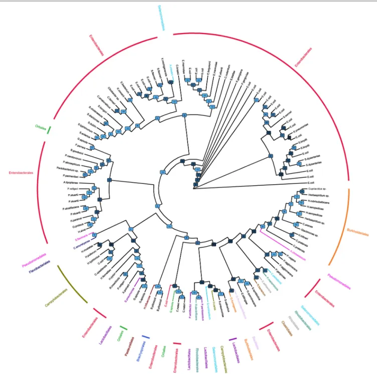
The phylogenetic tree with alignment of bacterial α1,3GT sequences showed that sequences largely clustered with bacteria orders (Figure 1), indicating a classical transmission of α1,3GT genes. Enterobacterales (including E. coli) was the predominant order of bacteria bearing α1,3GT. As expected, genetic distance of bacterial α1,3GT to mammal α1,3GT sequences was important (data not shown, comparison with bovine, porcine, murine, canine, cat, monkey, sheep, rat, and human sequences of GGTA1 gene). Some bacteria appeared to bear at least 2 different α1,3GT
Montassier et al. α1,3-Galactosyltransferase in Gut Microbiome
FIGURE 1 | Phylogenic tree of bacteria bearing α1,3GT sequences. Phylogenetic tree of bacteria based on the α1,3-GalactosylTransferase sequences retrieved on KEGG and UniProt databases. Orders have been represented in different colors. The tree is represented as a cladogram; therefore, the interpretation of the groups is possible but the branch length has no meaning. Bootstrap confidence values are indicated on nodes.
gene sequences (for example E. coli) suggesting that several enzymes translated from different genes harbor α1,3GT function.
Identification of α1,3GT Sequences in Gut
Microbiome of Adult Individuals
Based on all α1,3GT bacteria sequences extracted from the ExPASy and KEGG databases, we then studied the presence
of corresponding sequences in the gut microbiome using the open access shotgun data of the two different cohorts of adult individuals.
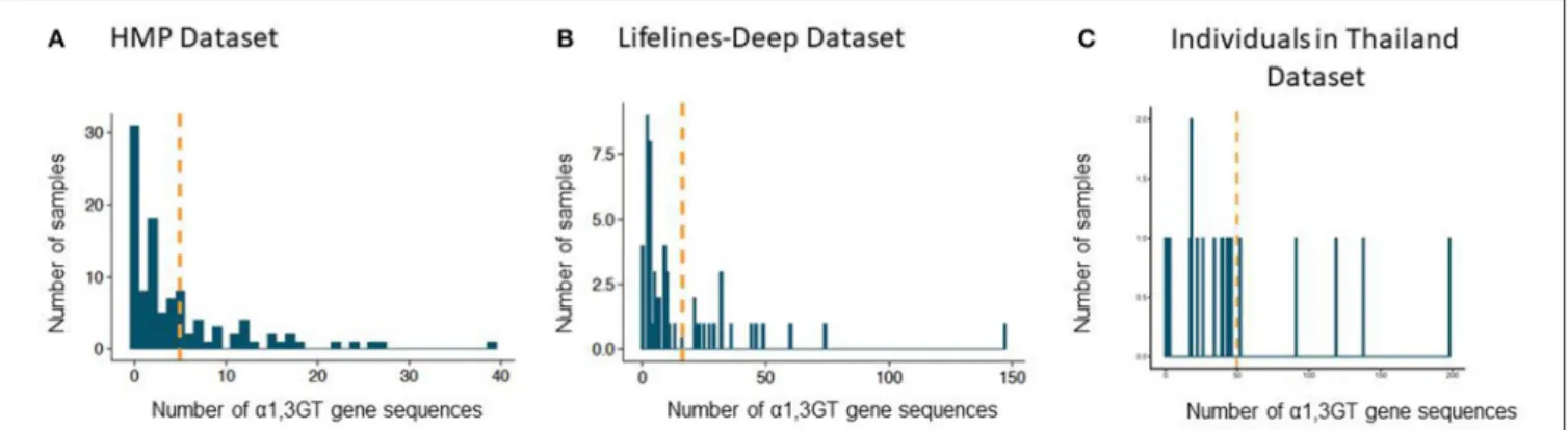
In the Human Microbiome Project (HMP) cohort, 75 individuals (71% of the studied population) exhibited α1,3GT sequences in their gut microbiome when a lenient 0.85 sequence identity cutoff was used. The mean number of sequences above
the homology threshold per subject was 5 (standard deviation: 7; range: 0–39) (Figure 2A). With a 0.935 sequence homology cutoff, the mean number sequences per subject α1,3GT was 2 (standard deviation: 4, range: 0–19).
In the more recent database from the LifeLines-DEEP cohort, we confirmed also the presence of α1,3GT sequences and found these sequences in 53 subjects (93%) with a 0.85 identity cutoff (Figure 2B). The mean number of sequences per subject was 16 (standard deviation: 24; range: 0–147). With a 0.935 cutoff, the mean number of compatible α1,3GT sequences was 11 (standard deviation: 19, range: 0–116).
In another recent database from a multi-generational Asian American cohort, where we only selected individuals living in Thailand (n = 20), we and found α1,3GT sequences in 19 subjects (95%) with a 0.85 identity cutoff (Figure 2C). The mean number of sequences per subject was 50 (standard deviation: 50; range: 0–147). With a 0.935 cutoff, the mean number of compatible
α1,3GT sequences was 11 (standard deviation: 19,
range: 0–198).
α
1,3GT Positive Bacteria in Gut
Microbiome of Adult Individuals
The α1,3GT sequences allowed us to identify a first bacteria map of the human gut microbiome which exhibited α1,3GT genes. Using BURST, we found that optimal lowest common ancestor taxonomy assignment mostly associated the α1,3GT homology to sequences originating from the Enterobacteriaceae family, mostly Escherichia coli; the Pasteurellaceae family, mostly Haemophilus influenza; the Streptococcus genus, mostly Streptococcus salivarius and Streptococcus pneumoniae; and the Lactobacillaceae family, with most Pediococcus and Lactobacillus genera (Table 1). Most, but not all, bacteria species displaying the α1,3GT gene sequences were classified as Gram-negative. Interestingly, gram-negative bacteria bear a complex lipopolysaccharide (LPS) in the outer leaflet of their membrane, which can harbor α1,3Gal antigen.
α
1,3GT Positive Gut Microbiome of Healthy
Individuals Exhibit α1,3GT Activity and
α
1,3Gal Antigen Production
To confirm our bioinformatics sequence analysis on gut microbiome from healthy individuals, we tested by ELISA the presence of α1,3Gal antigen on human stool samples. First, a validation was performed on human cells (peripheral
blood mononuclear cells) and rat splenocytes, used,
respectively, as negative and positive controls. As shown in
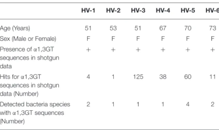
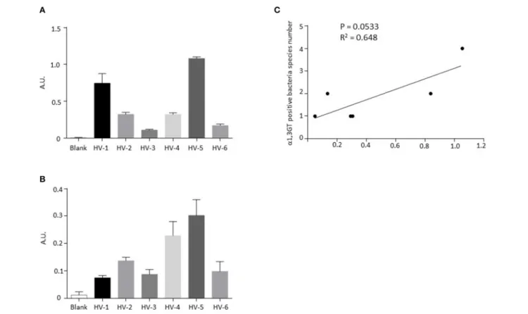
Supplementary Figure 1A, IB4 lectin, specific for α1,3Gal antigen, bound to rat cells and not to human cells and the presence of α1,3Gal antigen by ELISA was only detected in rat cells (Supplementary Figure 1B). Stool samples from six healthy volunteers (HV), all characterized as positive for bacterial
α1,3GT gene sequences (Table 2), exhibited α1,3Gal antigen
production (Figure 3A) and α1,3GT activity (Figure 3B). There was no significant correlation between the number of hits for α1,3GT sequences in shotgun data and the amount of Gal antigen in stools in this small cohort (data not shown). However,
there was a tendency for positive correlation (P = 0.0533, R2
= 0.648, Figure 3C) between the number of bacteria species
harboring α1,3GT sequences and the amount of Gal antigen in the stools detected by ELISA, suggesting that the extend of diversity of bacteria with α1,3GT in the microbiota, influences the production of Gal antigen.
DISCUSSION
Here, we describe for the first time the distribution of α1,3GT gene homologs in the human gut microbiome expected to encode the Gal antigen using a meta-analysis of two publicly available shotgun data sets including 163 adults from United States and The Netherlands. This method, contrary to 16S RNA analysis of
gut microbiota which we previously used (11), directly detects
the presence of α1,3GT gene sequences instead of relying on
taxonomic prediction (29). We showed that sequences with an
85 or 93.5% homology with the reference α1,3GT gene sequences
FIGURE 2 | Presence of sequences with α1,3GT sequence homology in the gut microbiome of adult individuals from two cohorts. (A) Number of hits for α1,3GT sequences in the HMP dataset (106 individuals), mean number = 5. (B) Number of hits for α1,3GT sequences in DEEP lifelines dataset (57 individuals), mean number =16. (C) Number of hits for α1,3GT sequences in Asian American dataset (20 individuals), mean number = 50. BURST was used with sequence identity homology cutoff of 0.85. The orange line represents the mean number of α1,3GT homologs present per patient.
Montassier et al. α1,3-Galactosyltransferase in Gut Microbiome
TABLE 1 | Most frequent hits for taxonomy. Taxonomy
(BURST based lowest common ancestor taxonomy assignment on the basis of a 85% of homology with the α1,3GT sequence)
Number and ranking of homologous α1,3GT sequences k__Bacteria;p__Proteobacteria;c__Gammaproteobacteria;o__Enterobacterales;f__Enterobacteriaceae;g__Escherichia;s__Escherichia coli;t__ 2141 k__Bacteria;p__Proteobacteria;c__Gammaproteobacteria;o__Enterobacterales;f__Enterobacteriaceae 1792 k__Bacteria;p__Proteobacteria;c__Gammaproteobacteria;o__Pasteurellales;f__Pasteurellaceae;g__Haemophilus;s__Haemophilus influenzae;t__ 251 k__Bacteria;p__Proteobacteria;c__Gammaproteobacteria;o__Enterobacterales;f__Enterobacteriaceae;g__Escherichia 147 k__Bacteria;p__Proteobacteria;c__Gammaproteobacteria;o__Enterobacterales;f__Enterobacteriaceae;g__Escherichia;s__Escherichia
fergusonii;t__Escherichia fergusonii ATCC 35469
130 k__Bacteria;p__Firmicutes;c__Bacilli;o__Lactobacillales;f__Streptococcaceae;g__Streptococcus;s__Streptococcus
salivarius
81 k__Bacteria;p__Proteobacteria;c__Gammaproteobacteria;o__Enterobacterales;f__Hafniaceae;g__Hafnia;s__Hafnia
alvei;t__Hafnia alvei ATCC 13337
68 k__Bacteria;p__Firmicutes;c__Bacilli;o__Lactobacillales;f__Lactobacillaceae;g__Pediococcus;s__Pediococcus_pentosaceus 64 k__Bacteria;p__Firmicutes;c__Bacilli;o__Lactobacillales;f__Streptococcaceae;g__Streptococcus;s__Streptococcus_pneumoniae 61 k__Bacteria;p__Bacteroidetes;c__Bacteroidia;o__Bacteroidales;f__Tannerellaceae;g__Parabacteroides;s__Parabacteroides distasonis;t__ 68 k__Bacteria;p__Bacteroidetes;c__Bacteroidia;o__Bacteroidales;f__Tannerellaceae;g__Parabacteroides 64 k__Bacteria;p__Proteobacteria;c__Gammaproteobacteria;o__Enterobacterales;f__Enterobacteriaceae;g__Escherichia;s__Escherichia coli 52 k__Bacteria;p__Firmicutes;c__Bacilli;o__Lactobacillales;f__Lactobacillaceae;g__Lactobacillus;s__Lactobacillus
delbrueckii;t__Lactobacillus delbrueckii subsp. bulgaricus
32 k__Bacteria;p__Proteobacteria;c__Gammaproteobacteria;o__Enterobacterales;f__Hafniaceae;g__Hafnia 32 k__Bacteria;p__Bacteroidetes;c__Bacteroidia;o__Bacteroidales;f__Prevotellaceae;g__Prevotella;s__[Hallella]
seregens;t__Hallella seregens ATCC 51272
31 k__Bacteria;p__Bacteroidetes;c__Bacteroidia;o__Bacteroidales;f__Prevotellaceae;g__Prevotella;s__Prevotella sp. P6B1;t__ 30 k__Bacteria;p__Proteobacteria;c__Gammaproteobacteria;o__Enterobacterales;f__Hafniaceae;g__Hafnia;s__Hafnia alvei 23 k__Bacteria;p__Firmicutes;c__Clostridia;o__Clostridiales;f__Eubacteriaceae;g__Eubacterium;s__[Eubacterium]
eligens;t__[Eubacterium] eligens ATCC 27750
21
k__Bacteria;p__Bacteroidetes;c__Bacteroidia;o__Bacteroidales;f__Prevotellaceae;g__Prevotella 19
A lowest common ancestor assignment and an α1,3GT sequence identity cutoff of 85% in the HMP, DEEP-lifelines, Asian American datasets were used for the analysis.
TABLE 2 | Characteristics of healthy volunteers.
HV-1 HV-2 HV-3 HV-4 HV-5 HV-6
Age (Years) 51 53 51 67 70 73
Sex (Male or Female) F F F F F F
Presence of α1,3GT sequences in shotgun data + + + + + + Hits for α1,3GT sequences in shotgun data (Number) 4 1 125 38 60 11
Detected bacteria species with α1,3GT sequences (Number)
2 1 1 1 4 2
which encode the α1,3-Gal antigen were restricted to a small number of gut microbiota, dominated by the Enterobacteriaceae family. High homology with the α1,3GT gene sequences was
also identified in the Haemophilus and Streptococcus genera, which represent <1% of the human gut microbiome in relative
abundance (30). Altogether, our approach suggests that only
a small fraction of the gut microbiome of adult individuals is able to produce the Gal antigen. Our study fits with previous observations that indirectly suggested that gut microbiome was the main source of production of the Gal antigen (18) resulting in the rise of the anti-Gal within the first year of life. Interestingly, in α1,3GT-KO mice, modifications of gut microbiome followed during the first months of life were related to anti-Gal Ab levels (31).
However, our estimation likely underestimates the actual proportion of the microbiota able to produce the Gal antigen as also suggested by the presence of anti-Gal antibodies in most of the adult individuals tested. Indeed, it is possible that a fraction of adult individuals in whom no high homology of known α1,3GT sequences were identified (29 and 7% in the two cohorts, respectively) corresponds to “false negative,” owing the high diversity of the sequences of α1,3GT in bacteria
FIGURE 3 | Detection of α1,3-Gal antigen and α1,3GT activity in stools from healthy donors exhibiting bacterial α1,3GT gene sequences in their shotgun analysis. (A) Levels of α1,3-Gal antigen detected by ELISA in stools from 6 healthy volunteers. (B) Activity of α1,3GT enzymes detected by ELISA in stools from six healthy volunteers. (C) Correlation between the number of detected bacteria species harboring α1,3GT gene sequences and the amounts of Gal antigen in stools.
and not characterized viruses/parasites in the microbiome. Moreover, not all genes coding α1,3GT have been identified in bacteria as such. For instance, in Neisseria meningitidis,
anti-Gal antibodies bind to pili from these bacteria (32) but the
corresponding enzyme with α1,3GT activity is not described. Besides, enzymes previously thought to be unimodal are now being annotated with multiple modalities. Indeed, two genes coding other glycosyltransferases were recently described as coding bispecific transferases exhibiting α1,3GT activity: wciN in Streptococcus pneumoniae (33) and WclR in Escherichia coli (34) (Supplementary Table 1).
Although anti-Gal Abs are found in most individuals, there is a substantial dispersion of their levels attributed to several demographic variables, including ABO blood groups, as well as age, sex, geographic location during childhood, and diet
(35, 36). Another important variable may be the exposure to
different types and levels of microorganisms of the healthy microbiota. Several bacterial pathogens able to synthetize α1,3-Gal such as Escherichia coli (34), Streptococcus pneumonia (33) Salmonella (16) or parasites may also contribute to immunization in the childhood or adulthood in specific pathological contexts. Moreover, inflammatory bowel diseases and gastrointestinal infections are particularly associated with adherent-invasive
Escherichia coli (37–39). Oral inoculation of α1,3GT gene KO
mice with this bacterium induces production of anti-Gal Abs
(40). Moreover, this bacterium readily binds human anti-Gal Abs (13). Altogether, our data suggest that α1,3GT positive bacteria of the gut microbiota, generate anti-Gal antibodies previously described to involve a substantial fraction of the B cell repertoire (4) and may also shape the dispersion of blood anti-Gal antibody levels. However, our present study only portraits a first map of the bacteria of gut microbiota harboring α1,3GT sequences allowing the synthesis of α1,3Gal epitope. Beside the nature of the bacteria themselves, which may influence the immunogenicity of the α1,3GT in the gut, other parameters, particularly the direct assessment of the actual amounts of α1,3GT antigen in the gut microbiota, would be important to correlate with the levels of blood anti-Neu5Gc in the future. Unfortunately, such study would require large cohorts of individuals with both blood and gut microbiota samples. We could only provide here preliminary results based on a few cases suggesting that the level of diversity of the bacteria harboring α1,3GT gene sequences have a borderline correlation with the levels of α1,3Gal in the stools.
We have recently suggested that the low level of anti-Gal reported in multiple sclerosis (10) could be linked to the low level of α1,3GT positive flora also observed in their gut microbiome (11). It is possible that manipulation of anti-Gal antibodies can find some utility in human autoimmune diseases and particularly in multiple sclerosis, a disease of unknown precise etiology. The present study is thus a step toward a possible demonstration that
Montassier et al. α1,3-Galactosyltransferase in Gut Microbiome
these decreased levels were governed by abnormal microbiota. One can hypothesize that supplementing multiple sclerosis patient diet with Enterobacteriaceae or other α1,3GT positive species could be considered in the future. However, currently, one cannot speculate on a direct role of either the anti-Gal antibodies or rather that anti-Gal could be a marker of a microbiota alteration directly interfering in this disease (41). Furthermore, the link between the anti-Gal antibody levels and the amount of bacterial displaying α1,3GT activity is currently speculative.
Taken together, our data may open new avenues for delineating targeted strategies to eventually modulate the level
of anti- α1,3Gal antibody response in some diseases (10, 11),
attenuate food allergy to α1,3Gal antigen, or decrease the risk of deleterious effects of anti-α1,3Gal Abs after implantation of
animal derived bio-devices (42) such as bovine or porcine
bio-prosthetic heart valves or help fighting some infections due to Gal displaying microorganisms. Manipulating the microbiome to decrease the level of anti- α1,3Gal Abs may be also useful for xenotransplantation, as using genetically engineered GGTA1 gene KO animal donors in the clinic is not yet permitted.
CONCLUSIONS
This study described for the first time the gut bacteria species bearing α1,3-GT gene sequences: the Enterobacteriaceae family, including Escherichia coli, Pasteurellaceae genera, Haemophilus influenza, and Lactobacillus species. These bacteria were detected in the gut microbiome of most individuals. Moreover, this bioinformatics analysis was confirmed by biological tests on human stool samples. Gal antigens and α1,3-Galactosyltransferase activity were detected in healthy stools of individuals exhibiting α1,3-Galactosyltransferase bacterial sequences in their shotgun data. These data could offer more target strategies to modulate anti-Gal antibodies in patients confronted to animal derived biotherapeutics or biodevices and could represent an important issue for xenotransplantation and auto-immune diseases in the future.
DATA AVAILABILITY STATEMENT
All datasets generated for this study are included in the article/Supplementary Material.
AUTHOR CONTRIBUTIONS
EM was in charge of shotgun data analysis and alignment of sequences. EM, GA-G, and QL performed bioinformatics analysis of shotgun data. CM performed ELISA and flow cytometry experiments. YT and ED collected fecal samples in healthy individuals. BR performed the research of α1,3-GT sequences. VD, AG, and RG performed phylogenic tree building. LB, NV, and SL made a substantial contribution to data management and analysis. P-AG, DL, and AN were involved in revising the manuscript critically and made a substantial contribution to the final manuscript. LB and J-PS designed all the experimental work, coordinated the study. EM, J-PS, and LB wrote the manuscript.
FUNDING
This work was realized in the context of the LabEX IGO program supported by the National Research Agency via the investment of the future program ANR-11-LABX-0016-01. We thank INSERM and University of Nantes for their institutional support. AG was supported by IHU CESTI.
ACKNOWLEDGMENTS
We are most grateful to the Genomics and Bioinformatics Core Facility of Nantes (GenoBiRD, Biogenouest) for its technical support. We thank Marck Fraudeau for his script classifying α1,3-GT sequences by name. We are most grateful to Prof Uri Galili for his advices and helpful comments.
SUPPLEMENTARY MATERIAL
The Supplementary Material for this article can be found online at: https://www.frontiersin.org/articles/10.3389/fimmu. 2019.03000/full#supplementary-material
Supplementary Figure 1 | Confirmation of α1,3-Gal antigen presence in rat cells and not in human cells. (A) Detection of α1,3-Gal antigen on cell surface using IB4 lectin coupled with FITC by flow cytometry. (B) Level of α1,3-Gal antigen detected by ELISA on cell lysates.∗
p <0.05, Kruskal-Wallis test.
Supplementary Table 1 | List of bacteria harboring α1,3GT genes. When available, the gene name and the KEGG orthology numbers are indicated.
REFERENCES
1. Galili U. Significance of the evolutionary α1,3-galactosyltransferase (GGTA1) gene inactivation in preventing extinction of apes and old world monkeys. J Mol Evol. (2015) 80:1–9. doi: 10.1007/s00239-014-9652-x
2. Tangvoranuntakul P, Gagneux P, Diaz S, Bardor M, Varki N, Varki A, et al. Human uptake and incorporation of an immunogenic nonhuman dietary sialic acid. Proc Natl Acad Sci USA. (2003) 100:12045–50. doi: 10.1073/pnas.2131556100
3. McKenzie IF, Patton K, Smit JA, Mouhtouris E, Xing P, Myburgh JA, et al. Definition and characterization of chicken Gal alpha(1,3)Gal antibodies. Transplantation. (1999) 67:864–70. doi: 10.1097/00007890-1999032 70-00013
4. Galili U, Anaraki F, Thall A, Hill-Black C, Radic M. One percent of human circulating B lymphocytes are capable of producing the natural anti-Gal antibody. Blood. (1993) 82:2485–93. doi: 10.1182/blood.V82.8.2485.2485 5. Cooper DK, Koren E, Oriol R. Oligosaccharides and
discordant xenotransplantation. Immunol Rev. (1994)141:31–58. doi: 10.1111/j.1600-065X.1994.tb00871.x
6. Kuwaki K, Tseng YL, Dor FJ, Shimizu A, Houser SL, Sanderson TM, et al. Heart transplantation in baboons using α1,3-galactosyltransferase gene-knockout pigs as donors: initial experience. Nat Med. (2005) 11:29–31. doi: 10.1038/nm1171
7. Yilmaz B, Portugal S, Tran TM, Gozzelino R, Ramos S, Gomes J, et al. Gut microbiota elicits a protective immune response against malaria transmission. Cell. (2014) 159:1277–89. doi: 10.1016/j.cell.2014.10.053
8. Avila JL, Rojas M, Galili U. Immunogenic Gal alpha 1−3Gal carbohydrate epitopes are present on pathogenic American trypanosoma and leishmania. J Immunol. (1989) 142:2828–34.
9. Takeuchi Y, Porter CD, Strahan KM, Preece AF, Gustafsson K, Cosset FL, et al. Sensitization of cells and retroviruses to human serum by (α1-3) galactosyltransferase. Nature. (1996) 379:85–8. doi: 10.1038/379085a0 10. Le Berre L, Rousse J, Gourraud PA, Imbert-Marcille BM, Salama A, Evanno
G, et al. Decrease of blood anti-α1,3 Galactose Abs levels in multiple sclerosis (MS) and clinically isolated syndrome (CIS) patients. Clin Immunol. (2017) 180:128–35. doi: 10.1016/j.clim.2017.05.006
11. Montassier E, Berthelot L, Soulillou JP. Are the decrease in circulating anti-α1,3-Gal IgG and the lower content of galactosyl transferase A1 in the microbiota of patients with multiple sclerosis a novel environmental risk factor for the disease? Mol Immunol. (2018) 93:162–5. doi: 10.1016/j.molimm.2017.09.016
12. Naso F, Gandaglia A, Bottio T, Tarzia V, Nottle MB, d’Apice AJ, et al. First quantification of alpha-Gal epitope in current glutaraldehyde-fixed heart valve bioprostheses. Xenotransplantation. (2013) 20:252–61. doi: 10.1111/xen.12044 13. Galili U, Mandrell RE, Hamadeh RM, Shohet SB, Griffiss JM. Interaction between human natural anti-alpha-galactosyl immunoglobulin G and bacteria of the human flora. Infect Immun. (1988) 56:1730–7.
14. Hamadeh RM, Jarvis GA, Zhou P, Cotleur AC, Griffiss JM. Bacterial enzymes can add galactose alpha 1,3 to human erythrocytes and creates a senescence-associated epitope. Infect Immun. (1996) 64:528–34.
15. Pradel E, Parker CT, Schnaitman CA. Structures of the rfaB, rfaI, rfaJ, and rfaS genes of Escherichia coli K-12 and their roles in assembly of the lipopolysaccharide core. J Bacteriol. (1992) 174:4736–45. doi: 10.1128/jb.174.14.4736-4745.1992
16. Endo A, Rothfield L. Studies of a phospholipid-requiring bacterial enzyme. I. Purification and properties of uridine diphosphate galactose: lipopolysaccharide alpha-3-galactosyl transferase. Biochemistry. (1969) 8:3500–7. doi: 10.1021/bi00837a003
17. Hamadeh RM, Jarvis GA, Galili U, Mandrell RE, Zhou P, Griffiss JM. Human natural anti-Gal IgG regulates alternative complement pathway activation on bacterial surfaces. J Clin Investig. (1992) 89:1223–35. doi: 10.1172/JCI115706 18. Manez R, Blanco FJ, Diaz I, Centeno A, Lopez-Pelaez E, Hermida M,
et al. Removal of bowel aerobic gram-negative bacteria is more effective than immunosuppression with cyclophosphamide and steroids to decrease natural α-galactosyl IgG antibodies. Xenotransplantation. (2001) 8:15–23. doi: 10.1034/j.1399-3089.2001.00082.x
19. Buonomano R, Tinguely C, Rieben R, Mohacsi PJ, Nydegger UE. Quantitation and characterization of anti-Galα1-3Gal antibodies in sera of 200 healthy persons. Xenotransplantation. (1999) 6:173–80. doi: 10.1034/j.1399-3089.1999.00023.x
20. Human Microbiome Project C. Structure, function and diversity of the healthy human microbiome. Nature. (2012) 486:207–14. doi: 10.1038/nature11234 21. Vangay P, Johnson AJ, Ward TL, Al-Ghalith GA, Shields-Cutler RR, Hillmann
BM, et al. US immigration westernizes the human gut microbiome. Cell. (2018) 175:962–72.e910. doi: 10.1016/j.cell.2018.10.029
22. Sievers F, Wilm A, Dineen D, Gibson TJ, Karplus K, Li W, et al. Fast, scalable generation of high-quality protein multiple sequence alignments using Clustal Omega. Mol Syst Biol. (2011) 7:539. doi: 10.1038/msb.2011.75
23. Goujon M, McWilliam H, Li W, Valentin F, Squizzato S, Paern J, et al. A new bioinformatics analysis tools framework at EMBL-EBI. Nucleic Acid Res. (2010) 38:W695–9. doi: 10.1093/nar/gkq313
24. Stamatakis A. RAxML version 8: a tool for phylogenetic analysis and post-analysis of large phylogenies. Bioinformatics. (2014) 30:1312–3. doi: 10.1093/bioinformatics/btu033
25. Yu G, Lam TT, Zhu H, Guan Y. Two methods for mapping and visualizing associated data on phylogeny using Ggtree. Mol Biol Evol. (2018) 35:3041–3. doi: 10.1093/molbev/msy194
26. Yu G, Smith D, Zhu H, Guan Y, Lam T. ggtree: an r package for visualization and annotation of phylogenetic trees with their covariates and other associated data. Methods Ecol Evol. (2016) 8:28–36. doi: 10.1111/2041-210X.12628 27. Reimer LC, Vetcininova A, Carbasse JS, Sohngen C, Gleim D, Ebeling C, et al.
BacDive in 2019: bacterial phenotypic data for High-throughput biodiversity analysis. Nucleic Acid Res. (2019) 47:D631–6. doi: 10.1093/nar/gky879
28. Al-Ghalith GA, Hillmann B, Ang K, Shields-Cutler R, Knights D. SHI7 is a self-learning pipeline for multipurpose short-read DNA quality control. mSystems. (2018) 3:e00202-17. doi: 10.1128/mSystems. 00202-17
29. Langille MG, Zaneveld J, Caporaso JG, McDonald D, Knights D, Reyes JA, et al. Predictive functional profiling of microbial communities using 16S rRNA marker gene sequences. Nat Biotechnol. (2013) 31:814–21. doi: 10.1038/nbt.2676
30. Lloyd-Price J, Mahurkar A, Rahnavard G, Crabtree J, Orvis J, Hall AB, et al. Strains, functions and dynamics in the expanded Human Microbiome Project. Nature. (2017) 550:61–6. doi: 10.1038/nature23889
31. Bello-Gil D, Audebert C, Olivera-Ardid S, Perez-Cruz M, Even G, Khasbiullina N, et al. The formation of glycan-specific natural antibodies repertoire in GalT-KO mice is determined by gut microbiota. Front Immunol. (2019) 10:342. doi: 10.3389/fimmu.2019.00342
32. Hamadeh RM, Estabrook MM, Zhou P, Jarvis GA, Griffiss JM. Anti-Gal binds to pili of Neisseria meningitidis: the immunoglobulin A isotype blocks complement-mediated killing. Infect Immun. (1995) 63:4900–6.
33. Han W, Cai L, Wu B, Li L, Xiao Z, Cheng J, et al. The wciN gene encodes an α-1,3-galactosyltransferase involved in the biosynthesis of the capsule repeating unit of Streptococcus pneumoniae serotype 6B. Biochemistry. (2012) 51:5804–10. doi: 10.1021/bi300640b
34. Chen C, Liu B, Xu Y, Utkina N, Zhou D, Danilov L, et al. Biochemical characterization of the novel α-1, 3-galactosyltransferase WclR from Escherichia coli O3. Carbohydr Res. (2016) 430:36–43. doi: 10.1016/j.carres.2016.04.012
35. Kumar G, Satyananda V, Fang J, Zhou H, Fujita M, Ekser B, et al. Is there a correlation between anti-pig antibody levels in humans and geographic location during childhood? Transplantation. (2013) 96:387–93. doi: 10.1097/TP.0b013e3182992a84
36. He Y, Wu W, Zheng HM, Li P, McDonald D, Sheng HF, et al. Regional variation limits applications of healthy gut microbiome reference ranges and disease models. Nat Med. (2018) 24:1532–5. doi: 10.1038/s41591-018-0164-x 37. Sevrin G, Massier S, Chassaing B, Agus A, Delmas J, Denizot J, et al.
Adaptation of adherent-invasive E. coli to gut environment: Impact on flagellum expression and bacterial colonization ability. Gut Microbes. (2018) 1:1–17. doi: 10.1080/19490976.2017.1421886
38. Lashermes A, Boudieu L, Barbier J, Sion B, Gelot A, Barnich N, et al. Adherent-invasive E. coli enhances colonic hypersensitivity and P2X receptors expression during post-infectious period. Gut Microbes. (2018) 9:26–37. doi: 10.1080/19490976.2017.1361091
39. Elhenawy W, Oberc A, Coombes BK. A polymicrobial view of disease potential in Crohn’s-associated adherent-invasive E. coli. Gut Microbes. (2018) 9:166–74. doi: 10.1080/19490976.2017.1378291
40. Posekany KJ, Pittman HK, Bradfield JF, Haisch CE, Verbanac KM. Induction of cytolytic anti-Gal antibodies in a-1,3-galactosyltransferase gene knockout mice by oral inoculation with Escherichia coli O86:B7 bacteria. Infect Immun. (2002) 70:6215–22. doi: 10.1128/IAI.70.11.6215-6222. 2002
41. Wekerle H. Nature plus nurture: the triggering of multiple sclerosis. Swiss Med Wkly. (2015) 145:w14189. doi: 10.4414/smw.2015.14189
42. Park CS, Oh SS, Kim YE, Choi SY, Lim HG, Ahn H, et al. Anti-alpha-Gal antibody response following xenogeneic heart valve implantation in adults. J Heart Valve Dis. (2013) 22:222–9.
Conflict of Interest:The authors declare that the research was conducted in the absence of any commercial or financial relationships that could be construed as a potential conflict of interest.
Copyright © 2020 Montassier, Al-Ghalith, Mathé, Le Bastard, Douillard, Garnier, Guimon, Raimondeau, Touchefeu, Duchalais, Vince, Limou, Gourraud, Laplaud, Nicot, Soulillou and Berthelot. This is an open-access article distributed under the terms of the Creative Commons Attribution License (CC BY). The use, distribution or reproduction in other forums is permitted, provided the original author(s) and the copyright owner(s) are credited and that the original publication in this journal is cited, in accordance with accepted academic practice. No use, distribution or reproduction is permitted which does not comply with these terms.