Université de Montréal
Characterization of a Novel Class of Anti-HCV Agents
Targeting Protein-Protein Interactions
par Alex Park
Programme de microbiologie et immunologie Faculté de Médecine
Mémoire présenté à la Faculté de Médecine
en vue de l’obtention du grade de maîtrise en sciences (M.Sc.) en microbiologie et immunologie
option générale
Septembre, 2016
Résumé
Le virus de l’hépatite C (VHC) est un agent causateur de maladies du foie important responsable d’une pandémie affectant près de 180 millions d’individus mondialement. L’absence de symptômes dans les premières années d’infection entraîne des diagnostics tardifs qui empêchent la prise en charge rapide des patients avant l’apparition d’une fibrose et, dans près de 16 % des cas d’infection, d’une cirrhose.
En exploitant les interactions protéine-protéine membranaires, des essais utilisant la technologie BRET, dans les cellules vivantes, ont été précédemment optimisés afin d’établir le réseau complet des interactions du VHC. En utilisant les fondements de cette étude, un essai à haut débit dans les cellules vivantes a été réalisé pour identifier de nouveaux composés anti-VHC ciblant une nouvelle interaction NS3/4A-NS3/4A. Approximativement 110,000 petites molécules ont été criblées pour leurs effets sur l’homodimérization de NS3/4A et ont été classées par rapport à leur spécificité et à leur puissance contre le VHC. Au terme de cette étude, UM42811 a été identifié comme un activateur potentiel de l’interaction NS3/4A-NS3/4A offrant une activité antivirale prometteuse dotant une excellente fenêtre thérapeutique. Par la suite, un séquençage exhaustif des virus, soumis à un traitement de UM42811, a permis d’établir le profil de résistance du VHC contre ce composé. Grâce à cette fine cartographie, il a été possible d’identifier un nouveau mécanisme d’inhibition de NS3/4A qui est indépendant de son activité protéase.
En utilisant les données de notre groupe sur les interactions VHC-hôte, il a été possible de continuer la caractérisation fonctionnelle du composé UM42811 en étudiant son effet sur les interactions potentiellement bénéfiques à la persistance virale. Pour ce faire, les protéines associées au transport nucléaire et mitochondriale qui sont des interactants de choix de NS3/4A ont été priorisées. Parmi ces facteurs de l’hôte, l’étude de karyopherin subunit beta 1 (KPNB1) et de heat shock protein 60 (HSP60) a été priorisée. De façon intéressante, les expériences de co-immunoprécipitation ont démontré que UM42811 était capable de prévenir l’interaction KPNB1-NS3/4A ainsi que l’interaction HSP60-NS3/4A. De plus, les études
fonctionnelles et les analyses d’immunobuvardage de type western ont démontré que l’interaction KPNB1-NS3/4A avait des effets délétères sur l’induction des gènes stimulés par l’interféron (ISG). Finalement, il a été démontré que KPNB1 est possiblement clivé par NS3/4A suggérant la présence potentielle d’un mécanisme de subversion ou d’échappement.
En bref, cette étude démontre la puissance des stratégies impliquant les interactions protéine-protéine dans les cellules vivantes pour l’identification de nouveaux composés inhibiteurs, caractérise un nouveau mécanisme d’inhibition anti-VHC et révèle la possibilité d’un nouveau mécanisme d’évasion du système immunitaire.
Mots-clés : virus de l’hépatite C, VHC, antiviraux à action directe, ADD, criblage à haut débit, BRET, interaction protéine-protéine, résistance
Abstract
Hepatitis C virus (HCV) is an important causative agent for liver diseases and is responsible for a worldwide pandemic affecting roughly 180 million individuals worldwide. Late diagnosis following the progression to fibrosis and to cirrhosis, in nearly 16% of chronic infections, is attributed to the absence of symptoms in the first years of infection.
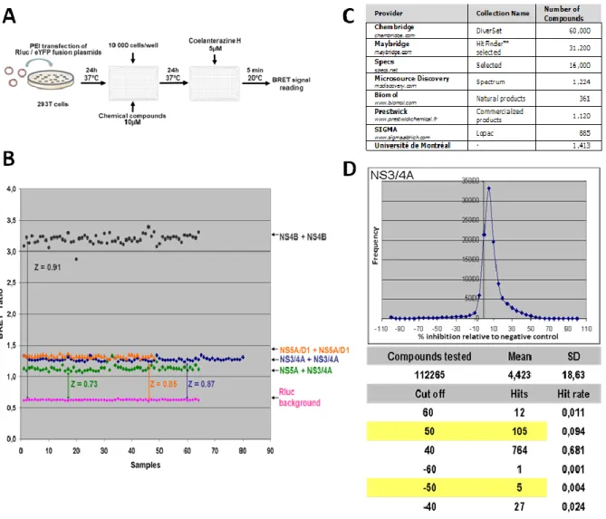
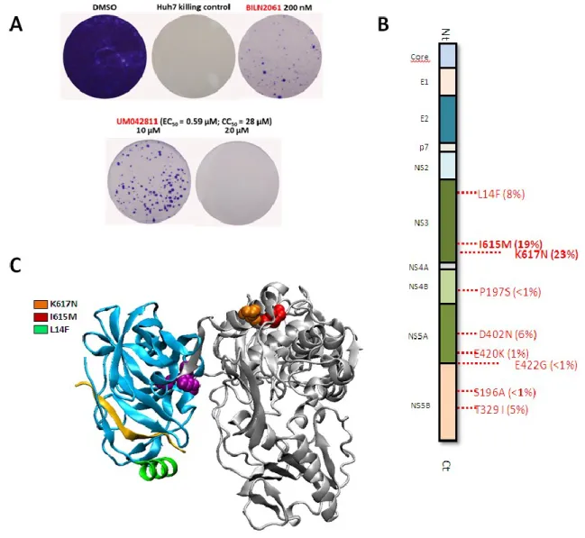
By exploiting membrane protein-protein interactions (PPI), live cell assays using bioluminescence resonance energy transfer (BRET) technology have previously been optimized to complete a comprehensive hepatitis C virus (HCV) protein interaction network. Using the groundwork laid by this network study, a high-throughput assay (HTS) cell-based assay was implemented to identify novel inhibitory compounds targeting an unreported NS3/4A-NS3/4A interaction. Approximately 110,000 compounds from a small-molecule collection were screened to monitor modulation of NS3/4A homodimerization and were discriminated based on specificity and potency. UM42811 was identified as a potential NS3/4A-NS3/4A interaction activator and found to have a promising antiviral activity boasting an excellent therapeutic window. Combined deep sequencing and mutation mapping have yielded a resistance profile based on statistical and functional probability pointing towards a novel inhibitory mechanism targeting the HCV NS3/4A independent from protease activity inhibition.
Data from an HCV to host protein interaction network generated by our group was used to analyze alternative effects of UM42811 on interactions which potentially benefit viral persistence. NS3/4A-specific host interactors were heavily associated with nuclear and mitochondrial transport based on Gene Ontology (GO). Among these specific interactors, karyopherin subunit beta 1 (KPNB1) and heat shock protein 60 (HSP60) were selected for further study. Interestingly, co-immunoprecipitation experiments revealed that UM42811 was able to prevent both KPNB1-NS3/4A and HSP60-NS3/4A interactions. Moreover, functional and western analysis revealed the KPNB1-NS3/4A interaction to have deleterious effects on
interferon stimulated gene (ISG) induction. Unexpectedly, analysis revealed a putative NS3/4A mediated cleavage of KPNB1.
Overall, this study demonstrates the strength of cell-based PPI strategies in the identification of novel HCV antiviral compounds, characterizes a novel inhibitory mechanism for HCV and reveals a potentially novel viral immune evasion mechanism.
Keywords : Hepatitis C virus, HCV, direct-acting antivirals, DAA, high-throughput screening, HTS, bioluminescence resonance energy transfer, BRET, protein-protein interaction, PPI, resistance
Table of Contents
Résumé ... i
Abstract ... iii
Table of Contents ... v
List of Tables ... viii
List of Figures ... ix
List of Abbreviations & Acronyms... x
Acknowledgements ... xiv
Introduction ... 1
1. Global Significance ... 2
1.1. Virus Discovery ... 2
1.2. Worlwide Prevalence ... 3
1.3. Genetic Variance, Genotypic Distribution and Origins ... 5
2. HCV Lifecycle ... 8
2.1. Viral Entry ... 8
2.2. Genome Translation and Replication ... 9
2.3. Assembly and Release ... 11
2.4. Innate Response and Immune Evasion ... 12
3. Treatment Evolution ... 14
3.1. Direct Acting Antiviral (DAA) – Design Strategies ... 16
3.1.1. NS3/4A Serine Protease ... 16
3.1.2. NS5B RNA-Dependent RNA Polymerase... 20
3.1.3. NS5A Phosphoprotein ... 22
3.2. Host-Directed HCV Inhibitors – Design Strategies ... 25
3.2.1. RNA-Based Inhibitors ... 25
3.2.3. Immunomodulators ... 27
3.3. Current Combination Therapies ... 28
4. Models for the Study of HCV ... 31
4.1. Animal Models... 31
4.2. Cell Culture Based Systems ... 32
5. Bioluminescence Resonance Energy Transfer (BRET) ... 35
Hypothesis & Objectives ... 37
Experimental Procedures ... 38 Cell culture ... 38 Expression vectors ... 38 BRET assays ... 38 HCV replication ... 39 EC50 and CC50 assays ... 39
Selection of HCV replicon escape resistant variants ... 40
Production of mutated HCV enzymes ... 40
shRNA gene silencing... 41
Functional firefly luciferase assays ... 41
Western blot analysis ... 42
Co-immunoprecipitation ... 42
Results ... 43
1. Investigation of pairwise interactions between HCV proteins ... 43
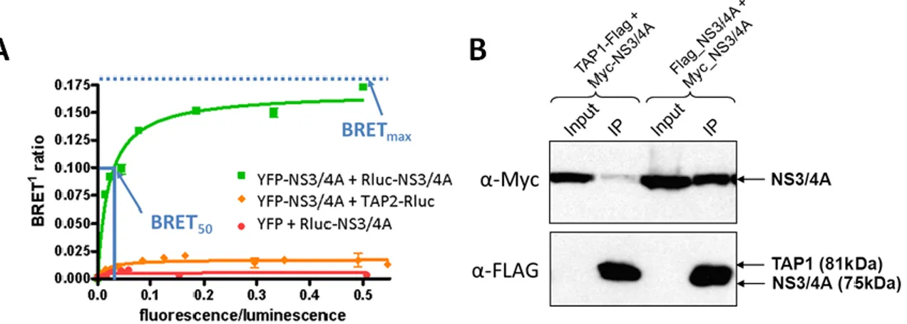
2. Homodimerization of the NS3/4A heterodimer ... 46
3. Implementation of a BRET HTS assay ... 48
4. Validation and antiviral characterization of lead compound UM42811 ... 52
5. Characterization of UM42811 resistance profile reveals mutations located at the surface of the C-terminal NS3 helicase subdomain ... 55
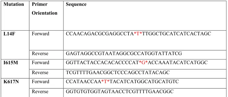
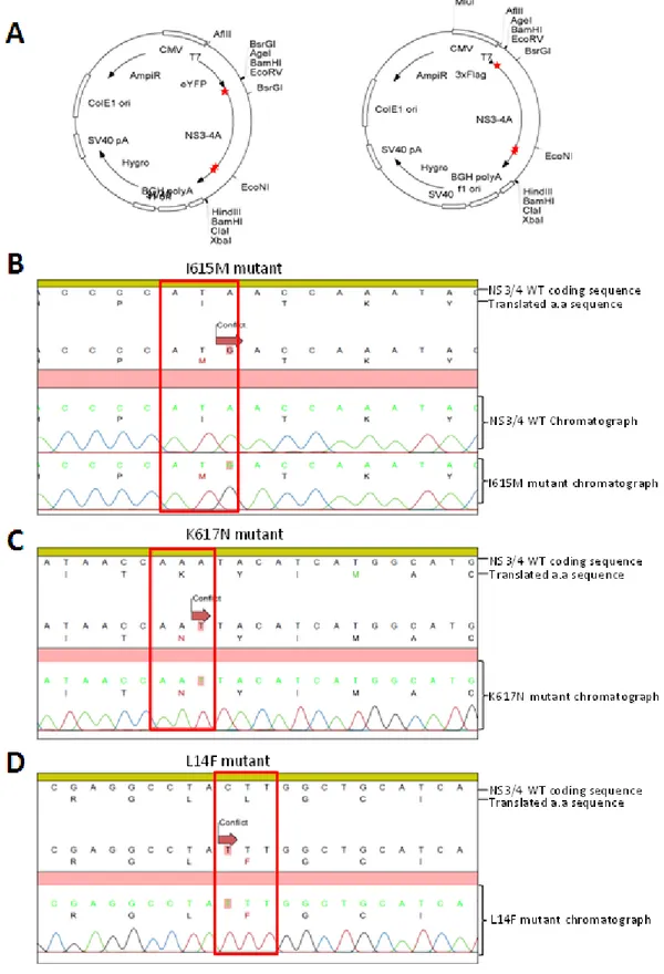
6. Generation of NS3/4A mutant fusion proteins and of mutant HCV replicon DNA precursors ... 58
8. Functional consequences of a UM42811/BILN2061-sensitive NS3/4A-KPNB1
interaction ... 65
9. Differential HSP60-IRF3-NS3/4A interaction configurations under UM42811 or BILN2061 treatment ... 70
Discussion ... 75
Perspectives... 81
Conclusion ... 83
Bibliographie... i
Annex 1: Spliceosome SNRNP200 Promotes Viral RNA Sensing and IRF3 Activation of Antiviral Response. ... i
Annex 2: HCV NS3/4A Protease Inhibitors and the Road to Effective Direct-Acting Antiviral Therapies. ... ii
List of Tables
List of Figures
Figure 1. Worldwide estimated prevalence of HCV and genotype distribution ... 7 Figure 2. HCV lifecycle – Potential points of intervention ... 30 Figure 3. Overview of cell culture based systems for the study of HCV. ... 34 Figure 4. Bioluminescence resonance energy transfer identifies novel HCV protein-protein interactions... 45 Figure 5. Specific interaction between NS3/4A heterodimers ... 47 Figure 6. Identification of potential NS3/4A PPI modulators through a BRET HTS assay 50 Figure 7. Validation and antiviral characterization of lead compound UM42811 ... 54 Figure 8. Characterization of UM42811 resistance profile reveals mutations located at the surface of the C-terminal NS3 helicase subdomain ... 57 Figure 9. Mutated NS3/4A expression vectors and mutated replicon sequences ... 60 Figure 10. Point mutations at putative resistant sites of UM42811 may affect the NS3/4A heterodimeric homodimers - UM42811binding reduces precursor interaction induced by BILN2061…… ... 64 Figure 11. NS3/4A interacts with KPNB1, but is disrupted by both UM42811 and the protease inhibitor BILN2061. ... 68 Figure 12. NS3/4A protease activity is required for the inhibition of KPNB1-mediated ISG56 induction following IFN-α 2A stimulation. ... 69 Figure 13. NS3/4A interacts with HSP60, but is disrupted by UM42811. ... 72 Figure 14. Quantitative changes in HSP60 expression have no effect on IFN-𝛽 induction following SeV infection. ... 73 Figure 15. Hypothesized interaction configuration between NS3, HSP60 and IRF3 in the context of UM42811 and BILN2061 treatment ... 74
List of Abbreviations & Acronyms
ACCA Aminocyclopropane carboxylic acid ACSL3 Acyl-CoA synthethase 3ALT Alanine aminotransferase ApoE Apolipoprotein E
ARFGAP1 ADP Ribosylation Factor GTPase Activating Protein 1 BRET Bioluminescence resonance energy transfer
CD81 Cluster of differentiation 81 cLD Cytosolic lipid droplet CLDN1 Claudin-1
CypA Cyclophilin A
DAA Direct-acting antiviral
DGAT1 Diacylglycerol acyltransferase-1 DMV Double-membrane vesicle DNA Deoxyribonucleic acid dsRNA Double-stranded RNA
EGFR Epidermal growth factor receptor EMCV Encephalomyocarditis virus EphA2 Ephrin receptor A2
ER Endoplasmic reticulum
ESCRT Endosomal-sorting complex required for transport eYFP enhanced Yellow fluorescent protein
Fluc Firefly luciferase GAG Glycosaminoglycan
GO Gene ontology
GFP Green fluorescent protein IDU Injection drug user
IRIC Institute of Research in Immunology and Cancer HAV Hepatitis A virus
HBV Hepatitis B virus
HCC Hepatocellular carcinoma HCV Hepatitis C virus
HIV Human immunodeficiency virus HNF4α Hepatocyte nuclear factor 4α HSP60 Heat shock protein 60 HTA Host-targeted agents HTS High throughput screening IFN Interferon
IFN-λR1 Interferon-λ receptor chain 1 IFNAR1 Interferon-α/β receptor alpha chain IFNAR2 Interferon- α/β receptor beta chain IKK-α IκB kinase-α
IL-10R2 Interleukin-10 receptor chain 2 IRES Internal ribosome entry site IRF3 Interferon regulatory factor 3 IRF7 Interferon regulatory factor 7 ISG Interferon-stimulated gene ISRE IFN-stimulated response element JAK1 Janus kinase 1
JFH-1 Japanese patient with fulminant hepatitis kDa kiloDalton
KPNA1 Karyopherin (importin) subunit alpha 1 KPNB1 Karyopherin (importin) subunit beta 1
LD Lipid droplet
LDLR Low-density lipoprotein receptor (LDLR) LuLD Luminal lipid droplet
MAPK Mitogen-activated protein kinase MAVS Mitochondrial antiviral signalling miR-122 microRNA 122
MTP Microsomal triglyceride transfer protein Myd88 Myeloid differentiation primary response 88 NANBH Non-A Non-B hepatitis
NF-κB Nuclear factor kappa-light-chain-enhancer of activated B cells NI Nucleoside inhibitor
NNI Non-nucleoside inhibitor NPC1L1 Niemann-Pick C1-like 1 NS Non-structural
NTR Non-translated region OAS Oligoadenylate synthethase OCLN Occludin
PAMP Pathogen-associated molecular pattern PEG-IFN Pegylated interferon
PI4KIIIα Phosphatidylinositol 4-kinase III α PI4P Phosphatidylinositol-4-phosphate PIP Pipecolinic acid
PKR Protein kinase R
PLA2G4 MAPK-regulated cytosolic phospholipase A2 PPI Protein-protein interaction
PRR Pattern-recognition receptor
qPCR Real-time polymerase chain reaction RBV Ribavirin
RdRp RNA-dependent RNA-polymerase RIG-I Retinoic acid-inducible gene 1 RLR RIG-I-like receptor
Rluc Renilla luciferase RNA Ribonucleic acid SeV Sendai virus shRNA short hairpin RNA SOC Standard of care
ssRNA Single-stranded RNA
STAT Signal transducer and activator of transcription SVR Sustained virologic response
TAP1 Transporter associated with Antigen Processing 1 TfR1 Transferrin Receptor 1
TG Triglyceride
TIC Tetrahydroisoquinoline-3-carboxylic acid TIR Toll-interleukin receptor
TLR Toll-like receptor
TRIF TIR-domain-containing adapter-inducing interferon-β UTR Untranslated region
VAP-A Vesicle-associated membrane protein-associated protein A VAP-B Vesicle-associated membrane protein-associated protein B VLDL Very low density lipoprotein
WHO World Health Organization YB-1 Y-box-binding protein 1 YFP Yellow fluorescent protein
Acknowledgements
Having started as summer undergraduate intern, I am very grateful that Dr. Daniel Lamarre took a chance with my inexperience and provided me with numerous resources available in his laboratory to encourage my growth as an independent and critical thinker. The tools and techniques used in his laboratory stretch far and wide and provide hands-on experience in a multitude of disciplines great for any aspiring researcher. Dr. Lamarre’s experience and vast knowledge has never failed to be insightful and has definitely been an example to follow in the refinement of my critical thinking ability and ambition. It is without question that his guidance and support have been instrumental in making my journey possible. I would also like to thank Martin Baril, former research associate in Dr. Lamarre’s lab, for being an inspiring mentor during much of my early days as a research intern and eventual M.Sc student. Research can be tough at times, but Martin never failed to provide the much needed boost in moral with his advice and optimism. His outlook and clear-headedness have left an imprint on my own attitude when faced with complex situations and his positivism and support have been missed dearly since his departure.
To past and current student colleagues of mine, Bridget Gagné, Michael Meloche, Salwa Es-Saad, and Bassim Mohamed, I will say that I have truly appreciated the sense of community you have all brought to this relatively small laboratory and I am grateful to have shared my experience with individuals with such ambition and resolve. My relationship with you all will be cherished. To Nicolas Tremblay, secretly master barista and O’ Wise One of R9.200 whose experiences surprisingly surpass expectation, I say: “thank you”. Thank you for your advice in all things regarding life and science in general. Thank you for making work days enjoyable with your quirky personality and impromptu adventures. Finally, thank you for taking all this time out of your own despite being under no obligation do so.
I cannot end my acknowledgements without thanking my family, friends and significant other whose support made difficult situations all the more bearable and without which any of this would be possible. Whether my future leads me towards a path in research or a diverging one, my appreciation for the scientific method and its virtues have increased exponentially and I would like to thank everyone for this experience.
1. Global Significance
1.1. Virus Discovery
The historical background surrounding the discovery of Hepatitis Cvirus (HCV) is quite interesting and is undeniably important to fully appreciate the global significance of the virus. In the early 1960s, Hepatitis A virus (HAV) and Hepatitis B virus (HBV) were the only established causative agents of viral hepatitis though other viral agents such as cytomegalovirus and Epstein-Barr virus were known to cause liver damage as a generalized infection characteristic(1).
Before the advent of specific serological assays, clinical and epidemiological features distinguished type A from type B hepatitis; while hepatitis A was characterized by an acute infection of short incubation period transmitted via the oral-fecal route, hepatitis B was defined by a blood-borne infection of long incubation period (2). It was only after the development of specific antigen and antibody testing and the subsequent findings of patients with viral hepatitis lacking positive serologies for hepatitis A and B that, in the mid-70s, a Non-A Non-B hepatitis (NANBH) was described (1, 2). Though the identification of NANBH-specific antibodies did not occur until 1985 (3), standard HAV and HBV testing along with the use of elevated serum alanine aminotransferase (ALT) levels as a specific marker for hepatitis quickly linked patient onset of NANBH with contaminated blood transfusions (4, 5).
In 1989, following the long-awaited full isolation of a complementary DNA (cDNA) clone derived from a NANBH genome along with the discovery of a positive-stranded RNA virus of 10 000 nucleotides, hepatitis C virus was, for the first time, used in place of NANBH (6).
1.2. Worlwide Prevalence
Following the official discovery of HCV in 1989, the World Health Organization (WHO) released a report attributing over 90% of NANBH cases to the virus (7). It was also estimated, at the time, that approximately 100 million people worldwide were chronically infected (7). The global prevalence estimates have since increased, but have not changed significantly over recent years, partly due to the lack of new and more accurate data. As of late, assessments are still approximating a global prevalence of 235:10,000 or roughly 160 million chronically infected individuals (8).
Many cases of HCV infections go undiagnosed as acute infections are symptomatic in only an estimated 15-30% of cases (9) and as nearly a quarter of all acute infections are spontaneous cleared (10). Left untreated, chronically infected individuals are at risk of developing cirrhosis with an estimated probability of 16% after 20 years and that probability increases exponentially with prolonged infection (11). Similarly, hepatocellular carcinoma (HCC) develops in 1-3% of these cases 30 years post-infection (12). Looking at its contribution in annual deaths, in 2010, about half a million worldwide deaths were attributed to HCV infection representing, but not restricted to, 28% of all cirrhosis-related and 26% of all HCC-related deaths (13).
While the global burden of HCV-associated advanced liver disease is useful to depict the gravity of the HCV pandemic, the actual trends relating to the evolution of chronic infection within given countries provide interesting insights towards predicting future problem areas. When considering age-specific prevalence data (14, 15), three broad patterns consistent with temporal patterns of HCV incidence, heavily related to iatrogenic exposure or injectable drug use, can be defined.
The first pattern describes countries where HCV is endemic and where there is very little sign of decreasing prevalence. Egypt is one such example and is characterized by very
and inadequate screening methods prior to blood transfusions (16). Over the 20 years leading to 2020, given its long incubation period, HCV-related mortality is predicted to increase at least 2.4 fold with more than 20 000 HCV-related predicted deaths in 2020(17).
The second pattern describes countries such as Japan and Italy where peak HCV incidence occurred several decades ago, often through iatrogenic exposure, and where current HCV prevalence and incidence are low due to proper screening and disease control. In these countries, HCV-related mortality is likely already on the decline(18, 19).
The last of the three patterns describes countries that generally have low HCV prevalence but have an increased prevalence in middle age groups likely due to later trends of injection drug use (20). In these countries, including USA, Australia and several countries of Western Europe, trends of HCV-related advanced liver disease are expected to follow those seen in Japan except with a considerable lag given that HCV infections only peaked at the turn of the millennia (20, 21). Because the development of HCV-associated liver complications occurs a number of decades after initial infection, in the USA, annual liver-related deaths are projected to increase until 2030 (21).
Over 20 years have passed since the discovery of HCV, it is clear that HCV is of global importance, establishing itself as a widespread global health issue, and reinforces the need of proper interventions for its prevention and control.
1.3. Genetic Variance, Genotypic Distribution and Origins
HCV like many RNA viruses is extremely prone to mutations; human hosts are capable of producing up to 1012 virions a day (22) with a calculated 3.5 × 10 -5 mutations per replication cycle (23). While bad news for the host, high genetic diversity is generally important for the evolution of the virus as it potentially confers immune escape, vaccine evasion, drug resistance and new host adaptability mechanisms (23).
HCV is made up of seven phylogenetic clades or genotypes, each with their own subtypes. The total sequence divergence within these subcategories is approximated at ~30% and at ~20% respectively (24). Moreover, natural mutations occurring within a chronically infected individual produce a closely related but heterogeneous population of HCV isolates referred to as quasispecies. Though genotype switching is uncommon, very rarely and in some special cases it has been observed (25, 26). Though no definitive link between genotype and pathogenicity has been made (27), identifying the genotype of an infection is relevant for treatment regimen in addition to providing important insight for HCV epidemiology.
Because genotyping is based on the sequence divergence within highly conserved subgenomic regions, namely E1, core, NS5B and 5’UTR (28), determining whether the mutability of HCV alone is enough to account for the existence of different genotypes and subtypes is, to certain extent, dependent on the origins of the virus. Although the concepts of non-human primate and equine origins have been explored (29) and although multiple cross-species transmission events could explain important sequence discrepancies between genotypes, the true origins of the virus have yet to be elucidated. Though the differentiation of genotypes does not necessarily resolve the true origin of HCV, it certainly provides some insight into the circulation and the recent spread of HCV on the basis of distinctive genotypic features: affected risk groups and geographical distribution (29).
Genotype 1, 2 and 3 are relatively widespread worldwide. However, among the three, genotype 1 has the greatest geographical distribution as it affects much of the developed western world including most of North America (30), Northern and Western Europe (31). Genotypes 4, 5 and 6 have more restricted geographical distributions, but do not seem to display any less genetic diversity than genotypes 1 to 3. Genotype 4 is found predominantly in the Middle East (32). Genotype 5 is almost exclusively found in South Africa (33). Genotype 6 is common to Southeast Asia (34). Not much is known about genotype 7, but it is believed to originate from Central Africa (35).
Because transmission from mother to child or sexual contact is largely inefficient (15), the widespread use of blood transfusion and other parenterally delivered treatments, none of which were common risk factors prior to the Second World War, is perhaps not coincidently in concordance with genotype 1b, 2a and 2b being mostly prevalent in the older population of Europe and Asia (29). In parts of Europe, increases in genotype 3a (36, 37), which typically infects injection drug users (IDUs) (29), may reflect the reduced contribution of iatrogenic transmission and the increased transmission through injection drug use.
Despite similar intragenotypic diversity, discrepancies within genotype-specific distribution patterns has been suggested to be the combined result of recent epidemic spread into new risk groups overlaid on top of a much older circulation of HCV(29). Globally prevalent infections such as 1a, 1b, and 3a may fortuitously be the most successful variants to enter previously unexposed susceptible individuals through parenteral transmission.
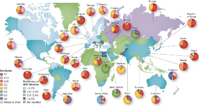
Figure 1. Worldwide estimated prevalence of HCV and genotype distribution
Visual representation of estimated region-specific prevalence of HCV infection and region-specific distribution of HCV genotypes. Countries of the middle east like Egypt have the highest prevalence of HCV infection and are predominantly characterized by genotype 4 infections. While genotype 1 has the largest worldwide distribution, genotypes 4, 5, and 6 are for the most part region specific affecting Southeast Asia, South Africa, and Egyptian middle east respectively. (permission from Hajarizadeh B, et al., Nature Reviews Gastroenterology &
2. HCV Lifecycle
2.1. Viral Entry
During primary infection, HCV particles are transported through the blood stream until they reach the basolateral surfaces of hepatocytes. There, the virus becomes concentrated by interacting with host factors with low affinity prior to interacting with subsequent essential entry factors. This initial low-affinity attachment involves interaction between host factors such as the heparan sulfate proteoglycans syndecan-1 and syndecan-4 and low-density lipoprotein receptor (LDLR) and components of the viral particles such as apolipoprotein E (apoE) and the envelope glycoproteins E1 and E2 (39-41).
The steps following this initial attachment phase are partially understood and involve a number of proposed host factors of which 4 are generally accepted: scavenger receptor B1 (SRB1) (42), tetraspanin CD81 (43), and tight junction proteins claudin-1 (CLDN1) (44) and occludin (OCLN) (45). Other factors include the cholesterol transporter Niemann-Pick C1-like 1(NPC1L1) (46), transferrin receptor 1 (TfR1) (47), and ephrin receptor A2 (EphA2) (48). Interestingly, SRB1 seems to play an intermediary role between the attachment and post-attachment phases of viral entry. Though SRB1 has demonstrated primary interaction with virion apolipoproteins, its lipid transfer activity appears to be the determining factor for productive viral entry (49, 50).
CD81, probably the most recognized among entry factors, once bound with E2 prime low pH-dependent fusion of viral endocytosis (51). Co-receptor complex formation between CD81 and CLDN1, which occurs more so at the basolateral membrane than at tight junctions, appears to be required however (52) and precedes its co-internalization with HCV particles during clathrin-dependent endocytosis. The discrepancy found within the localizations of these co-receptor complexes possibly implicates epidermal growth factor receptor (EGFR). EGFR, through downstream signaling pathways, promotes the lateral diffusion of CD81 and thus
facilitates the formation of the CD81-CLDN1 complex (53). While the precise role of OCLN in the HCV life cycle has yet to be clearly defined, cell-to-cell spread has been reported as the principle mode of HCV transmission (54). Following fusion, the HCV genome is released into the cytosol where translation and replication can begin.
2.2. Genome Translation and Replication
Original analyses of the amino acid sequences extrapolated from the nucleotide sequences of HCV initially revealed a gene organization similar to those of flaviviruses. HCV was later assigned to a new genus, Hepacivirus, within the Flaviviridae family and is therefore a Baltimore class IV single stranded RNA virus of positive polarity (+ssRNA).
The HCV genome contains a single open reading frame (ORF) and rather than a 5’cap or a 3’ poly A tail has highly structured RNA elements within its 5′ and 3′ non-translated regions (NTRs) to protect itself and assist with translation initiation (55). The 5′NTR contains an internal ribosomal entry site (IRES) to initiate translation (56) in addition to a microRNA 122 (miR-122) binding site (57). Contribution of miR-122, a liver-specific microRNA, has been shown to be indispensable to HCV replication by slowing exonuclease Xrn1 mediated degradation of HCV RNA (58) through its association with Argonaute 2 at the 5’end (59). Conversely, though no definitive consensus has been reached, the 3’ untranslated region (UTR) has been shown to have stem loop structures which, in conjunction with an upstream
5BSL3.2 stem loop within the NS5B coding region, are equally as important for HCV replication and translation (60, 61).
HCV genomic RNA is translated into a single polyprotein and is subsequently processed by viral and host encoded proteases into 10 mature proteins: core, E1, E2, p7, NS2, NS3, NS4A, NS4B, NS5A, and NS5B (62). Host signal peptidases and signal peptide peptidases process the structural proteins of HCV by mediating the cleavage at the core/E1, E1/E2, E2/p7 and p7/NS2 junctions. Moreover, the NS2 cysteine protease, whose activity is
enhanced by the N-terminus of NS3, mediates the cleavage of the NS2/NS3 junction freeing the NS3/4A serine protease. NS3/4A processes most of the non-structural (NS) proteins by mediating the cleavage of the NS4A/4B, NS4B/5A, and NS5A/5B junctions while also cleaving itself and other host factors (see chapter 2.4).
Once the polyprotein is fully processed, HCV induces massive rearrangements of intracellular membranes creating a sophisticated micro-environment called the “membranous web” to replicate its genome (55). This membranous web is particular to HCV and has been shown to be predominantly characterized by double-membrane vesicles (DMV) (63). While NS3/4A, NS4B, NS5A or NS5B alone can induce some membrane remodeling, the cooperative effect of each is required for a fully functional ultrastructure. There are, however, particular structural contributions which are differentially attributed to these NS proteins. NS4B’s oligomerization capacity has been suggested to be responsible for the scaffolding of membranous vesicles (64) while NS5A has been shown to be uniquely responsible for the induction of DMVs (65).
To follow suit, nascent genomic RNA is translated to produce either new viral polyproteins or RNA intermediates necessary for replication of genomic RNA. While RNA synthesis is the primary responsibility of the RNA-dependent RNA polymerase (RdRp) NS5B, lipid droplets (LD), triacylglycerides and cholesteryl esters deposits encapsulated by a phospholipid monolayer, are thought to play a key role in the coordination of viral RNA synthesis and the production of infectious particles (66).
2.3. Assembly and Release
The final stages of the HCV lifecycle require the careful spatiotemporal coordination of viral genomic RNA and structural proteins for the maturation of viral particles (67). A peculiarity of HCV morphogenesis is its complex interconnection with lipid metabolism. As a consequence, a plethora of host factors have arose in literature reporting varying degrees of contribution to HCV morphogenesis. For instance, host factors involved in very low-density lipoprotein (VLDL) synthesis pathways such as microsomal triglyceride transfer protein (MTP) (68), acyl-CoA synthetase 3 (ACSL3) (69) and hepatocyte nuclear factor 4α (HNF4α) (70) have been shown to play a role in the process.
LDs are established key players within viral assembly. While they are normally distributed throughout the cytoplasm in uninfected cells, HCV infection induces their perinuclear accumulation (71) and consequently their proximity to replication machinery. The interaction between HCV core protein, which forms the nucleocapsid, and LDs is essential for the recruitment of viral factors implicated in assembly (66) as confirmed by mutational studies in which this interaction is abrogated (72, 73). Cellular factors have also been shown to influence this core-LD association. For instance, host factors like diacylglycerol acyltransferase-1 (DGAT1) (74), an enzyme involved in lipid synthesis, and MAPK-regulated cytosolic phospholipase A2 (PLA2G4) (75), an enzyme involved in lipolysis, are reported to be involved in core trafficking to LDs. IκB kinase-α (IKK-α) has also been shown to influence this interaction through downstream lipogenic gene induction pathways (76).
While trafficking of core to LDs is vital for viral assembly, the coordination of other viral factors to assembly sites near LDs is also necessary (66). This includes HCV glycoproteins E1 and E2 that form a non-covalent heterodimer otherwise retained in the ER (77). NS2’s interaction with these factors and viroporin p7 was reported to assist in this matter (78, 79).
Other NS proteins have also been shown to contribute to the assembly phase. Among them, NS5A emerges as a central player. While its interaction with LDs has been shown to be required for assembly (66), its phosphorylation at a specific serine residue by casein kinase II was shown to prompt the transition between replication and assembly (80). Similarly, NS3/4A, through its interaction with Y-box-binding protein 1 (YB-1), has been shown to influence the equilibrium between RNA replication and the production of infectious particles (81).
Following assembly, HCV particles are released through the secretory pathway (82) where the endosomal-sorting complex required for transport (ESCRT) pathway, exploited by many enveloped viruses (83)has also been proposed to play a role. It has been suggested that neutralization of acidic compartments by HCV p7 during these later stages protects newly formed particles (84).
2.4. Innate Response and Immune Evasion
The coevolution of hosts and viruses has granted the cell many mechanisms to recognize and defend against viral pathogens. In response to HCV infection in particular, there is a strong induction of type I IFNs and of IFN stimulated genes (ISGs). This response begins with the detection of foreign invaders through pattern recognition receptors (PRRs) (85). In the case of HCV infection, these specifically involve RIGI and Tolllike receptors (TLR)1, -3, and -7. Following recognition, signal transduction continues through adaptor proteins TIR-domain-containing adapter-inducing interferon-β (TRIF), myeloid Differentiation Primary Response 88 (MYD88) and mitochondrial antiviral-signaling protein (MAVS). These effectors then activate the transcription factors IRF3, IRF7 and NF-κB and consequently lead to their nuclear translocation. This allows the induction and transcription of type I and type III IFNs, whose secretion will alert neighboring cells. Although both type I and type III IFNs share a number of biological properties, they remain relatively distinct. For instance, while peroxisomal MAVS can induce type III IFNs (86), it is unable to induce type I IFNs (87).
Secreted IFNs are then detected by IFN receptor complexes (IFNAR1/IFNAR2 complex for type I IFNs and IFN-λR1/IL-10R2 complex for type III IFNs (88)) with IFN-λ receptors being more restricted to epithelial cells than the ubiquitously expressed IFN-α/β receptors (89). Signal transduction from either complex continues through the JAK/STAT pathway demonstrating differences in kinetics (90) and ultimately culminating in the induction and transcription of ISGs. The transcription of inflammatory cytokines, PRRs and effector proteins keeps cells on high alert and maintains their antiviral state (85).
In response, HCV has developed many strategies to deal with host defenses and to allow it to establish a persistent infection. Among the better characterized mechanisms is the protease activity of the NS3/4A protein responsible for the cleavage of MAVS (91) and TRIF (92). As a result, the RIG-I and TLR3 mediated early IFN response are inhibited. While NS3/4A predominantly affects the early IFN response, core has been shown to play a role in hindering the late response by inhibiting the JAK/STAT pathway through its interaction with STAT1 (93) and through the induction of suppressor cytokines SOCS1 and SOCS3 (94, 95). Additionally, HCV glycoprotein E2 (96) and NS5A (97) have been shown to inhibit IFN signalling through their interaction with ISG.protein kinase R (PKR) Beyond its interaction with PKR, NS5A has been shown to interact with another ISG 2’5’ oligoadenylate synthethase (OAS) (98) achieving the same effect. Despite these evasion mechanisms, the activation of the IFN response remains detectable in patients. A rapid IFN response preceding the effect of viral evasion mechanisms and the lack of infection in all hepatocytes has been suggested to be responsible for these observations (99).
3. Treatment Evolution
HCV is a prime example that accurately represents the collective contribution of rigorous research to the evolution of treatment and prospective eradication of disease. From early type I IFN-based therapies riddled with side-effects which hinder treatment adherence to modern combination therapies which approach the pan-genotypic, treatment of HCV infection has a long and complex history. For HCV treatments, a therapy is considered effective when sustained virologic response (SVR) is achieved. SVR is described as the state where the virus remains undetectable following a period, typically 24 weeks, after treatment is concluded.
The Schering-Plough Corporation was the first to introduce an HCV treatment with their IFNα-2b recombinant, Intron A, being approved by the FDA in 1991 (100). Roche Pharmaceuticals would follow suit with their IFNα-2a treatment, Roferon A, being approved by the FDA in 1996. The IFN mono-therapy, however, had a poor therapeutic response with less than 20% of treated individuals achieving SVR (101). Merck, who would later acquire the Schering-Plough Corporation, brought on much needed improvements with the introduction of pegylated-IFN (PEG-IFN) and the addition of ribavirin (RBV) to treatment therapy. RBV was Originally discovered in 1972 as a guanosine analogue with broad spectrum activity against a number of RNA and DNA viruses (102), RBV was commercialized for hepatitis C treatment under the name Rebetol in 1998. On the other hand, the conjugation of IFN with polyethylene glycol allowed protection from proteolytic breakdown and thus increased its biological half-life alleviating difficulties associated with treatment adherence (103). In 2001, these milestones would establish the standard of care (SOC) for the next decade. Because genotype 1 patients were classically considered the most difficult to treat, therapeutic efficacy was still suboptimal allowing but 40% of infected individuals to attain SVR (104). In light of this, attempts continued to introduce better therapeutic regimens.
With the growing body of information surrounding the HCV life cycle and the role of individual viral proteins, the next revolution in treatment came in the form of direct-acting antivirals (DAAs). The first evidence of the potential of these novel anti-HCV agents
specifically designed against essential viral enzymes was with the discovery of BILN 2061. Even though its clinical development was interrupted due to some evidence of cardiotoxicity at high doses in rhesus macaques, the efficacy of BILN 2061 in humans established the first proof-of-concept for an NS3 protease inhibitor (105). With the momentum gained from these findings, in 2011, the first generation of DAAs boceprevir (Merck) and telaprevir (Vertex) were finally approved, introduced and added to the previous PEG-IFN/RBV regimen. These new triple therapies led to higher SVR rates, but still had the adverse effects of IFN-based treatment (106). Triple therapy with boceprevir or telaprevir quickly fell out of favor with the introduction of new wave DAAs. For instance, sofosbuvir (Gilead) which was approved by the FDA at the end of 2013 in combination with PEG-IFN/RBV raised the standards by achieving SVR rates approaching 90% (107).
Competing groups gradually developed and introduced a plethora of increasingly potent DAAs using different drug design strategies (detailed in chapter 3.1) often inspired by each other. Eventually culminating in late 2014, a new generation of all oral IFN-free combination therapies became available. Boasting excellent SVR rates, high genetic barrier to resistance as a result of the multi-pronged approach all without the undesirable IFN associated side effects, combination DAA therapies (detailed in chapter 3.3) mark the success of years of research from discovery of a virus to near optimal treatment design.
3.1. Direct Acting Antiviral (DAA) – Design Strategies
In principle, every step of the HCV replication cycle is a potential target for drug design. However, early technical limitations such as a lack of a robust replication system
(discussed in chapter 4.2) and of structural information for HCV historically made designing DAAs difficult. Coincidentally, fortunate discoveries coupled with systematic research chronologically lead NS3/4A protease, NS5B polymerase, and NS5A phosphoprotein to become the primary and most successful targets for HCV-specific DAAs though other viral targets such as NS4B (108) and p7 (109) have been explored.
3.1.1. NS3/4A Serine Protease
The NS3/4A serine protease is a heterodimeric enzyme that belongs structurally to the trypsin superfamily and is comprised of the N-terminal domain of the NS3 as well as the NS4A protein. It is unique, however, within its classification, for its requirement of a viral cofactor and of a structural zinc atom (110).
While the N-terminal domain of NS3 is responsible for the protease function, the C-terminal two-thirds of NS3 function as an RNA helicase belonging to the DExH family (111). Though its exact function within the HCV lifecycle is unclear, based on functional homology, it may be involved in RNA folding/remodeling (112), polymerase processivity (113), and/or genome encapsidation (114). Whether the helicase activity functions as a monomer or oligomer and whether either form is more functionally fit is debatable though some evidence indicates that which form NS3 takes is dependent on relative concentrations of enzyme to substrate (115).The NS4A protease cofactor consists only of 54 residues making it a relatively small protein. The N-terminal hydrophobic region, predicted to form a transmembrane α-helix, is involved in anchoring the heterodimeric complex to the membrane while amino acids 21-34 are directly implicated in the interaction with NS3 and are absolutely required (116). Crystal structures of NS3 with and without cofactor revealed that NS4A optimizes the orientation of
the catalytic triad residues and thereby stabilizes the protease domain of NS3 and increases enzymatic activity (117).
The main functional purpose of NS3/4A is the cleavage of the viral polyprotein at the junctions between NS3/NS4A, NS4A/NS4B, NS4B/NS5A and NS5A/NS5B. The liberated viral proteins are a product of trans-cleavage events, while NS3 and NS4A result from an intramolecular cis-cleavage. These junctions possess a consensus sequence of Asp/Glu-(Xaa)4
-Cys/Thr ↓ Ser/Ala-(Xaa)2-Leu/Trp/Tyr (118). This consensus sequence agreed with later
studies that characterized the requirement of decamer peptide substrates spanning P6-P4’ (Schechter & Berger nomenclature (119)) and defined preferences for an acidic residue in P6, cysteine in P1, serine or alanine in P1’, and a hydrophobic residue in P4’ (120-122). The NS3/4Aprotease lacks several surface loops at the substrate binding cleft found in other serine proteases (110). As a result, the solvent-exposed and relatively featureless substrate binding site posed a great challenge in the design of small molecule inhibitors and selection of non-peptidic candidates from compound collections.
3.1.1.1. NS3/4A Protease Inhibitors (-previr)
Based on the enzymatic features derived mainly from X-Ray crystallography studies, three distinct strategies for developing inhibitors against the NS3/4A serine protease were contemplated. These strategies consisted of interfering with either the NS3/NS4A interaction or the binding of zinc, and preventing substrate binding to the active site. The first two strategies are still, however, considered extremely difficult (123) and drug candidates of this category are virtually nonexistent. For instance, nonpeptidic small molecule inhibitors such as certain benzimidazole-based compounds inhibit the enzyme by exploiting its interaction with zinc (124). Though noncompetitive allosteric inhibitors, emerging mainly from random screening, could have potential within combination therapies, due to the nature of the method by which they are identified, the necessity of extensive safety assessments makes their development problematic.
With the insights gained from the design of human immunodeficiency virus (HIV) protease inhibitors, and with the large body of structural data surrounding the enzyme and its substrate, active site inhibitors were the first to be successfully developed and continue to be considered the most promising and widely used approach. Though, initially, the absence of a well-defined substrate binding site had raised concerns for the design of low molecular weight inhibitors, early breakthroughs lead to the emergence of two main mechanistic classes of active site-based inhibitors, namely covalent and product analogue inhibitors(125).
3.1.1.1.1. Covalent Inhibitors
Often referred to as transition state analogues or serine-trap inhibitors, covalent NS3/4A inhibitors are product-based inhibitors conceived through the replacement of the scissile amide bond with an electrophilic warhead. When the catalytic serine (S139) of the protease attacks the electrophilic warhead, a chemical formation which mimics and locks the transition state of peptide bond cleavage is generated. Despite classic electrophile-based covalent inhibitors functioning via an irreversible mechanism and despite speculations of clinical restrictions due to the possibility of unspecific irreversible binding (126), NS3/4A protease inhibitors of this mechanistic class were the first to be approved by the FDA. Notably, of the various electrophilic warhead alternatives, the early success of both boceprevir (Merck) and telaprevir (Vertex) is owed in part to the unusual mechanism by which linear α-ketoamide derivative inhibitors allow reversible binding to the catalytic site of the viral enzyme(127).
3.1.1.1.2. Product Analogues
This mechanistic class is comprised of competitive, reversible, mostly macrocyclic, noncovalent inhibitors whose drug design efforts were initiated by early findings regarding the susceptibility of the NS3/4A protease to feedback inhibition (128, 129).
With structural studies characterizing substrate specificity, and the importance of the P1 residue (see chapter 3.1.1.) to the potency and specificity of serine protease ligands (130), the major challenge for the development of N-terminal product-like inhibitors was designing proper chemically stable replacements for the P1 sulfhydryl group. Amino acid substitutes whether those with small hydrophobic side chains or those with larger side chains, both lead to a loss of potency either due to reduced contact surface area or steric incompatibility (128).
Eventually, the continuing research efforts culminated in the successful replacement of the unstable P1 residue with aminocyclopropane carboxylic acid (ACCA) derivatives (131, 132). The addition of a macrocyclic ring that connected the side chain of the P1 and the P3 residues and the stepwise optimization which followed proved to be useful in improving affinity and specificity, while preserving bioavailability characteristics of small molecules (133). Ultimately this resulted in the discovery of the compound BILN 2061 or ciluprevir (105). Macrocyclization was truly a breakthrough in NS3/4A protease inhibitor design, as many subsequent drug candidates, though varying in C-terminal moieties for improved pharmacokinetics, retain essentially the same core P1 ACCA and accompanying structures.
Simeprevir (Janssen), paritaprevir (AbbVie), and grazoprevir (Merck) are notable examples for this mechanistic class of protease inhibitors and are showcased within upcoming combination therapies.
Rather than focus on the N-terminal product, certain groups explored the C-terminal side of the scissile bond for competitive inhibition. These groups observed that P1’ substitutions with proline, tetrahydroisoquinoline-3-carboxylic acid (TIC) or pipecolinic acid (PIP) generated noncleavable substrate analogues (120). Despite boasting equally high potency and selectivity, development of clinical candidates was never fully engaged due to their large molecular weights and hence their poor pharmacokinetics.
3.1.2. NS5B RNA-Dependent RNA Polymerase
As the RNA-dependent RNA polymerase of a positive stranded RNA virus, NS5B is required for the synthesis of negative stranded RNA intermediate and subsequently that of positive polarity RNA genomes. Early in vitro studies using purified NS5B had already described the enzymatic activity of NS5B as being indiscriminate, but primer-dependent (134). Further progress in regards to HCV NS5B was hindered by difficulties associated to its poor solubility, a consequence of being a component of a membrane-bound complex (135). Interestingly, while the full-length enzyme displayed rather poor catalytic activity (136) the removal of the dispensable C-terminal hydrophobic tail, consisting of 21 residues and responsible for the ER membrane targeting of NS5B (137), resulted in enhanced enzymatic activity (138) and most importantly enhanced solubility which greatly facilitated the determination of 3D structures.
The unliganded crystal structure of NS5B, reported by several groups (139-141), had revealed unique structural features which could be manipulated in later drug design. While NS5B acquired the overall classic “right hand” shape, which included fingers, palm and thumb subdomains, it differed from the majority of cellular and viral polymerases which adopted a “half-open right hand” architecture. Due to the presence of two extended loops that span finger and thumb domains near the active site, NS5B adopted a more compact shape. The presence of a unique β-hairpin in the thumb subdomain which protrudes into the active site is another structural feature particular to NS5B. This may allow greater discrimination for template binding in a model which has previously been characterized as rather unspecific.
While in vitro evidence supports a highly processive “copy back” mechanism, in which 3’-terminal-OH group of the template is used as a primer for polymerization (134), it is generally agreed that NS5B proceeds through de novo initiation since the alternative would lead to an eventual loss of terminal sequences that are indispensible for proper HCV replication (142).
3.1.2.1. NS5B Polymerase Inhibitors (-buvir)
Due to the nature of viral replication and absence of RNA-dependent RNA polymerases in uninfected cells, targeting viral polymerases is invariably a straightforward choice in a drug design strategy and is unsurprisingly the rationale behind the majority of early approved antiviral drugs (143). For HCV, though an alternative mechanistic approach has been explored (144), polymerase inhibitors fall into two primary categories: nucleoside/nucleotide inhibitors (NI) and non-nucleoside/nucleotide inhibitors (NNI).
The NS5B polymerase has multiple binding sites that can be targeted for inhibition among which the catalytic site is the most phylogenetically conserved (145). Because NIs induce premature elongation termination by acting as substrate analogues that bind competitively to the active site of the enzyme, NIs boast a high genetic barrier with only the S282T mutation being reported to confer resistance (145, 146). Sofosbuvir (Gilead), a phosphoramidate prodrug that becomes an active uridine nucleotide analogue after triphosphorylation, is an example of a NI of current interest.
NNIs, however, can bind 4 different allosteric binding sites, located within the canonical thumb and palm domains (147, 148), and distort the precise geometry of the active site to significantly impair enzymatic function. Because these allosteric sites are not as conserved as the catalytic site, NNIs are more susceptible to resistance mutations. The thumb domain contains two binding pockets, the upper thumb (thumb I) and lower thumb (thumb II) which are characterized by distinct non overlapping resistance patterns (149). On the other hand, the palm domain contains partially overlapping palm I and II sites that also have an overlap in their resistance profiles (149). Though the design of these inhibitors is not quite as structured as that of NS3/4A protease inhibitors, palm I site inhibitors do have a recurrent structural benzothiadiazine-containing theme (149). Dasabuvir (AbbVie), an aryl dihydrouracil derivative, is an example of a palm I site directed NNI currently used in combination therapies.
3.1.3. NS5A Phosphoprotein
NS5A is a zinc-binding phosphoprotein spanning 447 amino acids (150) that is described to be phosphorylated at several different regions by distinct kinases and, as a consequence (151-153), exists in two forms designated p56 and p58 based on electrophoretic mobility. Though functional differences between its forms have yet to be elucidated, essential host factor and NS5A interactant lipid kinase phosphatidylinositol 4-kinase III alpha (PI4KIIIα) has been shown regulate its phosphorylation (154).
Structurally, the amino-terminus of NS5A includes an amphipathic α-helix, which is responsible for anchoring to the ER and ER-derived membranes such as lipid droplets (LDs) (155).This amphiphathic helix, comprised of the first 31 residues of NS5A, is also the only structural feature conserved in all HCV genotypes (156). NS5A can be divided into three distinct domains which are separated by linker regions (150). While domain I (D-I) residues were confirmed by biochemical assays to be involved in NS5A dimerization and RNA binding (157), domains II (D-II) and III (D-III) are thought to be responsible for NS5A’s large network of host interactants by fault of their intrinsically disordered and hence flexible nature (158, 159).
NS5A’s interaction with PI4KIIIα has been identified as having a central role in HCV replication as it induces the accumulation of phosphatidylinositol-4-phosphate (PI4P) within the membranous web and without which dramatic changes in ultrastructural morphology occur (160). Cyclophilin A through its contribution to de novo formation of double-membrane vesicles (DMV) (65) was also demonstrated to be a key host interactant essential for optimal HCV replication and has its own dedicated class of inhibitors (details in chapter 3.2.2.). ADP Ribosylation Factor GTPase Activating Protein 1 (ARFGAP1) (161), a GTPase-activating protein, and vesicle-associated membrane protein-associated protein A (VAP-A) and VAP-B (162) have also been shown to interact with NS5A with varying contributions to HCV replication.
Despite intrinsically lacking in enzymatic activity (163), NS5A is indispensible to HCV replication and its promiscuous ability to interact with numerous host factors is likely the source of its relevance.
3.1.3.1. NS5A Inhibitors (-asvir)
Classically, it was thought that NS5A was undruggable given the lack of characterized enzymatic activity (163). However, high throughput screening (HTS) using cell-based HCV replication systems serendipitously allowed the discovery of an early monomeric candidate which after subsequent chemical optimizations eventually led to dimeric daclatasvir (Bristol-Myers Squibb) (164-166). The same study noted rapid emergence of key resistance mutations within the NS5A coding region and confirmed the inhibitor’s target. A subsequent study determined that previously identified Y93H and L31V amino acid substitutions within NS5A to roughly confer a 25 fold increase in resistance to daclatasvir individually, but a 15 000 fold increase when combined (167). These findings were also related to emerging resistance mutations in clinical settings further suggesting a specific drug binding site.
Prior to the dimeric design of NS5A inhibitors, were a number of monomeric molecules which varied greatly in chemical scaffolds (166). Though emerging treatment-induced mutations were also mapped at the homodimer interface in the crystal structure of NS5A, these inhibitors were not as effective as their eventual successor becoming optimal within the nanomolar range rather than the picomolar range (166).With the encouraging clinical results from daclatasvir, NS5A inhibitor design trends shifted towards utilizing a dimeric pharmacophore, featuring a conjugated bis-biaryl core terminated by peptidic caps (166). The current trend in NS5A inhibitor designs utilizes a dimeric pharmacophore, featuring a linear conjugated bis-biaryl core terminated by peptidic caps (166). While peptidic caps are relatively conserved among various drug candidates, the chemical composition of core linkers varies widely with a tendency to remain within range of 15-18 Å (166). There is however a preference for imidazole-proline coupling for the junction between the core and
The exact mechanism by which inhibition is achieved remains elusive with no definitive consensus even on whether inhibitor binding is symmetrical or asymmetrical within the NS5A homodimer (168). However, putative molecular mechanisms have been suggested to explain inhibition on HCV replication. It has proposed that this DAA class disrupts the function of new replication complexes rather than affecting preformed complexes while also causing the redistribution of NS5A to lipid droplets (LD) (169). Others have proposed mechanisms at the stage of assembly and release revolving around host factors such as PI4KIIIα and TIP47. These suggested mechanisms are unlikely exclusive and probably need to be combined to explain the biphasic clinical response to these DAAs (170).
3.2. Host-Directed HCV Inhibitors – Design Strategies
Ingenuity is decidedly not lacking in HCV antiviral therapies. While DAAs are largely successful at inhibiting the virus, these strategies do not compare to the diversity found in host-directed HCV inhibitors or host-targeted agents (HTAs).
3.2.1. RNA-Based Inhibitors
The concept of utilizing RNA molecules as therapeutic agents was initially incited from the discovery that the entire HCV genome was translated into a single polyprotein via an internal ribosome entry site (IRES) (171). From there, the potential of trans-cleaving ribozymes (172) and antisense RNAs(173) as potential inhibitors of viral translation lead to the identification of ISIS 14803, a 20-base antisense oligonucleotide inhibitor (174), which was ultimately dropped during phase 1b clinical trials after generating inconclusive data. Eventually, the discovery of the liver-specific microRNA 122 (miR-122) and its possible role in HCV replication (57) reintroduced the potential of RNA-based treatments. With host miR-122 as a target, another antisense oligonucleotide inhibitor was developed. Miravirsen (Roche), having shown moderate efficacy in treating chronically infected individuals (175), is currently undergoing phase II clinical trials as a monotherapy.
3.2.2. Cyclophilin A Inhibitors
Two classes of cyclophilin A inhibitors exist: classical cyclosporine and non-immunosuppressive cyclosporine derivatives. The requirement of cyclophilin A for HCV replication was only truly demonstrated when the mechanistic prerequisite was shown to be its peptidyl-prolyl isomerase activity (176, 177) and when HCV NS5A was determined to be its viral ligand (178). As the cyclophilin A-NS5A interaction is conserved across genotypes, these
Though its initial discovery is not merited directly to anti-HCV efforts, these discoveries definitely emphasized the potential of cyclosporine in HCV therapeutics. For instance, cyclosporine A, a cyclic peptide of 11 amino acids, was originally sought after for its immunosuppressive activity which was achieved through a ternary complex formation between cyclosporine A-bound cyclophillin A and calcineurin (179). Its immunosuppressive ability was not favorable in the context of its anti-HCV ability, and so chemical modifications lead to the development non-immunosuppressive cyclosporine derivatives like alisporivir (Debiopharm), and N-methyl-4-isoleucine cyclosporine or NIM811 (Sandoz Pharmaceuticals) (180). Unlike its parent molecule, these derivatives while retaining its binary complex formation with cyclophilin A were unable to bind calcineurin. Normally, the N-methyl leucine at position 4 of cyclosporine occupies the calcineurin binding pocket however its substitution with N-ethyl valine or N-methyl-isoleucine allows respectively alisporivir (debiorivir) and NIM811 to be non-immunosuppressive (181, 182).
Though some evidence attributed cyclosporine sensitivity to HCV NS2 (183), discrepancies in cyclosporine sensitivity between replicons lacking NS2 and full replicative systems which have NS2 were later described to be due to reduced replication competence
because NS2-mediated polyprotein cleavage is the rate-limiting step in polyprotein processing (65).
3.2.3. Entry Inhibitors
Evidence for anti-HCV agents targeted at viral entry is varied and plentiful. The continuing development of entry inhibitors has brought forth considerable insight towards the early stages of the viral cycle (see chapter 2.1. for details). Entry inhibitors can act at the specific stages of viral entry whether it is at initial attachment, post-binding entry or at the level of viral endocytosis/membrane fusion. Because this mostly involves some form of disruption or modification to the interaction between HCV envelope glycoproteins and host factors, as with other host-targeted agents, entry inhibitors tend to have a high genetic barrier
to resistance with largely HCV genotype-independent potency, but pose a greater risk of simultaneous cellular toxicity.
Oriented towards the attachment and post-binding entry phases are: host-derived peptides like soluble low-density lipoprotein receptor (LDLR) (184), heparin-derived molecules (185), recombinant human L-ficolin (186), and claudin-1 (CLDN1)-derived molecules (187); virus-derived peptides such as p7 ion channel-derived molecules (188); glycan-binding synthetic molecules such as lectin cyanovirin-N (189), and boronic acid-modified lipid nanoparticles (190); structural mimic molecules like imidazole-based compounds (191); and natural compounds like epigallocatechin gallate (192) which is commonly found in green tea extracts. Though most candidates are in the in vitro stages of testing, a few have emerged in clinical trials. The savenger receptor B1 (SRB1) antagonist ITX 5061 (iTherX), an arylketoamide-based compound, is the most promising candidate of its class (193, 194) and is currently undergoing phase II of clinical trials. Because of its differing inhibitory mechanism, it also shows potential in combination with conventional DAAs (195).
Lesser characterized entry inhibitors are: those that target the acidification mechanism of virion-cell membrane fusion like vacuolar ATPase inhibitors concanamycin A and bafilomycin A (196); and those that disturb the lipid balance required for membrane fusion such as phenothiazines (197) and indole derivatives (198).
3.2.3. Immunomodulators
Improving tolerability, efficacy and or pharmacokinetic properties of existing treatments and characterizing other immunomodulatory candidates are venues which have also been explored by certain research groups. More specifically affecting hepatocytes and consequently reducing haematological side effects, interferon analogues such as PEG-IFN-lambda (199) and ribavirin analogues such as the taribavirin prodrug (200) have both undergone documented trials. More recent candidates which have been found to indirectly promote HCV elimination by bolstering the innate immune responses are nitazoxanide, a
receptor (TLR)-7 agonists (201) which mediate endogenous interferon and cytokine release through the myd88 adaptor.
3.3. Current Combination Therapies
In December 2013, the approval of the polymerase inhibitor sofosbuvir (Gilead) in combination with PEG-IFN/RBV brought forth new optimism for IFN-free therapy. Boasting broad genotype coverage with high SVR rates ranging from 82% to 100% (202), the idea of dropping PEG-IFN in favor of DAA substitutes proved promising. Combined with protease inhibitor simeprevir (Janssen) or NS5A inhibitor daclatasvir (Bristol-Myers Squibb) with or without ribavirin, novel therapies which included sofosbuvir quickly demonstrated SVR rates well above previous standards covering beyond genotype 1 and proving efficacious in both naïve and previously treated patients (203, 204).
From late 2014 and onwards, proprietary therapies using a combination of pre-approved or novel re-optimized DAAs are likely to dominate the HCV therapeutic scene. Though other design strategies (see chapter 3.2.) may continue to improve with future iterations, they are unlikely to be competitive. Outside of cost considerations, current combination therapies have the clear advantage boasting excellent SVR rates across the board and conferring more confidence in their safety when compared to host-directed inhibitors.
Currently, a number of combination therapies are available with more recent ones incorporating second-generation DAA iterations which are designed for broader genotype coverage (205). These all-oral IFN-free therapies achieve SVR rates approaching 100% within their designated genotypes, last typically between 12 to 24 weeks as opposed to the traditional 24 to 48 weeks with IFN-based treatments, and demonstrate excellent treatment adherence and good tolerability profiles (206-214). In October 2014, the combination therapy Harvoni® (Gilead) was approved by the FDA for treatment of HCV genotype 1 and subsequently approved for genotypes 4, 5, and 6. It is comprised of first-generation NS5A inhibitor ledipasvir and first-generation nucleoside inhibitor sofosbuvir. In December 2014, Viekira