Veno-venous extracorporeal CO
2removal improves pulmonary
hemodynamics in a porcine ARDS model
P. Morimont1,2, J. Guiot1,2, T. Desaive2, V. Tchana-Sato2, N. Janssen2, A. Cagnina2, D. Hella3, F. Blaffart3, J.-O. Defraigne2,4, B. Lambermont1,2
1Medical Intensive Care Unit, University Hospital of Liege, Liege, Belgium 2GIGA-Research, Cardiovascular Sciences, University of Liege, Liege, Belgium 3School of Perfusion, University of Liege, Liege, Belgium
4Department of Cardiothoracic Surgery, University Hospital of Liege, Liege, Belgium
Correspondence
P. Morimont, Medical Intensive Care Unit, Department of Medicine, University Hospital of Liege, 4000 Liege, Belgium
E-mail: ph.morimont@chu.ulg.ac.be Conflicts of interest
This study was supported by Maquet Getinge Group, Germany (supply of equipment). Funding
This study was supported by a grant from the Leon Fredericq Foundation of the University of Liege.
Submitted 21 January 2015; accepted 22 January 2015; submission 23 October 2014. Citation
Morimont P, Guiot J, Desaive T, Tchana-Sato V, Janssen N, Cagnina A, Hella D, Blaffart F, Defraigne J-O, Lambermont B. Veno-venous extracorporeal CO2removal improves
pulmonary hemodynamics in a porcine ARDS model. Acta Anaesthesiologica Scandinavica 2015
doi: 10.1111/aas.12497
Background: Protective lung ventilation is recommended in
patients with acute respiratory distress syndrome (ARDS) to mini-mize additional injuries to the lung. However, hypercapnic acidosis resulting from ventilation at lower tidal volume enhances pulmo-nary hypertension and might induce right ventricular (RV) failure. We investigated if extracorporeal veno-venous CO2removal therapy could have beneficial effects on pulmonary circulation and RV function.
Methods: This study was performed on an experimental model of
ARDS obtained in eight anaesthetized pigs connected to a volume-cycled ventilator. A micromanometer-tipped catheter was inserted
into the main pulmonary artery and an admittance
micr-omanometer-tipped catheter was inserted into the right ventricle. RV–arterial coupling was derived from RV pressure-volume loops. ARDS was obtained by repeated bronchoalveolar lavage. Protective ventilation was then achieved, and the pigs were connected to a pump-driven extracorporeal membrane oxygenator (PALP, Maquet, Germany) in order to achieve CO2removal.
Results: ARDS induced severe hypercapnic acidosis. Systolic
pul-monary artery pressure significantly increased from 29.6± 1.8 to 43.9± 2.0 mmHg (P < 0.001). After the PALP was started, acidosis was corrected and normocarbia was maintained despite protective ventilation. Pulmonary artery pressure significantly decreased to 31.6± 3.2 mmHg (P < 0.001) and RV–arterial coupling significantly improved (RV–arterial coupling index = 1.03± 0.33 vs. 0.55 ± 0.41,
P< 0.05).
Conclusion: Veno-venous CO2removal therapy enabled protective
ventilation while maintaining normocarbia during ARDS. CO2
removal decreased pulmonary hypertension and improved RV func-tion. This technique may be an effective lung- and RV-protective adjunct to mechanical ventilation.
Editorial comment: what this article tells us
In adult respiratory distress syndrome, hypercarbia and increased pulmonary vascular resistance can place strain on the right heart. The findings in this large animal experimental model show that extracorporeal carbon dioxide elimination in this setting can lead to improvement in pulmonary vascular resistance and right ventricular–arterial coupling.
Acta Anaesthesiologica Scandinavica (2015)
Acute respiratory distress syndrome (ARDS) is responsible for injuries to the alveolar epithelium and microvascular endothelium, which result in severe hypoxemia, decreased pulmonary compli-ance, and increased pulmonary vascular resis-tance (PVR).1,2 Acute pulmonary hypertension
increases right ventricular (RV) afterload.
Positive-pressure ventilation, required to correct ARDS-induced hypoxemia, further increases pul-monary hypertension and RV afterload, leading to acute cor pulmonale and RV failure.3,4 Moreover, mechanical ventilation induces additional lung injuries due to overdistention, repeated stretch to the alveoli, and increased inflammatory mediator levels.5–7
Despite new strategies in mechanical ventilation, ARDS continues to be a devastating disease.8
Mortality rates for ARDS decreased over time but still remain around 40%, which, in large part, is caused by hemodynamic complications to this syndrome.9,10
In order to reduce deleterious effects of positive-pressure ventilation, pro-tective ventilation strategies have been devel-oped, showing improvement in the outcome of mechanically ventilated patients. The ARDSnet investigators reported a 25% reduction in mortal-ity with a ventilation strategy involving limita-tion of mean tidal volume to 6 ml/kg, as compared with a more traditional tidal volume of 12 ml/kg.11However, low tidal volumes required by such ventilation strategies increase hypercap-nia associated to ARDS. Most clinicians seldom use very low tidal volumes in practice because of the clinical acceptability of this ‘permissive’ hypercapnia. Indeed, despite some potential ben-eficial anti-inflammatory effects, it is well estab-lished that hypercapnic acidosis (HCA) has deleterious effects, in particular by contributing to increase constriction within the pulmonary vasculature.4,12–14
Thus, modern care for ARDS requires decision to maximally reduce ventilator settings to ensure lung protection and reduce exacerbation of lung injury while facing the deleterious consequences of this intervention. In order to supplement or replace the lung function and to avoid ventilator-induced lung injury, gas exchange via an extra-corporeal device has been developed.15
Such a device may make it possible to avoid mechanical ventilation altogether in selected patients.16 Extracorporeal membrane oxygenation (ECMO) allows blood oxygenation and carbon dioxide
(CO2) removal but requires high blood flow and, as a result, placement of large cannulas. More-over, this technique is too costly for routine use as a lung rest strategy in adult ARDS patients.15 When compared with oxygenation, removal of
CO2 from blood can be accomplished at lower
blood flows. Indeed, due to its high solubility and diffusing capacity in blood, high amount of CO2 can be removed from a venous blood sample as compared with the small amount of oxygen that can be added.17,18 As a result, less invasive veno-venous devices have been specifically designed for CO2 removal with high gas exchange effi-ciency at relatively low blood flow rates (400– 1500 ml/min). The invasiveness of these devices, called ‘low flow extracorporeal veno-venous CO2 removal therapy (ECCO2RT)’, is reduced by use of percutaneous dual lumen catheters, compa-rable with catheterization for dialysis. The aim of our study was to determine if ECCO2RT used at early stage of ARDS could have beneficial effects on pulmonary circulation and improve RV func-tion in a pig model.
Material and methods
All experimental procedures and protocols used in this investigation were reviewed and approved by the ethical committee of the Medical Faculty of the University of Liege and conformed to the Guide
for the Care and Use of Laboratory Animals published
by the US National Institutes of Health (NIH pub-lication no. 85-23, revised 1996). Primary end points were changes in pulmonary artery pressure (PAP) and RV–arterial coupling. Experiments were performed from November 2013 to April 2014 on eight healthy pure Pietran pigs of either sex, weighing from 21 to 33 kg. The animals were premedicated with intramuscular administration of tiletamine (250 mg) and zolazepam (250 mg). Anesthesia was then induced and maintained by a continuous infusion of sufentanil (0.5μg/kg/h)
and pentobarbital (5 mg/kg/h). Spontaneous
movements were prevented by cisatracurium bésilate (0.5 mg/kg/h). After endotracheal intu-bation via a cervical tracheostomy, the pigs were connected to a volume-cycled ventilator (Servo 300, Maquet, Rastatt, Germany) set to deliver a tidal volume of 10 ml/kg at a respiratory rate of
20 breaths/min with an inspired O2 fraction
5 cm H2O. End-tidal CO2 measurements (Cap-nomac, Datex, Helsinki, Finland) were used to monitor the adequacy of ventilation. The pulmo-nary trunk was exposed via a median sternotomy. A micromanometer-tipped catheter (Scisense, London, Canada) was inserted into the main pul-monary artery through a stab wound in the RV outflow tract in order to measure PAP. Pressure in the left auricle (Pla) was measured with a micro-manometer-tipped catheter (Scisense) inserted into the cavity through the left atrial appendage. Systemic arterial blood pressure (SAP) was moni-tored via a micromanometer-tipped catheter (Sci-sense) inserted into the aorta through the left carotid artery. RV volume was measured from admittance catheterization.19
A 5-F, 5-electrode admittance micromanometer-tipped catheter (Sci-sense) was inserted through the RV infundibu-lum into the RV and positioned so that all electrodes were in the RV cavity. A central venous line was inserted into the right jugular vein and placed inside the superior vena cava. A 4F
fluid-filled catheter (Pulsiocath, Pulsion Medical
System, Munich, Germany) was inserted into the right femoral artery for pulse pressure analysis. A 6F Fogarty balloon catheter (Baxter Healthcare Corp., Oakland, CA, USA) was advanced into the inferior vena cava through a right femoral venotomy. Inflation of this balloon produced a gradual preload reduction.
Experimental protocol
After surgical preparation, the animals were allowed to stabilize for 60 min (‘baseline state’). Baseline hemodynamic recording was performed, including PAP, Pla, mean arterial blood pressure, and heart rate (HR). ARDS was achieved by repeated bronchoalveolar lavage (37°C, 60 ml/kg of 0.9% saline solution). Ventilator settings were reduced to achieve ‘protective ventilation’ (tidal volume = 6 ml/kg) after the induction of ARDS. When PaO2/FiO2 was < 200 and PaCO2> 60 mmHg, hemodynamic recording was per-formed (‘ARDS state’) and ECCO2RT was started.
Hemodynamic recording was repeated after
30 min of ECCO2RT (‘ECCO2RT state’). ECCO2RT was stopped after 60 min. A last hemodynamic recording was performed 30 min after ECCO2RT was stopped (‘OFF state’). Arterial blood gas analysis was performed at each stage of the
experiment. End-tidal CO2 was continuously
monitored. Fluid administration with Hartmann’s solution was continuously controlled by preload responsiveness. When pulse pressure variation
was ≤ 11%, animals were considered as
adequately filled. A warming blanket and a heated operating table were used to prevent hypothermia.
Description and insertion of the ECCO2RT
ECCO2RT was achieved with the PALP module
(Cardiohelp, Maquet), which consists of a unit in which gas exchange and pump take place with an integrated control console. The system was inter-faced with the pig through an inflow catheter (13F) inserted into the inferior vena cava and an outflow catheter (10F) inserted into the superior vena cava. The pump withdrew venous blood from the inferior vena cava, which, after CO2 removal, was re-infused into the right atrium through the superior vena cava. The PALP unit was primed with 345 ml of normal saline. Plastic tubing provided by the manufacturer was imme-diately connected to each venous catheter, and the PALP unit was started. Pump speed was adjusted to reach an aspiration pressure of 60 mmHg and sweep gas flow was set to the maximum value (10 l/min).
Heparin bolus was given before the PALP con-nection and assessed by the activated clotting time (ACT; in seconds) using a Hemochron Sig-nature Microcoagulation System (International Technidyne, Edison, NJ, USA). The target ACT was 180 s.
Data collection
All analog signals were continuously digitized and recorded (Notocord, Paris, France): HR (beats per minute), SAP (mmHg), PAP (mmHg), Pla (mmHg), RV end-systolic volume (ml), and RV end-diastolic volume (ml). PALP blood flow (l/min) was directly recorded from the PALP console.
Arterial tension of oxygen (PaO2, mmHg), CO2 (PaCO2, mmHg), and arterial pH were measured at each stage of the experiment (Baseline, ARDS, ECCO2RT, OFF).
Extravascular lung water (EVLW) and pulmo-nary vascular permeability index (PVPI) were
measured using a transpulmonary thermodilution method at baseline, after ARDS and at the end of the experiment.20
Transpulmonary thermodilu-tion was not performed during PALP therapy because of the right atrium reinfusion.
Data analysis
Maximal and minimal PAP defined systolic and diastolic pressures, respectively. Systolic ejection interval (ts) was measured from the foot of the pulmonary arterial pressure wave to its incisura, and the diastolic interval was td = T – ts, where T is the cardiac cycle length. Pulmonary blood flow (PBF) was derived from RV stroke volume given by RV admittance catheter and HR. RV contractil-ity was assessed by end-systolic elastance (Ees). The slope of the end-systolic pressure (ESP)– volume relation was obtained from transient occlusions of the inferior vena cava using the Fogarty balloon, during apnea. Pulmonary arte-rial elastance (Ea) was assessed from the ratio of RV ESP minus Pla to RV stroke volume (SV). Mean PVR was calculated as the ratio of mean PAP minus Pla divided by PBF.
Statistical analysis
Changes in hemodynamic parameters and blood gas data at each stage of the experiment were evaluated by a repeated-measures analysis of variance followed by Scheffé post-hoc tests. Cor-relation analysis was performed using Pearson’s correlation coefficient. Data were expressed as mean ± standard deviation.
Results
EVLW and PVPI increased during ARDS
(229± 95 ml to 441 ± 95 ml, P < 0.01 and from 2.21± 0.49 to 4.40 ± 0.49, P < 0.001, respectively).
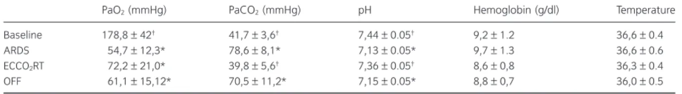
Arterial blood gases and acid-base equilibrium during the experiment are depicted in Table 1. Appropriate hypercapnia was obtained in all pigs. When compared with baseline, mean PaCO2 more than doubled during the ARDS, returned to baseline when the PALP was started, and
increased again when ECCO2RT was stopped.
PaO2 decreased during ARDS. The arterial pH
decreased during ARDS, returned to normal
values during ECCO2RT, and decreased again
when the PALP was stopped.
Hemodynamic data are depicted in Table 2. The amount of fluid (Hartmann’s solution)
used to achieve adequate filling was
1.08± 0.18 l. Mean blood flow in the PALP was
645± 84 ml/min. Systolic PAPs increased by
more than half during ARDS, decreased during ECCO2RT, and increased again after the PALP was stopped (Figs 1 and 2). Changes in PAPs were highly correlated with changes in PaCO2 (r = 0.87) and with changes in pH (r = −0.84) resulting from PALP therapy. Changes in PAPs
were poorly correlated with changes in PaO2
due to PALP therapy (r = −0.55). Similarly, PVR increased during ARDS, decreased during
ECCO2RT, and increased again after the
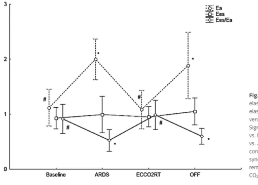
PALP was stopped (Table 2). Cardiac index (CI) and Pla did not change during the whole experiment. SV decreased during ARDS and increased during PALP therapy. Ea near doubled during ARDS, returned to normal values during
ECCO2RT and increased again when the
PALP was stopped (Fig. 3). RV contractility, assessed with Ees, increased when the PALP was stopped (Fig. 3). Ventriculo–arterial cou-pling, assessed by the ratio of Ees on Ea decreased under the unit during ARDS, come back at or around the unit during ECCO2RT and decreased again when the PALP was stopped (Fig. 3).
Table 1 Arterial blood gas data.
PaO2(mmHg) PaCO2(mmHg) pH Hemoglobin (g/dl) Temperature
Baseline 178,8± 42† 41,7± 3,6† 7,44± 0.05† 9,2± 1.2 36,6± 0.4
ARDS 54,7± 12,3* 78,6± 8,1* 7,13± 0.05* 9,7± 1.3 36,6± 0.6
ECCO2RT 72,2± 21,0* 39,8± 5,6† 7,36± 0.05† 8,6± 0,8 36,3± 0.4
OFF 61,1± 15,12* 70,5± 11,2* 7,15± 0.05* 8,8± 0,7 36,0± 0.5
*Significant difference vs. baseline at P< 0.05;†Significant difference vs. ARDS at P< 0.05. All data are means ± standard deviation. Basal, baseline
Table 2 Hemodynamic data.
HR sAP CI SV Pla PAPm PVR Ea Ees Ees/Ea
Baseline 73± 11† 104± 25 2,11 ± 0,59 30,1 ± 10,6† 7,5± 1.9 19,8 ± 3,9† 6,57± 3,50† 1,12± 0,42† 0,93± 0,25 0.92 ± 0,27†
ARDS 103± 25* 105 ± 11 2,29 ± 0.48 21,7 ± 5,3* 6,5± 3,9 32,8 ± 6,2* 11,92 ± 3,40* 1,99 ± 0,45* 0,99 ± 0,42 0,52 ± 0,20* ECCO2RT 82± 17 91± 11 2,58 ± 0.67 32,0 ± 9,2† 8,1± 1,9 23,8 ± 4,5† 6,76± 3,36† 1,08± 0,42† 0,95± 0.22 0,98 ± 0,27†
OFF 76± 11 95± 11 2,24 ± 0.45 28,1 ± 6,4 9,1± 2,1 32,7 ± 7,0* 11,56 ± 5,68* 1,88 ± 0,76* 1,05 ± 0,31 0,60 ± 0,14* *Significant difference vs. baseline at P< 0.05;†significant difference vs. ARDS at P< 0.05. All data are means ± standard deviation. HR, heart rate;
sAP (mmHg), systolic arterial pressure (mmHg); CI (l/min/m2), cardiac index; SV (ml), stroke volume; Pla (mmHg), left atrial pressure; PAPm (mmHg),
mean pulmonary arterial pressure; PVR(mmHg/min/l/m2), indexed mean pulmonary vascular resistance; Ea (mmHg/ml), pulmonary arterial
elas-tance; Ees (mmHg/ml), RV end-systolic elaselas-tance; Ees/Ea, RV ventriculo–arterial coupling index. Basal, baseline conditions; ARDS, acute respiratory distress syndrome; ECCO2RT, extracorporeal CO2removal therapy; OFF, stop of extracorporeal CO2removal.
Fig. 1. Evolution of systolic pulmonary artery pressure (PAPs). *Significant difference vs. baseline at P< 0.05,#significant difference vs.
ARDS at P< 0.05. Baseline, baseline conditions; ARDS, acute respiratory distress syndrome; ECCO2RT, extracorporeal CO2
removal therapy; OFF, stop of extracorporeal CO2removal.
Fig. 2. Example of the time course of systolic pulmonary artery pressure (PAPs). Baseline, baseline conditions; ARDS, acute respiratory distress syndrome; ECCO2RT, extracorporeal
CO2removal therapy; OFF, stop of
Discussion
This is the first study precisely analyzing the
respiratory and hemodynamic effects of CO2
removal with an extravascular device in an experimental model of ARDS. The findings were:
1. CO2 removal during ARDS with the PALP
device was very effective to achieve normocar-bia and to normalize acidosis, while maintain-ing protective ventilation.
2. Pulmonary vascular tone was significantly reduced and RV–arterial coupling was improv-ed during ECCO2RT.
Artificial lung support systems are medical devices designed to supplement or replace the respiratory function of the natural lung. ECMO was introduced for treatment of neonatal respira-tory failure. ECMO is an effective but costly and very invasive technique for selected patients with severe pulmonary failure. ECMO requires highly trained staff and is currently used in adults only in a few centers. Several simpler devices have been developed allowing a reduction in minute ventilation, reduced airway pressure, improved PaO2-to-FIO2 ratio, and improved survival in animal models of ARDS. Recently, Batchinsky et al. demonstrated that such simpler devices can provide a 50% reduction in minute ventilation while maintaining normocarbia and may be an
effective lung-protective adjunct to mechanical ventilation.21
Our results showed that the PALP
device was efficient for CO2 removal during
ARDS, enabling lung protective ventilation at low tidal volume. Alteration of the permeability of the alveolar-capillary membrane during ARDS was demonstrated by impaired gas exchanges and increased PVPI.22 The device achieved correction of HCA in less than 15 min in all pigs. The PALP technology uses the same technique as ECMO and is composed of a centrifugal pump, a micro-porous hollow-fiber oxygenator, two venous can-nulas, and tubing. The gas exchange membrane is very efficient and specifically designed for CO2 removal with low resistance to blood flow. However, as compared with ECMO, cannulas are substantially smaller with almost the same size as dialysis cannulas.
The main finding of our study was a significant reduction in PAP and PVR during ECCO2RT. This reduction was clearly related to the correction of HCA. HCA enhances pulmonary vasoconstric-tion.12,23
Several clinical studies demonstrated that HCA causes an increase in mean PAP in ARDS.4,24,25
Our study is concordant with those findings with a significant increase in PAP during ARDS. However, the relative roles of hypercapnia and acidosis in the mechanism of pulmonary
vasoconstriction remain unclear. Acute PHT
increases RV afterload,26,27 which individually
Fig. 3. Evolution of pulmonary arterial elastance (Ea, mmHg/ml), RV end-systolic elastance (Ees, mmHg/ml) and RV ventriculo–arterial coupling index (Ees/Ea). Significant differences: *Significant difference vs. baseline at P< 0.05,#significant difference
vs. ARDS at P< 0.05. Baseline, baseline conditions; ARDS, acute respiratory distress syndrome; ECCO2RT, extracorporeal CO2
removal therapy; OFF, stop of extracorporeal CO2removal; RV, right ventricular.
and collectively with microvascular obstruction effects of positive-pressure ventilation, and HCA exacerbate RV failure in ARDS.4Acute cor pulmo-nale in ARDS patients is associated with high mortality rates.28 Impaired RV function at early stage of ARDS may be underdiagnosed, and yet it might be the harbinger of a downward spiral in the patient’s condition.4 We previously estab-lished that PVR and RV ejection fraction are poor indicators of RV–arterial performance.26 RV– arterial coupling is beneficial for cardiovascular performance and is assessed by the ratio of Ees to Ea, where Ees and Ea characterize the RV system and the pulmonary vascular system, respectively.29
Ees precisely determines the con-tractility of the right ventricle independently of the loading conditions, and Ea is a precise measure of RV afterload. Indeed, afterload is not only characterized by PVR, but also results from a dynamic interplay among resistance, compliance, and wave reflection.26
The ratio of Ees to Ea
reflects the mechano-energetic aspects of
RV–vascular coupling. It can be demonstrated that efficiency of energy transfer from the RV to the pulmonary circulatory system is optimal when Ees/Ea = 2, whereas RV stroke work is maximal when Ees/Ea = 1. However, when Ees/ Ea< <1, there is a mismatch between RV contrac-tility and pulmonary vascular load.29 In ARDS patients, increased RV afterload is responsible for increased Ea, whereas Ees may decrease because of HCA, hypoxia, and often associated sepsis, leading to uncoupling between the right ventricle and the pulmonary circulation, and finally pre-cipitating RV failure.30 Therapies should be ideally oriented to restore the coupling between the heart and pulmonary vasculature by avoiding any increase in pulmonary vascular tone as well as depression in RV contractility.24,30 Our study showed that Ea was significantly increased during ARDS and was significantly reduced during ECCO2RT. The effects of HCA on myocar-dial function are contradictory. Acidosis decreases myocardial contractility because hydrogen ions inhibit Ca2+
influx into the myocardial fiber.31 However, hypercapnia causes coronary vasodila-tation leading to better myocardial perfusion and performance.32
We did not observe significant
changes in Ees during ARDS and during
ECCO2RT. During ARDS, the animals were
unable to maintain an optimal ventriculo–arterial
coupling despite a trend to increase RV contrac-tility, and Ees/Ea ratio dropped below the unit. The ECCO2RT, however, helped to restore a ratio around the unit mainly by lowering Ea. When the PALP was stopped, we again observed an RV–arterial coupling index (Ees/Ea ratio) lower than the unit. Uncoupling between RV and pul-monary arterial system resulted in decreased SV, whereas CO did not change because of increased HR.
In ARDS, pulmonary vasoconstriction may not result only from HCA, but also from other factors
like hypoxia or inflammatory mechanisms.
Hypoxic pulmonary vasoconstriction is a widely conserved, homeostatic, and adaptive vasomotor response to alveolar hypoxia, which redistributes blood to optimally ventilated lung segments by an active process of vasoconstriction.33,34
This response to hypoxia is strongly enhanced when HCA is superimposed.34
Conversely, improved blood oxygenation without changes in alveolar oxygenation should not affect PVR. However, changes in PaCO2 indirectly influenced changes in alveolar oxygenation by the alveolar gas equa-tion, and this mechanism could have played a role in pulmonary vasomotor tone.34 In our study,
changes in PaO2 due to ECCO2RT were
non-significant and poorly correlated with changes in
PAP, whereas changes in PaCO2 and pH were
highly significant and highly correlated with changes in PAP. Similarly, when the PALP was stopped, pulmonary vasoconstriction occurred again, whereas PaO2did not significantly change.
Blood temperature slightly, but
non-significantly, decreased along the experiment. Hemoglobin tended to decrease during PALP therapy, which may be explained by hemodilu-tion resulting from the priming of the device (Table 1). As a result, neither temperature nor hemoglobin changes should have significantly influenced CO2exchanges during the experiment. Others methodological issues should be taken into account: The experimental model of ARDS used in this study is different from ARDS observed in human beings. Although injury to the pulmonary circulation has been clearly demon-strated in human ARDS,35,36
our model induced lung injury without direct aggression to the pul-monary vasculature, allowing this study to focus on the effects of hypercapnia on pulmonary circu-lation and RV function.
In conclusion, use of the PALP device for ECCO2RT in a pig model of ARDS allowed pro-tective ventilation while maintaining normocar-bia. The PALP device made it possible to rapidly initiate extracorporeal lung support with a rela-tively low invasive extracorporeal lung support
technology, thus avoiding ventilator-induced
barotrauma, which should have been necessary to limit hypercapnia. Moreover, this device showed beneficial hemodynamic effects by reducing pul-monary vascular constriction related to HCA in ARDS. The beneficial effect on Ea improved RV–arterial coupling and consequently RV func-tion in this experimental model of ARDS.
Acknowledgement
This study was supported by a grant from the Leon Fredericq Foundation of the University of Liege.
References
1. Ware LB, Matthay MA. The acute respiratory distress syndrome. N Engl J Med 2000; 342: 1334–49.
2. Zapol WM, Snider MT. Pulmonary hypertension in severe acute respiratory failure. N Engl J Med 1977; 296: 476–80.
3. Vieillard-Baron A, Schmitt JM, Augarde R, Fellahi JL, Prin S, Page B, Beauchet A, Jardin F. Acute cor pulmonale in acute respiratory distress syndrome submitted to protective ventilation: incidence, clinical implications, and prognosis. Crit Care Med 2001; 29: 1551–5.
4. Lheritier G, Legras A, Caille A, Lherm T,
Mathonnet A, Frat JP, Courte A, Martin-Lefevre L, Gouello JP, Amiel JB, Garot D, Vignon P.
Prevalence and prognostic value of acute cor pulmonale and patent foramen ovale in ventilated patients with early acute respiratory distress syndrome: a multicenter study. Intensive Care Med 2013; 39: 1734–42.
5. Protti A, Cressoni M, Santini A, Langer T, Mietto C, Febres D, Chierichetti M, Coppola S, Conte G, Gatti S, Leopardi O, Masson S, Lombardi L, Lazzerini M, Rampoldi E,
Cadringher P, Gattinoni L. Lung stress and strain during mechanical ventilation: any safe threshold? Am J Respir Crit Care Med 2011; 183: 1354–62. 6. Chiumello D, Carlesso E, Cadringher P, Caironi P,
Valenza F, Polli F, Tallarini F, Cozzi P, Cressoni M,
Colombo A, Marini JJ, Gattinoni L. Lung stress and strain during mechanical ventilation for acute respiratory distress syndrome. Am J Respir Crit Care Med 2008; 178: 346–55.
7. Slutsky AS, Ranieri VM. Ventilator-induced lung injury. N Engl J Med 2013; 369: 2126–36. 8. Rubenfeld GD, Caldwell E, Peabody E, Weaver J,
Martin DP, Neff M, Stern EJ, Hudson LD. Incidence and outcomes of acute lung injury. N Engl J Med 2005; 353: 1685–93.
9. Phua J, Badia JR, Adhikari NK, Friedrich JO, Fowler RA, Singh JM, Scales DC, Stather DR, Li A, Jones A, Gattas DJ, Hallett D, Tomlinson G, Stewart TE, Ferguson ND. Has mortality from acute respiratory distress syndrome decreased over time?: a systematic review. Am J Respir Crit Care Med 2009; 179: 220–7.
10. Bull TM, Clark B, McFann K, Moss M. Pulmonary vascular dysfunction is associated with poor outcomes in patients with acute lung injury. Am J Respir Crit Care Med 2010; 182: 1123–8.
11. ARDSnet. Ventilation with lower tidal volumes as compared with traditional tidal volumes for acute lung injury and the acute respiratory distress syn-drome. The Acute Respiratory Distress Syndrome Network. N Engl J Med 2000; 342: 1301–8. 12. Stengl M, Ledvinova L, Chvojka J, Benes J,
Jarkovska D, Holas J, Soukup P, Sviglerova J, Matejovic M. Effects of clinically relevant acute hypercapnic and metabolic acidosis on the cardiovascular system: an experimental porcine study. Crit Care 2013; 17: R303.
13. Ijland MM, Heunks LM, van der Hoeven JG. Bench-to-bedside review: hypercapnic acidosis in lung injury–from ‘permissive’ to ‘therapeutic’. Crit Care 2010; 14: 237.
14. Croinin O, Ni D, Chonghaile M, Higgins B, Laffey JG. Bench-to-bedside review: permissive hypercapnia. Crit Care 2005; 9: 51–9.
15. Peek GJ, Mugford M, Tiruvoipati R, Wilson A, Allen E, Thalanany MM, Hibbert CL, Truesdale A, Clemens F, Cooper N, Firmin RK, Elbourne D. Efficacy and economic assessment of conventional ventilatory support versus extracorporeal
membrane oxygenation for severe adult respiratory failure (CESAR): a multicentre randomised controlled trial. Lancet 2009; 374: 1351–63. 16. Del. Sorbo L, Ranieri VM. We do not need
mechanical ventilation any more. Crit Care Med 2010; 38: S555–8.
17. Barrett KBS, Boitano S, Brooks H. Ganong’s review of medical physiology. New York: McGraw Hill, 2003.
18. Park M, Costa EL, Maciel AT, Silva DP,
Friedrich N, Barbosa EV, Hirota AS, Schettino G, Azevedo LC. Determinants of oxygen and carbon dioxide transfer during extracorporeal membrane oxygenation in an experimental model of multiple organ dysfunction syndrome. PLoS ONE 2013; 8: e54954.
19. Kutty S, Kottam AT, Padiyath A, Bidasee KR, Li L, Gao S, Wu J, Lof J, Danford DA, Kuehne T. Validation of admittance computed left ventricular volumes against real-time three-dimensional echocardiography in the porcine heart. Exp Physiol 2013; 98: 1092–101.
20. Monnet X, Anguel N, Osman D, Hamzaoui O, Richard C, Teboul JL. Assessing pulmonary permeability by transpulmonary thermodilution allows differentiation of hydrostatic pulmonary edema from ALI/ARDS. Intensive Care Med 2007; 33: 448–53.
21. Batchinsky AI, Jordan BS, Regn D, Necsoiu C, Federspiel WJ, Morris MJ, Cancio LC. Respiratory dialysis: reduction in dependence on mechanical ventilation by venovenous extracorporeal CO2 removal. Crit Care Med 2011; 39: 1382–7.
22. Kushimoto S, Endo T, Yamanouchi S, Sakamoto T, Ishikura H, Kitazawa Y, Taira Y, Okuchi K, Tagami T, Watanabe A, Yamaguchi J, Yoshikawa K, Sugita M, Kase Y, Kanemura T, Takahashi H, Kuroki Y, Izumino H, Rinka H, Seo R, Takatori M, Kaneko T, Nakamura T, Irahara T, Saito N.
Relationship between extravascular lung water and severity categories of acute respiratory distress syndrome by the Berlin definition. Crit Care 2013; 17: R132.
23. Laffey JG, Engelberts D, Kavanagh BP. Buffering hypercapnic acidosis worsens acute lung injury. Am J Respir Crit Care Med 2000; 161: 141–6. 24. Weber T, Tschernich H, Sitzwohl C, Ullrich R, Germann P, Zimpfer M, Sladen RN, Huemer G. Tromethamine buffer modifies the depressant effect of permissive hypercapnia on myocardial
contractility in patients with acute respiratory distress syndrome. Am J Respir Crit Care Med 2000; 162: 1361–5.
25. Thorens JB, Jolliet P, Ritz M, Chevrolet JC. Effects of rapid permissive hypercapnia on hemodynamics, gas exchange, and oxygen transport and
consumption during mechanical ventilation for the acute respiratory distress syndrome. Intensive Care Med 1996; 22: 182–91.
26. Morimont P, Lambermont B, Ghuysen A, Gerard P, Kolh P, Lancellotti P, Tchana-Sato V, Desaive T, D’Orio V. Effective arterial elastance as an index of pulmonary vascular load. Am J Physiol Heart Circ Physiol 2008; 294: H2736–42.
27. Viitanen A, Salmenpera M, Heinonen J. Right ventricular response to hypercarbia after cardiac surgery. Anesthesiology 1990; 73: 393–400. 28. Boissier F, Katsahian S, Razazi K, Thille AW,
Roche-Campo F, Leon R, Vivier E, Brochard L, Vieillard-Baron A, Brun-Buisson C,
Mekontso Dessap A. Prevalence and prognosis of cor pulmonale during protective ventilation for acute respiratory distress syndrome. Intensive Care Med 2013; 39: 1725–33.
29. Sagawa KML, Suga H, Sunagawa K. Cardiac contraction and the pressure–volume relationship. New York: Oxford University Press, 1988.
30. Morimont P, Lambermont B, Desaive T, Blaffart F, Dauby PC, Defraigne J-O. Right ventriculoarterial coupling in acute respiratory distress syndrome (ARDS) and expected benefits of CO2 removal therapy. J Crit Care 2013; 28: 887–1114.
31. Orchard CH, Kentish JC. Effects of changes of pH on the contractile function of cardiac muscle. Am J Physiol 1990; 258: C967–81.
32. Nomura F, Aoki M, Forbess JM, Mayer JE Jr. Effects of hypercarbic acidotic reperfusion on recovery of myocardial function after cardioplegic ischemia in neonatal lambs. Circulation 1994; 90: II321–7.
33. Moudgil R, Michelakis ED, Archer SL. Hypoxic pulmonary vasoconstriction. J Appl Physiol (1985) 2005; 98: 390–403.
34. Meyer P. Effet des gaz respiratoires sur la
circulation pulmonaire. In: Meyer P ed. Physiologie humaine, 2nd edn. Paris: Flammarion Medecine – Sciences, 1983: 1180–1.
35. Price LC, McAuley DF, Marino PS, Finney SJ, Griffiths MJ, Wort SJ. Pathophysiology of pulmonary hypertension in acute lung injury. Am J Physiol Lung Cell Mol Physiol 2012; 302: L803–15.
36. Greene R, Zapol WM, Snider MT, Reid L, Snow R, O’Connell RS, Novelline RA. Early bedside detection of pulmonary vascular occlusion during acute respiratory failure. Am Rev Respir Dis 1981; 124: 593–601.