University of Montreal
Bioassay-guided antidiabetic potentials of Devil’s club (Oplopanax horridus) preparations from the traditional pharmacopeia of the Squamish and other first nations of British
Columbia. By
Nyruz Ramadan Elahmer
Biomedical Sciences Program Faculty of medicine
Submission to the Faculty of Graduate and Postdoctoral Studies In order to obtain the degree of Master of Science (MSc)
Biomedical Sciences Program
Experimental Medicine Division
February 2018
University of Montreal
Faculty of Graduate and Postdoctoral Studies
This thesis entitled:
Bioassay-guided antidiabetic potentials of Devil’s club (Oplopanax horridus) preparations from the traditional pharmacopeia of the Squamish and other first nations of British
Columbia
Presented by: Nyruz Ramadan Elahmer
Has been evaluated by a jury composed of the following persons:
Dr. Mohamed Benderdour, chairperson-Rapporteur
Dr. Jacques Thibodeau, jury Member
Résumé
L'approche ethnobotanique a été utilisée pour identifier les espèces de plantes
médicinales utilisées par les Premières nations du Canada pour traiter les symptômes du Diabète de type 2 (DT2). Le bois piquant (Oplopanax horridus (Sm.) Miq.) a été identifié comme l'une de ces plantes qui ont des activités anti-diabétiques. Le but de ce mémoire de maîtrise était d'évaluer la stimulation potentielle par les préparations de l'écorce interne de la plante sur le transport du glucose dans les cellules C2C12 et l'inhibition de l'enzyme G6Pase dans les
hépatocytes H4IIE en utilisant des bioessais in vitro. Différentes préparations du bois piquant ont été utilisées. Les préparations traditionnelles de plantes médicinales sont souvent faites avec de l'eau chaude, de sorte que l'extrait d'eau chaude de la plante a été utilisé pour imiter les méthodes traditionnelles. En outre, l’extrait alcoolique à 80% de la plante a aussi été utilisé pour maximiser l'extraction des composés végétaux. D'autres fractions phytochimiques obtenues à l’aide de solvants de polarité croissante (hexanes, dichlorométhane -DCM, acétate d'éthyle, méthanol et eau) et un composé pur (acide chlorogénique) ont également été utilisés pour obtenir une bonne image des composés actifs du bois piquant. Le résultat de cette étude a montré que les extraits d'éthanol, DCM et hexanes stimulaient significativement le transport du glucose dans les cellules C2C12 après 18 h d'incubation avec des pourcentages de stimulation respectivement de 204 ± 4%, 201 ± 14% et 197 ± 8% au-dessus du témoin négatif (véhicule, DMSO). Par ailleurs, une inhibition statistiquement significative de l'activité de la G6Pase (-24 ± 4% par rapport au témoin négatif) a été observée lorsque la fraction de DCM a été testée. Ainsi, en ce qui concerne
l'homéostasie du glucose, les résultats ont confirmé que plusieurs préparations d'écorce interne du bois piquant stimulaient significativement le transport du glucose musculaire et inhibaient l'activité hépatique de la glucose-6-phosphatase (G6Pase). D'autres études utilisant l'approche du fractionnement phytochimique sont nécessaires pour isoler les composés et les comparer avec leurs mélanges pour permettre une meilleure compréhension de l'effet synergique et antagoniste et pour comprendre le mécanisme d'action des plantes et des cibles moléculaires.
Mots-clés:
Diabète de type 2, transport du glucose, G6Pase, médecine traditionnelle, homéostasie du glucose, produits de santé naturels.
Abstract:
An ethnobotanical approach has been used to identify medicinal plant species used by Canadian First Nations to treat Type 2 diabetes (T2D) symptoms. Devil's Club (Oplopanax
horridus (Sm.) Miq.) was identified as one of these plants reported to possess anti-diabetic
properties. The aim of this Masters thesis was to evaluate Devil’s club inner bark potential stimulation of glucose transport in C2C12 cells and inhibition of G6Pase enzyme in H4IIE hepatocytes using in vitro bioassays. Different preparations of devil’s club were used.
Traditional preparations of medicinal plants are often made with hot water, so a hot water extract of the plant was used to mimic the traditional methods. Also, 80% ethanol extract of the plant was used to maximize the extraction of plant compounds. Other fractions prepared with solvents of increasing polarity (Hexanes, dichloromethane –DCM, ethyl acetate, methanol, water) and a pure compound (chlorogenic acid) were used to begin unraveling devil’s club active compounds. The results of this study showed that ethanol extract, DCM, and hexanes fractions significantly stimulated glucose transport in C2C12 cells after 18 h incubation with stimulation percentages of 204±4%, 201±14%, and 197±8% above DMSO vehicle control, respectively. Meanwhile, a statistical significant inhibition in glucose-6-phosphatase (G6Pase) activity (-24±4% compared with vehicle control) was observed when DCM fraction was tested. Hence, with respect to glucose homeostasis, the results confirmed that several preparations of devil’s club inner bark significantly stimulated muscle glucose transport and inhibited hepatic G6Pase activity. Further studies using the phytochemical fractionation approach are needed to isolate compounds and compare them together with their mixtures to enable better understanding of the synergistic and antagonistic effect and to understand the plant’s mechanisms of action and molecular targets. Keywords:
Type 2 diabetes, glucose transport, G6Pase, traditional medicine, glucose homeostasis natural health products.
Table of Contents:
Table of Contents ... III List of tables ... IV List of figures ... V List of abbreviations ... VI Acknowledgement ... VIII Chapter 1: Introduction ... 1 1. Introduction ... 2
1.1 Energy homeostasis and glucose homeostasis ... 5
1.2 Diabetes mellitus ... 10
1.2.1 Diabetes definition and symptoms. ... 10
1.2.2 Diabetes diagnosis and classification. ... 11
1.2.3 Pathogenesis of T2D ... 15
1.2.4 Diabetes treatment ... 16
1.3 Devil's club (Oplopanax horridus) ... 22
1.4 Objectives of the study ... 34
Chapter 2: Methodology ... 36
Chapter 3: Result... 44
Chapter 4: Discussion and conclusion ... 50
Chapter 5: Reference ... 57
List of tables:
Introduction:
Table 1: Other Specific Types of diabetes ... 13
Table 2: Some therapeutic targets in T2D ... 18
Table 3: Summary of Medicinal Uses of Devil’s Club (Oplopanax horridus) ... 22
Table 4: Phytochemical and pharmacological differences between Oplopanax species… ... 26
Result: Table 5: list of maximum non-toxic dose of OH preparations… ... 45
Discussion: Table 6: The polarity of each solvent that was used… ... 54
List of figures:
Introduction:
Figure 1: Devil's Club ... 4
Figure 2: Gluconeogenesis and glycolysis ... 8
Figure 3: Pathophysiology of hyperglycaemia and free fatty acid… ... 16
Figure 4: Seven polyynes isolated from Oplopanax horridus… ... 28
Figure 5: Glycosides and other compounds of Devil’s club… ... 29
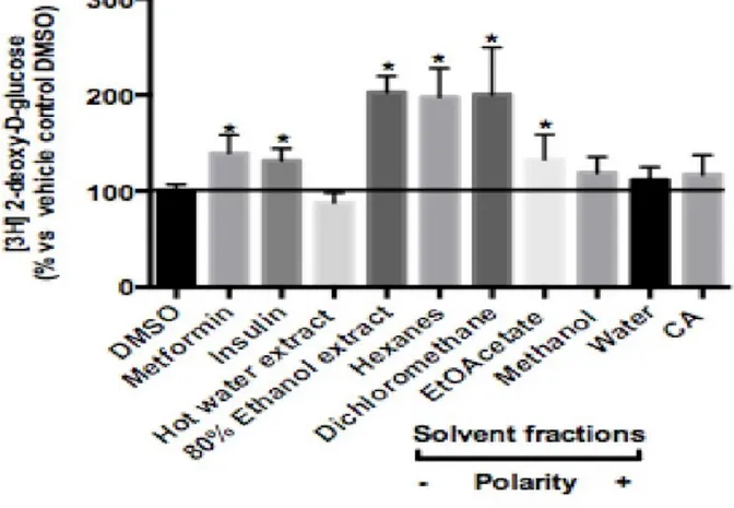
Result: Figure 6: Effects of the plant preparations on muscle glucose transport… ... 46
List of abbreviations: CEI: Cree of Eeyou Istchee
CIHR-TAAM: Canadian Institutes of Health Research Team in Aboriginal Antidiabetic Medicines
CA: Chlorogenic acid
ATP: Adenosine triphosphate G6P: Glucose-6-phosphate F6P: Fructose-6-phosphate PEP: Phosphoenolpyruvate
PEPCK: Phosphoenolpyruvate carboxykinase G6Pase: Glucose-6-phosphatase
SGLT: Sodium dependent glucose co-transporter GLUT: Facilitative glucose transporters
PI3K: Phosphatidylinositol-3-kinase OGTT: Oral glucose tolerance test OH: Oplopanax horridus
IFG: Impaired fasting glucose IGT: Impaired glucose tolerance GDM: Gestational diabetes mellitus IR: Insulin resistance
FFAs: Free fatty acids
CAM: Complementary and alternative medicine ROS: Reactive oxygen species
OS: Oxidative stress
TEAC: Trolox equivalent antioxidant capacity assay OH: Oplopanax horridus
GC-MS: Gas chromatography–mass spectrometry HPLC: High-performance liquid chromatography
SPE-HPLC: Solid-phase extraction- High-performance liquid chromatography NSCLC: Non-small cell lung cancer cells
GRAS: Generally recognized as safe product GU: Glucose uptake
HGP: Hepatic glucose production DCM: Dichloromethane
DMEM: Dulbecco’s modified Eagle medium FBS: Fetal Bovine Serum
DMSO: Dimethyl sulfoxide BSA: Bovine serum albumin MetS: Metabolic syndrome EE: Ethanol extraction HWE: Hot water extraction
Acknowledgement:
I would like to express my sincere gratitude to Dr. Pierre S. Haddad for his continuous help and support of my master study, and for his patience, too. Also, I would like to thank the lab team for their helping and cooperation. Special thanks go to the Libyan Minister of Higher Education and Scientific Research, which offered me the scholarship giving me a chance to pursue my academic degree. Last but not least, I would like to appreciate my parents for all their love and support. Also, I would like to thank my small family (my husband and my son) for their endless love and support through my life. To them I dedicate this thesis.
Chapter 1:
1 Introduction:
Diabetes is identified as one of the most common chronic metabolic diseases (Shaw et al., 2010). As a growing global health problem, the prevalence of diabetes has increased in recent decades in adults (Shaw et al., 2010; Whiting et al., 2011). Prevalence of type 2 diabetes is expected to significantly increase from 171 million people to 366 million from 2000 to 2030, respectively (Wild et al., 2004). In Canada, prevalence adjusted to the national population is assumed to climb up from 10.2% in 2013 to 11.7% in 2035 (Guariguata et al., 2014).
It is known that the prevalence of type 2 diabetes varies widely by race and ethnicity, and some Canadian First Nations communities are particularly and disproportionately affected (Ayach & Korda, 2010). For instance, diabetes is still recognized as one of the most severe medical disorders in the Cree (Kuzmina et al., 2010). As a subpopulation of the Canadian Cree, made up of approximately 18535 individuals today (Secrétariat aux affaires autochtones, 2015), the Cree of Eeyou Istchee (CEI; Eastern James Bay area of Quebec) is spread into 9 Nations throughout northern Quebec (Brassard et al., 1993). In 2009, an annual diabetes report showed that the age-adjusted prevalence of diabetes in Cree of Eeyou Istchee (CEI) of northern Quebec population was 20.6% while it was 4.9% in the general Quebec population (Harris et al., 1997). Moreover, the Cree population is also characterized by high rates of diabetes complications (Légaré, 2004). For instance, kidney problems affected 58%, retinopathy affected 11%, neuropathy affected 12%, and vascular complications affected 13% of participants in a 2002 study (Légaré, 2004). On the other hand, British Columbia is home to 198 First Nations, which make about one-third of all First Nations in Canada (Government of Canada, 2010). In 2002, an
increasing, notably for the age group over 35 years and among women (Johnson et al., 2002). Approximately one half of the population of this study was identified as having not less than one diabetic complication with prevalence similar to the rest of other First Nations (Johnson et al., 2002). In the same study, the gestational diabetes prevalence of First Nations of British
Columbia was 28 cases per 1,000 live-births, which is higher than the prevalence of the general population of British Columbia (18 per 1,000 live-births), increasing the risk of diabetes in their offspring (Johnson et al., 2002). However, the prevalence of gestational diabetes of British Columbia First Nations was lower than the prevalence of the James Bay Cree of northern Quebec (128 per 1,000 live-births) (Johnson et al., 2002).
On the other hand, urbanization in developing countries has changed lifestyle
dramatically, which causes an increase in adverse outcomes of noncommunicable disorders like type 2 diabetes (Guariguata et al., 2014). Rapid and drastic transitions in lifestyle have also appeared in Indigenous communities across North America over the last 50 years, affecting profoundly their health status (Harris et al., 1997). Despite concomitant modernization of medical treatments, there are still difficulties to provide optimal therapy for type 2 diabetes (Young et al., 2000). Consequently, to help manage this problem and, in the case of Indigenous populations, provide culturally relevant alternatives, new strategies are required.
More than 400 plants are considered as being used medicinally by Indigenous peoples of eastern Canada, and 105 of these plants are classified as having known biochemical compounds that have medicinal potential (Arnason et al., 1981). De Laguna ranks devil's club (Oplopanax horridus (Sm.) Miq.; Araliaceae) as the most important medicinal plant of all (De Laguna, 1972). In a literature review of medicinal plants that are used to treat symptoms of diabetes by Cree Nations, Devil's club was classified as an anti-diabetic plant (Downing, 2010). Also, devil’s club
inner bark is known to be used as an anti-diabetic agent by Squamish and other First Nations of British Columbia, including Haida, Heiltsuk, Nuxalk, and Sechelt (Lantz et al., 2004). The earliest study on the antidiabetic activity of Oplopanax showed that an aqueous extract of the root bark of the plant exerted a hypoglycemic action in rabbits (Large & Brocklesby, 1938). Additionally, another study also mentioned the hypoglycemic effect of an infusion of the roots of Oplopanax (MacDermot, 1949). However, few scientific studies have been done to prove the plant’s hypoglycemic effect and explain the underlying mechanisms.
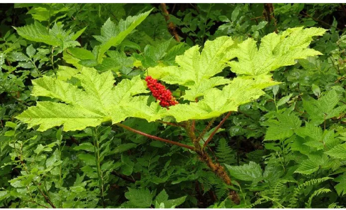
Figure 1: Devil's Club. “Image re-used with permission of the rights holder, Alaska Floats My Boat website, see appendix 1.” (Harvesting Devil's Club Root, 2013).
Consequently, this master thesis aims to evaluate the antidiabetic activities of various extracts, fractions and pure compounds derived from Oplopanax horridus. These preparations were tested on skeletal muscle cells in culture to determine their effect on glucose transport.
Hepatocytes in culture were then used to evaluate the inhibition action of the plant preparations on a critical enzyme of gluconeogenesis, namely glucose-6-phosphatase, which indicates the potential to reduce hepatic glucose production.
Energy homeostasis and glucose homeostasis:
To control energy homeostasis, metabolic processes provide the necessary and appropriate amount of energy is required to meet body needs (Röder et al., 2016). There are two main phases of metabolic processes, namely, anabolic metabolism (gluconeogenesis, glycogenesis, lipogenesis, and protein synthesis) and catabolic metabolism (glycolysis, glycogenolysis, lipolysis, and proteolysis) (Röder et al., 2016). The energy needed can be provided by the oxidation or degradation of proteins, fats, or carbohydrates (Röder et al., 2016). Extra energy can be stored as glycogen and fat such as triglycerides (TG) (Röder et al., 2016).
Glucose, a monosaccharide molecule, is the critical fuel in mammals to generate adenosine triphosphate (ATP) (Owen et al., 1967). Even though most human tissues utilize proteins and fats as an energy source in the absence of glucose, the brain essentially only uses glucose (Owen et al., 1967). In the case of low blood glucose concentrations (hypoglycemia), seizures, loss of consciousness and death can occur (Röder et al., 2016). In contrast, high blood glucose levels (hyperglycemia) might cause blindness, renal failure, and vascular disease (Röder et al., 2016). Consequently, blood glucose levels should be maintained in a limited range in a process called glucose homeostasis. Through the different pancreatic hormones, especially insulin (the anabolic hormone) and glucagon (the catabolic hormone), the pancreas controls the blood glucose levels in the very limited range of 4-6 mM (Röder et al., 2016). Glucose homeostasis is achieved by opposing and balancing the actions of insulin and glucagon (Röder et al., 2016). On the one hand, when blood glucose levels are under the lower limit, glucagon is secreted from α-cells to stimulate
hepatic glycogenolysis and to promote hepatic and renal gluconeogenesis, reaching the normal blood glucose levels by increasing endogenous blood glucose levels (Freychet et al., 1988). On another hand, rising in exogenous glucose levels promotes insulin release from β-cells, and insulin stimulates glycogenesis, lipogenesis, and the incorporation of amino acids into proteins to reach the normal blood glucose levels (Komatsu et al., 2013). As the central organ, the liver also involved in glucose homeostasis (Röder et al., 2016). The liver can maintain blood glucose levels via glucose production through gluconeogenesis and glycogenolysis and glucose storage through glycogenesis (Röder et al., 2016).
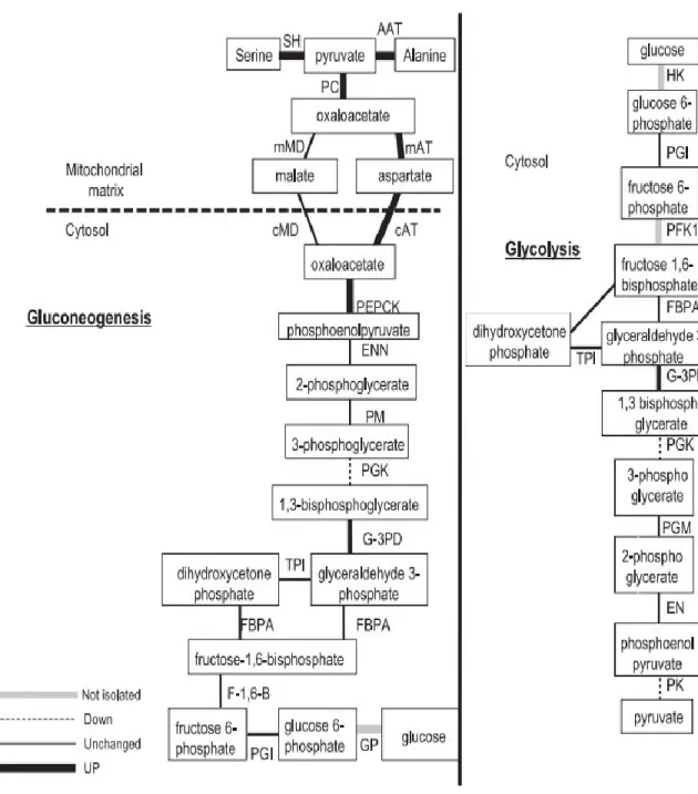
A summary about gluconeogenesis and glucose uptake will be discussed, as this master project deals with just the study of Devil’s club effects on hepatic gluconeogenesis and glucose transport in skeletal muscle cells,
Gluconeogenesis and glycolysis:
Both gluconeogenesis and glycolysis are controlled by intercellular and intracellular signals (Berg et al., 2002). Glycolysis is the multiple reactions process that converts anaerobically one molecule of glucose to two molecules of pyruvate with net production of two molecules of adenosine triphosphate (ATP) (Berg et al., 2002). Firstly, hexokinase phosphorylates glucose into glucose-6-phosphate (G6P) (Garrett & Grisham, 2010). Secondly, glucose phosphate isomerase converts G6P to fructose-6-phosphate (F6P) (Garrett & Grisham, 2010). Then, F6P is converted to fructose-1,6-bisphosphate by phosphofructokinase (Garrett & Grisham, 2010). Eventually, phosphoenolpyruvate (PEP), which is formed from fructose-1,6-bisphosphate, is converted into pyruvate by pyruvate kinase (Garrett & Grisham, 2010). Then, pyruvate might undergo anaerobic fermentation into ethanol (alcoholic fermentation) or lactate (lactic acid fermentation) or aerobic
During starvation or intense exercise, gluconeogenesis is put in place to generate glucose from non-carbohydrate precursors (pyruvate and lactic acid), mainly in the liver or kidney, in order to maintain glucose homeostasis (Berg et al., 2002). Gluconeogenesis is composed of a sequence of enzyme-catalyzed reactions and initiates in the cytoplasm or mitochondria, based on the substrate being used (Nordlie et al., 1999). As the first rate-limiting step, gluconeogenesis is launched by a carboxylation reaction of pyruvate in the mitochondria to generate oxaloacetate, which undergoes decarboxylation and phosphorylation reaction to release PEP in the presence of the cytosolic enzyme phosphoenolpyruvate carboxykinase (PEPCK) (Barthel et al., 2001). This step is followed by a sequence of reaction that is almost equivalent to reversed glycolysis except that fructose-1,6-bisphosphate is converted into F6P by fructose-1,6-bisphosphatase, which is considered as another rate-limiting enzyme of gluconeogenesis (Barthel et al., 2001). Lastly, G6P is hydrolyzed into glucose by glucose-6-phosphatase (G6Pase) that plays an essential role as the last rate-limiting enzyme of gluconeogenesis (Barthel et al., 2001) (Figure 2).
Glucose transport:
Both glucose synthesized within the body and dietary glucose should be relocated from the circulation to the target cells by different processes, including glucose transfer across plasma membranes via integral transport proteins (Wood & Trayhurn, 2003). Integral transport proteins comprise two distinct groups whose components have been addressed over the last two decades, namely: (i) sodium-dependent glucose co-transporter (SGLT) (Wright, 2001); (ii) the facilitative glucose transporters (GLUT) (Wood & Trayhurn, 2003). The SGLTs, members of Na-
dependent transporters family (gene name SLC5A), transport glucose with different affinities and capacities by a secondary active transport mechanism (Wood & Trayhurn, 2003). Therefore, glucose is transported into cells against its concentration gradient by coupling it to Na+, the
electrochemical gradient which is maintained by the Na+–K+ ATPase pump (Wood & Trayhurn,
2003). This active glucose transport exists across the proximal renal tubules and the luminal membrane of small intestinal cells (Wood & Trayhurn, 2003). The first cloned SGLT type is the high-affinity and low-capacity SGLT1, which is expressed predominantly on renal proximal straight tubules (S3 cells) and the apical membranes of small-intestinal absorptive cells (enterocytes) (Wood & Trayhurn, 2003; Hediger et al., 1987). The second SGLT is SGLT2, which is expressed essentially on the apical membrane of renal convoluted proximal tubules (S1 and S2 cells) and carries out glucose transport with low affinity and high capacity (Wells et al., 1992; Kanai et al., 1994). In the kidney, the bulk of filtered glucose is reabsorbed through SGLT2, and the remaining glucose is transported via SGLT1 to prevent any glucose loss in the urine (Wood & Trayhurn, 2003).
The GLUT family, gene name SLC2A, contains 14 members, which can be classified into 3 classes based on their functional characteristics (Gould & Holman, 1993). GULT4 is the
primary glucose transporter in the skeletal muscle cells (Zorzano et al., 2005). However, GLUT1 counts only 5% of the total expression of glucose transporters (Zorzano et al., 2005). In the resting state, GLUT4 occurs predominantly in intracellular membrane compartments (Bryant et al., 2002). Upon insulin stimulation, muscle contraction and/or hypoxia, GLUT4 is
translocated to the cell surface to increase glucose transport by 10 to 20 times (Bryant et al., 2002). Additionally, skeletal muscle cells display insulin-dependent glucose uptake which is mediated by phosphatidylinositol-3-kinase (PI3K) pathway or an insulin-independent mechanism which intervenes in the case of muscle contraction and/or hypoxia (Azevedo et al., 1995; Nesher et al., 1985; Wallberg-Henriksson & Holloszy, 1985).
Diabetes mellitus:
Definition and symptoms:
Diabetes is known as a metabolic disease of multiple aetiology associated with
hyperglycemia resulting from a deficiency in insulin action, insulin secretion, or both (WHO, 1999). The long-lasting hyperglycemia of diabetes is characterized by long-term dysfunction, and damage to various organs, particularly the kidneys, heart, eyes, and blood vessels (American Diabetes Association, 2014). Diabetes is developed from several pathogenic processes ranging from autoimmune destruction of β-cells, which causes insulin secretion deficiency, to other abnormalities that cause insulin resistance (IR) (American Diabetes Association, 2014).
Diabetes symptoms are characterized by thirst, blurring of vision, polyuria, and weight loss (WHO, 1999). Diabetes can lead to ketoacidosis or a non–ketotic hyperosmolar state in severe cases, which might lead to coma, stupor, and death (WHO, 1999). Even when diabetes
symptoms are not existing or not severe, hyperglycaemia can cause functional or pathological changes that might be present for a long-term (WHO, 1999).
Diabetes diagnosis and classification:
The Second World Health Organization Expert Committee on Diabetes Mellitus and the National Diabetes Data Group of the USA together produced criteria and systems for diabetes diagnostic tests and classification from 1979 to 1980 (Alberti & Zimmet, 1998). Little change was done by WHO in following years (Alberti & Zimmet, 1998). However, due to new
knowledge regarding the aetiology of diabetes and more information on the predictive value of diabetes complications, WHO and the American Diabetes Association Expert Committee later re-examined diagnostic criteria and diabetes classification to identify fixed criteria for diabetes (Alberti & Zimmet, 1998).
According to WHO in 1999, when fasting plasma glucose levels reach more than 7.0 mmol/L (126 mg/dL) or plasma glucose reaches more than 11.1 mmol/L (200 mg/dL) after 2 hours of an oral glucose tolerance test (OGTT), the case can be diagnosed as diabetes (WHO, 1999). However, Impaired fasting glucose (IFG) and impaired glucose tolerance (IGT) are known as pre-diabetic tests (WHO, 1999). While IFG is positive when fasting glucose level fall in the range of 6.1 to 6.9 mmol/L (110 and 125 mg/dL), IGT is positive when plasma glucose level ranges from 7.8 mmol/ L to 11.1 mmol/L (140 mg/dL to 200 mg/dL) after 2 hours of OGTT (WHO, 1999).
Diabetes can be categorized into four classes based on their aetiology and their distribution in different populations: type 1 diabetes, type 2 diabetes, type 3 diabetes (others specific types), and type 4 diabetes (gestational diabetes) (American Diabetes Association,
2006). However, each class of diabetes is caused by a deficiency in insulin production and/or in insulin action to decrease blood glucose, which leads to hyperglycemia (American Diabetes Association, 2006).
Type 1 diabetes:
The process of β cells destruction, which might ultimately cause diabetes mellitus, is involved in Type 1 diabetes, in which insulin secretion insufficiency occurs (Alberti & Zimmet, 1998). This type of diabetes, which is also known as insulin-dependent diabetes or juvenile diabetes, accounts for 5-10% of the cases found in children (American Diabetes Association, 2011). This type is usually identified by the existence of anti-GAD, islet cells or insulin
antibodies in the blood that illustrate the autoimmune mechanisms that cause β cells destruction (Alberti & Zimmet, 1998). However, no indications for autoimmune process are demonstrable in some populations, especially non-Europids, and this case is classified as idiopathic Type 1 diabetes (Alberti & Zimmet, 1998). Type 1 diabetes can be treated with exogenous insulin treatment coupled with lifestyle management (Atkinson & Eisenbarth, 2001).
Type 2 diabetes:
Type 2 diabetes, or insulin-independent diabetes, is characterized by insulin resistance that also yields insulin deficiency (Alberti &Zimmet, 1998). Even though T2D is known as the most common type of diabetes with approximately 90-95% of all kinds of diabetes (American Diabetes Association, 2011), the etiology of this form of diabetes is not yet known (Alberti & Zimmet, 1998). T2D has well-known cofounders, the most important of which being obesity, age and lack of physical activity (Ansari, 2009). Most T2D patients suffer from obesity while non-obese patients suffer from metabolic disorders and increasing of abdominal fat (Ansari,
2009). Due to its gradual development, this type of diabetes can go undiagnosed for a long time (Ansari, 2009). Most cases are treated with oral hypoglycemic drugs to manage blood glucose, in conjunction with lifestyle modifications (UK Prospective Diabetes Study (UKPDS) Group, 1998). Initially, insulin treatment might not be needed (UK Prospective Diabetes Study (UKPDS) Group, 1998). However, when beta-cell mass becomes insufficient, insulin therapy regularly occurs (UK Prospective Diabetes Study (UKPDS) Group, 1998).
Type 3 diabetes:
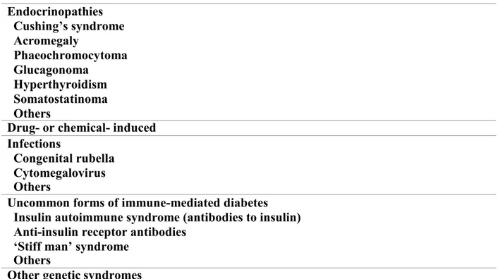
Other specific less common types of diabetes may be caused by different disorders such as malnutrition, endocrinopathies, drugs, genetic defects, infections, and other illnesses (Bell & Polonsky, 2001) (Table 1). For instance, fibrocalculous pancreatopathy is classified as
malnutrition-related diabetes mellitus (Alberti & Zimmet, 1998).
Other specific types of diabetes Genetic defects of beta-cell function
Chromosome 20, HNF4 α (MODY1) Chromosome 7, glucokinase (MODY2) Chromosome 12, HNF1 α (MODY3) Chromosome 13, IPF1 (MODY4) Mitochondrial DNA 3243 mutation Others
Genetic defects in insulin action Type A insulin resistance Leprechaunism
Rabson-Mendenhall syndrome Lipoatrophic diabetes
Others
Diseases of exocrine pancreas Fibrocalculous pancreatopathy Pancreatitis Trauma/ pancreatectomy Neoplasia Cystic fibrosis Haemochromatosis Others
Endocrinopathies Cushing’s syndrome Acromegaly Phaeochromocytoma Glucagonoma Hyperthyroidism Somatostatinoma Others
Drug- or chemical- induced Infections
Congenital rubella Cytomegalovirus Others
Uncommon forms of immune-mediated diabetes Insulin autoimmune syndrome (antibodies to insulin) Anti-insulin receptor antibodies
‘Stiff man’ syndrome Others
Other genetic syndromes
Table 1: Other specific types of diabetes “Table reused with permission of the rights holder, John Wiley and Sons, see appendix 1.” (Alberti & Zimmet, 1998).
Type 4 diabetes:
As a carbohydrate intolerance disorder, gestational diabetes mellitus (GDM) results in hyperglycaemia, itself related to the placental hormones being implicated in the increase of insulin resistance during pregnancy (Alberti & Zimmet, 1998; Desoye & Hauguel-de Mouzon, 2007). Women who have diabetes mellitus before pregnancy and get pregnant do not have gestational diabetes, but they have diabetes mellitus (Alberti & Zimmet, 1998). GDM is observed in 2-10% of all pregnancies (Desoye & Hauguel-de Mouzon, 2007). GDM confounding factors include a history of glucose intolerance, older women, history of large gestational age babies, and certain high-risk ethnic groups, and it has thus been proposed to
screen pregnant women at high risk of hyperglycaemia during the first trimester to detect undiagnosed diabetes mellitus (Alberti & Zimmet, 1998).
Pathogenesis of T2D:
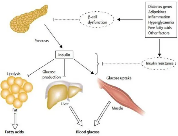
Type 2 diabetes mellitus gradually evolves into long-term hyperglycaemia, in conditions ranging from insulin resistance (IR) related to obesity to frank pancreatic β cell dysfunction (Stumvoll et al., 2005). Different mechanisms have been proposed for β cell dysfunction, such as lipotoxicity, glucotoxicity, and amyloid formation for β cell dysfunction, whereas
inflammatory cytokines, adipokines, increased non-esterified fatty acids, and mitochondrial dysfunction appear to be responsible for insulin resistance (Stumvoll et al., 2005). T2D is being a chronic and progressive disorder, insulin resistance causes an elevation of blood glucose, especially in the postprandial phase, which in turn stimulates increased insulin release from β- cell that cause hyperinsulinemia; a phenomenon known as β cell compensation (Butler et al., 2003). In time, β cells eventually lose their capacity to produce insulin, which leads to β cell insufficiency (Butler et al., 2003). Insulin resistance occurs due to failures in the biologic responses to insulin, which reduce the ability of insulin to decrease hepatic glucose production, to promote the generation of triglycerides, to stimulate glucose uptake in muscle and adipose tissue – leading to diminished peripheral glucose utilization – and to decrease lipolysis – leading to increase of free fatty acids (FFAs) efflux (Utriainen et al., 1998; Vaag et al., 1995; Kelsey et al., 2013) (Figure 3).
Figure 3: Pathophysiology of hyperglycaemia and increased circulating free fatty acids, “Figure reused with permission of the rights holder, Elsevier, see appendix 1” (Stumvoll et al., 2005).
Diabetes treatment:
The major thrust behind the selection of treatments in T2D management consists in protecting diabetic patients from diabetes complications (Stumvoll et al., 2005). Insulin resistance (IR) has a major impact on T2D pathogenesis, particularly on cardiovascular complications, so the treatment must be aimed mainly towards improvement in the biological response to insulin (Stumvoll et al., 2005). Consequently, the cornerstone of T2D therapy involves moderate exercise, lifestyle interventions and weight loss; these clearly decreasing the
risk of glucose intolerance and cardiovascular diseases caused by the so-called metabolic syndrome (Diabetes Prevention Program Research Group, 2002; Tuomilehto et al., 2001). However, advances in the understanding of insulin production and of molecular pathways by which the hormone modulates intermediary metabolism provide insights into novel therapeutic interventions for T2D (Saltiel, 2001). The main challenge is to develop pharmacological agents that avoid interfering with other molecular pathways, which might cause severe adverse effects (Saltiel, 2001) (Table 2). As a result, it is crucial to fully understand cellular location, tissue distribution, and isotype selectivity and kinetics during the selection of modulators of receptors, enzymes, or macromolecular interactions (Saltiel, 2001). As reported in 2008 by the Canadian Diabetes Association Clinical Practice Guidelines Committee, diabetic patients should be advised to adopt a healthier lifestyle (optimal nutrition and moderate physical activity) and be prescribed one or more of five groups of T2D medications (Bhattacharyya et al., 2009). The latter include oral hypoglycemic agents (OHAs), insulin replacement treatment for advanced stages, lipid-lowering drugs to decrease LDL-cholesterol, low-dose aspirin to protect from thrombosis, and antihypertensive drugs (angiotensin II receptor antagonists & angiotensin- converting enzyme inhibitors) to manage blood pressure (Bhattacharyya et al., 2009). In
contrast, other practitioners and researchers recommend some alternative medicines, which have synergistic effects (Crawford, 2009; John et al., 2003).
Table 2: Major therapeutic targets considered in T2D “Table reused with permission of the rights holder, Elsevier, see appendix 1” (Saltiel, 2001).
As mentioned, this Masters project aims to study the inhibitory action of Oplopanax
horridus on hepatic gluconeogenesis, as well as the plant’s potential capacity to stimulate
skeletal muscle glucose transport. In this context, insulin and metformin are used as positive controls and, hence, a summary about insulin and biguanides, notably metformin, will be provided.
Oral hypoglycemic agents OHAs:
Although lifestyle management might prevent T2D symptoms, optimal lifestyle intervention is difficult to adhere to (Kurtz, 1990; Kravitz et al., 1993). Consequently, pharmacological therapy is often required to control T2D hyperglycemia. Different studies related to T2D treatments suggest that successful hypoglycemic treatment like OHAs decreases microvascular risks, such as retinopathy and nephropathy (UKPDS, 1998; Vijan et al., 1997).
However, the impact of OHA therapy on macrovascular complications, such as myocardial infarction and stroke is still controversial (UKPDS, 1998; Pitale et al., 2000; Stettler et al., 2006). OHAs can be categorized into biguanides, α-glucosidase inhibitors, insulin secretagogues,
insulin sensitizers and others (Krentz & Bailey, 2005). Single or multiple OHAs can be used to treat T2D symptoms (Krentz & Bailey, 2005).
As a first line treatment, pharmacological therapy of type 2 diabetes (non-insulin- dependent diabetes) is chiefly carried out by metformin, a biguanide antidiabetic medication (Sirtori & Pasik, 1994). Metformin has been available for more than 30 years, but its detailed mechanism of action was only understood a few years ago (Sirtori & Pasik, 1994). Metformin acts mainly to suppress hepatic gluconeogenesis by insulin-independent pathways, inhibiting hepatic extraction of different substrates like lactate, opposing glucagon effect and decreasing hepatic glucose-6- phosphatase activity (Wiernsperger & Bailey, 1999). Because it acts independently of insulin, it effectively enhances the action of insulin and has been termed an insulin sensitizer (Wiernsperger & Bailey, 1999). Additionally, metformin enhances insulin- stimulated glucose uptake in skeletal muscle cells by stimulating the translocation of insulin- sensitive GLUT4 transporters to the cell membrane by insulin-independent pathways (Lee et al., 2011). Also, metformin enhances the functions of insulin and glucose-sensitive transporters, suppresses fatty acid oxidation, and reduces hyper-triglyceridaemia, consequently decreasing energy sources for gluconeogenesis (Wiernsperger & Bailey, 1999). Metformin was considered as potentially dangerous due to the possible induction of lactic acidosis, which might lead to a fatal outcome (Sirtori & Pasik, 1994). However, a study shows that this risk is negligible, especially when appropriate care is provided when clinical risks of lactic acidosis are suspected (Sirtori & Pasik, 1994).
Insulin therapy:
As a progressive disease, some T2D patients resort to insulin therapy once their glycemia is no longer adequately controlled by OHAs (Mudaliar & Edelman, 2001). Uncontrolled blood glucose levels worsen insulin resistance in peripheral tissues like skeletal muscle, leading to decreased glucose uptake and other physiological processes, hence perpetrating a vicious circle of high blood glucose levels (Mudaliar & Edelman, 2001). Short-term insulin treatment
improves insulin sensitivity by improving glucose control and decreasing glucose toxicity that causes insulin resistance (Mudaliar & Edelman, 2001).
Complementary and alternative medicine (CAM):
Although different conventional treatment choices are available, diabetes adverse
outcomes are still common (Health Canada, 2002). Conventional diabetes treatments represent a huge portion of expenditures in US healthcare, amounting to an estimated 25% of the total healthcare budget in 1998 (Yeh et al., 2002). With all of these dissatisfactions with modern therapies, diabetic patients have demonstrated a growing interest in complementary and
alternative medicine (CAM) (Yeh et al., 2002). The interest is being notable, not only among the general public, but also among researchers, educators, and healthcare providers (Berman et al., 1999; Bloomgarden, 2001).
Regarding Canadian Indigenous populations, traditional treatment is generally associated with medical practices developed before European colonization that brought forth new diseases and Western therapies (Johnston, 2002). In recent surveys of northern Quebec, including James Bay Cree and Inuit populations, many of the plants that were reported by Arnason et al. in the Canadian Journal of Botany in 1981 are identified as anti-diabetic plants (Fraser et al., 2007;
Leduc et al., 2006). This includes 20 plants from Whapmagoostui and 16 plants from Mistissini that are used by Cree Elders for the treatment of symptoms of diabetes (Downing, 2010). Many of these plants have notable antioxidant activity (Downing, 2010). In addition, the plant part, selection, and preparation play an important role in plant medical activities (Fraser et al., 2007; Leduc et al., 2006). Conventional treatments usually act on a specific metabolic defect while diabetes is comprised of several defects (Tiwari & Rao, 2002). However, the traditional
medicinal plants might be more useful due to their phytochemical synergistic effects (Downing, 2010).
Antioxidants provide an optimal defense against diabetes outcomes (Downing, 2010), so a given plant’s antioxidant activity can contribute to its beneficial effects in the context of diabetes (Ame et al., 1993). This helps return the production of reactive oxygen species (ROS) toward normal values and hence protects various human metabolism processes from damage to key proteins, lipids, and carbohydrates (Downing, 2010). Normally, the human body generates different enzymes that neutralize the ROS and hence their adverse effects (Ame et al., 1993). In contrast, generation of these enzymes in diabetic patients is insufficient, and the amount of ROS in diabetics is much more than normal, leading to oxidative stress (OS) (Ame et al., 1993; Johansen et al., 2005). Significantly, phenolic compounds like tannins are usually quantified to evaluate the antioxidant activities of the plant due to their strong antioxidant effect (Fraser et al., 2007; Spoor et al., 2006). During an in vitro study, devil’s club extract exhibited significant antioxidant effects in hydroxyl free radical scavenging activity in Trolox equivalent antioxidant capacity (TEAC) assay and dose-dependent inhibition of nitric oxide generation (Tai et al., 2006). Although the mechanism of action of the antioxidant activity of devil’s club is still lacking, this evidence supports a biological activity of devil’s club that is relevant for diabetes
(Tai et al., 2006). Additionally, different studies have mentioned the use of Devil's club (Oplopanax horridus) for diabetes (Downing, 2010; Large & Brocklesby, 1938; MacDermot, 1949).
Devil’s Club (Oplopanax horridus):
The Devil’s Club, Oplopanax horridus (Sm.) Miq., is also known as Echinopanax
horridum (Sm.), Panax horridum Sm., and Fatsiu horrida (Sm.) (Smith, 1983). The plant is an
understory shrub that thrives in the Pacific Northwest of North America (Calway et al., 2012). It occurs in dense, old growth forests, developing large layering groups of the plant (Lantz & Antos, 2002). Devil’s club is descended from Araliaceae family, which is the same family of many famous plants like American ginseng, so it is also known as “Alaskan ginseng” or “Pacific ginseng” (Lantz et al., 2004; Chan et al., 2011). Three different species are included within
Oplopanax which are O. horridus, O. japonicus, and O. elatus (Wang et al., 2010). However, O. elatus and O. japonicus are found in eastern Asia and Japan, respectively (Wang et al., 2010), so
they are not included in this research. Devil's club has a history of use mainly by Pacific Indigenous peoples (Calway et al., 2012). It has been used by more than 38 cultural-linguistic groups to treat up to 34 different medical issues and to assist during spiritual practices (Lantz et al., 2004) (Table 3). Oplopanax horridus (OH) has been characterized as having potential antidiabetic, antibacterial, antiviral, and cancer chemo-preventive effects (Calway et al., 2012). However, OH phytochemical studies are quite limited (Calway et al., 2012).
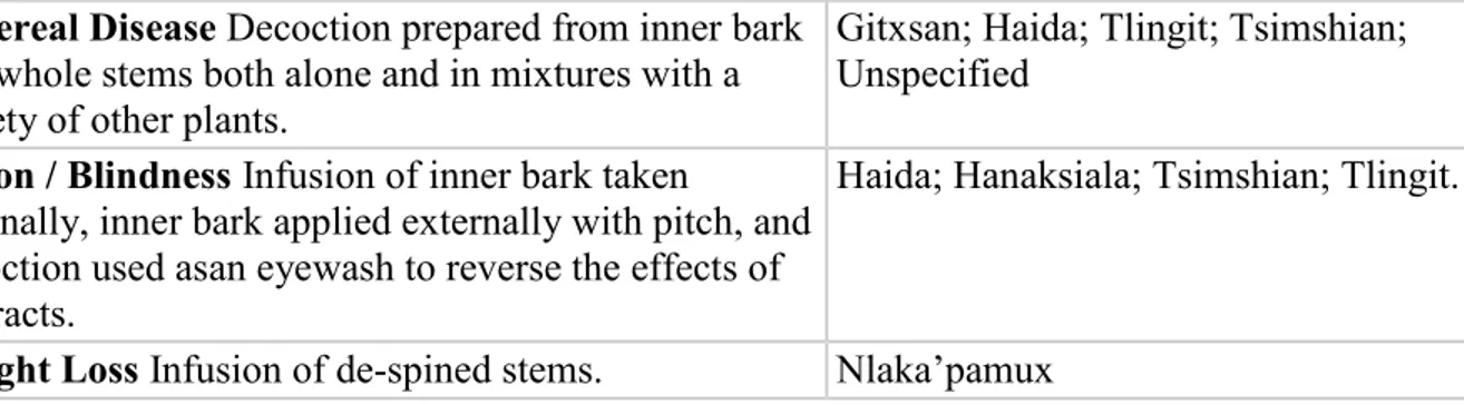
Summary of Medicinal Uses of Devil’s Club (Oplopanax horridus)
Medicinal Uses Cultural Linguistic Group
Appetite Stimulant Infusion of inner bark. Nlaka’pamux; Secwepemc; Squamish. Arthritis / Rheumatism Infusion or decoction of inner
bark, pounded leaves and sometimes roots, inner bark
Alutiiq; Carrier; Ditidaht; Gitxsan; Haida; Halkomelem; Hanaksiala;
used in bath/steam bath, inner bark chewed, crushed root used as poultice, and whole stems used to beat rheumatic limbs as counter-irritant.
Makah; Oweekeno; Nuu-chah-nulth; Stl’atl’imx; Nuxalk; Sahaptin; Sechelt; Sekani; Squamish; Stl’atl’imx; Tlingit; Tsimshian; Unspecified.
Birth Control Decoction of roots. Métis.
Blood Purifier Decoction of inner bark. Carrier; Nlaka’pamux. Broken Bone Decoction of inner bark. Alutiiq; Gitxsan; Haida.
Cancer Infusion of inner bark. Alutiiq; Gitxsan; Haida; Tlingit; Tsimshian.
Childbirth / Menstruation Inner bark mashed and swallowed, or decoction of inner bark taken as purgative to expel afterbirth, to start post-partum menstrual flow, regulate menstruation, and for cramps.
Alutiiq; Carrier; Hanaksiala; Lushootseed; Makah; Secwepemc; Tlingit.
Diabetes Infusion or decoction of inner bark and
sometimes roots, both alone and in mixtures. Cree; Haida; Halkomelem; Heiltsuk; Metis; Nlaka’pamux; Nuxalk; Sechelt; Secwepemc; Squamish; Stl’atl’imx; Straits Salish; Tsimshian.
Diphtheria Infusion of roots applied externally. Sekani. Emetic / Purgative Decoction or infusion of inner bark
prepared in water or seal oil, both alone and in mixtures, roots chewed and the inner bark sometimes swallowed.
Alutiiq; Carrier; Eyak; Gitxsan; Haisla; Haida; Makah; Nuxalk; Tlingit; Tsetsaut; Unspecified; Wet’suwet’en.
Fertility Unspecified. Unspecified.
Fever Decoction of inner bark. Tanaina; Unspecified. Flu Infusion of inner bark, alone and in mixtures, and
the inner stem bark chewed. Alutiiq; Gitxsan; Haida; Nlaka’pamux; Tanaina; Tsimshian; Tlingit; Wet’suwet’en.
Gall Stones Infusion of inner bark. Haida; Tlingit. Haemorrhaging and Blood Disorders Infusion of
inner bark, alone and in mixture, and berries pounded into paste taken internally.
Comox; Hanaksiala.
Heart Disease Berries pounded into paste taken internally.
Alustiiq; Hanaksiala; Wet’suwet’en. Insanity Introduced into the system by beating with
stems.
Haida; Tsimshian; Tlingit.
Internal Infections Infusion of inner bark. Haida; Tanaina; Tsimshian; Tlingit; Unspecified.
Laxative Infusion or decoction of inner bark prepared
Nlaka’pamux; Nuxalk; Tanaina; Tlingit; Tsimshian; Unspecified. Lice and Dandruff Pounded berries rubbed on hair
and scalp. Haida; Oweekeno.
Lymph Trouble (Dropsy) Ash of inner bark. Alutiiq.
Measles Decoction of inner bark. Halkomelem; Tlingit. Pain Relief, Analgesic Decoction of inner bark, inner
stem bark mixed with oil and eaten, dried inner bark laid into tooth cavity, steam bath with inner bark.
Alutiiq; Gitxsan; Haida; Kwakwaka’wakw; Nuxalk; Oweekeno; Tlingit; Tsimshian. Perfume, Baby Talc Unspecified. Makah.
Pneumonia Decoction or infusion of inner bark, and inner bark used in steam baths with a variety of additional plants.
Alutiiq; Squamish; Tlingit.
Respiratory Ailments, Coughs, Colds Decoctions and infusions prepared from inner stem bark, whole stems and sometimes roots, inner bark also chewed, used in sweat baths, and burned and dampened and worn around the neck.
Alutiiq; Eyak; Gitxsan; Haida; Halkomelem; Hanaksiala; Okanagan; Oweekeno; Nlaka’pamux; Okanagan; Sahaptin; Secwepemc; Squamish; Tagish; Tanaina; Tlingit; Tsimshian; Unspecified; Wet’suwet’en.
Skin Wash Infusion or decoction of roots used as a
general wash for acne, skin disease, dandruff, etc. Alutiiq; Comox; Gitxsan; Sechelt; Sekani; Tlingit. Sores (Swellings, Cuts, Boils, Burns, and External
Infections) Inner bark, or infusion of, used externally as a poultice or wound dressing or rubbed over sore, dried inner bark pulverized with pitch or burnt to ash and mixed with oil or grease (sometimes salmonberries and dog feces) and applied externally, berries pounded into a paste and applied externally, decoction of root applied externally, and sliver of bark placed in wound to prevent infection.
Alutiiq; Carrier; Eyak; Gitxsan; Haida; Hanaksiala; Kwakwaka’wakw;
Makah; Nlaka’pamux; Nuxalk; Sechelt; Tanaina; Tlingit; Tsimshian; Unspecified; Wet’suwet’en.
Stomach Trouble / Pains, Ulcers Infusion or decoction of inner bark or paste made from berries taken internally.
Gitxsan; Haida; Hanaksiala; Kwakwaka’wakw; Nlaka’pamux; Nuxalk; Squamish; Tanaina; Tlingit; Unspecified.
Tonic Infusion or decoction of inner bark or sometimes roots, inner bark chewed, and bark ash infused.
Ditidaht; Gitxsan; Haida;
Halkomelem; Nlaka’pamux; Nisga’a; Nuu-chah-nulth; Oweekeno; Tlingit; Sechelt; Unspecified; Wet’suwet’en. Unspecified Use, General Sickness Unspecified. Alutiiq; Carrier; Ktunaxa; Gitxsan;
Nlaka’pamux; Nuxalk; Oweekeno; Quileute; Sechelt; Tlingit; Tsimshian.
Venereal Disease Decoction prepared from inner bark and whole stems both alone and in mixtures with a variety of other plants.
Gitxsan; Haida; Tlingit; Tsimshian; Unspecified
Vision / Blindness Infusion of inner bark taken
internally, inner bark applied externally with pitch, and decoction used asan eyewash to reverse the effects of cataracts.
Haida; Hanaksiala; Tsimshian; Tlingit.
Weight Loss Infusion of de-spined stems. Nlaka’pamux
Table 3: Summary of Medicinal Uses of Devil’s Club (Oplopanax horridus) “Table reused with permission of the rights holder, FAO copyright, see appendix 1” (Lantz et al., 2004).
Distribution:
As mentioned above, OH is distributed in the Pacific Northwest of North America, starting from Alaska to the Pacific Coast down to Idaho, Oregon, Washington, British Columbia and Montana located in the east and south to the southwestern Yukon Territory (Calway et al., 2012; Hitchcock et al., 1961). Also, there are scattered populations of this plant located around the islands in Lake Superior and Michigan. (Calway et al., 2012; Hitchcock et al., 1961).
Cultivation:
Devil’s club is occasionally cultivated as an ornamental landscaping plant (Calway et al., 2012). It is discontinuously cultivated on farms to conserve the natural stands from unorganized harvest (Luna, 2001). It is usually collected from the wild for traditional remedial practices (Calway et al., 2012). Because OH grows slowly, unorganized harvesting might have a negative impact on plant populations (Calway et al., 2012). Also, the ecological imbalance due to
extreme logging might impact the plant population, too (Lantz et al., 2004).
Morphological identification has been used traditionally for OH discrimination (Calway et al., 2012). On one hand, OH has morphological differences that can identify it from others. Devil’s club might grow up to 5 m in height with almost same length in the roots, which grow shallowly underneath the ground (Smith, 1983). OH is described by a densely thorny stem that can grow to 3 cm in diameter, holding many greenish-white flowers, which can reach 6 mm long and appear in June (Smith, 1983). From the late summer through the winter, scarlet berries appear in size 6-10 mm without any reported use (Hulten, 1968; Viereck & Little, 1975). On another hand, OH has a strong similarity with other species like O. elatus, which differs at only 2-3 positions of nuclear ribosomal DNA sequence when compared to OH DNA sequence (Artyukova et al., 2005). This similarity illustrates why many OH chemical components are similar to O. elatus components (Calway et al., 2012). Indeed, Zhao et al. reviewed the HPLC fingerprints of both O. horridus and O. elatus, and they found 90% similarity between them (Zhao et al., 2008). However, the main differences between the three species are shown in Table 4.
Table 4: Phytochemical and pharmacological differences between Oplopanax species “Table reused with permission of the rights holder, Springer Nature, see appendix 1” (Calway et al., 2012).
Phytochemical studies:
As mentioned above, Devil’s club is strongly related to American ginseng, same family member, but OH has different chemical constituents than ginseng (Calway et al., 2012). The main bioactive constituents in ginseng are triterpene glycosides known dammarane saponins (Qi et al., 2011; Si et al., 2011) which do not exist in any Oplopanax species (Calway et al., 2012). However, other types of triterpene glycosides are isolated from OH, which vary based on what part of the plant is used (Calway et al., 2012). Significantly, the main chemical constituents of OH are glycosides, polyynes, polyenes, and lignans (Calway et al., 2012).
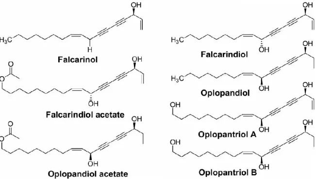
Mainly, most of the phytochemical studies of OH have focused on OH root bark where several polyynes were identified (Calway et al., 2012). Five polyynes were found in OH in 1997, including falcarinol; falcarindiol; oplopandiol acetate; oplopandiol; and 9,17-
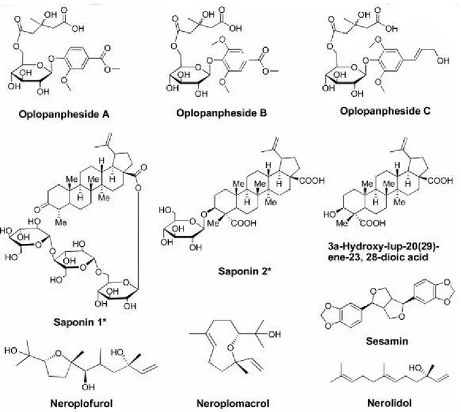
octadecadiene-12,14-diyne-1,11,16-triol 1-acetate (Kobaisy et al., 1997). These five polyynes have been related to the antimycobacterial activity of OH (Lantz et al., 2004; Kobaisy et al., 1997; Liang et al., 2000). Later, two other polyynes were isolated; namely, oplopantriol A and B (Huang et al., 2010b). (Figure 4). Moreover, different glycosides were identified from OH: (1) Two lupane-form saponins (24-nor-3-oxo-lup-20(29)-en-28-oic acid-28-O-α-L-rhamnopyranosyl and 3α-hydroxy-lup-20(29)-ene-23, 28-dioic acid-3-O-β-D-glucopyranoside (1’” →4”)-β-D- glucopyranosyl (1”→6')-β-D-glucopyranoside) (Calway et al., 2012), (2) lupane aglycone (3α- hydroxy-lup-20(29)-ene-23,28-dioic acid) which was found in the leaves (Liu et al., 2010), (3) three phenolic glycosides (oplopanphesides A – C), which were identified in the root bark
(Huang et al., 2011). Other compounds were isolated from the stem bark of OH, including neroplomacrol and neroplofurol, two sesquiterpenes, a lignan compound, and sesamin (Inui et al., 2010), while a polyene compound nerolidol was isolated from the root bark (Huang et al., 2010a). Isolated compounds are illustrated in Figure 5.
By using GC-MS, the main composition of the essential oil from the stem and root of OH was nerolidol with a content of over 50% of the oil (Huang et al., 2010a; Gruber et al., 2004). In another study, the main constituents of volatile oil from the leaves of OH were identified as phytol and 2-methyl-6-p-methylbenzene-2-heptene with concentrations of 34.4% and 8.13%, respectively (Li et al., 2009). By using the HPLC fingerprint method, polyynes were not found to be main components of OH berries (Wang et al., 2010). By using an online SPE-HPLC, six polyynes were identified in all OH root bark samples (Huang et al., 2010a).
Figure 4: Seven polyynes isolated from Oplopanax horridus “Figure reused with permission of the rights holder, Springer Nature, see appendix 1” (Huang et al., 2010b).
Figure 5: Glycosides and other compounds of Devil’s club “Figure reused with permission of the rights holder, Springer Nature, see appendix 1” (Calway et al., 2012).
Pharmacological studies:
OH is known as a treatment for many ailments of Indigenous people, such as type II diabetes, respiratory problems, and others (Calway et al., 2012). Similarly to ginseng use, the OH root bark is considered as the most desirable part for pre-clinical studies of the plant (Jung et al., 2011). Some of OH therapeutic usages are listed below.
Antibacterial:
As the most important use, OH has shown antibacterial activities, especially
antimycobacterial, in several studies (Calway et al., 2012). In counter-current chromatography- based analysis and antiviral screening study, OH had particular antimycobacterial activities against Mycobacterium that cause tuberculosis and leprosy in humans (Kobaisy et al., 1997; Inui et al., 2007; McCutcheon et al., 1995). Hence, OH is widely used to deal with internal
infections, specifically tuberculosis, among Indigenous populations (Calway et al., 2012). In a GC-MS analysis of OH, two polyynes, namely falcarindiol and oplopandiol, are found to be the main effective constituents for OH antimycobacterial activities (Inui et al., 2010). In another study, other different polyynes (falcarinol, (Z)-9,17-octadecadiene-12,14-diyne-1,11,16-triol 1- acetate, and oplopandiol) showed antimycobacterial activities against both Mycobacterium avium and Mycobacterium tuberculosis (Kobaisy et al., 1997). In the same study, the polyynes showed an effectiveness against two Gram-negative bacteria (Escherichia coli DC2 and Pseudomonas
aeruginosa Z61), two Gram-positive bacteria (Staphylococcus aureus and Bacillus subtilis), and
the yeast Candida albicans (Kobaisy et al., 1997). Finally, the most effective constituent among the tested polyynes was falcarinol (Kobaisy et al., 1997).
As the most extensive pharmacological research of OH, anticancer studies have been performed to assess the plant’s anticancer abilities (Calway et al., 2012). In an in vivo study, azoxymethane-induced neoplasia was inhibited by nerolidol, a component of OH extract
(Wattenberg, 1991). In an in vitro study, the growth of ovarian cancer and other cancer cells was inhibited by using the root bark extract of OH (Tai et al., 2010; Tai et al., 2006). To date, several studies have been specified on colorectal cancer (Calway et al., 2012). Also, OH extract has an anticancer effect on different organs, such as breast and lung (Calway et al., 2012).
In an in vitro study, OH stem extract exhibited significant antiproliferative effects on colorectal cancer cells compared with berry extracts (Wang et al., 2010). The stem extract stimulated cell apoptosis, as well as cyclin A expression and stopped tumor cells in S- and G2/ M-phases (Wang et al., 2010). In the same study, OH stem extract exerted more significant antiproliferative actions on a breast cancer cell line than OH berry extract did (Wang et al., 2010). In another study, stem extract demonstrated more anti-lung cancer bioactivity comparing with berry extract (Wang et al., 2010).
As the most commonly used part, OH root bark extracts were tested on colorectal tumor cells, and only the hydrophobic fractions exhibited significant antiproliferative effects, which include early and late apoptosis (Li et al., 2010). To discover the active compounds in OH hydrophobic fractions of root bark, polyynes bioactivities were tested (Sun et al., 2010). Those polyynes showed inhibition effects on colon tumor cells, so it was concluded that OH root bark anti-tumor effects are likely associated with those hydrophobic components (Sun et al., 2010). In an in vitro study, 70% and 100% ethanol fractions of OH root bark had potent apoptotic and antiproliferative effects on breast cancer cells more than the total extract while the 30% ethanol and water fractions significantly increased cell proliferation at concentrations > 100 μg/ml (Sun
et al., 2010). Consequently, this result suggests that hydrophilic fractions should be taken away to reach the desirable activities (Sun et al., 2010). Also, the same study has reported that the 70% and 100% ethanol fractions of OH root barks demonstrated more potent antiproliferative activities on non-small cell lung cancer (NSCLC) cells than the total extract (Sun et al., 2010). Significantly, falcarindiol was also identified as the most potent antiproliferative agent in hydrophobic fractions of OH root bark (Sun et al., 2010). In an in vitro study, the mechanism of action of OH root bark extract might be related to the ability of the plant to induce cancer cell apoptosis and to regulate cell cycle transition (Li et al., 2010).
Antidiabetic:
As a one of OH reported common uses, OH has shown anti-diabetic effects (Calway et al., 2012). Despite the fact that a study performed by Thommasen, et al. has reported that there were not any significant hypoglycemic effects of OH by using OH tea (Thommasen et al., 1990), an in vivo study, was carried out on white Belgian hares, has proven the activity of OH in
lowering blood glucose levels (Large & Brocklesby, 1938). However, the largest evidence in support of the anti-diabetic properties of Devil’s club come from traditional use by Indigenous people (Thommasen et al., 1990). As a result, performing more studies to evaluate the effects of OH on blood glucose and its mechanisms of actions are needed, and examining this is the objective of this research.
Monograph of Devil's club:
There are no official regulations on Devil’s club cited on the Health Canada website, but the following information is a result of a systematic review of scientific research data reviewed by volunteers of the Natural Standard Research Collaboration.
Dose:
*Adults:
There are no official guidelines for the safe and effective dose for Oplopanax horridus. All forms of OH preparation (decoctions, infusions, and tinctures) have been traditionally used. In traditional use, 15-30 drops of tincture (dry 1:5, fresh 1:2, both 60% alcohol) three times per day or 1-3 fluid ounces of cold infusion three times per day have been used. For anti-diabetic effects, 1.4-1.6 ml/ kg of an aqueous extract has been applied. For colds, weight gain, and other disorders, 125 ml before main meals have been used. For analgesia (pain relief), OH raw inner bark is chewed and spit on wounds and fracture to inhibit pain and swelling. To reduce
infection, dried inner bark can be used after rubbed to a pulp. To decrease swellings, ointment of stem ashes mixed with grease has been used (NATURAL MEDICINES, n.d).
*Children:
There is lack in data concerning optimal dose of Devils club for children use (NATURAL MEDICINES, n.d).
Allergies:
Commonly, the spines on the leaves and stems of OH are considered as a cause of topical allergic reaction (NATURAL MEDICINES, n.d).
Side effects and warnings:
Although Oplopanax horridus is not mentioned by the US Food and Drugs
Administration (FDA) as safe (GRAS) product, the American Herbal Products Association mentions it as Class 1, which identifies herbs that can be safely consumed if used appropriately.
Diarrhea was observed in one case where an aqueous extract of inner root bark was consumed. Also, chronic intake of OH infusion might lead to excessive weight gain. As mentioned, the spines of the leaves and stems may cause allergy. OH can lower blood glucose levels, so caution is recommended for patients who have diabetes, hypoglycemia, or taking drugs or other herbs that affect blood sugar (NATURAL MEDICINES, n.d).
Pregnancy and Breastfeeding:
Because of the lack of scientific support, OH is not advised in pregnant or breastfeeding women (NATURAL MEDICINES, n.d).
Drug, herbs, and dietary supplements interactions:
As mentioned before, OH has a power to decrease blood glucose levels. Consequently, it might have a synergistic effect with other medications like OHAs, insulin, or other herbs that lower blood sugar, so blood glucose monitoring is necessary by qualified healthcare
professionals during Devil's club intake (NATURAL MEDICINES, n.d).
Based on the evidence from some experimental studies and the traditional use of Devil’s club by Indigenous people that support the anti-diabetic activities of the plant, this research hypothesized that the water and 80% ethanol root bark extracts and some other fractions would show good anti-diabetic effects on different cell lines, namely the H4IIE rat hepatoma cell line and C2C12 murine skeletal myoblasts cell line.
Objectives of the study:
As seen, several Indigenous groups traditionally use Oplopanax horridus as an anti- diabetic plant, but there are not enough scientific studies or evidence that support its
hypoglycemic activities. Since the CIHR-TAAM team has begun collaborating with the Squamish Nation of British Columbia in 2016, the community advised us of their interest in studying the plant’s antidiabetic potential using cell-based bioassays (Haddad, P.S., personal communication, January 12,2018). As the traditional preparations are usually based on hot water, a hot water extract of OH root bark was prepared, alongside the more classical 80 % ethanol extract commonly used by phytochemists. Also, to begin understanding the chemical components that could underlie the biological activity, several solvent fractions were also prepared from OH root bark, whereas the pure compound chlorogenic acid (CA), known to be present, was also tested. All the raw extracts, fractions, and the compound were tested in two different in vitro bioassays; namely, to assess glucose uptake (GU) potentiation in skeletal muscle cells (C2C12) and Glucose-6-Phosphatase (G6Pase) inhibition in hepatic cells (H4IIE). Through glucose transporter 4 (GLUT4), skeletal muscle is the main tissue involved in
postprandial glucose disposal (representing around 80% of glucose uptake), causing a decrease in blood glucose levels (Ferrannini et al., 1988). Moreover, as a rate-limiting enzyme for the final step of gluconeogenesis and glycogenolysis, G6Pase inhibition decreases hepatic glucose production (HGP) and blood glucose levels (Boustead et al., 2004). Hence, these two bioassays evaluate biological activities that are very pertinent to systemic glucose homeostasis.
Chapter2:
Material and methods:
Plant material and extraction:
Inner bark samples of Oplopanax horridus was collected in a culturally respectful manner by Drs. Pierre Haddad and Alain Cuerrier in partnership with Leigh Joseph and Shirley Lewis from Squamish Frist Nation in British Columbia, Canada. Then, the collected inner barks were air dried and sent to the University of Ottawa, where they were cleaned and ground using a Wiley Mill (Arthur H. Thomas, Swedesboro, USA) with a 2-millimetr filter. The produced plant powder was extracted in two different methods: the first (standard phytochemical) method used 80% ethanol (10 mL/g dry material) and extraction was carried out two times for 24 h using a mechanical shaker (this ethanol extract will hereafter be designated as EE); the second method (mimicking Indigenous traditional preparation) used boiling water for 75min (this hot water extract will hereafter be designated as HWE). In both methods, extracts were filtered with Whatman paper. Extracts were subsequently dried using a rotary evaporator followed by lyophilization. All lyophilized extracts were conserved at 4 °C in a desiccator and kept away from light.
Oplopanax fractionation scheme:
In order to begin understanding the chemical nature of Oplopanax active principles, a serial chemical fractionation approach was used. Hence, the inner bark samples of OH were fractionated by applying a series of organic solvents of increasing polarity; namely hexanes, dichloromethane (DCM), ethyl acetate, methanol and water. The following scheme was used:
50g ground inner bark of Oplopanax horridus in 500mL Hexanes.
Shaken at 200rpm for 1 hour.
Filtrate isolated with suction filtration
Residue collected by re-dissolved and re-extracted in 500mL more of hexanes (200rpm, 1h)
Filtrate isolated with suction filtration.
Hexanes fractions combined, rotovapped to remove solvent. Hexane fraction = 1.2231g, 2.45% yield.
Residue collected by re-dissolved and re-extracted in 500mL of Dichloromethane (DCM) Shaken at 200rpm for 1 hour.
Filtrate isolated with suction filtration
Residue collected by re-dissolved and re-extracted in 500mL more of DCM (200rpm, 1h)
Filtrate isolated with suction filtration.
DCM fractions combined, rotovapped to remove solvent. DCM fraction = 1.4769g, 2.95% yield.
Residue collected by re-dissolved and re-extracted in 500mL of Ethyl acetate. Shaken at 200rpm for 1 hour.
Filtrate isolated with suction filtration
Residue collected by re-dissolved and re-extracted in 500mL more of ethyl acetate (200rpm, 1h)
Filtrate isolated with suction filtration.
Ethyl acetate fractions combined, rotovapped to remove solvent. Ethyl acetate fraction = 0.3679g, 0.74% yield.
Residue collected by re-dissolved and re-extracted in 500mL of Methanol Shaken at 200rpm for 1 hour.
Filtrate isolated with suction filtration
Filtrate isolated with suction filtration.
Methanol fractions combined, rotovapped to remove solvent. Methanol fraction = 2.9067g, 5.81% yield.
Residue collected by re-dissolved and re-extracted in 500mL of miliQ water. Shaken at 200rpm for 1 hour.
Filtrate isolated with suction filtration
Residue collected by re-dissolved and re-extracted in 500mL more of water (200rpm, 1h)
Filtrate isolated with suction filtration.
Water fractions combined, freeze dried to remove liquid. Water fraction = 4.1g, 8.2% yield.
Materials:
The H4IIE rat hepatoma cell lines, cells passage 5, and C2C12 murine skeletal myoblasts, cells passage 4, were acquired from the American Type Culture Collection (ATCC, Manassas, USA). Cell culture media was purchased from Invitrogen Life Technologies (Burlington,
Canada) and Wisent (St. Bruno, Canada). Cytotoxicity Detection Kit was purchased from Roche (South San Francisco, CA). Other reagents were purchased from Sigma-Aldrich (Oakville, Canada), unless otherwise specified.
Cell Culture (C2C12 murine myoblasts):
C2C12 muscle cells were grown in high-glucose Dulbecco’s modified Eagle medium (DMEM) supplemented with 10% Horse Serum (HS), 10% Fetal Bovine Serum (FBS), and 0.5 % penicillin-streptomycin antibiotics in a humidified atmosphere of 5% CO2/95% air at 37°C.
Firstly, the cells were cultured in Petri dishes with proliferation medium replaced every two days until cells reached 80% of confluence. Then, the cells were passaged into 12-well plates with
proliferation medium until they reached 60-70% of confluence. Then, cells were differentiated for a period of 7 days into myotubes in FBS-free DMEM containing 2% HS and 0.5 %
antibiotics prior to experiments. On day 6 of differentiation, cells were treated for 18 h with the treatment (the extracts, the fractions, the pure compound, and controls) to perform glucose uptake assay.
Cell Culture of H4IIE (rat hepatoma):
H4IIE cells were grown in high glucose Dulbecco’s Modified Eagle’s Medium (DMEM) supplemented with 10% FBS and 0.5% antibiotics (PS: Penicillin 100 U/mL, Streptomycin 100 𝜇g/mL). The cells were incubated at 37°C, 5% CO2 until reaching 90% confluence and treated
18 h with Oplopanax extracts, fractions, pure compound, and controls to perform hepatic glucose production assay.
Cell viability test:
Cell viability was evaluated by using a Cytotoxicity Detection Kit. C2C12 murine and H4IIE rat hepatoma cells were seeded in 24-well plates, cultured to 80- 90% confluence in proliferative medium (and C2C12 cells were differentiated for 7 days), and treated for 18 h with
Oplopanax extracts, fractions, pure compound, and controls. Cells were then treated with serial
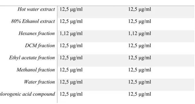
dilutions (100, 50, 25, and 12.5 μg/ml) of OH extracts, fractions, and pure compound. Because of its higher toxicity, the hexane fraction was further diluted until 1.12 μg/ml. Subsequently, cell culture media for each condition (in duplicate or triplicate) were collected separately
(extracellular LDH), and then attached cells were lysed with culture medium containing 1% Triton X-100, for 10 min (intracellular LDH). All samples were collected in Eppendorf tubes, and lysed samples were centrifuged at 250 xg at 4ºC for 10 minutes. Emitted fluorescence was