CCTA being widely used in other clinical applications such as myocardial function or dynamic perfusion.
Recent advance
The rate of CCTA failure has substan-tially diminished as a result of recent ven-dor-specific technological advances in temporal, spatial and contrast resolution, noise reduction and arrhythmia detec-tion. Due to these improvements, and de-spite radiation dose concerns, CCTA may yet become the one-shot-stop examina-tion for both coronary artery disease and advanced applications such as perfusion assessment. Obviously this prospect warrants further clinical investigation. Bibliography
1. Budoff M.J., Dowe D., Jollis J.G., Gitter M., Sutherland J., Halamert E., Scherer M., Bellinger R., Martin A., Benton R., Delago A., Min J.K.: Diagnostic performance of 64-multi-CCTA depends on operator awareness of
these potential pitfalls.
CCTA image quality determinants and artifacts
CCTA is technically complex and sub-ject to constraints that may result in im-age artifacts. Key factors determining CCTA image quality include temporal resolution, spatial resolution, contrast resolution and noise.
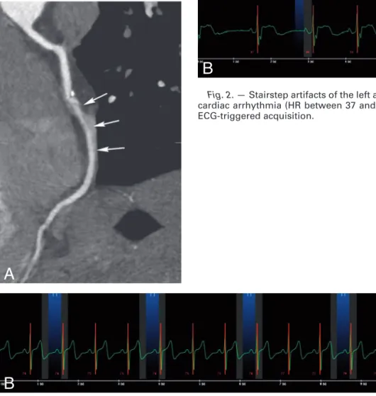
Artifacts that may reduce image quality include blurring (Fig. 1), stairstep (Fig. 2) and blooming artifacts, low attenuating, streak, windmill or rod artifacts, vessel tracking artifacts, the omission of the region of interest, pseudostenosis and non-depiction of one or several coronary artery segments.
Insufficient tissue-contrast, limited spatial and temporal resolution, and radiation dose have until now prevented JBR–BTR, 2013, 96: 401-412.
PROCEEDINGS OF THE CARDIAC – CT COURSE held in Brussels on 16.03.2013
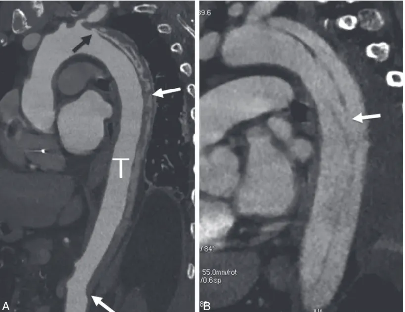
Fig. 1. — Cardiac motion-related blurring artifact of the second segment of the right
coronary artery (arrows in A) due to high heart rate (average heart rate of 75 beats per minute in B) in this prospective ECG-triggered acquisition.
Fig. 2. — Stairstep artifacts of the left anterior descending artery (arrows in A) due to
cardiac arrhythmia (HR between 37 and 51 beats per minute in B) in this prospective ECG-triggered acquisition.
Cardiac CT angiography: technical as-pects in 2012
O. Ghekiere1, A. Nchimi2
With the advent of the multidetector computed tomography (CT), cardiac computed tomography angiography (CCTA) has become a viable non-invasive method to rule out obstructive coronary artery diseases (CAD) due to its excellent negative predictive value. CCTA is mainly indicated in those patients presenting with clinically low-to-intermediate pretest likelihood for CAD.
Despite its high diagnostic accuracy for detection of obstructive coronary ar-tery disease, insufficient temporal, spatial or contrast resolution may sometimes provide incomplete or even misleading images of some coronary segments. These image artifacts are the main cause for misinterpretation regarding the pres-ence of CAD and the successful use of
A
B
A
aortic valve dysfunction. Consequently, when a dilated ascending aorta is ob-served in a young adult, a bicuspid aortic valve has to be considered (Fig. 3). Sec-ondary to wear and tear, a bicuspid valve can present with thickened and partially calcified cusps at a relatively young age. Nevertheless, the amount of calcification by itself cannot be adequately used as a predictive marker for valve dysfunction (Fig. 3). Finally, a bicuspid valve can be associated with other congenital anoma-lies, including an aortic coarcatio.
A morphologically normal aortic root incorporates three sinuses of Valsalva of equal size and a tricuspid valve. In analo-gy with the variation in numbers of cusps, significant discrepancy in the develop-ment of these sinuses and cusps can exist (Fig. 4). As such, an aortic valve with a hypoplastic cusp and associated smaller sinus can consequently mimick a ‘true’ bicuspid valve (Fig. 5). CT has proven to be an excellent tool in the evaluation of valve morphology, often acting compli-mentary to the echocardiography-derived evaluation. While magnetic resonance (MR) imaging and echocardiography are traditionally the methods of choice in the evaluation of aortic valve dysfunction, several studies have demonstrated the ability of ECG-gated CT studies to cor-rectly assess both aortic valve stenosis and regurgitation (Fig. 6). In contrast to the mentioned traditional imaging mo-dalities, this evaluation requires an addi-tional radiation exposure which can be substantial. Therefore, we do not recom-mend this in routine practice, its use bet-ter reserved for application on a case-by-case basis.
detector row coronary computed to-mographic angiography for evalua-tion of coronary artery stenosis in individuals without known coronary artery disease: results from the pro-spective multicenter ACCURACY (As-sessment by Coronary Computed To-mographic Angiography of Individuals Undergoing Invasive Cor-onary Angiography) trial. J Am Coll
Cardiol, 2008, 52: 1724-1732.
2. Vanhoenacker P.K., Heijenbrok-Kal M.H., Van Heste R., Decramer I., Van Hoe L.R., Wijns W., Hunink M.G.: Di-agnostic performance of multidetec-tor CT angiography for assessment of coronary artery disease: meta-analy-sis. Radiology, 2007, 244: 419-428. 3. Kroft L.J., de Roos A., Geleijns J.:
Artifacts in ECG-synchronized MDCT coronary angiography. AJR, 2007, 189: 581-591.
4. Choi H.S., Choi B.W., Choe K.O., Choi D., Yoo K.J., Kim M.I., Kim J.: Pitfalls, artifacts, and remedies in multi- detector row CT coronary angi-ography. Radiographics, 2004, 24: 787-800.
5. Nicol E.D., Arcuri N., Rubens M.B., Padley S.P.: Considerations when in-troducing a new cardiac MDCT ser-vice. Avoiding the pitfalls. Clin Radiol, 2008, 63: 355-369.
6. Nakanishi T., Kayashima Y., Inoue R., Sumii K., Gomyo Y.: Pitfalls in 16-de-tector row CT of the coronary arteries.
Radiographics, 2005, 25: 425-438.
7. Mahesh M., Cody D.D.: Physics of cardiac imaging with multiple-row detector CT. Radiographics. 2007;27: 1495-1509.
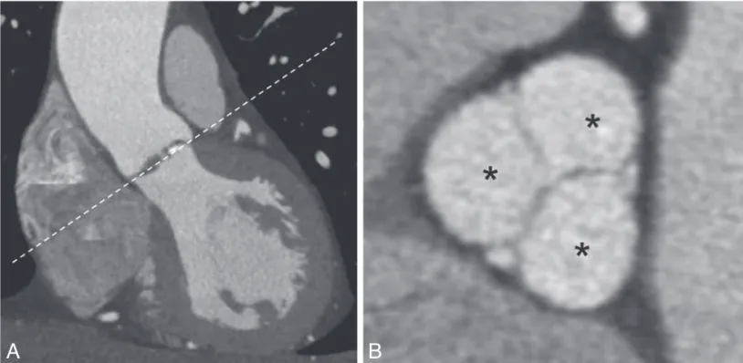
Fig. 1. – The aortic root has a double-oblique orientation, but a rough initial cross-sectional reformation can be obtained by
align-ing the imagalign-ing plane in an oblique fashion along the aortic valve cusps (dashed line) on a coronal reformatted image (A). The aortic root has three sinuses of Valsalva (asterisk in B), and contains a normally tricuspid aortic valve.
1. Department of Radiology, CHC Clinique St-Joseph, Liège Belgium, Department of Radiology, CHU Sart Tilman Liège, Belgium.
Imaging of the aortic root and ascend-ing aorta
R. Salgado, P. M. Parizel1 Introduction
In recent years there has been increas-ing interest in the non-invasive imagincreas-ing of the aortic root and ascending aorta. This is in part due to the exploration of the capabilities of CT-imaging in the as-sessment of the aortic valve, including the evaluation of aortic valve stenosis and insufficiency. Recent advances in transcatheter valve implantation tech-niques targeted at the aortic valve have also spurred interest in the non-invasive evaluation of the complex anatomy of the aortic root.
Finally, the ascending aorta is a some-times underestimated source of acute chest pain.
Aortic valve and root
While the aortic valve has traditionally three cusps (Fig. 1), many variations can exist. Up to 1-2% of the population has a bicuspid valve (Fig. 2). Its presence has potential important clinical consequenc-es, as a bicuspid aortic valve is more sus-ceptible to premature degeneration, and is associated with a varying degree of
PROCEEDINGS OF THE CARDIAC – CT COURSE 403
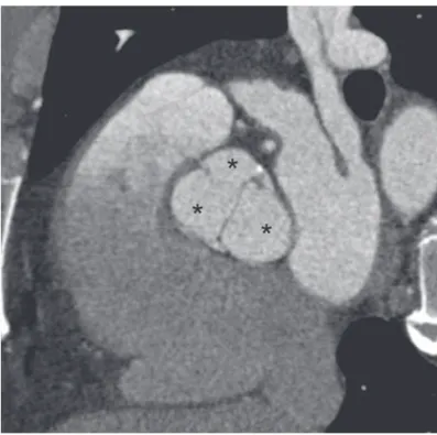
Fig. 2. – Double-oblique reformatted cross-sectional
contrast-enhanced CT image in a 42-year-old man demonstrating the typical appearance of a bicuspid aortic valve. Note that there are only two sinuses of Valsalva (asterisk).
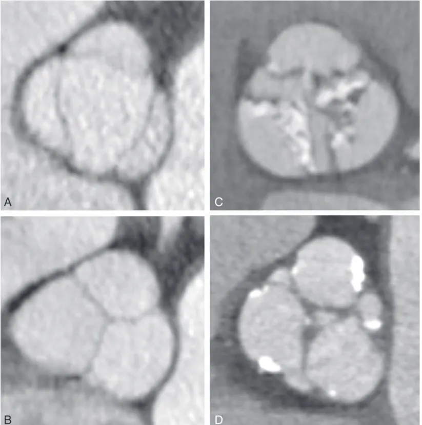
Fig. 4. – A 52-year-old man with an acquired functionally
bi-cuspid valve. The cross-sectional reformatted contrast-enhanced CT image reveals three sinuses of Valsalva (asterisk), but only two cusps. This functionally bicuspid appearance can be the end result of fusion between two cusps, in this case secondary to a decade-old valve infection. As such, originally tricuspid aortic valves can become functionally bicuspid.
Fig. 3. – Coronal contrast-enhanced CT image demonstrating
a premature degenerated bicuspid valve in a 36-year-old woman with extensive valve calcifications and thickened cusps. Note the dilated ascending aorta, a common associated finding second-ary to the hemodynamic effects of aortic valve dysfunction.
Fig. 5. – A 65-year-old man with a hypoplastic left coronary
cusp on a cross-sectional contrast-enhanced CT image through the aortic valve. The hypoplastic left coronary cusp and left sinus of Valsalva make this valve appear almost bicuspid. Many varia-tions exist in the morphology of the aortic valve. While CT can deliver excellent image quality, the associated radiation expo-sure and the lack of true functional information makes it not suit-able for routine valve evaluation. Consequently, the application of CT for the evaluation of valve morphology must be restricted on a case-by-case basis, and always in conjunction with the find-ings of echocardiography and magnetic resonance imaging.
contrast to the circular contour which was always assumed using two-dimen-sional techniques like echocardiography. Current research concentrates, among others, in establishing the optimal inte-gration of different imaging tools for achieving optimal transcatheter valve size selection for a given patient, and to subsequently prevent or minimize para-valvular regurgitation.
raphy, most notable transesophageal ul-trasound, has been successfully used in different trials for the evaluation of annu-lar dimensions, it is now widely recog-nized that the excellent three-dimension-al capabilities of CT can make a significant contribution in optimizing annular sizing. Several CT-based studies of the annular dimensions have indicated an oval annu-lar morphology in a majority of cases, in Pre-operative evaluation of the aortic
annulus
With the development of transcatheter valve implantation techniques for treat-ment of critical aortic valve stenosis, con-siderable attention is currently given to the optimal pre-procedural evaluation of the aortic annulus dimensions using non-invasive techniques. While
echocardiog-Fig. 6. – Evaluation of aortic valve stenosis and insufficiency using CT. Panel A illustrates the normal opening of the valve during
systole. Panel B conversely demonstrates the only slight systolic valve opening in an 82-year-old man with critical aortic valve ste-nosis. Panel C shows the normal closing of the aortic valve in the end-diastolic phase of the cardiac cycle. Panel D reveals in the same cardiac phase a residual opening corresponding with aortic valve insufficiency. Functional aortic valve evaluation is traditionally reserved for echocardiography and magnetic resonance imaging. CT is, despite its excellent planimetric capabilities, not routinely indicated herefore.
B
D
PROCEEDINGS OF THE CARDIAC – CT COURSE 405
lapping features as revealed by ECG-gated multidetector-row CT angio-graphy. Insights Imaging, 2012, 3: 561-571.
2. Goffinet C., Kersten V., Pouleur A.C., et al.: Comprehensive assessment of the severity and mechanism of aortic regurgitation using multidetector CT and MR. Eur Radiol, 2010, 20: 326-336 3. Feuchtner G.M., Müller S., Bonatti J., et al.: Sixty-four slice CT evaluation of aortic stenosis using planimetry of the aortic valve area. Am J Roent
genol, 2007, 189: 197-220.
4. Lehmkuhl L., Foldyna B., Von Aspern K., et al.: Inter-individual variance and cardiac cycle dependency of aortic root dimensions and shape as as-sessed by ECG-gated multi-slice com-puted tomography in patients with severe aortic stenosis prior to trans-catheter aortic valve implantation: is there is only an entry site in the aortic
wall, and no detectable re-entry site. Con-sequently, this stationary blood forms a thrombus in the aortic wall, with a ‘false’ lumen that is typically semicircumferen-tial and smaller than the main true lumen.
It is furthermore important to recog-nize that this situation can evolve to a classic aortic dissection. This can occur when one or several re-entry sites devel-op along the course of the diseased aortic wall (Fig. 7). On such occasion, there is transition to two channels of flowing blood, with often the false lumen having a larger cross-sectional diameter than the true lumen.
Bibliography
1. Ueda T., Chin A., Petrovitch I., et al.: A pictorial review of acute aortic syndrome: discriminating and over-Acute aortic syndrome
The term ‘acute aortic syndrome’ is used to indicate the trilogy of acute aortic dissection, intramural hematoma and penetrating atherosclerotic ulceration as aortic-based causes of acute chest pain. However, contemporary criticism points out that the distinction between these three causes is conceptually flawed, as especially acute aortic dissection and in-tramural hematoma most probably rep-resent two separate prep-resentations of the same clinical entity. Most important, it is now preferred to use ‘intramural hema-toma’ as a descriptive term, indicating stationary blood within the aortic wall. This stationary blood is formed as blood from the main (true) lumen enters the aortic wall through a small and often on CT or MR imaging unnoticeable defect in the intima layer. As is often the case,
Fig. 7. – Evolution from an intramural hematoma to a classic dissection in a 32-year-old man with acute chest pain. On admission,
the sagittal contrast-enhanced CT image (A) reveals a clear intramural hematoma (white arrows), with a small entry site (black arrow) just distally from the left subclavian artery, and no clear re-entry site. This image shows the clear distinction between one channel of flowing blood (large true lumen T), and one channel of stationary blood (intramural hematoma). However, after 6 days a new CT examination now shows several entry- and re-entry sites with two channels of flowing blood and a consequently larger false lumen. This changed appearance now represents a classic form of (in this case type-B) aortic dissection, and illustrates the varying morphol-ogy of the acute aortic syndrome.
ablation can be used to thermally destroy atrial foci suspect for initiation of these arrhythmias. These procedures are often aided by cardiac CT by identifying the ex-act pulmonary vein anatomy, of which there are many variants (large or small venoatrial junctions, common ostia or accessory pulmonary veins).
In end-stage heart failure, cardiac re-synchronization therapy (CRT) has been shown to increase survival and provide systematic relief of symptoms. This pro-cedure requires positioning of a pacing lead near the inferolateral wall of the LV. The main factor determining the success of a transvenous LV lead implantation is cardiac anatomy, particularly of the coro-nary venous system, which is known to be highly variable. With only a small ad-aptation of the regular cardiac scanning algorithm, many studies have shown reli-able visualization of the cardiac venous anatomy and accurate quantification on the dimensions of the ostium of the coro-nary sinus and the diameter of the target veins.
Imaging of myocardial infarction and ischemia
Similar to what is more commonly performed in an MRI setting, cardiac CT can be are acquired during the first pass of a bolus of contrast agent, theoretically allowing hypoperfused areas within the myocardium regions to appear with low attenuation.
Although currently perfusion CT is still largely investigational and still under de-velopment in preclinical animal models, a good accuracy to detect myocardial in-farction has been observed. In similar studies, delayed phase scanning showed delayed hyperenhancement in the infarct area.
schemes only acquire information about the second part of the cardiac cycle, thus disallowing any functional evaluation. A motivated choice has to be made by the radiologist, based on the clinical history and depending on what information is to be gained, whether to choose for a low-dose scan or a full functional 4D evalua-tion.
Imaging of cardiac valves
Homologous to the situation described for cardiac function, the high spatial reso-lution allows for detailed analysis of the cardiac valves. Cardiac CT provides im-portant information on the presence of (peri)valvular and annular calcifications, variations in the number of valve leaflets, and recently more specifically on the aor-tic valve anatomy, geometry, and valve area in patients considered for transarte-rial aortic valve implantation (TAVI) (Fig. 1).
In selected cases, imaging of artificial valves and assist devices can be consid-ered, since the new fast acquisition algo-rithms and artifact-reducing reconstruc-tion kernels allow accurate anatomical and functional visualization in case of valve dysfunction.
The exact value of valvular assess-ment by cardiac CT is largely still under investigation, re-emphasized by its grad-ing in the recent Appropriateness Criteria for Cardiac Computed Tomography and Cardiac Magnetic Resonance Imaging, from the American College of Cardiology, as an ‘uncertain indication’ (Fig. 2). Imaging of the atria and the cardiac venous system
In patients with drug-resistant atrial fibrillation, transcatheter radiofrequent it crucial for correct sizing? Int J
Cardiovasc Imaging, 2013, 3: 693-703.
5. Achenbach S., Delgado V., Hausleiter J., et al.: SCCT expert consensus document on computed tomography imaging before transcatheter aortic valve implantation (TAVI)/transcathe-ter aortic valve replacement (TAVR).
J Cardiovasc Comput Tomogr, 2012,
6: 366-380.
1. Department of Radiology, Antwerp University Hospital, Edegem, Belgium.
Non-coronary applications of cardiac CT (with MR correlation)
S. Dymarkowski1
The high diagnostic accuracy and high negative predictive value of cardiac mul-tidetector row computed tomography (MDCT) to evaluate coronary artery ste-noses is very well known, and as such, this technique has been put forth as the most promising non-invasive exam. Additionally, the full volumetric dataset allows for analysis of a wide range of other cardiac pathologies.
Left ventricular volumes and systolic function can be accurately assessed, if both systolic and diastolic frames are acquired. The same holds true for evaluation of (artificial) cardiac valves. Furthermore, accurate high-resolution visualization of the atria, pulmonary veins and the cardiac venous system is often sought by the cardiologist performing electrophysiological procedures.
The newest technological advances allow these exams to be performed at relatively low radiation doses. This brings novel applications such as stress- perfusion imaging and cardiac dual energy imaging into the picture and opens the door towards CT imaging of myocardial infarction and ischemia. Cardiac function assessment
Assessment of global systolic function can be achieved by reconstructing a 4D retrospective gated spiral acquisition in different phases throughout the cardiac cycle (typically a phase every 10% of the RR interval) to identify end-diastolic and end-systolic phases. By delineating the endocardial surface and by using the Simpson method, the LV volumes are ob-tained. The accuracy of MDCT-derived functional parameters has been exten-sively investigated and compared with other imaging modalities such as gated single-photon emission computed to-mography (SPECT) and magnetic reso-nance imaging (MRI) and has been found to be both a very reproducible and accu-rate means to quantify LV volumes and LV ejection fraction.
In recent years however, the habit of obtaining retrospectively gated scans has strongly declined, since newer acquisi-tion algorithms such as the ‘step and shoot’ prospective gating method and the emergence of single heartbeat acqui-sitions have allowed for a dramatic de-crease in radiation dose, but these
Fig. 1. — Circulite left ventricular assist device. 3D VRT
PROCEEDINGS OF THE CARDIAC – CT COURSE 407
pericardial disease on diastolic function, or the functional significance of pericar-dial thickening are rarely assessable by CT, where this is rather comfortably done in echocardiography and cardiac MRI. Conclusions
Cardiac CT can be considered as a valuable non-invasive imaging modality for the study of different non-coronary cardiac diseases. The ability to perform high quality functional analyses of ven-tricular and valvular function has been extensively proven, but must be looked at skeptically, since this comes at a signifi-cant cost in terms or radiation dose.
Cardiac CT is also being increasingly solicited as the preferred modality for planning of different minimally invasive procedures, such as transcatheter ablation of arythmogenic foci within the atria or TAVI and CRT implantations.
The use of CT for investigation of myocardial scar formation and ischemia visualization is still in its investigational phase.
Bibliography
1. Nasis A., Mottram P.M., Cameron J.D., Seneviratne S.K.: Current and Evolving Clinical Applications of Multidetector Cardiac CT in Assess-ment of Structural Heart Disease. Radiology, 2013, 267: 11-25.
2. Hendel R.C., Patel M.R., Kramer C.M., et al.: ACCF/ACR/SCCT/SCMR/ASNC/ NASCI/SCAI/SIR 2006 appropriate-ness criteria for cardiac computed to-mography and cardiac magnetic res-onance imaging: a report of the American College of Cardiology Foundation Quality Strategic Direc-tions Committee Appropriateness Criteria Working Group, American College of Radiology, Society of Car-diovascular Computed Tomography, of atrial fibrillation and adjacent to areas
of infarction tissue in patients with isch-emic heart disease. These thrombi ap-pear as non-attenuating mural masses, usually in high contrast to the iodine within the cardiac chambers. Other mass-es, such as primary and secondary tu-mors, or mimics of normal anatomy, may be readily detected by CT, but may prove to be difficult to characterize (Fig. 3). In these cases MRI is the preferred imaging modality since its multiparametric ap-proach allows for more accurate tissue characterization.
Pericardial diseases, expressed as fo-cal or generalized thickening, contrast en-hancement in inflammatory conditions and calcifications in more chronic set-tings are easy to investigate with cardiac CT, especially in scenarios where echo-cardiography is known to underperform, e.g. poor acoustic window due to consti-tution or in the immediate postoperative setting. Nevertheless, the influence of A significant coronary stenosis will
usually not show any signs of decreased perfusion in a resting state. Therefore, also the feasibility of performing adenos-ine or dipyridamole stress myocardial perfusion imaging has been explored and has shown potential to provide informa-tion on stress-induced ischemia in select-ed patients. Issues of patient safety, unfa-miliarity with monitoring requirements and difficulties in acquiring reliable data in patients with drug induced tachycardia during CT have so far limited its wide spread use in clinical practice.
Imaging of myocardial masses and peri-cardial diseases
A cardiac mass can often be detected as an incidental finds on cardiac CT per-formed for other indications. The majori-ty of these lesions are cardiac thrombi, usually located in the left atrium or left atrial appendage in patients with history
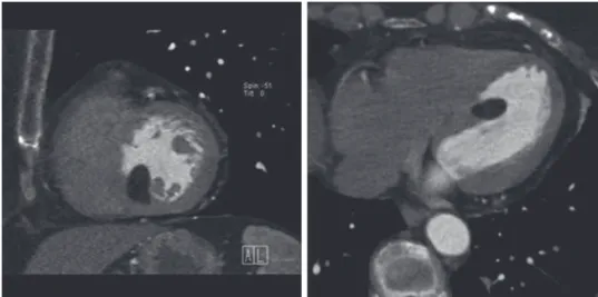
Fig. 3. — Short-axis (left) and transverse (right) CT image of a cardiac lipoma, arising
from the inferobasal interventricular septum.
Fig. 2. — Systolic (A) and diastolic (B) image of an artificial mitral valve that shows absence of proper opening of the posterior
leaflet.
tain” in presence of low pre-test probabil-ity and “appropriate” in patients with in-termediate and high pre-test likelihood (evidence level 7 out of 9 for both) exclud-ing subjects with ST-segment elevation and/or positive cardiac enzymes in which examination was considered inappropri-ate (6).
A similar consensus was recently re-ported by the Italian sub society of cardi-ac radiology in which use of CCTA in pa-tients with non-assessable ECG and /or equivocal laboratory biomarkers was scored as class II clinical indication (i.e. diagnostic information of clinical rele-vance provided) in presence of ACP whereas the triple rule out protocol appli-cation was regarded as potentially useful but still investigational (7).
The “Triple Rule Out” Protocol
Ideal CCTA protocol should represent the best compromise between the need for a comprehensive and reliable evalua-tion of all thoracic cardiovascular struc-tures at the lowest radiation dose possible. Recent technological advances enable an accurate and detailed imaging of coro-nary arteries, thoracic aorta and pulmo-nary arteries in a single contrast-en-hanced ECG-gated CT scan.
Although the examination is poten-tially feasible even on 16-slice CT scan-ner, it is recommended to use last gener-ation machines (64-slice or greater) implemented by radiation dose reduction tools and dedicated cardiac post-process-ing software.
Use of a preliminary unenhanced pro-spectively ECG-gated acquisition cover-ing the entire thoracic aorta might be helpful to provide information regarding distribution and amount of coronary ar-tery calcium and to eventually detect hy-perdense vascular rims reflecting intra-mural aortic hematoma at the cost, however, of an increased patient’s expo-sure.
The choice to apply retrospective or prospective ECG-synchronization tech-niques is conditioned by the available technology and patient’s heart rate, keep-ing in mind the general concept that the combination of a stable and sinus heart rhythm and a heart rate of less than 65-70 beats per minute (depending on the scan-ner gescan-neration) is mandatory to obtain adequate image quality.
Radiation doses of retrospectively gat-ed 64-slice CT range between 7 to 14 mSv when dose modulation strategies are ap-plied whereas in prospectively ECG-gat-ed triggering, a significant effective dose reduction is realistically feasible with a dose of 5 to 7 mSv, less than that from nuclear myocardial perfusion imaging. Recently, a high pitch dual-spiral cardio-thoracic comprehensive protocol using a last generation dual source CT scanner has been proposed in patients with undif-ferentiated acute chest pain, allowing to minimize patient’s individual exposure to an average 3.8 mSv.
Heart-rate control can be achieved in most of the cases intravenously adminis-tering a short-acting beta-blocker (like 4-8% of patients with myocardial
infarc-tion or acute coronary syndrome who are inappropriately discharged from the ED due to the presence of normal initial car-diac enzyme levels associated with non-diagnostic/non-specific electrocardio-graphic (ECG) and echocardioelectrocardio-graphic alterations (2).
Furthermore, the growing pressure of legal disputes for malpractice and the reasonable clinical caution in excluding potentially lethal syndrome is a common cause of unnecessary diagnostic proce-dures and precautionary prolonged hos-pitalization with obvious economical im-plications.
Although an acute coronary syndrome is ultimately diagnosed in only 10 to 15% of patients who present with ACP, it has been estimated that the mean length of hospitalization is 4.6 days, with an aver-age cost of approximately $23,000 per patient in the US an annual cost over $3 billion (3).
Primary objectives of social health systems in Europe and US are to speed-up diagnostic algorhythm, improve diag-nostic accuracy and, obviously, reduce management costs.
In this clinical setting, contrast-en-hanced coronary computed tomographic angiography (CCTA) has emerged as a promising non-invasive diagnostic tool, with an high sensitivity and specificity and a ~100% negative predictive for the exclusion of coronary artery disease, po-tentially allowing to improve clinical care of those patients.
A role of CCTA in supporting the triage of patients with suspected acute coronary syndrome has been hypothesized since early 2000’ (4) and its impact on costs and health care has been subject of an exten-sive recent literature, nevertheless unani-mous consensus was not achieved.
In particular, great emphasis was given to the so-called “triple rule out” protocol that offers in one-stop-shop exam a complete evaluation of thoracic cardiovascular structure, revealing three severe acute potentially lethal disorders (ischemic heart disease, acute aortic disease or pulmonary embolism).
Present article will discuss different acquisition protocols, clinical results and economical implications of CCTA in the clinical setting of ACP.
Clinical indications
According to current American Heart Association (AHA) guidelines, appropri-ateness of CCTA in patients presenting with symptoms of ACP has been indicat-ed as “uncertain” in both patients pre-senting with persistent ECG ST-segment elevation following exclusion of myocar-dial infarction or with ACP of uncertain cause (score 6 out of 9 for both indica-tions) and inappropriate in presence of a definite diagnosis of myocardial infarc-tion (5).
The Asian Society of Cardiovascular Imaging (ASCI) guidelines, categorized clinical appropriateness to the exam ac-cording to pre-test probability of disease. Use of CCTA was indicated as “uncer-Society for Cardiovascular Magnetic
Resonance, American Society of Nuclear Cardiology, North American Society for Cardiac Imaging, Society for Cardiovascular Angiography and Interventions, and Society of Inter-ventional Radiology. J Am Coll Car
diol 2006;48:1475–1497.
3. Hurlock G.S., Higashino H., Mochizuki T.: History of cardiac computed to-mography: single to 320-detector row multislice computed tomography. Int
J Cardiovasc Imaging. 2009;25 Suppl
1:31-42.
4. Tops L.F., Krishnàn S.C., Schuijf J.D., Schalij M.J., Bax J.J.: Noncoronary Applications of Cardiac Multidetector Row Computed Tomography. JACC
Cardiovasc Imaging, 2008, 1: 94-106
5. Schuleri K.H., George R.T., Lardo A.C.: Applications of cardiac multide-tector CT beyond coronary angiogra-phy. Nat Rev Cardiol, 2009, 6: 699-710.
6. Rodríguez-Granillo G.A., Ingino C.A., Lylyk P.: Myocardial perfusion imag-ing and infarct characterization usimag-ing multidetector cardiac computed to-mography. World J Cardiol, 2010, 2: 198-204.
7. Leipsic J., Gurvitch R., Labounty T.M., Min J.K., Wood D., Johnson M., Ajlan A.M., Wijesinghe N., Webb J.G.: Mul-tidetector computed tomography in transcatheter aortic valve implanta-tion. JACC Cardiovasc Imaging, 2011, 4: 416-429.
8. Jongbloed M.R., Lamb H.J., Bax J.J., et al.: Noninvasive visualization of the cardiac venous system using multi-slice computed tomography. J Am
Coll Cardiol, 2005, 45: 749-753.
9. Wang Z.J., Reddy G.P., Gotway M.B., Yeh B.M., Hetts S.W., Higgins C.B.: CT and MR imaging of pericardial dis-ease. Radiographics, 2003, 23: S167-180.
1. Department of Radiology, UZ Leuven, Leuven, Belgium.
Acute chest pain and CT: current in-sights & cost-effectiveness
M. Francone, N. Galea, R. Rosati, I. Carbone1
Acute chest pain (ACP) is a common reason of admission at the emergency departments (ED), not infrequently repre-senting a source of challenging dilemmas for clinicians and anxiety for patients with relevant legal implications related to in-appropriate therapeutic management (1). The list of differential diagnosis of ACP is extensive and ranges from benign musculoskeletal disorders to life-threat-ening conditions requiring immediate treatment such as acute coronary syn-drome, aortic dissection, rupture of the esophagus, perforating peptic ulcer, pulmonary embolism and tension pneumo thorax.
Current diagnostic algorithms may re-liably rule out serious disorders in most of the cases however there is still persis-tence of an unacceptable rate of about
PROCEEDINGS OF THE CARDIAC – CT COURSE 409
Main findings emerged from literature were the following:
1) Use of CCTA allows a higher rate of pa-tient’s discharge from ER with a short-er length of stay as compared to the standard of care (SOC) (14). Litt and colleagues (3) reported a remarkable 27% difference in the rate of early dis-charge of patients who underwent CCTA as compared to SOC with a sig-nificantly reduced median length of ER stay of 18.0 hours vs 24.8 hours. Similar results were found in a large metanaly-sis of four large trials reported reporting a significant reduction patient’s stay, including both ED or total hospital stay (15).
2) The majority of authors found a cost saving ranging between 15% and 38% when use of CCTA was compared to perfusion imaging as reference
stan-dard (12).
Moreover, detection of regional myo-cardial perfusion defect has an additional diagnostic value to CCTA, reducing rates of false positive an improving the posi-tive predicting value from 67% to 90.1%, if compared to rest sestamibi single-pho-ton emission CT myocardial perfusion imaging (SPECT-MPI) (13).
Cost-effectiveness
Analysis of CCTA cost-effectiveness in the clinical setting of ACP has been a sub-ject of interest in a large number of re-cently published papers, either based on simulated models of clinical and econom-ic outcomes and on large patient’s co-horts or metanalysis.
metoprolol 5-20 mg or esmolol 10 mg/ mL) few minutes prior to the examination excluding patients with general contrain-dications to the drug (asthma, sinus bra-dycardia and hypotension, atrioventricu-lar block etc). Preliminary administration of sublingual nitroglycerin could also be useful to dilate coronary arteries.
Test bolus or bolus tracking protocol are strongly advisable to determine the optimal timing of contrast injection and when bolus tracking is selected we sug-gest to use a threshold of 120-140 HU and a region of interest placed within the as-cending aorta. Contrast medium adminis-tration protocol should be adapted to the covered surface of body, however a bolus of 80–110 mL of contrast medium at a rate of 4-5.0 mL/s followed by a 40 mL saline flush is usually adequate to obtain an optimal vascular enhancement within the entire acquisition window.
Further technical aspects about the CT scan protocol are provided in a large expert consensus recently published (8). Images interpretation
Radiological report of patients admitted with ACP should be promptly prepared and communicated to the referring clini-cal team of the ED in order to speed-up diagnostic workup; utilization of a pre-filled initial and final patient’s report form has even been suggested on this re-gard (4).
A complete evaluation of both cardiac and non-cardiac structures needs to be provided, to rule out pulmonary embolism and aortic dissection and to exclude non-vascular causes of chest pain potentially affecting patient’s management (eg. pneumotorax, pneumonia, pleural or peri cardial effusion).
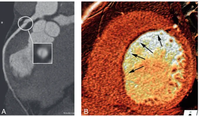
Coronary arteries evaluation should include detailed description of location, extent and morphological features of plaques, with particular attention to soft density lesions and to the so-called “napkin-ring sign” with is characterized by the presence of a ringlike attenuation pattern resembling the thin-cap fibroath-eroma recognized as a precursor lesion for plaque rupture (9) (Fig. 1).
A strong prognostic value of CCTA was recently demonstrated, independent of traditional risk factors and calcium scoring, in a patient’s cohort of 457 sub-ject presenting with ACP in which the ab-sence of plaques was associated with no clinical event in a follow-up period of 11 months (10).
Comprehensive cardiac evaluation should combine morphological informa-tion obtained from coronary arteries analysis with functional (ejection fraction, regional wall motion) and perfusional data, in order to correlate non-invasive angiographic findings with status of cor-responding vascular territory (Fig. 2) (11). Combined evaluation of regional wall motion abnormalities and myocardial perfusion defects has shown to improve detection of acute myocardial infarction with a sensitivity of 94% and specificity of 97%, using cardiac biomarkers and single photon emission CT (SPECT) myocardial
Fig. 1. — CCTA in a 63-year-old patient admitted at the ER due to recent atypical
an-gina, presenting with negative ECG and enzymes. Curved planar reconstruction show the presence of an eccentric soft tissue lesion located in mid left anterior descending artery, causing moderate luminal stenosis (A). An high grade lesion is also located at the origin of left posterior descending artery (C) and was successfully treated with cor-onary stenting. Corresponding selective corcor-onary angiography images are displayed in figures B and D.
A
C
B
Conclusions
The very high negative predictive value of CCTA for CAD minimizes risk of undetected acute coronary syndromes providing relevant impact on patient’s prognosis and clinical management (3, 16).
CCTA in ACP is a fast, feasible, and ro-bust imaging technique which is becom-ing widely available in the ER with an ac-ceptable dose exposure when using last generation scanners combined with ap-propriate dose reduction strategy. It al-lows a rapid patient’s discharge with sig-nificant health-care costs reduction and improved individual therapeutic manage-ment.
On the other hand, the presence of an high-grade stenosis does not necessarily reflect an acute coronary syndrome and non-invasive angiographic findings should be necessarily combined with comple-mentary functional/perfusional tests to rule-out underlying myocardial ischemia due to the assumption that coronary physiology is the critical decision point for consideration of revascularization.
Additional limitations to widespread utilization of CCTA in this setting rely on the current limited availability of dedicat-ed cardiac imaging specialist and on the still high number of false positive cases related to the presence of extensive vas-cular calcifications with intraluminal beam hardening artifacts.
The future is anyway probably going in the direction of CCTA as acknowledged by clinical guidelines and appropriate-ness criteria of the main national and in-ternational medical societies and wit-nessed by the extensive literature vs 1,397 undergoing SOC, with
subse-quent cost increase of 3.9% in case of hospitalization of CCTA patients (17). In positive CCTA patients, therefore, the increase of downstream testing and procedures may cause a cumula-tive mean cost of care similar to the SOC group.
SOC, mostly in patients with low- to intermediate-risk of disease (14-16) (Fig. 3).
3) CCTA allows higher detection of coro-nary arteries disease with subsequent increase of percutaneous and stenting procedures as emerged from the pooled data of a large study popula-tion 1,869 patients undergoing CCTA
Fig. 2. — Left anterior descending artery post-traumatic focal dissection in a 26-year-old patient following a
severe motorbike accident (A). Corresponding thin-slice volume rendering reconstruction shows the presence of a large perfusion defect located in the mid anterior-septal left ventricular wall perfectly matching distribution territory of the dissected vessel (B, arrows).
Fig. 3. — Bar chart showing analysis of CCTA cost
effective-ness compared to standard of care. Data are plotted from refer-ence #15 and derived from the metanalysis of 4 large clinical trials focused on patients with ACP. ROMICAT II study results are differentiated in discharged (ER only) vs hospitalized (hosp) patients.
PROCEEDINGS OF THE CARDIAC – CT COURSE 411
Heart valve disease (stenosis or insuf-ficiency) is common and affects mainly the aortic and mitral valve. There is no known method (medication, lifestyle or otherwise) that halts or reverses the dis-ease process. If the is valve severely ste-notic or insufficient, heart valve replace-ment (or repair in case of mitral valves) with a PHV is inevitable (1).
Two types of prosthetic heart valves are used: mechanical and biological PHV. Mechanical PHV are composed of metal alloys and/or carbon whereas biological PHV are composed of pericardium or ani-mal tissue. Biological PHV do often have a supporting frame work that contains metal elements. Mechanical PHV are de-signed to last lifelong but also require lifelong anticoagulation therapy. Biologi-cal PHV have the advantage of being much less thrombogenic and thus do not require anticoagulation. However, they are prone to wear and have a limited (10-20 years) lifespan.
Although often being a lifesaving intervention, having a PHV presents the patient with a chronic disease with associated morbidity (bleeding due to anticoagulation, endocarditis and pros-thesis dysfunction) and reduced life expectancy (1). Other modes of PHV dysfunction include obstruction by pannus formation underneath the PHV and thrombosis.
Echocardiography and fluoroscopy (for mechanical PHV) are the techniques used for PHV assessment (1, 2). Unfortu-nately they are often unable to determine the cause of PHV dysfunction (2). There is a role for cardiac CT to assess PHV and determine the cause of dysfunction as a complementary technique to echocardio-graphy and fluoroscopy (3, 4).
PHV imaging by CT
Image quality
In the last 5 years the abilities of CT for PHV assessment have become subject of intense research. Our own research line on PHV imaging combines in-vitro and patient studies. The in-vitro studies use a dedicated perfusion set-up to optimize scan protocols aimed to reduce radiation exposure and at the same time reduce PHV induced artefacts.
Using this set-up we have demonstrat-ed that most PHV types generate only a limited amount of artefacts and have a good image quality on CT (Figure 1) (5). Artefacts can be reduced using prospec-tive acquisitions, higher KV settings and iterative reconstruction (6-8). Only Bjork Shiley and Sorin mono leaflet PHV that contain cobalt chromium generate exten-sive artefacts that prohibits assessment with CT (3,5).
Obstructive dysfunction
Obstructive dysfunction of a PHV man-ifests itself as an increased pressure gradient over the PHV on echocardiogra-phy (1, 2). The most important etiologies that need to be differentiated between are pannus, thrombus and patient pros-thesis mismatch. The latter is basically strongly supporting its utilization in large
clinical trials.
References
1. Kolansky D.M.: Acute coronary syn-dromes: morbidity, mortality, and pharmacoeconomic burden. Am J
Managed Care, 2009, 15: S36-41.
2. Hoffmann U., Pena A.J., Cury R.C., et al.: Cardiac CT in emergency depart-ment patients with acute chest pain.
Radiographics : a review publication
of the Radiological Society of North America, Inc 2006, 26: 963-78; discus-sion 979-980.
3. Litt H.I., Gatsonis C., Snyder B., et al.: CT angiography for safe discharge of patients with possible acute coronary syndromes. New Engl J Med, 2012, 366: 1393-1403.
4. White C., Read K., Kuo D.: Assess-ment of chest pain in the emergency room: what is the role of multidetec-tor CT? Eur J Radiol, 2006, 57: 368-372.
5. Taylor A.J., Cerqueira M., Hodgson J.M., et al.: ACCF/SCCT/ACR/AHA/ ASE/ASNC/NASCI/SCAI/SCMR 2010 appropriate use criteria for cardiac computed tomography. A report of the American College of Cardiology Foundation Appropriate Use Criteria Task Force, the Society of Cardiovas-cular Computed Tomography, the American College of Radiology, the American Heart Association, the American Society of Echocardiogra-phy, the American Society of Nuclear Cardiology, the North American Soci-ety for Cardiovascular Imaging, the Society for Cardiovascular Angiogra-phy and Interventions, and the Soci-ety for Cardiovascular Magnetic Res-onance. J Am Coll Cardiol, 2010, 56: 1864-1894.
6. Asci C.C.T., Group C.M.R.G.W., Tsai I.C., et al.: ASCI 2010 appropriateness criteria for cardiac computed tomog-raphy: a report of the Asian Society of Cardiovascular Imaging Cardiac Computed Tomography and Cardiac Magnetic Resonance Imaging Guide-line Working Group. Int J Cardiovasc
Imag, 2010, 26 Suppl 1: 1-15.
7. di Cesare E., Carbone I., Carriero A., et al.: Clinical indications for cardiac computed tomography. From the Working Group of the Cardiac Radiol-ogy Section of the Italian Society of Medical Radiology (SIRM). Radiolo
gia Medica, 2012, 117: 901-938.
8. American College of Cardiology Foundation Task Force on Expert Consensus D, Mark D.B., Berman D.S., et al.: ACCF/ACR/AHA/NASCI/ SAIP/SCAI/SCCT 2010 expert consen-sus document on coronary computed tomographic angiography: a report of the American College of Cardiology Foundation Task Force on Expert Consensus Documents. J AmColl
Cardiol, 2010, 55: 2663-2699.
9. Kashiwagi M., Tanaka A., Shimada K., et al.: Distribution, frequency and clinical implications of napkin-ring
sign assessed by multidetector com-puted tomography. J Cardiol, 2013. 10. Nance J.W. Jr., Schlett C.L., Schoepf
U.J., et al.: Incremental prognostic value of different components of coro-nary atherosclerotic plaque at cardiac CT angiography beyond coronary cal-cification in patients with acute chest pain. Radiology, 2012, 264: 679-90. 11. Francone M., Carbone I., Danti M., et
al.: ECG-gated multi-detector row spiral CT in the assessment of myo-cardial infarction: correlation with non-invasive angiographic findings.
Eur Radiol, 2006, 16: 15-24.
12. Cury R.C., Feuchtner G.M., Batlle J.C., et al.: Triage of patients presenting with chest pain to the emergency de-partment: implementation of coro-nary CT angiography in a large urban health care system. AJR, 2013, 200: 57-65.
13. Feuchtner G.M., Plank F., Pena C., et al.: Evaluation of myocardial CT per-fusion in patients presenting with acute chest pain to the emergency de-partment: comparison with SPECT-myocardial perfusion imaging. Heart, 2012, 98: 1510-1517.
14. Hoffmann U., Truong Q.A., Schoenfeld D.A., et al.: Coronary CT angiography versus standard evalua-tion in acute chest pain. New Engl J
Med, 2012, 367: 299-308.
15. Hulten E., Pickett C., Bittencourt M.S., et al.: Outcomes after coronary com-puted tomography angiography in the emergency department: a sys-tematic review and meta-analysis of randomized, controlled trials. J Am
Coll Cardiol, 2013, 61: 880-892.
16. Hoffmann U., Bamberg F.: Is comput-ed tomography coronary angiogra-phy the most accurate and effective noninvasive imaging tool to evaluate patients with acute chest pain in the emergency department?: CT coro-nary angiography is the most accu-rate and effective noninvasive imag-ing tool for evaluatimag-ing patients presenting with chest pain to the emergency department. Circulation
Cardiovascular Imag, 2009, 2: 251-263;
discussion 263.
17. Hulten E., Pickett C., Bittencourt M.S., et al.: Meta-analysis of coronary CT angiography in the emergency de-partment. Eur Heart J Cardiovasc
Imag, 2013.
1. Department of Radiological, Oncologi-cal and PathologiOncologi-cal Sciences, “Sapienza” University of Rome, Rome, Italy.
Prosthetic heart valve assessment: a novel application for cardiac CT
Ricardo P.J. Budde
Grant: This work is supported by a grant from the Dutch Heart Foundation (NHS- 2009B014)
This article is a brief summary of the presentation about prosthetic heart valve (PHV) imaging by CT given by the author at the recent cardiac-CT course 2013 in Brussels.
Mali W.P.Th.M., Herwerden van L.A., Chamuleau S.A.J.: Diagnostic chal-lenges in evaluation of left-sided prosthetic heart valve dysfunction.
Nat Rev Cardiol, 2011, 17: 466-78.
5. Symersky P., Budde R.P.J., Prokop M., Mol de B.A.J.M.: Multidetector-Row computed tomography imaging characteristics of mechanical pros-thetic valves. J Heart Valve Disease, 2011 20: 216-222.
6. Habets J., Symersky P., Mol de B.A.J.M., Mali W.P.Th.M., Leiner T., Budde R.P.J.: A novel iterative recon-struction algorithm allows reduced dose multidetector-row CT imaging of mechanical prosthetic heart valves.
Int J Cardiovasc Imag, 2012, 28:
1567-75.
7. Symersky P., Habets J., Westers P., Mol de B.A.J.M., Prokop M., Budde R.P.J.: Prospective ECG-triggering re-duces prosthetic heart valve induced artifacts compared with retrospective ECG-gating on 256-slice CT. Eur Radi
ology, 2012, 22: 1271-7.
8. Habets J., Symersky P., Leiner T., Mol de B.A.J.M., Mali W.P.Th.M., Budde R.P.J.: Artifact reduction strategies for prosthetic heart valve CT imaging.
Int J Cardiovasc lmaging, 2012, 28:
2099-2108.
9. Symersky P., Budde R.P.J., Mol de B.A.J.M., Prokop M.: Comparison of multidetector-row computed tomo-graphy to echocardiotomo-graphy and fluo-roscopy for the evaluation of patients with mechanical prosthetic valve ob-struction. Am J Cardiol, 2009, 104: 1128-34.
10. Fagman E., Perrotta S., Bech-Hanssen O., Flinck A., Lamm C., Olaison L., Svensson G.: ECG-gated computed tomography: a new role for patients with suspected aortic prosthetic valve endocarditis. Eur Radiol, 2012, 22: 2407-14.
11. Taylor A.J., Cerqueira M., Hodgson J.M., Mark D., Min J., O’Gara P., et al.: ACCF/SCCT/ACR/AHA/ASE/ASNC/ NASCI/SCAI/SCMR 2010 appropriate use criteria for cardiac computed tomography. J Am Coll Cardiol, 2010, 56: 1864-94.
1. Department of Radiology, University Medical Center Utrecht, Utrecht, the Netherlands.
Conclusion
PHV assessment is a promising new application for cardiac CT that can pro-vide complementary information to echo-cardiography and fluoroscopy. CT can be especially valuable to determine the cause of obstructive PHV dysfunction and determine the extent of PHV endocarditis. Acknowledgements
The work presented is performed by the valve imaging group in the University Medical Center Utrecht and the Academi-cal MediAcademi-cal Center Amsterdam that is composed of Radiologists, Cardiologists and Cardiac Surgeons.
References
1. Vahanian A., et al.: Guidelines on the management of valvular heart dis-ease (version 2012): Joint Task Force on the Management of Valvular Heart Disease of the ESC and the EACTS.
Eur Heart J, 2012, 33: 2451-2496.
2. Girard S.E., Miller F.A. Jr., Orszulak T.A., Mullany C.J., Montgomery S., Edwards W.D., Tazelaar H.D., Malouf J.F., Tajik A.J. Reoperation for pros-thetic aortic valve obstruction in the era of echocardiography: trends in diagnostic testing and comparison with surgical findings. J Am Coll Car
diol, 2001, 37: 579-84.
3. Habets J., Mali W.P.Th.M., Budde R.P.J.: MDCT angiography in the eval-uation of prosthetic heart valve dys-function. Radiographics, 2012, 32: 1893-905.
4. Habets J., Budde R.P.J., Symersky P., Brink van den R.B.A., Mol de B.AJ.M., too small a PHV for the size of the patient
and is a diagnosis by exclusion. Differen-tiation between thrombus and pannus is difficult by echocardiography but clini-cally extremely important (2). Pannus re-quires reoperation whereas thrombosis can be treated with thrombolysis. CT is able to visualize both pannus and throm-bus and the location of the lesion can be used to discriminated between both (9). However, no data on the diagnostic accu-racy of CT is available from large patient series yet.
Endocarditis
Endocarditis is an especially devastat-ing complication after PHV implantation that is associated with high mortality and morbidity. CT is helpful to establish the presence and extent of periannular com-plications such as mycotic aneu-rysms (10). Due to the complex anatomy, CT has an advantage over echocardiogra-phy due to its ability to reconstruct imag-es in any dimag-esired imaging plane. This in-formation can be vital to the surgeon to optimize pre-operative planning. Guidelines
So what are the current recommenda-tions for the use of CT for PHV assess-ment? The 2010 appropriate use criteria state that the use of CT is appropriate for “Characterization of prosthetic cardiac valves” (11): In the most recent ESC guideline on valvular heart disease the authors state: “CT provides useful addi-tional information if valve thrombus or pannus are suspected (1). In our review article (4) we propose a flowchart for the use of CT depending on the specific etiol-ogy of PHV dysfunction.
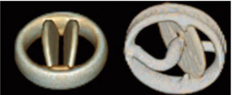
Fig. 1. — Volume rendered CT images of a bileaflet