Université de Montréal
Étude sur les fonctions in vivo des GEFs DOCK chez
les mammifères
par Mélanie Laurin
Programmes de Biologie Moléculaire Faculté de Médecine
Thèse présentée à la Faculté des études supérieures en vue de l’obtention du grade de PhD
en Programmes de Biologie Moléculaire
Septembre, 2013
Résumé
Dock1 (aussi nommé Dock180) est le membre prototypique de la famille Dock d’activateurs des petites GTPases de la famille Rho. Dock1 agit au sein d’une voie de signalisation conservée au cours de l’évolution et des études génétiques ont démontré que les orthologues de Dock1, myoblast city (mbc) chez la drosophile et Ced-5 chez le nématode, activent Rac dans divers processus biologiques. Notamment, mbc est un important régulateur de la fusion des myoblastes lors de la formation des fibres musculaires de drosophile. Mbc est aussi essentiel à la migration collective d’un groupe de cellules, appelées cellules de bordures, lors de leur migration dans la chambre de l’oeuf suite à l’activation de récepteurs à activité tyrosine kinase (RTK). La migration collective des cellules de bordures récapitule certains des événements observés lorsque des cellules tumorales envahissent le tissu environnant lors de la formation de métastases. Chez les mammifères, des études réalisées en lignées cellulaires suggèrent que Dock1 est aussi un régulateur du cytosquelette lors de la migration cellulaire. De plus, certaines études ont démontré que la voie Dock1/Rac agit en aval de RTKs lors de l’invasion de cellules de glioblastome. Néanmoins, les fonctions in vivo de Dock1 chez les mammifères demeurent méconnues et le but de cette thèse est d’identifier et de caractériser certaines de ses fonctions.
Guidés par la fonction de mbc, nous démontrons dans l’objectif no 1 un rôle essentiel pour ce gène au cours du développement embryonnaire grâce à la caractérisation d’une souris Dock1 knock-out. Des défauts sévères de fusion des myoblastes sont observés en absence de l’expression de Dock1 et ils contribuent à la réduction de la masse musculaire des souris mutantes. De plus, nous avons constaté une contribution du gène Dock5, un membre de la famille Dock proche de Dock1, au développement des fibres musculaires.
Dans l’objectif no 2, nous avons observé que des hauts niveaux d’expression de DOCK1 corrèlent avec un mauvais pronostic chez les patientes atteintes de cancer du sein possédant une forte expression du gène codant pour le RTK HER2. Une surexpression ou une amplification du locus codant pour le récepteur HER2 est associée à près de 20% des cas de cancer du sein. Les cancers de ces patientes développent fréquemment des métastases et sont associés à un mauvais pronostic. Des études biochimiques ont révélé que DOCK1 interagit avec le récepteur HER2 dans des cellules de cancer du sein. De plus, DOCK1 est essentiel à l’activation de RAC et à la migration cellulaire induite par HER2 dans ces cellules. L’utilisation d’un modèle de cancer du sein médié par HER2 chez la souris combiné avec l’inactivation du gène Dock1 dans les glandes mammaires, nous a permis d’identifier Dock1 et Rac comme de nouveaux effecteurs de la croissance tumorale et de la formation de métastases régulées par l’oncogène HER2.
Nous concluons que l’utilisation de différents modèles de souris knock-out pour le gène Dock1 nous a permis d’identifier des fonctions clés de ce gène. Tout comme son orthologue mbc, Dock1 joue un rôle important lors du développement embryonnaire en régulant notamment la fusion des myoblastes. Nos études ont également contribué à démontrer un important degré de conservation des mécanismes moléculaires de fusion entre les espèces. De plus, DOCK1 agit en aval du RTK HER2 et son expression dans les cellules épithéliales de glandes mammaires contribue au développement tumoral et à la formation de métastases induits par cet oncogène.
Abstract
Dock1 (also known as Dock180) is the prototypical member of the Dock family of Rho GTPase activators (RhoGEFs). Genetic studies in Drosophila and C. elegans have demonstrated that Dock1 orthologues act upstream of the Rac GTPase to activate it during various biological processes. Myoblast city (mbc), Dock1 ortholog in the Drosophila, is an important regulator of myoblast fusion during muscle fiber formation. Moreover, mbc regulates the collective migration of a cluster of border cells downstream of the activation of some tyrosine kinase receptors (RTKs). Migration of border cells is often view as a model for studying the invasive migration of cancer cells during metastasis development. Work done in cell lines also suggests that Dock1 is an important cytoskeletal regulator that controls cell migration. The Dock1/Rac pathway was also shown to act downstream of some RTKs to promote the invasion of glioblastoma cells. Yet, the in vivo functions of Dock1 in mammals are still poorly understood and the identification and characterization of some of these functions is the main objective of my thesis.
Guided by the function of mbc, in Aim #1 we revealed that Dock1 is essential to embryonic development by characterizing a Dock1 knock-out mouse model. A deficiency in myoblast fusion was observed in Dock1-null embryos which led to a reduction in their muscle mass. Furthermore, we uncovered a contribution of the other
Dock1-related GEF, Dock5, to myofiber development.
In Aim #2 a correlation between high level of DOCK1 expression and a poor prognosis in HER2+ breast cancer patients was revealed. Amplification or overexpression of the HER2 receptor tyrosine kinase is associated with near 20% of breast cancer cases. The presence of this genetic abnormality correlates with a poor prognosis and the development of metastasis. Biochemical and in vitro studies led us to identify that DOCK1 interacts with HER2 and is essential to HER2-mediated RAC activation and migration. The use of a HER2 breast cancer mouse model with Dock1
inactivation in the mammary gland led us to identify DOCK1-RAC signaling as novel effectors in HER2-mediated tumor growth and metastasis.
We conclude that the use of Dock1 mouse models allowed us to identify some of the key functions regulated by this gene in vivo. Much like its ortholog mbc, Dock1 is essential to embryonic development and regulates myoblast fusion. Our study also reveals important degree of conservation of the mechanisms that regulate fusion between species. In addition, DOCK1 acts downstream of the HER2 RTK in mammary epithelial cells where it contributes to the progression of breast cancer pathology and the formation of metastasis induced by this oncogene.
Préface
Cette thèse est un manuscrit rédigé par articles. Elle contient 2 articles publiés et un manuscrit en préparation pour soumission. La thèse est divisée en 3 chapitres suivis d’une discussion.
CHAPITRE 1 : INTRODUCTION
La section 1.1 du Chapitre 1 contient un article de revue. Une version modifiée de ce manuscrit sera soumise comme article de revue à Genes & Development. Les sections 1.2 et 1.3 contiennent une revue de la littérature sur le développement musculaire et le cancer du sein respectivement.
CHAPITRE 2 : LA RACGEF ATYPIQUE DOCK180 (DOCK1) RÉGULE LA FUSION DES MYOBLASTES IN VIVO
Ce chapitre contient un article publié :
Laurin M, Fradet N, Blangy A, Hall A, Vuori K and Côté JF (2008). The atypical Rac
activator Dock180 (Dock1) regulates myoblast fusion in vivo. PNAS 105, 15446-15451
CHAPITRE 3 : LA RACGEF DOCK1 EST UN IMPORTANT RÉGULATEUR DU DÉVELOPPEMENT MÉTASTATIQUE DANS LE CANCER DU SEIN HER2+
Ce chapitre contient un article publié :
Laurin M, Huber J, Pelletier A, Houalla T, Park M, Fukui Y, Haibe-Kains B, Muller WJ
and Côté JF (2013). Rac-specific guanine nucleotide exchange factor DOCK1 is a critical regulator of HER2-mediated breast cancer metastasis. PNAS 110, 7434-7439
Table des matières
Préface... vi
Liste des tableaux ... x
Liste des figures... xi
Liste des abréviations ... xiv
INTRODUCTION...1
CHAPITRE 1...2
1.1 LES FONCTIONS IN VIVO DES GEFs DOCK CHEZ LES MAMMIFÈRES ....3
Insights into the biological functions of DOCK GEFs from in vivo studies ...4
Abstract...5
Rho GTPases’ regulation and signaling...6
Dock GEFs in development ...12
Dock1 and Elmo1 contribute to cardiovascular development...12
Dock1 regulates Rac in the neuronal growth cone: from attraction to repulsion ...14
Dock3 integrates actin and microtubule dynamics to promote axon outgrowth ...16
Tug of war between Akt and PP2: activation of Rac by Dock6 is controlled by phosphorylation during axonal outgrowth...17
Dock7 regulates pigmentation and metabolism...18
GEF-independent functions of Dock7 in neurogenesis: controling interkinetic nuclear migration...18
Unraveling the role of the Dock1 pathway in myoblast fusion ...19
Bone resorption: Dock5 promotes osteoclast adhesion ...20
DOCK family in disease ...22
The Adams-Oliver Syndrome and misregulation of CDC42/RAC: genetic mutations in DOCK6 and ARHGAP31 in familial cases ...22
DOCK1 as a promoter of cell invasion downstream of oncogenic kinases...24
1.2.1 Le développement musculaire chez la drosophile...30
1.2.2 La spécification des progéniteurs musculaires chez la drosophile ...30
1.2.3 La fusion des myoblastes chez la drosophile ...32
1.2.3.1 Les événements cellulaires observés lors de la fusion ...32
1.2.3.2 Les voies de signalisation nécessaires à la fusion...33
1.2.4 Le développement musculaire chez les vertébrés...41
1.2.4.1 Le développement musculaire chez la souris et le poulet ...41
1.2.4.2 Le développement musculaire chez le poisson zèbre. ...44
1.2.5 La fusion des myoblastes chez les mammifères ...47
1.2.5.1 Les événements cellulaires observés lors de la fusion ...47
1.2.5.2 Les voies de signalisation régulant la fusion des myoblastes...48
1.2.6 Conclusion...58
1.3 LES GLANDES MAMMAIRES ET LE CANCER DU SEIN ...59
1.3.1 Les glandes mammaires ...60
1.3.2 Le cancer du sein ...63
1.3.3 Le récepteur HER2 et le cancer du sein...66
1.3.3.1 HER2 et la famille de récepteur à l’EGF ...66
1.3.3.2 HER2 et le développement des glandes mammaires ...67
1.3.3.3 Le cancer du sein HER2+...69
1.3.3.4 Les modèles de cancer du sein induits par l’oncogène HER2 ...70
1.3.4 Le développement des métastases...77
1.3.4.1 La cascade métastatique...77
1.3.4.2 Les modes de migration cellulaire ...78
1.3.5 HER2 et la signalisation en aval dans le cancer du sein ...84
1.3.6 DOCK1 et les récepteurs à activité tyrosine kinase...88
1.3.7 Conclusion...90
1.4 LES OBJECTIFS DE RECHERCHE...91
1.4.1 Objectif 1 (Chapitre 2)...91
1.4.2 Objectif 2 (Chapitre 3)...92
RÉSULTATS...93
LA RACGEF ATYPIQUE DOCK180 (DOCK1) RÉGULE LA FUSION DES
MYOBLASTES IN VIVO...94
Contributions Figures ...95
The atypical Rac Activator Dock180 (Dock1) regulates myoblast fusion in vivo...96
Abstract ...97
Introduction...98
Results ...101
Discussion ...109
Materiel and Methods...113
Acknowledgments ...114
Supplemental Information...115
CHAPITRE 3...132
LA RACGEF DOCK1 EST UN IMPORTANT RÉGULATEUR DU DÉVELOPPEMENT MÉTASTATIQUE DANS LE CANCER DU SEIN HER2+...132
Contributions Figures ...133
RAC-specific guanine nucleotide exchange factor DOCK1 is a critical regulator of HER2-mediated breast cancer metastasis ...135
Abstract ...136
Introduction...137
Results ...139
Discussion ...151
Materiel and Methods...154
Acknowledgements ...155
Supplemental Information...156
DISCUSSION...186
Liste des tableaux
CHAPITRE1Table 1.I In vivo models of Dock GEFs...11
CHAPITRE 2
Table 2.SI Viability of embryos and pups...129 Table 2.SII E14.5 embryos and P21 pups derived from Dock1+/-Dock5+/- X Dock1
+/-Dock5+/- crosses...130 Table 2.SIII PCR primers for different procedures ...131
CHAPITRE 3
Table 3.SI RNA-Seq analysis overview...184 Table 3.SII Primers for different procedures ...185
Liste des figures
CHAPITRE 1Figure 1.1.1 Dock GEF Family ...8
Figure 1.1.2 Structural basis of Dock GEF activity, localization and signaling...9
Figure 1.1.3 Elmo scaffolds: spatio-temporal orchestrators of Dock-mediated Rac activation...10
Figure 1.1.4 The role of Dock GEFs in development...13
Figure 1.1.5 Docks in neurogenesis ...15
Figure 1.1.6 Eliminating apoptotic cells via the Elmo-Dock1-Rac pathway: implications in tissue homeostasis. ...21
Figure 1.1.7 Moving ameboid or mesenchymal? DOCKs tip the balance...23
Figure 1.1.8 DOCK1 in cancer...26
Figure 1.2.1 Le muscle squelettique et son architecture...29
Figure 1.2.2 La spécification des progéniteurs musculaires chez la drosophile ...31
Figure 1.2.3 Les événements cellulaires observés lors de la fusion des myoblastes de drosophile...34
Figure 1.2.4 Schéma des voies de signalisation impliquées dans la fusion des myoblastes de drosophile ...35
Figure 1.2.5 La myogenèse chez les mammifères ...42
Figure 1.2.6 Les vagues de myogenèse...46
Figure 1.2.7 Les voies de signalisation impliquées la fusion des myoblastes chez les mammifères...57
Figure 1.3.1 Anatomie de la glande mammaire des humains...61
Figure 1.3.2 Le développement des glandes mammaires chez la souris ...62
Figure 1.3.3 Les récepteurs de la famille de l’EGF, leurs ligands et les voies de signalisation activées par ces récepteurs...68
Figure 1.3.4 HER2 et les variants NEU oncogéniques ...73
Figure 1.3.5 La cascade métastatique...79
Figure 1.3.6 Les modes de migration lors de l’invasion tumorale ...80
Figure 1.3.7 La migration mésenchymale en 5 étapes ...83
CHAPITRE 2 Figure 2.1 Generation and analysis of Dock1 and Dock5 mutant mice ...102
Figure 2.2 Expression profile and characterization of the skeletal muscle in the absence of Dock1 and Dock5...103
Figure 2.3 The absence of Dock1 affects myoblast fusion in vivo ...105
Figure 2.4 Genetic interactions between Dock1 and Dock5 in muscle development ...108
Figure 2.S1 Disruption of Dock1 and Dock5 genes in mice...123
Figure 2.S2 Expression profile of Dock1 and Dock5 during mouse development .... ...124
Figure 2.S3 General and dramatic reduction of muscle mass in embryos lacking Dock1...126
Figure 2.S4 Loss of Dock1 does not affect determination of myoblast and migration of muscle progenitors...127
Figure 2.S5 Proliferative and apoptotic status of muscle cells in Dock1-null embryos ...128
CHAPITRE 3 Figure 3.1 DOCK1, a negative prognostic factor for human breast cancer survival, activates RAC and promotes cell migration downstream of HER2 ...141
Figure 3.2 Dock1 contributes to HER2 tumorigenesis in a mouse model of breast cancer ...144
Figure 3.3 Dock1 regulates HER2-mediated lung metastasis ...146
Figure 3.4 Identification of a Dock1-null gene signature enriched in interferon response genes ...148
Figure 3.S1 DOCK1 is expressed in human breast cancer ...166 Figure 3.S2 DOCK1, is phosphorylated by SRC kinase downstream of HER2 activation...167 Figure 3.S3 Portrait of Rho GTPase regulation in Neu-induced tumors ...169 Figure 3.S4 Generation of a Dock1 conditional knockout mouse ...171 Figure 3.S5 Dock1 signaling regulates tumor growth by promoting cell proliferation and blocking apoptosis ...172 Figure 3.S6 Dock1 regulates c-Jun and Stat3 activation in HER2-driven MINs and Stat1 expression levels in HER2-driven tumors ...173 Figure 3.S7 Reduced levels and activation of Stat1 in HER2-driven tumors in the absence of Dock1 expression...174 Figure 3.S8 Dock1 is expressed in lung metastasis ...175 Figure 3.S9 Deletion of Dock1 expression in NIC+ tumor does not affect white blood cell recruitment...176 Figure 3.S10 Dock1 is essential for HER2-mediated lung metastasis in experimental metastasis assay ...177 Figure 3.S11 Components upstream of Dock1 in the integrin-signaling pathways are activated correctly in Dock1-null tumors...178 Figure 3.S12 Validation of the differential expression data obtained by
RNA-sequencing using Q-PCR analysis...180 Figure 3.S13 Oncogenic HER2 elevates interferon-response gene expressions in culture ...181 Figure 3.S14 Expression levels of some Dock1flx/flx-signature genes are predictive of
disease free survival in HER2+ breast cancer...182
Figure 3.S15 Model of signaling downstream of Dock1 in NIC+ MINs and tumors. ...
Liste des abréviations
ADAM12 : ADAM METALLOPEPTIDASE DOMAIN 12
α-PIX : RAC/CDC42 GUANINE NUCLEOTIDE EXCHANGE FACTOR (GEF) 6
ANTS : ANTISOCIAL
AOS : Adams-Oliver Syndrome
ARF6 : ADP-RIBOSYLATION FACTOR 6
ARHGP31 : RHO GTPASE ACTIVATING PROTEIN 31
BAI : BRAIN-SPECIFIC ANGIOGENESIS INHIBITOR
BDNF : BRAIN-DERIVED NEUROTROPHIC FACTOR
BNIP2 : BCL2/ADENOVIRUS E1B 19KDA INTERACTING PROTEIN 2
BOC : BROTHER OF CDO
β-PIX : RHO GUANINE NUCLEOTIDE EXCHANGE FACTOR (GEF7)
BRAG2 : BREFELDIN-RESISTANT ARF-GEF 2 PROTEIN
BSA : Bovine serum albumin
BST2 : BONE MARROW STROMAL CELL ANTIGEN 2
CDC42 : CELL DIVISION CYCLE 42
CDO : CELL ADHESION MOLECULE-RELATED/DOWN-REGULATED BY
ONCOGENES
CD31 : PLATELET/ENDOTHELIAL CELL ADHESION MOLECULE 1
CD45 : PROTEIN TYROSINE PHOSPHATASE, RECEPTOR TYPE, C
CED : CELL DEATH
CMPK2 : CYTIDINE MONOPHOSPHATE (UMP-CMP) KINASE 2, MITOCHONDRIAL
CRK : V-CRK SARCOMA VIRUS CT10 ONCOGENE HOMOLOG
CRK-L : V-CRK SARCOMA VIRUS CT10 ONCOGENE HOMOLOG
(AVIAN)-LIKE
CRMP-2 : COLLAPSIN RESPONSE MEDIATOR PROTEIN 2
CSN2 : CASEIN BETA
CSN1S1 : CASEIN ALPHA S1
CXCL12 : CHEMOKINE (C-X-C MOTIF) LIGAND 12
CXCR4 : CHEMOKINE (C-X-C MOTIF) RECEPTOR 4
DBL : DIFFUSE B-CELL LYMPHOMA
DCC : DELETED IN COLORECTAL CARCINOMA
DDX60 : DEAD (ASP-GLU-ALA-ASP) BOX POLYPEPTIDE 60A
DH : DBL-HOMOLOGY
DHR : DOCK-HOMOLOGY REGION
DHX58 : DEXH (ASP-GLU-X-HIS) BOX POLYPEPTIDE 58
DOCK : DEDICATOR OF CYTOKINESIS
DSG1C : DESMOGLEIN 1 GAMMA
DUF : DUMBFOUNDED
E : Embryonic day
EHD2 : EH-DOMAIN CONTAINING 2
EGF : EPIDERMAL GROWTH FACTOR
EGFRvIII : EPIDERMAL GROWTH FACTOR RECEPTOR VARIANT III
ER : ESTROGEN RECEPTOR
ES : Embryonic stem
ESR1 : ESTROGEN RECEPTOR 1 (ALPHA)
FAK : FOCAL ADHESION KINASE
FARP1 : FERM, RHOGEF (ARHGEF) AND PLECKSTRIN DOMAIN PROTEIN 1
(CHONDROCYTE-DERIVED)
FBLN5 : FIBULIN 5
FBS : Fetal bovine serum
FCM : Fusion competent myoblasts
FLX : Flox allele
FM : Founder myoblast
FuRMAS : Fusion-Restricted-Myogenic-Adhesive-Structure
GAP : GTPase Activating Protein
GBP3 : GUANYLATE BINDING PROTEIN 3
GBP7 : GUANYLATE BINDING PROTEIN 7
GDI : Guanine nucleotide dissociation Inhibitor
GDP : Guanosine diphosphate
GEF : Guanine nucleotide exchange factors
GO : Gene ontology
GPCR : G Protein-coupled receptor
GSK3β : GLYCOGEN SYNTHASE KINASE 3 BETA
GTP : Guanosine triphosphate
H&E : Hematoxylin and eosin
HER1 : EPIDERMAL GROWTH FACTOR RECEPTOR 1
HER2 : V-ERB-B2 ERYTHROBLASTIC LEUKEMIA VIRAL ONCOGENE
HOMOLOG 2, NEURO/GLIOBLASTOMA DERIVED ONCOGENE HOMOLOG (HUMAN)
HER3: V-ERB-B2 ERYTHROBLASTIC LEUKEMIA VIRAL ONCOGENE
HOMOLOG 3, NEURO/GLIOBLASTOMA DERIVED ONCOGENE HOMOLOG (HUMAN)
HER4: V-ERB-A ERYTHROBLASTIC LEUKEMAIA VIRAL ONCOGENE
HOMOLOG 4, NEURO/GLIOBLASTOMA DERIVED ONCOGENE HOMOLOG (HUMAN)
HERC6: HECT DOMAIN AND RLD 6
HBS : HIBRIS
HGF : HEPATOCYTE GROWTH FACTOR
HRG : HEREGULIN
IHC : Immunohistochemistry
IFIH1 : INTERFERON-INDUCED WITH HELICASE C DOMAIN 1
IFI272IA : INTERFERON, ALPHA-INDUCIBLE PROTEIN 27 LIKE 2A
IFI44 : INTERFERON-INDUCED PROTEIN 44
IFI47 : INTERFERON GAMMA INDUCIBLE PROTEIN 47
IKK : INHIBITOR OF KAPPAB KINASE
IL-4 : INTERLEUKIN-4
INF : INTERFERON
INM : Interkinetic nuclear migration
ITG : INTEGRIN
IRF9 : INTERFERON REGULATORY FACTOR 9
IRGM1 : IMMUNITY-RELATED GTPASE FAMILY M MEMBER 1
IRRE-C : IRREGULAR-CHIASM-C
ISG15 : ISG15 UBIQUITIN-LIKE MODIFIER
KIRRE : KIN OF IRRE
KIRREL : KIN OF IRRE LIKE
KO : Knock-out
LALBA : LACTALBUMIN, ALPHA
LARG : RHO GUANINE NUCLEOTIDE EXCHANGE FACTOR (GEF) 12
LBX1 : LADYBIRD HOMEOBOX 1
LGALS3BP : LECTIN, GALACTOSIDE-BINDING, SOLUBLE, 3 BINDING PROTEIN LGALS9 : LECTIN, GALACTOSE BINDING, SOLUBLE 9
LTR : Long terminal repeat
MBC : MYOBLAST CITY
MHC : MYOSIN HEAVY CHAIN
MIN : Mammary intraepithelial neoplastic lesions
MLC : MYOSIN LIGHT CHAIN
MMP12 : MATRIX METALLOPEPTIDASE 12
MMTV : MOUSE MAMMARY TUMOR VIRUS
MRF4 : MUSCLE-SPECIFIC REGULATORY FACTOR 4
MX1 : MYXOVIRUS (INFLUENZA VIRUS) RESISTANCE 1
MX2 : MYOVIRUS (INFLUENZA VIRUS) RESISTANCE 2
MYF5 : MYOGENIC FACTOR 5
NCK2 : NCK ADAPTOR PROTEIN 2
NEPH1 : NEPHRIN-LIKE PROTEIN 1
NEPH2 : NEPHRIN-LIKE PROTEIN 2
NEPH3 : NEPHRIN-LIKE PROTEIN 3
NEU : V-ERB-B2 ERYTHROBLASTIC LEUKEMIA VIRAL ONCOGENE
HOMOLOG 2 (RAT)
NFATC2 : NUCLEAR FACTOR OF ACTIVATED T CELLS, CYTOPLASMIC,
CALCINEURIN DEPENDENT 2
NF-KB : NUCLEAR FACTOR OF KAPPA-B
NIC : NEUNDL2-5-IRES-CRE
NMuMG : Normal murine mammary gland epithelial cell
OAS1A : 2’-5’ OLIGOADENYLATE SYNTHETASE 1A
OAS1B : 2’-5’ OLIGOADENYLATE SYNTHETASE 1B
OAS2 : 2’-5’ OLIGOADYNYLATE SYNTHETASE 2
OBSL1 : OBSCURIN-LIKE 1
PARP12 : POLY (ADP-RIBOSE) POLYMERASE FAMILY, MEMBER 12 PARP14 : POLY (ADP-RIBOSE) POLYMERASE FAMILY, MEMBER 14
PAX3 : PAIRED BOX GENE 3
PAX7 : PAIRED BOX GENE 7
PBS : Phosphate buffer saline
PCR : Polymerase chain reaction
PDGF : PLATELET-DERIVED GROWTH FACTOR
PFA : Paraformaldehyde
PGI2 : PROSTACYCLIN
PH : Pleckstrin Homology
PI3K : PHOPHATIDYLINOSITOL 3-KINASE
PIP3 : PHOSPHATIDYLINOSITOL 3,4,5-TRIPHOSPHATES
PSMB8 : PROTEASOME (PROSOME, MACROPAIN) SUBUNIT, BETA TYPE, 8
(LARGE MULTIFUNCTIONAL PEPTIDASE 7)
P-REX : PHOSPHATIDYLINOSITOL-3,4,5-TRIPHOSPHATE-DEPENDENT RAC
EXCHANGE FACTOR 2
p190RHOGEF : Rho guanine nucleotide exchange factor (GEF) 28
PS : PHOSPHATIDYLSERINE
RAC : RAS-RELATED C3 BOTULINUM TOXIN SUBSTRATE 1
RAS : RAT SARCOMA VIRAL ONCOGENE HOMOLOG
RHO : RAS-LIKE GTP-BINDING PROTEIN RHO
RHOA : RAS HOMOLOG FAMILY MEMBER A
RHOG : RAS HOMOLOGY GROWTH-RELATED
RGC : Radial glial cells
RNA : RIBONUCLEIC ACID
ROLS : ROLLING PEBBLES
RPL34 : RIBOSOMAL PROTEIN L34
RST : ROUGHEST
RSAD2 : RADICAL S-ADENOSYL METHIONINE DOMAIN CONTAINING 2
RTK : Receptor tyrosine kinase
RTP4 : RECEPTOR (CHEMOSENSORY) TRANSPORTER PROTEIN 4
SING : SINGLE-BAR
SLFN2 : SCHLAFEN 2
SLFN8 : SCHLAFEN 8
SMOC1 : SPARC RELATED MODULAR CALCIUM BINDING 1
SNS : STICKS AND STONES
SOS1 : SON OF SEVENLESS HOMOLOG 1 (DROSOPHILA)
SPF : Specific pathogen Free
SRC : V-SARCOMA (SCHMIDT-RUPPIN A-2) VIRAL ONCOGENE
HOMOLOG
STAT1 : SIGNAL TRANSDUCER AND ACTIVATOR OF TRANSCRIPTION 1
STAT3 : SIGNAL TRANSDUCER AND ACTIVATOR OF TRANSCRIPTION 3
(ACUTE-PHASE RESPONSE FACTOR)
TACC3 : TRANSFORMING, ACIDIC COILED-COIL CONTAINING PROTEIN 3
TARG : Targeted
TEB : Terminal end bud
TIAM1 : T CELL LYMPHOMA INVASION AND METASTASIS 1
TRIO : TRIO RHO GUANINE NUCLEOTIDE EXCHANGE FACTOR
UBA7 : UBIQUITIN-LIKE MODIFIER ACTIVATING ENZYME 7
UNC5B : UNC-5 HOMOLOG B
UPAR : UROKINASE PLASMINOGEN ACTIVATOR RECPTOR
USP18 : UBIQUITIN SPECIFIC PEPTIDASE 18
VASP : VASODILATOR-STIMULATED PHOSPHOPROTEIN
VEGF : VASCULAR ENDOTHELIAL GROWTH FACTOR
WASP : WISKOTT-ALDRICH SYNDROME PROTEIN
WAVE1 : WISKOTT-ALDRICH SYNDROME PROTEIN FAMILY MEMBER 1
WIP : WASP-INTERACTING PROTEIN
WISH : Whole mount in situ hybridization
WT : Wild-type
XAF1 : XIAP ASSOCIATED FACTOR 1
Remerciements
Je tiens à remercier toutes les personnes qui, de près ou de loin, ont participé à ce travail.
Je tiens plus particulièrement à remercier mon directeur de recherche Dr Jean-François Côté pour son soutien tout au long de ma thèse de doctorat. Je le remercie d’avoir été un aussi bon mentor et d’avoir su transmettre sa rigueur, son enthousiasme, son ouverture et sa passion pour la recherche académique. Je le remercie pour son acharnement à travailler constamment de façon à créer les meilleures opportunités pour chacun de ses étudiants. Merci JF pour ta patience, pour ton écoute, et ta persévérance à faire sortir le meilleur de moi-même, même quand disons-le je n’y croyais pas de mon côté!
Je souhaite aussi à remercier les membres de mon comité de doctorat; Dr Jean Vacher, Dr David Hipfner et Dr Paul Holland. Ils m’ont aidé à développer mon esprit critique et je suis reconnaissante pour leurs recommandations qui mon permis de progresser dans mes différents projets de recherche. J’aimerais aussi remercier les membres de mon comité de thèse pour avoir accepté de juger et critiquer ce manuscrit.
Je tiens à remercier nos collaborateurs, Dr Anne Blangy et Dr Allan Hall pour leur support lors du projet de recherche sur le rôle de DOCK1 dans la fusion des myoblastes. Je remercie aussi nos collaborateurs, Dr Morag Park, Dr William Muller, Dr Benjamin Haibe-Kains et Dr Yoshinori Fukui pour leur contribution au projet de recherche sur le rôle de DOCK1 dans le cancer du sein.
J’aimerais enfin remercier les membres des plateformes de l’IRCM pour leur aide à la réalisation de ces projets de recherche, plus particulièrement Dominic Filion en microscopie, Annie Vallée, Geneviève Brindle et Dominique Lauzier en histologie et Odile Neyret en biologie moléculaire. Je remercie aussi Suzie Riverin et Claudia Toulouse pour leur aide précieuse à la gestion des colonies de souris.
J’aimerais remercier les membres présents et passés du laboratoire Côté pour leur contribution aux différents projets grâce aux discussions, mais aussi et surtout pour avoir su créer et maintenir une super atmosphère de travail.
Merci à Nad, en qui je serai toujours reconnaissante d’avoir été l’instigatrice de mon opportunité à joindre le laboratoire Côté! Merci Nad pour ton aide irremplaçable dans tous mes projets et pour avoir été bien plus qu’une collègue de travail pendant toutes ces années!
Merci à Mani, ma « soul sister »! I can’t imagine what those years would have been without you. I have so much to thank you for that I don’t know where to start…Thanks for paving the way for me, thanks for understanding my higher level of sarcasm, thanks for the late night choreography, thanks for the hip hop initiation, thanks for your support… This could go on forever so I guess I will finish by saying that I think you were right…you would be Batman!
Merci à Nou, Ari et Jen pour avoir été non seulement des supers collègues de travail, mais pour avoir été de véritables amies! Merci à Nou pour avoir été présente avec son support moral de toutes heures pendant les révisons. Contente miss que nos prédictions se soient révélées fausses! Merci à Ari pour son aide inestimable à la révision du papier et pour être toujours disponible pour discuter de « belle science »! Contente miss que nos vies aient finalement « matchées ». Merci à Jen pour avoir initié d’aussi belle façon le projet de cancer du sein. Contente Jen, que nos vies parallèles se soient finalement croisées! Je vous aime les filles!
Enfin, j’aimerais remercier les membres de l’équipe sportive la plus geek en ville, les PRIMERS Julien, Jen, Stephen, Ludi, Alex, American Steve, Joe, Aurèle, Sarah, Jin, Vas, Max, et Gab. Merci guys pour avoir contribué à ce que je sois aussi heureuse durant cette dernière année de thèse malgré l’intensité du travail, que se soit sur le terrain ou à l’extérieur du terrain pendant une pratique, une retraite, un bbq, un souper ou un show! Un merci particulier à Jules et Alex pour leur dévouement à tenter de nous expliquer comment faire un stack vertical, une cut et un flick qui ont de l’allure! PRIMERS HYBRIDIZE, PRIMERS AMPLIFY…GOOO PRIMERS!
Je remercie aussi les membres du club de la petite cuillère, Marie, Damien et MiniJen pour nos cafés du vendredi. Toujours agréable de débuter la fin de semaine avec vous à philosopher sur la vie, notre avenir et bien sûr la science toujours la science!
Je remercie JF Clément pour avoir toujours été là depuis le début de mon parcours de biologiste, mais aussi disons-le depuis le début de mon parcours de vie « adulte » ! Humm…
Enfin, je tiens à remercier plus particulièrement ma famille. Mon père et ma mère qui m’ont toujours encouragé et soutenu pendant mes études. Merci pour avoir toujours tenté de me faciliter la vie afin de me permettre de me concentrer sur mes études. Merci pour votre patiente et pour avoir toujours démontré le plus grand intérêt pour mes projets de recherche. Merci à ma grand-mère pour avoir toujours pris des nouvelles et avoir envoyé autant d’ondes positives. Merci à mon frère que je sollicite aux moindres travaux ou déménagement. Je vous aimexxxx.
1.1 LES FONCTIONS IN VIVO DES GEFs DOCK CHEZ LES
MAMMIFÈRES
Insights into the biological functions of DOCK GEFs from in
vivo studies
Mélanie Laurin1,2 and Jean-François Côté1-4
1Institut de Recherches Cliniques de Montréal (IRCM), Montréal, QC, Canada H2W
1R7.
2Département de Médecine (Programmes de Biologie Moléculaire), Université de
Montréal, Montréal, QC, Canada H3T 1J4.
3Département de Biochimie, Université de Montréal, Montréal, QC, Canada H3T 1J4. 4Division of Experimental Medicine, McGill University, Montréal, QC, Canada H3A
1A3.
Abstract
Rho GTPases are implicated in several aspects of embryonic development and aberrant regulation of their activity contributes to the progression of numerous diseases. The Rho guanine exchange factors (GEFs), which can be classified into Dbl and Dock subfamilies, mediate the spatio-temporal activation of Rho proteins and their ability to engage effector pathways. Recently, the combination of structural, biochemical and in vitro studies has allowed for major advancements in our understanding of the molecular mechanisms regulating the activity and function of atypical GEFs of the Dock family. In this review, we focus on genetic and mouse studies that have unearthed exciting key and unexpected roles for individual Dock family members in development and diseases.
Rho GTPases’ regulation and signaling
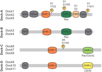
Rho proteins represent 22 members within the Ras superfamily of small GTPases. By orchestrating cytoskeleton remodeling, these molecular switches regulate numerous processes throughout embryonic development including axon guidance, migration of hematopoietic cells and fusion of myoblasts (1-3). Rho proteins are also implicated in diseases such as cancer progression where their abnormal regulation contributes to the ability of tumor cells to escape the primary tumor to form life-threatening metastases (4). Rho proteins continuously switch between inactive (GDP-bound) and active (GTP-bound) conformations; three groups of proteins tightly coordinate the cycling states of these GTPases. GTPase-activating proteins (GAPs) and Rho guanine nucleotide-dissociation inhibitors act as negative regulators by promoting their intrinsic GTPase activity or sequester them in the cytoplasm, respectively. The guanine nucleotide exchange factors (GEFs), which can be divided into Dbl and Dock subfamilies, act as positive regulators by mediating GDP to GTP exchange on Rho GTPases to ultimately promote their binding to specific effectors pathways (5). Dock-related GEFs form an 11 member subfamily in mammals. Dock1, initially named Dock180, is the prototypical member of this family and was originally identified as a binding partner to the adaptor protein Crk (6). From that point, the name Dock180 was used to designate this “180kDa protein Downstream of Crk. However, the literature was since modified so that “Dock” now stands for “Dedicator of cytokinesis”. This revision still remains obscure and unexplained in the field especially since no evidence suggests so far that Dock180 regulates cytokinesis. Members of the Dock subfamily of GEFs are characterized by the presence of two evolutionarily conserved Dock Homology Region (DHR) domains: the lipid-binding DHR-1 and the GEF DHR-2 modules. Members of this family can be further classified into 4 subgroups that each differ in their composition of additional protein domains and their specificity toward Rac or Cdc42 GTPases (Figure 1.1.1 page 8). In particular, novel regulatory phosphorylation sites and the Phosphatidic Acid-binding domain play key roles in spatio-temporal activation of these GEFs (Figure 1.1.1 page 8 and Figure 1.1.2
contribution of Elmo scaffold proteins in the spatio-temporal activation of Rac by a subset of Elmo-binding Dock GEFs in Figure 1.1.3 (Figure 1.1.3 page 10). In addition, recent genetic studies offered important insights into the in vivo roles played by Dock GEFs (Table 1.I page 11). In particular, the precise roles of Dock2 and Dock8 in immunology are being unraveled and these advances have recently been discussed in detail elsewhere (7). Here, we review the emerging and unsuspected biological functions controlled by Dock family members in development and diseases.
Dock1 Dock2 Dock5 SH3 Dock-A Dock3 Dock4 Dock-B Dock6 Dock7 Dock8 Dock-C Dock9 Dock10 Dock11 Dock-D SH3 helical DHR-1 DHR-1 DHR-1 DHR-1 DHR-2 DHR-2 DHR-2 DHR-2 PxxP PxxP PBR P D1 Y1811 D1 Y722 P P D6 S1194 PH Rac Rac Rac/Cdc42 Cdc42 D1 D2 D1 S1250 P
Figure 1.1.1 Dock GEF Family
Dock proteins, subdivided into four subfamilies, are characterized by the evolutionarily conserved Dock Homology Region (DHR)-1, mediating binding to PIP3, and DHR-2, encompassing the GEF activity towards Rac/Cdc42 GTPases (8). The N-terminus of Dock-A/B GEFs, including a SH3 domain, mediates their interaction with Elmo scaffolding proteins, while the C-terminal PxxP region coordinates interactions with SH3-containing adaptor proteins, such as Crk and Grb2 (8). Dock-D members have a N-terminal localized PH domain involved in phosphoinositides binding for membrane translocation (9, 10). Several studies have identified that Dock GEFs are post-translationally modified by kinases and phosphatases. Of in vivo relevance, phosphorylation of DOCK1 (D1) on Y722, Y1811 or S1250 increases its GEF activity towards RAC and is elevated in brain
cancers (11-13). Akt binds to Dock6 (D6) and phosphorylates its S1194 and inhibits by this means its GEF activity; while binding
PIP3
PDGF
i) GDP bound to nucleotide-binding site of the Rho GTPase
ii) DHR2-mediated release of GDP
iii) DHR2-mediated release of RhoGTPase GDP
switch 1 switch 2 P Mg guanosine P-loop switch 1 Dock2 Dock2 Dock2 Dock2 PA PIP3 Dock2 PIP3 Dock2 PIP3 PIP3
PIP3 PIP3 GTPRac
PDGFR PLD PC Dorsal ruffle Peripheral Ruffle ?
Mouse embryonic fibroblast Neutrophil
PA
Dock1 PBR binds to phosphatidic acid lipid (PA) during dorsal ruffle formation
Structural basis of DOCKs DHR-2 GEF activity Dock2 PBR binds to phosphatidic acid lipid (PA)
during neutrophil migration Chemokine gradient Chemokine gradient Rac GTP Dock1 Dock1 Rac GTP PA Dock1 Dock5 PIP3 PIP3 P P P guanosine switch 2 P-loop Lobe B Lobe C α10 insert (nucleotide sensor) α10N α10C switch 1 Mg P P guanosine switch 2 Lobe B Lobe C α10 insert disordered α10N α10C P-loop P Mg Val Val Nucleus Nucleus Nucleus A B C
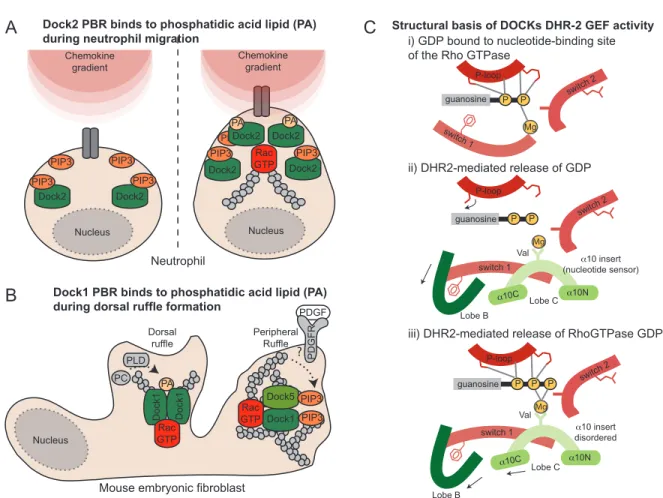
Figure 1.1.2 Structural basis of Dock GEF activity, localization and signaling.
The DHR-1 domain of Dock GEFs facilitates their recruitment at the membrane downstream of PI3-Kinase signaling by directly binding to PIP3. A polybasic region (PBR, see Figure 1) was also identified in Dock1 and Dock2 and suspected to bind PIP3 but recent advances instead suggest that it bind the signaling lipid phosphatidic acid lipid (PA). What is the role and biological relevance of this second lipid-binding activity? (A) Insight into this came from analyzing the cytokine-induced temporal produc-tion of these lipids in neutrophils. These studies disclosed that PIP3 is produced early following stimulation while PA production peaked later. Dock2 is critical for migration and its rapid and broad recruitment at the membrane where cytokine receptors are getting activated is mediated by a direct DHR-1/PIP3 interaction. Neutrophil polarization is next dependent on Phospholipase-D-dependent production of PA that serves to narrow the recruitment of Dock2 to the pseudopod via the PBR (15). It is therefore through such sequential lipid-binding events that Dock2 is ideally positioned to promote polarized neutrophil migration. (B) In fibroblasts, activation of PDFG receptor by its ligand promotes both peripheral membrane ruffles and dorsal ruffles, the later being dependent on the production of PA. PDGF treatment promotes a rapid PIP3-dependant recruitment of both Dock1 and Dock5 at the membrane to activate Rac and promote peripheral ruffles. PA, again produced sequentially to PIP3, narrows the localization of Dock1, but not Dock5 that does not contain a PBR, in the membrane to generate the characteristic PDGF-induced dorsal ruffles via Rac activation (16). It is therefore through such sequential lipid-binding events that Dock1 and Dock2 are ideally positioned in specialized membranes to promote polarized cytoskeletal dynamics. (C) The molecular basis of DOCK-mediated Rho proteins activation remained elusive until structural studies identified a novel mechanism exploiting a nucleotide sensor region in the DHR-2 of DOCK2, DOCK8 and DOCK9 that promotes the release of GDP. Crystal structures revealed that the DHR-2 folds in three lobes and Lobe B and Lobe C are important for activity and binding to the target GTPase. (i) GTPases tightly bind nucleotide and the Switch 1 region directly coordinates a magnesium molecule bound to GDP. (ii) Upon contact with a DOCK GEF, an α10 helix insert in the Lobe C of the DHR-2 domain (green), characterized by a conserved valine residue, mediates Mg++ exclusion while the Lobe B displaces the Switch 1 region to contribute to GDP release and exposing the
nucleotide-binding site. These interactions also disturb interactions of the GTPase P-loop with the nucleotide to facilitate nucleotide release. (iii) Binding of GTP, and its interaction with Mg++, induces conformation changes that uncouple the GEF from
DHR-2 PxxP PBR SH3 helical DHR-1 PxxP EAD PH ELMO EID RBD Rac GTP GTP ase X Arl4A RhoG Acf7
Basal state Cell stimulation
α βγ Nucleus Nucleus Cytoplasm Cytoplasm PIP3 PA α β DHR-2 PxxP PBR SH3 helical DHR-1 PxxP EAD PH ELMO EID RBD PxxP EAD PH ELMO EID RBD DHR-2 PBR PxxP SH3 helical DHR-1 Elmo Dock1 Elmo Dock2 Elmo Dock1 αβγ α β Cxcl12 Cxcr4 Cxcr4 Bai1 Bai1 PtdSer
Figure 1.1.3 Elmo scaffolds: spatio-temporal orchestrators of Dock-mediated Rac activation
Elmo proteins are established Dock-A/B binding partners (Figure 1.1.1). Although interaction between Docks and Elmos is not essential for Rac activation per se, complex formation is mandatory to achieve cytoskeleton remodeling (10). Recent studies highlight the key role played by Elmo in positioning Dock1 to discrete area of the cells to allow for Rac activation in a polarized fashion post cell stimulation. (A) At basal level, Elmo and Dock1 are proposed to be autoinhibited by intramolecular interactions between Elmo EID and EAD domains and between Dock SH3 and DHR-2 domains (20-22). While Elmo and Dock1 are believed to be in a constitutive complex in cells, it remains unclear whether other Dock-A/B members are pre-bound or rather exist as single molecules at basal state. Further studies will be essential to clarify the dynamic of the interaction between Elmos and Dock-A/B members. Cell stimulation may sequentially release the autoinhibition constraints on Elmo followed by activation of Dock1 and Rac signaling (20). In contrast, Elmo and Dock2 may exist as individual proteins that enter in complex post-stimulation. Formation of a complex is proposed to directly and mutually relieve both proteins from their inhibited state (22). Finally, another level of complexity is added through the ability of Dock proteins to homo- or hetero-dimerize by their DHR-2 (20). (B) Upon cell stimulation, the recruitment of Elmo/Dock1 complex at the membrane can be more precisely guided by Elmo’s repertoire of interacting proteins. Engagement of RhoG or Arl4A to Elmo’s Ras-Binding Domain (RBD) contributes to both the relief of Elmo autoinhibition and promotes positioning of Elmo/Dock1 proteins at the membrane for Rac-dependent cytoskeleton reorganization (21, 23). Elmo also directly interacts with the microtubule- and actin-binding spectraplakin Acf7 (24). Upon integrin stimulation, cells expressing Elmo and Acf7 formed long and persistent membrane protrusions. Mechanisti-cally, Elmo is required for Acf7 ability to capture and stabilize the microtubule network at the cell cortex during integrin-mediated cell spreading (24). Recruitment of Elmo at the membrane was also observed via activation of the GPCR Cxcr4 by the cytokine Cxcl12. In this system, the released G protein Gαi2 directly bound Elmo and promoted Rac activation and cell invasion (25). The importance of Elmo/Dock1/Rac signaling downstream of this prometastatic cytokine during breast cancer progression will warrant full investigation.
Dock-A
Dock-B
Dock-C
Dock-D
Elmo
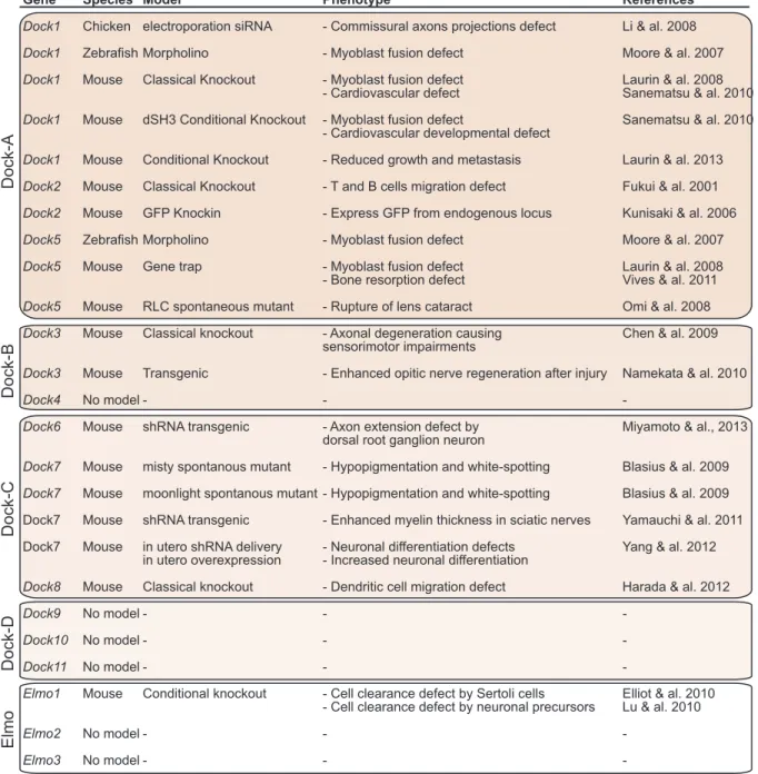
Gene Species Model Phenotype References
Dock1 Chicken electroporation siRNA - Commissural axons projections defect Li & al. 2008
Dock1 Zebrafish Morpholino - Myoblast fusion defect Moore & al. 2007
Dock1 Mouse Classical Knockout - Myoblast fusion defect Laurin & al. 2008
- Cardiovascular defect Sanematsu & al. 2010
Dock1 Mouse dSH3 Conditional Knockout - Myoblast fusion defect Sanematsu & al. 2010 - Cardiovascular developmental defect
Dock1 Mouse Conditional Knockout - Reduced growth and metastasis Laurin & al. 2013
Dock2 Mouse Classical Knockout - T and B cells migration defect Fukui & al. 2001
Dock2 Mouse GFP Knockin - Express GFP from endogenous locus Kunisaki & al. 2006
Dock5 Zebrafish Morpholino - Myoblast fusion defect Moore & al. 2007
Dock5 Mouse Gene trap - Myoblast fusion defect Laurin & al. 2008
- Bone resorption defect Vives & al. 2011
Dock5 Mouse RLC spontaneous mutant - Rupture of lens cataract Omi & al. 2008
Dock3 Mouse Classical knockout - Axonal degeneration causing Chen & al. 2009 sensorimotor impairments
Dock3 Mouse Transgenic - Enhanced opitic nerve regeneration after injury Namekata & al. 2010
Dock4 No model - - -
Dock6 Mouse shRNA transgenic - Axon extension defect by Miyamoto & al., 2013 dorsal root ganglion neuron
Dock7 Mouse misty spontanous mutant - Hypopigmentation and white-spotting Blasius & al. 2009
Dock7 Mouse moonlight spontanous mutant - Hypopigmentation and white-spotting Blasius & al. 2009 Dock7 Mouse shRNA transgenic - Enhanced myelin thickness in sciatic nerves Yamauchi & al. 2011 Dock7 Mouse in utero shRNA delivery - Neuronal differentiation defects Yang & al. 2012
in utero overexpression - Increased neuronal differentiation
Dock8 Mouse Classical knockout - Dendritic cell migration defect Harada & al. 2012
Dock9 No model - - -
Dock10 No model - - -
Dock11 No model - - -
Elmo1 Mouse Conditional knockout - Cell clearance defect by Sertoli cells Elliot & al. 2010 - Cell clearance defect by neuronal precursors Lu & al. 2010
Elmo2 No model - -
-Elmo3 No model - - -
Dock GEFs in development
Rho GTPases are viewed as essential molecular intermediates during development, yet the full impact of their upstream regulation through GEFs is only starting to be appreciated (26). Genetic screens in C. elegans and Drosophila shed light on the potential functions of Dock GEFs in mammals during development by regulating cell migration, myogenesis and clearance of apoptotic cells (8). Recent studies, summarized in Table 1.I (Table 1.I page 11), have moved away from cell culture models and focused on various vertebrate in vivo models to explore the biological functions of Dock GEFs. In this section, we focus on emerging functions of Docks in development.
Dock1 and Elmo1 contribute to cardiovascular development.
The chemokine Cxcl12 and its receptor Cxcr4, a member of the G-Protein Coupled Receptors (GPCRs) family, provide an important homing signal during the migration of endothelial progenitors, but the effector pathways involved in this system have remained elusive (27, 28). Characterization of two independent mutant mice revealed an essential role for Dock1 downstream of Cxcr4 during cardiovascular development (29) (Table 1.I page 11). Inactivation of Dock1 was lethal at birth and mutant mice displayed severe edema as a consequence of multiple cardiac abnormalities (27, 29). When Dock1-mutant hearts were explanted, endothelial cells failed to invade matrigel despite their ability to undergo epithelial to mesenchymal transition suggesting a key function for this GEF in migration. Rac activation, cytoskeletal changes and cell migration in Dock1-mutant explanted endothelial cells were impaired following treatment with Cxcl12, but interestingly not with Vegf, demonstrating a central signaling role for Dock1 downstream of Cxcr4 (29). While a role for Dock1 orthologs in migration is well established in lower organisms, these mouse studies were the first to uncover that Dock1 contributes in vivo to cell migration in mammals. It will be important to address how Dock1 molecularly connects to Cxcr4 receptor activation (Figure 1.1.4 page 13). Furthermore, as Cxcr4 is implicated in a plethora of
αβγ Dock1 Rac GDP Elmo Nucleus Elmo Dock1 Rac GTP Bai3 Nucleus Myoblast Myotube Bai3 Nucleus Nucleus A B Dock1 Rac GTP Nucleus Myoblast Myotube Nucleus Nucleus α βγ Rac GTP Elmo Endothelial Progenitor Nucleus Cxcl12 Dock1 ? Bai1 Apoptotic target Elmo Myoblast Myoblast Crk p130 Cas D Dock5 Rac GTP Dock5 αv β3 Rac GTP αv β3 Crk p130 Cas Rac GDP Nucleus Nucleus Nucleus P Sealing Zone Sealing Zone Nucleus Nucleus Nucleus Osteoclast Osteoclast Cxcr4 Cxcr4 C Endothelial Progenitor
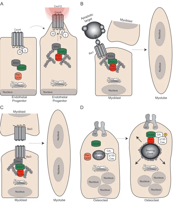
Figure 1.1.4 The role of Dock GEFs in development
(A) Dock1 is required downstream of Cxcl12-mediated activation of Cxcr4 to promote Rac activation and induce migration of endothelial progenitors. How the Elmo1/Dock1/Rac complex is coupled to Cxcr4 signaling remains to be determined. (B) Bai1’s ability to recognize apoptotic cells and interact with Elmo is proposed to trigger signaling that favors the fusion of myoblasts in order to help regenerate injured muscle tissue. (C) Bai3 is expressed by myoblasts and is essential for myoblast fusion. Activa-tion of Bai3 through a yet to be determined mechanism and its interacActiva-tion with Elmo is required for myoblast fusion. (D) In osteoclasts, Dock5 was shown to be essential dowstream of Integrin αυβ3 signaling to promote p130Cas phosphorylation and Rac activation, which is essential for the formation and maturation of the sealing zone.
developmental processes and diseases, it is possible that Dock1 contributes to other aspects of Cxcr4 signaling. Additional pathways provide guidance during vascular development (30). Among them, Netrin and its receptor Unc5B, promote the migration of endothelial cell progenitors through Rac activation (30, 31). In a zebrafish model, silencing dock1 or elmo1 robustly impaired Rac activation and ultimately vasculature development (31). Since Rac1 is critical for vascular development in mice, investigating the function of Elmo and Dock1 in Netrin-induced cardiac progenitor migration may reveal new pathways regulating this process (32). Collectively, these studies revealed specificity in the requirement of Elmo/Dock1 signaling downstream of different pro-migratory receptors to mediate Rac1 activation and migration of endothelial cells. A deeper characterization of the signaling pathway involved in developmental angiogenesis might also provide a better understanding of how these pathways could contribute to cancer progression by regulating tumor vascularisation.
Dock1 regulates Rac in the neuronal growth cone: from attraction to repulsion
Axon guidance is critical in brain development and deregulation of this process can have serious implications in mental diseases (33). Netrin, a classical guidance cue, acts via the receptor Dcc to induce Rac- and Cdc42-dependent attraction of commissural axons towards the neural tube floor plate (34, 35). Dock1 was shown to colocalize with Dcc in the growth cone following treatment with Netrin in vitro (Figure
1.1.5 page 15). Loss-of-function of Dock1 demonstrated the important role of this
GEF in Netrin-induced Rac activation leading to impaired axon outgrowth and abnormal commissural axons reorientation towards the guidance factor. These cell-based experiments hold true in vivo as electroporation of a siRNA targeting DOCK1 in the developing chicken neural tube caused misguidance of commissural axons (Table 1.I page 11). These data provided a long-sought mechanism to connect Dcc activation to Rac-mediated axon outgrowth and attraction towards Netrin. In contrast to Netrin/Dcc endorsed attraction, Ephrin-B3 favor hippocampal axonal repulsion (36). In this case, the adaptor protein Nck2 connects Ephrin-B3 to Dock1 to induce axon pruning (Figure 1.1.5 page 15).
DHR-2 DHR-1 PxxP SH3 Rac GTP FYN BDNF WAVE1 Nucleus axon growth Dock1 Rac GTP Dcc Netrin Dock1 Rac GTP Nck DHR-1 DHR-2 DHR-1 S1194 DHR-2 PP2A axon growth AKT axon growth PP2A AKT Nucleus Commisural neurons Attraction
Mossy fiber axon Prunning A P P GSKβ P CRMP-2 PIP3 ? S1194 P Dock7 Tacc3 Dock7 Tacc3 Dock7 Tacc3 Dock7 Tacc3 Dock7 Tacc3 Dock7 Tacc3 Tacc3 Tacc3 Tacc3 Tacc3 Tacc3 Basal Apical CTRL shRNA Dock7 B C D E RGC RGC RGC RGD Basal Progenitor Tacc3 RGC RGC RGC RGD RGD P Ephr Ephrin-B3 Rac GTP Dorsal root ganglion neurons NGF PI3K T rkA T rkB INM INM
Figure 1.1.5 Docks in neurogenesis
(A) In commissural neurons, Dock1 mediates Rac activation and promotes reorientation of the growth cone following activation of Dcc by the presence of a Netrin-1 gradient at the floor plate. (B) In the hippocampus, Ephrin-B3 mediates fiber axon pruning. After their engagement to their receptor, Ephrin-B3 molecules become phosphorylated and recruit the adaptor protein Nck2. Nck2 is essential to relocalize Dock1 to the membrane and induce Rac activation to promote repulsions of the axon. (C) In hippocampal neurons, TrkB activation by BDNF recruits Dock3 to the membrane in a Fyn kinase-dependent manner. Recruitment of Dock3 to the receptor is essential to promote Rac-dependent BDNF-induced axon outgrowth. Dock3 also directly binds WAVE1 through its DHR-1 domain to induce actin remodeling. Dock3 regulates the microtubule network through its intreraction with GSK3β and facilitates the phosphorylation of the kinase on an inhibitory site. Inhibition of GSK3β leads to the dephosphorylation of CRMP-2 and allows for microtubule elongation and axonal growth. (D) At early developmental stages, Dock6 is found in complex with PP2A via its binding to the DHR-2 domain. This interaction prevents phosphorylation of Dock6 on Ser1194 by Akt and allows axon growth.
At later developmental stages, the abundance of Akt increases. Akt interacts with Dock6 DHR-1 domain and phosphorylates it to prevent axon growth. (E) During cell cycle progression, the INM of RGCs along the apical to basal axis contributes to cell fate decision after division. Dock7, via its binding to Tacc3, regulates the speed of INM (left). Downregulation of Dock7 expression accelerates the baso-lateral to apical INM and leads to the proliferation of the radial glial progenitor (RGC) pool at the expense of
While these studies exploited in vivo and in vitro models, it remains unknown whether commissural and hippocampal axons are misguided in Dock1-null mice. Could abnormal DOCK1 and RAC signaling be at the base of some neuronal defects and mental illness? Future behavioral studies might shed light on the importance of this pathway in brain development.
Dock3 integrates actin and microtubule dynamics to promote axon outgrowth
Expression of Dock3 is largely restricted to neuronal tissues hinting towards a role for this GEF in brain development (37). Interestingly, transgenic animals overexpressing
Dock3 repaired injured optic nerves more effectively than control animals suggesting
an essential contribution by Dock3 in axonal growth (38) (Table 1.I page 11). In hippocampal neurons, activation of TrkB receptor tyrosine kinase by its ligand brain-derived neurotrophic factor (BDNF) promoted Rac activation via the recruitment of Dock3 to the membrane in a Fyn kinase-dependent manner for axonal growth (38) (Figure 1.1.5 page 15). Dock3 also directly regulates actin remodeling following BDNF stimulation by recruiting the actin nucleation-promoting factor WAVE1 through its DHR-1 domain (Figure 1.1.5 page 15). Whether the sole function of the DHR-1 of Dock3 is to bind WAVE1 or if it additionally serves a membrane recruitment function through PIP3-binding was not investigated. Dock3 was also shown to impact the
microtubule network via an interaction with Gsk3ß, a broad action kinase that is important in microtubule dynamics, which facilitates the phosphorylation of the kinase at an inhibitory site (39). Consequently, this sequestering and inhibition of Gsk3ß upon BDNF treatment lead to the dephosphorylation of CRMP-2 and allowed this protein to promote microtubule elongation and axonal growth (Figure 1.1.5 page 15). Further studies will be needed to fully understand the molecular mechanisms downstream of Dock3 that orchestrate crosstalk between the actin and microtubule cytoskeletons during axonal growth. The impact of Dock GEFs on the microtubules is also emerging from various studies and will need to be further investigated (Figure
1.1.3 page 10 and Figure 1.1.5 page 15). While overexpression of Dock3 in mice
degeneration and impaired sensorimotor functions (40) (Table 1.I page 11). In vivo, Dock3 was shown to control the activation of LIM kinase, and therefore, the phosphorylation of its target, cofilin. Reduced activation of the kinase was proposed to induce axonal dystrophy characterized by the accumulation of organelles, autophagic vacuoles and disorganized cytoskeletons (40). Pharmacological treatments aimed to increase DOCK3 signaling could be of therapeutic benefit for patients requiring axonal regeneration.
Tug of war between Akt and PP2: activation of Rac by Dock6 is controlled by phosphorylation during axonal outgrowth
The expression of Dock6 in dorsal root ganglions in vivo prompted for studies on its potential role in axon extension (14). Transgenic mice expressing shRNAs specific to
Dock6 in vivo showed a reduction in the length of peripheral axons at embryonic day
11 and also failed to form neuronal fibers extension in an injury model in vivo (14) (Table 1.I page 11). In agreement with in vivo observations, downregulation of Dock6 expression in dorsal root ganglion explants impaired axon outgrowth and the number of side branching. Likewise, knockdown of Rac decreased both the number of axons and branch points while Cdc42 knockdown only affected branching. These finding were in agreement with biochemical data suggesting that Dock6 is a Rac GEF in this system. At developmental stages when axons are growing, Dock6 is initially unphosphorylated and gradually becomes phosphorylated on Ser1194 and biochemical studies highlighted a GEF inhibitory function for this modification. In search for regulators of Dock6 phosphorylation, Akt was identified as a binding partner of the DHR-1 and was shown to directly phosphorylate Ser1194; in contrast, the phosphatase Pp2a interacted with the DHR-2 and was dephosphorylating this site (Figure 1.1.5
page 15). Accordingly, Dock6 was found in complex with Pp2a at early
developmental stages when low levels of Akt are present in dorsal root ganglion neurons, while high Akt expression and binding to Dock6 correlated with Dock6 phosphorylation at later time points when the axons had completed their migration.
phosphatase in the regulation of a Dock family member and is likely to emerge as an important mechanism in the regulation of other Dock GEFs.
Dock7 regulates pigmentation and metabolism
Two independent spontaneous mouse mutant lines, misty and moonlight (Table 1.I
page 11), display a similar hypo-pigmented and white-spotted phenotype owing to
genomic deletions in the Dock7 locus. Both mutations generate truncations in Dock7 that produce proteins lacking GEF activity (41). Surprisingly, the cellular and molecular basis for hypo-pigmentation have not yet been addressed in these models; an attractive hypothesis would be that Dock7 is involved in melanocytes development and migration. Further analyses of the misty mouse suggest that Dock7 could also be a critical regulator of bone remodeling and brown fat functions. Could these phenotypes be due to truncated proteins acting as dominant negatives or do they represent real biological processes regulated by Dock7? Clean mouse genetics would provide important insight into these questions.
GEF-independent functions of Dock7 in neurogenesis: controling interkinetic nuclear migration
Dock7 is highly expressed in the developing brain. Via its ability to regulate Rac-dependent microtubule dynamics through Stathmin phosphorylation, Dock7 contributes to the important decision facing a hippocampal neuron in dedicating one of its neurites to become an axon (8, 42). An additional important issue in neurogenesis is to address the molecular mechanisms orchestrating progenitor self-renewal versus differentiation. Dock7 is expressed in apical progenitors of the ventricular zone (VZ) and elegant mouse in utero electroporations of these cells were conducted to address its role in neurogenesis (43) (Table 1.I page 11). Knockdown of Dock7 led to proliferation of the radial glial progenitor (RGC) pool at the expense of reduced number of basal progenitors; conversely, ectopic expression of Dock7 forced their differentiation (Figure 1.1.5 page 15). During cell cycle progression, the
movement of RGCs nucleus along the apical to basal axis, a process termed interkinetic nuclear migration (INM), is emerging as an important mechanism in cell fate decision (44). In agreement with the phenotypes described above, downregulation of Dock7 accelerated the baso-lateral to apical INM, whereas its ectopic expression decreased this transition. Surprisingly, structure/function assays uncovered that Dock7 functions in a Rac-independent manner in this context. To this date, Dock7 is the only Dock GEFs reported to have GEF independent role in cells. Instead, Dock7 directly interacts with Tacc3 to promote microtubule growth between the centrosome and the nucleus providing a molecular explanation for the regulation of INM (Figure 1.1.5 page 15). By influencing RGCs INM, Dock7 could be a key protein involved in cell fate decision during neurogenesis. Surprisingly, misty and
moonlight mice did not display abnormal neurological behaviors and more detailed
analyses are therefore required to investigate if neurogenesis is altered in these models.
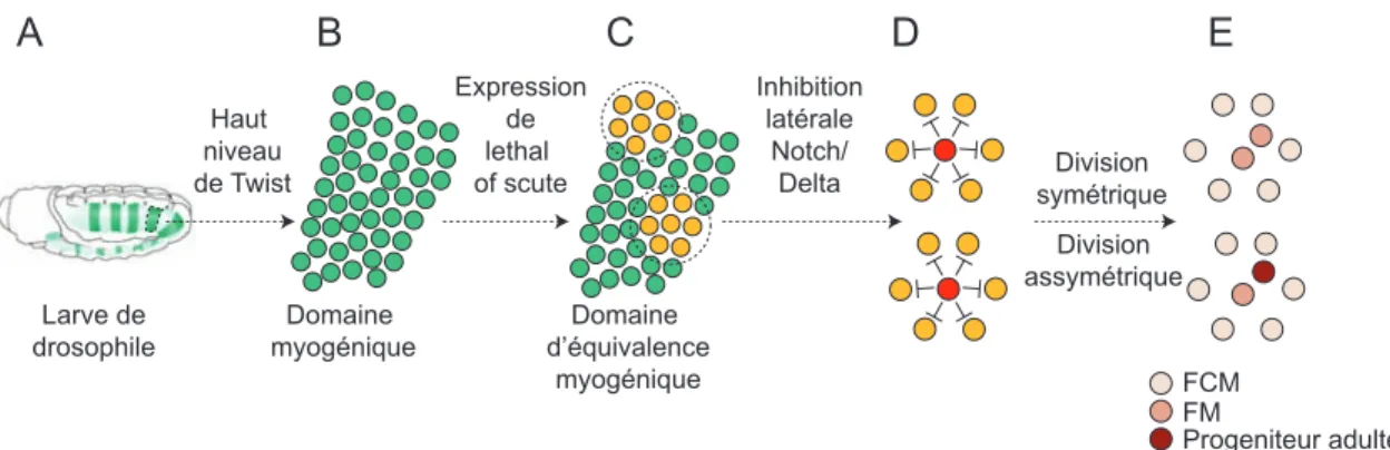
Unraveling the role of the Dock1 pathway in myoblast fusion
Successive rounds of myoblast fusion govern the formation of primary muscle fibers, yet this process is poorly understood at the molecular level in vertebrates (45). Genetic screens in Drosophila were instrumental to uncover cytoskeleton regulators, including myoblast city (ortholog of Dock1) and Rac, which control the fusion steps (45). Mutant mice for Dock1 and Rac1 were generated to address whether this pathway plays a universal role in myoblast fusion. Dock1 mutants died at birth and are characterized by a strong block in primary myoblast fusion both in vivo and ex
vivo (46) (Table 1.I page 11). Likewise, muscle-specific inactivation of Rac1, and also Cdc42, severely impaired myoblast fusion in vivo (47). Mechanistically, both Rac1
and Cdc42-null myoblasts were able to form cell:cell interactions, but the recruitment of F-actin, Vinculin and Vasp at the sites of contact was impaired (47). Identification of direct targets of Rac and precise understanding of actin dynamics is necessary to explain this unique fusion event. How is fusion triggered in vivo? Two receptors, Bai1
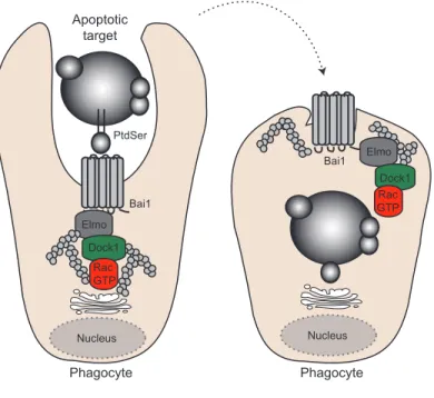
pathway through an interaction with Elmo (48) (Figure 1.1.4 page 13). Downregulation of Bai3 expressed by C2C12 myoblasts completely blocked fusion; and this phenotype could be rescued by re-expression of wildtype Bai3 but not by mutants unable to engage Elmo. In agreement with these data, uncoupling Bai3-Elmo interactions in vivo in muscle progenitors of chicken embryos prevented myoblast fusion. Furthermore, Bai1 was shown to promote apoptotic cell engulfment through the Elmo/Dock1 pathway (Figure 1.1.6 page 21) and its overexpression endorsed fusion in C2C12 myoblasts in an Elmo-dependent manner (49). Mice lacking Bai1 had smaller muscle fibers and were less efficient at repairing injured muscle tissue. As regeneration is likely to involve cell death, apoptotic cells themselves have been shown to activate Bai1 and promote cell fusion, thus providing a unique mechanism where tissue damage recognition and repair activity are coupled.
Bone resorption: Dock5 promotes osteoclast adhesion
Regulation of bone mass is under the control of osteoblast and osteoclast activities (50). Osteoclasts are multinucleated cells that tightly adhere to bones and are involved in their resorption through the secretion of proteolytic enzymes (50). The bone remodeling activity of osteoclasts is dependent on the proper assembly and disassembly of the sealing zone, an actin-rich ring structure, generated by the association of multiple podosomes (51). In the absence of Dock5 expression, the sealing zone is not established properly and the resorbing activity of Dock5-null osteoclast is impaired. Mechanistically, Dock5 was shown to be essential for the generation of this adhesive structure by promoting Integrin αvβ3 signaling via p130Cas phosphorylation and Rac activity (52) (Figure 1.1.4 page 13). In vivo,
Dock5 mutant mice displayed an increased trabecular bone mass, a characteristic of
improper bone resorption (Table 1.I page 11). Pharmacological targeting of the αvβ3/p130Cac/Dock5 pathway could represent a novel avenue to counteract the oscteoclastic activity in osteoporosis patients.