HAL Id: dumas-01643331
https://dumas.ccsd.cnrs.fr/dumas-01643331
Submitted on 21 Nov 2017
HAL is a multi-disciplinary open access archive for the deposit and dissemination of sci-entific research documents, whether they are pub-lished or not. The documents may come from teaching and research institutions in France or abroad, or from public or private research centers.
L’archive ouverte pluridisciplinaire HAL, est destinée au dépôt et à la diffusion de documents scientifiques de niveau recherche, publiés ou non, émanant des établissements d’enseignement et de recherche français ou étrangers, des laboratoires publics ou privés.
Proposition d’un modèle d’évaluation clinique de
sévérité des péri-implantites
Anthony Mazel
To cite this version:
Anthony Mazel. Proposition d’un modèle d’évaluation clinique de sévérité des péri-implantites. Chirurgie. 2017. �dumas-01643331�
ACADEMIE d’AIX-MARSEILLE
Proposition d’un modèle d’évaluation
clinique de sévérité des péri-implantites
THESE
Présentée et publiquement soutenue devant la
Faculté d’Odontologie de Marseille
(Doyen : Monsieur le Professeur Jacques D
EJOU)
Aix Marseille Université
(Président : Monsieur le Professeur Yvon B
ERLAND)
Le 13 octobre 2017
par
MAZEL Anthony
né le 03 janvier 1991
à La Seyne-sur-Mer
Pour obtenir le Diplôme d’Etat de Docteur en Chirurgie Dentaire
E
XAMINATEURS DE LAT
HESE:
Président : Monsieur le Professeur
M. RUQUET
Assesseurs : Monsieur le Professeur
M. DRANCOURT
Monsieur le Docteur
G. ABOUDHARAM
FACULTÉ D’ODONTOLOGIE
UNIVERSITÉ D’AIX-MARSEILLE
DOYENS HONORAIRES Professeur A. SALVADORI
Professeur R. SANGIUOLO†
Professeur H. ZATTARA
DOYEN Professeur J. DEJOU
VICE – DOYEN Professeur J.D. ORTHLIEB
CHARGÉ DES ENSEIGNEMENTS
DIRECTEUR DU DÉPARTEMENT DE FORMATION INITIALE
VICE – DOYEN Professeur C. TARDIEU
CHARGÉ DE LA RECHERCHE
DIRECTEUR DU DÉPARTEMENT DE LA RECHERCHE
DIRECTEUR DU DÉPARTEMENT DE FORMATION CONTINUE
CHARGÉS DE MISSION
Professeur V. MONNET-CORTI Professeur A. RASKIN
Docteur P. SANTONI Docteur F. BUKIET
RESPONSABLE DES SERVICES ADMINISTRATIFS
PROFESSEURS ÉMÉRITES
Madame C. BONNARD
Professeur J. J. BONFIL Professeur F. LOUISE Professeur O. HUE DOCTEURS HONORIS CAUSA DE L’UNIVERSITE D’AIX-MARSEILLE
PRÉSIDENT DE LA SECTION DE LA MÉDECINE DENTAIRE UNIVERSITÉ DE GENÈVE – SUISSE
J.N. NALLY 1972
DOYEN DE LA FACULTÉ DE CHIRURGIE DENTAIRE E. FOREST † 1973
UNIVERSITÉ DE PITTSBURGH – PENNSYLVANIE - USA
DOYEN DE LA FACULTÉ DE MÉDECINE
UNIVERSITE DE GENÈVE – SUISSE L.J. BAUME 1977
DOYEN HONORAIRE DE LA FACULTÉ DE CHIRURGIE DENTAIRE H. GOLDMAN † 1984
UNIVERSITÉ DE BOSTON - MASSACHUSSETTS – USA
56ème SECTION :
DÉVELOPPEMENT CROISSANCE ET PRÉVENTION
56.1 ODONTOLOGIE PÉDIATRIQUE
Professeur C. TARDIEU * Assistant V. MAGNAN
Maître de Conférences D. BANDON Assistant I. BLANCHET
Maître de Conférences A. CHAFAIE Assistant C. KHOURY
Assistant N. RENOU
56.2 ORTHOPÉDIE DENTO-FACIALE
Maître de Conférences J. BOHAR Assistant L. LEVY-DAHAN
Maître de Conférences E. ERARD Assistant S. MARION des ROBERT
Maître de Conférences J. GAUBERT Assistant C. MITLER
Maître de Conférences M. LE GALL * Assistant J. SCHRAMM
Maître de Conférences C. PHILIP-ALLIEZ Assistant A. PATRIS-CHARRUET Assistant M. BARBERO
56.3 PRÉVENTION - ÉPIDÉMIOLOGIE - ÉCONOMIE DE LA SANTÉ - ODONTOLOGIE LÉGALE
Professeur B. FOTI * Assistant J. SCIBILIA
Maître de Conférences D. TARDIVO
7ème SECTION :
SCIENCES BIOLOGIQUES, MÉDECINE ET CHIRURGIE BUCCALE
57.1 PARODONTOLOGIE
Professeur V. MONNET-CORTI * Assistant A. BOYER
Assistant A. MOREAU Assistant M. PIGNOLY Assistant V. MOLL
57.2 CHIRURGIE BUCCALE – PATHOLOGIE ET THÉRAPEUTIQUE - ANESTHÉSIOLOGIE – RÉANIMATION
Maître de Conférences D. BELLONI Assistant J. GARCONNET
Maître de Conférences J. H. CATHERINE * Assistant E. MASSEREAU Maître de Conférences P. ROCHE-POGGI Assistant E. QUINQUE
57.3 SCIENCES BIOLOGIQUES BIOCHIMIE, IMMUNOLOGIE, HISTOLOGIE, EMBRYOLOGIE, GÉNÉTIQUE, ANATOMO-PATHOLOGIE,
BACTÉRIOLOGIE, PHARMACOLOGIE
Maître de Conférences P. LAURENT Assistant C. LE FOURNIS 65ème SECTION : BIOLOGIE CELLULAIRE
Professeur I. ABOUT* (Responsable de la sous-section 57.3)
58ème SECTION :
SCIENCES PHYSIQUES ET PHYSIOLOGIQUES, ENDODONTIQUES ET PROTHETIQUES
58.1 ODONTOLOGIE CONSERVATRICE, ENDODONTIE
Professeur H. TASSERY Assistant B. BALLESTER
Maître de Conférences G. ABOUDHARAM Assistant L. ROLLET
Maître de Conférences F. BUKIET Assistant M. GLIKPO
Maître de Conférences C. PIGNOLY Assistant S. MANSOUR
Maître de Conférences L. POMMEL * Assistant H. DE BELENET
Maître de Conférences E. TERRER Assistant A. FONTES
Maître de Conférences M. GUIVARC’H
58.2 PROTHÈSE CONJOINTE, PROTHÈSE ADJOINTE PARTIELLE, PROTHÈSE TOTALE, PROTHÈSE MAXILLO-FACIALE
Professeur M. RUQUET
Maître de Conférences P. SANTONI * Assistant A. FERDANI
Maître de Conférences G. LABORDE Assistant A. REPETTO
Maître de Conférences M. LAURENT Assistant A. SETTE
Maître de Conférences A. TOSELLO Assistant C. NIBOYET
Maître de Conférences B.E. PRECKEL Assistant C. MENSE
Maître de Conférences P. TAVITIAN Assistant M. DODDS
Maître de Conférences G. STEPHAN
58.3 SCIENCES ANATOMIQUES ET PHYSIOLOGIQUES OCCLUSODONTOLOGIE, BIOMATÉRIAUX, BIOPHYSIQUE, RADIOLOGIE
Professeur J. DEJOU Assistant T. GIRAUD
Professeur J. D. ORTHLIEB * Assistant M. JEANY
Professeur A. RASKIN
Maître de Conférences A. GIRAUDEAU Maître de Conférences J. P. RÉ
Maître de Conférences B. JACQUOT
Remerciements
À Monsieur le Professeur RUQUET Michel
Docteur en Chirurgie Dentaire
Docteur de l’Université Aix-Marseille
Professeur des Universités
Praticien hospitalier des CSERD
Sous-section Prothèses
Vous avoir comme Président de mon jury est un grand honneur. Je vous
remercie pour votre enseignement de qualité au cours mon internat. En plus
de votre très grande compétence, j’ai découvert une personne avenante,
humble et disponible malgré votre emploi du temps très chargé.
Veuillez trouver ici la marque d’un profond respect et l’expression de mes
plus chaleureux remerciements.
À Monsieur le Docteur ABOUDHARAM Gérard
Docteur en Chirurgie Dentaire
Docteur de l’Université Aix-Marseille
Maître de Conférences des Universités
Praticien hospitalier des CSERD
Sous-section Odontologie Conservatrice, Endodontie
Je suis très heureux que vous ayez accepté de diriger mon travail. Vous ne
reculez jamais devant une tâche supplémentaire et vous l’assumez
admirablement. Merci de m’avoir soutenu et d’avoir été disponible chaque
fois qu’il le fallait.
J’ai beaucoup appris de vous durant ces trois années d’internat, vous m’avez
transmis l’amour de la CFAO. Je vous admire tant sur le plan professionnel
que personnel, vous m’avez appris à ne jamais baisser les bras, même dans
les coups durs. C’est un réel honneur de vous avoir comme Directeur de
Thèse.
À Monsieur le Docteur TAVITIAN Patrick
Docteur en Chirurgie Dentaire
Docteur de l’Université Aix-Marseille
Maître de Conférences des Universités
Praticien hospitalier des CSERD
Sous-section Prothèses
Cela a été un grand honneur pour moi de pouvoir apprendre à vos côtés lors
de ces années et je vous remercie de participer à ce jury. Votre grande
expérience clinique, humaine et votre humour font de vous un enseignant
unique et un modèle à suivre.
Veuillez trouver dans ce travail l’expression de mon immense admiration et
de mon profond respect.
À Monsieur le Professeur DRANCOURT Michel
Professeur des Universités
Praticien hospitalier
IHU Méditerranée Infection
Unité de Recherche sur les Maladies Infectieuses et Tropicales Émergentes
Habilitation à diriger des recherches
Je suis très heureux que vous ayez accepté de siéger au sein de ce jury. Votre
passion pour la microbiologie est communicative. J’apprécie beaucoup votre
spontanéité et votre humour. Travailler à vos côtés fut un réel plaisir.
Veuillez accepter mes sincères remerciements et ma vive gratitude.
TABLE DES MATIÈRES
I. INTRODUCTION ...1 II. ARTICLE “PERI‐IMPLANTITIS RISK FACTORS : A PROSPECTIVE EVALUATION” ...3 Abstract ... 4 Introduction ... 5 Methods ... 6 Model development ... 6 Bleeding and / or pus on probing ... 6
Pocket depth ... 6
Bone loss ... 7
Excessive cement ... 8
Oral Hygiene ... 9
Antecedents of Periodontal Diseases... 10
Tobacco smoking ... 11
Diabetes ... 12
Calculation method ... 13
Bleeding and / or pus on probing ... 13
Pocket depth ... 13
Bone loss ... 13
Antecedents of Periodontal Diseases... 13
Glycemic Status ... 13
Tobacco smoking ... 14
Oral hygiene ... 14
Excessive cement ... 14
The developed model ... 15
Low risk of peri-implantitis ... 15
Moderate risk of peri-implantitis ... 15
High peri-implantitis risk ... 15
Evaluation of the severity analysis model ... 16
Results ... 17 Discussion ... 18 Table and figure references ... 21 References ... 29 III. LA FLORE MICROBIENNE DES PÉRI‐IMPLANTITES ... 39 IV. ARTICLE “PERI‐IMPLANTITIS‐ASSOCIATED METHANOGENS: A PRELIMINARY REPORT” . 41 Abstract ... 42 Introduction ... 43 Patients and methods ... 44 Results ... 47 Discussion ... 48 Table and figure references ... 50 References ... 53 V. CORRÉLATION ENTRE LA DETECTION DE M. ORALIS ET LE NIVEAU DE RISQUE PÉRI‐ IMPLANTAIRE ... 55 VI. DISCUSSION ... 55 VII. CONCLUSION ... 59 VIII. ANNEXES ... 60 Annexe 1 : FORMULAIRE DE CONSENTEMENT ... 60 Annexe 2 : QUESTIONNAIRE PATIENT PERI‐IMPLANTITES ... 61 Annexe 3 : OUTIL DIAGNOSTIQUE EN LIGNE ... 62 IX. BIBLIOGRAPHIE ... I
I. INTRODUCTION
Lors de l’insertion d’un implant, après obtention de l'ostéo‐intégration, des complications inflammatoires d'origine infectieuses peuvent apparaître et affecter les tissus péri‐ implantaires. Le premier stade de ces complications est la mucosite ou inflammation de la muqueuse sans perte osseuse. La mucosite est réversible avec un traitement adapté (1). Le stade suivant des complications est la péri‐implantite. Cette complication est associée à une perte osseuse et n’est pas réversible. La prévalence des péri‐implantites est estimée à 10% des implants et 20% des patients au cours des 5/10 ans après la pose de l'implant (2,3). En général, on observe dans une péri‐implantite une perte osseuse marginale en forme de cratère autour de l’implant. Elle s’accompagne la plupart du temps par des manifestations cliniques diverses telles qu’une accumulation de plaque excessive, de saignement au sondage, de récession marginale et une difficulté à mettre en œuvre une hygiène appropriée. Cette pathologie représente un enjeu de santé publique car l’implantologie est devenue une technique de choix pour remplacer les dents manquantes et les données de l’industrie montrent une augmentation croissante de l’utilisation des dispositifs implantaires. On peut arguer de ces éléments une évolution parallèle du nombre de péri‐implantites.
Par ailleurs, si les stratégies de traitement ne montrent pas toutes une efficacité totale (9), différents points caractérisent une péri‐implantite. Une évaluation de la totalité des éléments cliniques accompagnant cette pathologie s’avère donc nécessaire pour adopter les décisions thérapeutiques appropriées (4). L’absence de gradation de la maladie ne permet pas de différencier les divers degrés de péri‐implantite ainsi que les facteurs de risques associés. Des classifications plus ou moins sommaires ont été publiées au fil des années ; parmi elles, celle de Froum et Rosen proposée en 2012 (5) présentant le même écueil que les autres : une absence de prise en compte de tous les paramètres influant sur la péri‐implantite. Il en résulte une confusion dans l’interprétation des données des études évaluant la prévalence, les traitements mis en place et leurs résultats thérapeutiques. Une évaluation standardisée paraît nécessaire pour mettre en œuvre les traitements les plus adaptés en fonction de la
progression de la pathologie. L’objectif de cette étude est de proposer un modèle d'évaluation original du risque de péri‐implantite en s’inspirant du modèle d’évaluation du risque parodontal publié par Chandra en 2007 (6).
Le travail constituant cette étude a été rédigé et est actuellement soumis pour publication à la revue : Oral Health and Preventive Dentistry
II. ARTICLE “PERI‐IMPLANTITIS RISK FACTORS :
A PROSPECTIVE EVALUATION”
Peri-implantitis risk factors: a prospective evaluation
Anthony Mazel2, Souad Belkacemi1, Patrick Tavitian2, Grégory Stéphan2, Delphine Tardivo2,
Jean Hugues Catherine2,Gérard Aboudharam12*
1 Aix-Marseille Université, CNRS, IRD, INSERM, AP-HM, URMITE, IHU Méditerranée-Infection, Marseille, France
2 UFR Odontologie, Aix-Marseille Université, Marseille, France
* Corresponding author: URMITE, IHU - Méditerranée Infection, 19-21 Boulevard Jean Moulin, 13005 Marseille, France. Phone: (+33) 4 13 73 24 01 Fax: (+33) 4 13 73 24 02
Email: gerard.aboudharam@univ-amu.fr
Acknowledgments: The authors thank Professor Michel Drancourt for his help in the design of this work and his advice
Abstract
Purpose: This study was carried out with the aim to create a score allowing an evaluation of
the risk of peri-implantitis evolution according to its severity and to validate this tool as a new model for peri-implantitis.
Materials et methods: Forty-three patients, 28 of whom were diagnosed with peri-implantitis
on at least one implant and 15 patients with implant therapy, were included prospectively in the study in the odontology department of “La Timone” Hospital at Marseille (France). Participants filled in a questionnaire and underwent a thorough clinical examination, a systematic retro-alveolar intra-oral radiography of the implant(s) and a 3D radiography. The following criteria were recorded: the number of implant faces showing bleeding and / or borehole suppuration, the pocket depth on at least two faces of the implant, bone loss as a function of the length of the implant evaluated on X-rays, the number of implant faces with plaque, the parameters required for the determination of excess cement (screwed or sealed prosthesis, burial of sealed prostheses), periodontal status, glycaemia and annual consumption of tobacco. Through Microsoft Excel®, each of these parameters was plotted on a radar chart.
Results: In the proposed evaluation model, 16/28 (57.1%) of cases were identified with high
peri-implantitis risk compared to only 2/28 (7.2%) of cases with low risk. The remaining 10/28 (35.7%) correspond to patients with moderate implantitis risk. All patients without peri-implantitis 15/15 (100%) were considered at low risk.
Conclusions: The observed results applied to the proposed evaluation model, constitute an
effective diagnostic tool in the peri-implantitis risk assessment. The systematic use of this tool allows an early detection and management of peri-implantitis to improve treatment.
Introduction
When inserting an implant, after obtaining osseointegration, inflammatory complications of infectious origin may appear and affect the peri-implant tissues. The first stage of these complications is mucositis or inflammation of the mucosa without bone loss. Mucositis is reversible with appropriate treatment (1). The next stage of complications is peri-implantitis. This complication is associated with bone loss and is not reversible. The prevalence of peri-implantitis is estimated at 10% of implants and 20% of patients within 5/10 years after implant placement with an increase in prevalence in case of tobacco or old periodontitis (2,3). A more recent study reported a prevalence of 8.8% at 10 years in 20% of patients followed for the evaluation of complications of peri-implant tissues without regular maintenance program (4). Serino observed in his study of partial edentulous patients that peri-implantitis observed were mostly related to accessibility to oral cleaning (5). The prevalence of peri-implantitis correlated with the number of implants inserted in an individual would increase but had no influence on the prevalence of mucositis (6). The peri-implantitis does not affect all patients similarly. Literature reviews observed a significant increase in the prevalence of peri-implantitis in subjects treated for periodontitis (7).
There is no real biological evidence of the disruption of integration nor a description of this phenomenon, Clementini in its systematic review and meta-analysis reveals a multifactorial etiology (8). In general, peri-implantitis has a marginal bone loss in the form of a crater around the implant. It is most often accompanied by various clinical manifestations, such as excessive plaque, bleeding during probing, marginal recession and difficulty in implementing appropriate oral hygiene.
This pathology represents a public health issue because implantology has become a technique of choice to replace lost teeth and industry data (unpublished) show an increase in the use of implant devices. One can argue that these elements are connected to the evolution of the number of peri-implantitis. Moreover, if treatment strategies do not show any total efficiency (9), different points characterize a peri-implantitis. An evaluation of all the clinical elements accompanying this pathology is therefore necessary to adopt the appropriate therapeutic decisions (10). The absence of gradation of the disease does not make it possible to differentiate the various levels of peri-implantitis as well as the associated risk factors. This results in confusion in the interpretation of data from studies evaluating the prevalence, treatment and outcomes of therapeutics (11). A standardized evaluation is needed to implement the most appropriate treatment based on the severity of peri-implantitis whose prognosis remains
uncertain. Moreover, peri-implantitis is a public health problem because the number of implants has been increasing in recent years (unpublished industry data) and probably also the number of peri-implantitis. In this work, we developed a new peri-implantitis evaluation score based on the periodontal risk assessment model (12).
Methods
Model development
A model of periodontal risk assessment (bleeding on probing, prevalence of residual periodontal pockets, loss of teeth, estimation of loss of periodontal support in relation to the patient's age, evaluation of systemic diseases, tobacco habits) was described in 2003 (13). A block diagram taking into account these parameters has been described. This first insufficient approach gave rise to a second functional diagram characterizing more precisely the periodontal score by adding as additional parameters: glycemic status, plaque accumulation, socio-economic factors and stress (12). We developed a new peri-implantitis score based on the one previously reported for periodontitis (12). This score included: bleeding and / or pus on probing, pocket depth, bone loss, excess cement, oral hygiene, history of periodontal disease, tobacco habits and glycemic status.
Bleeding and / or pus on probing
Peri-implantitis is an inflammatory disease affecting soft tissues and hard tissues around an osteo-integrated implant, this why evaluation of the presence of inflammation and bone loss are essential parameters for any diagnostic classification (14). In the classification proposed by Froum and Rosen (11), bleeding and / or pus on probing were mentioned as the best clinical indicators for determining the presence of inflammation. Bleeding can be easily assessed by indicating the presence or absence of bleeding after probing with a colored and graduated periodontal probe or a foam probe with a 0.4 mm diameter end on the 6 faces of the implant. The presence or absence of bleeding is determined by the occurrence of bleeding within 15 seconds following a light probing (15).
Pocket depth
Pocket depth may vary depending on the covering mucosa, the presence or absence of keratinized tissue, the type of restoration limiting the probing and also the pressure applied (16). In addition, it has been established that in the presence of inflammatory tissue, a periodontal
probe penetrates the tissues more deeply (17). Considering that peri-implant probing measurements are more sensitive to variations in force than the probing of periodontal pockets, it was recommended to apply a sampling pressure equivalent to 0.25 N (18).
Instead of using a probe equipped with a pressure sensitive device, a light probing is recommended around the implant. This sampling method, which was recommended to determine pocket depth for this classification, was found to be reproducible to ± 1 mm in more than 95% of cases (19). Excessive probing pressure, even on a complex of healthy soft tissues, can cause fiber breakage and therefore provide false-enhanced sounding mapping. However, a lightweight ≥ 4 mm sound is an appropriate clinical reference in assessing the healthy or pathological status of the peri-implantous mucosa (16). The clinical status of the peri-implant mucosa can therefore be determined by associating depths of probing and bleeding and / or pus with the probing on at least two faces of the implant.
Bone loss
Implants vary in shape and morphology. Numerous studies are based arbitrarily on a bone loss ≥ 1.8 mm (corresponding on average to the third coil of an implant) for the diagnosis of peri-implantitis (20-22). A vertical bone resorption of less than 0.2 mm after the first year of function of an implant is today a generally accepted criterion of success (23). However, accurate assessment of this height is difficult to measure clinically. Therefore, the diagnosis of peri-implantitis based on a crater ≥ 1.8 mm after one year seems difficult to apply by the clinicians (24). Standardized X-ray images can help determine the exact level of bone loss in relation to a fixed reference point (crown-implant junction or implant-abutment for example) but this is difficult to measure and compare in millimeters. The evaluation proposal is therefore based on a comparison of bone resorption over time, determined by changes in the percentage of bone loss relative to the length of the implant.
Following the example of Forum and Rosen's classification (11), several ranges of percentage of bone resorption relative to the length of the implant are found: <10%, 10% to 20%, 20% to 30%, 30% to 40%, 40% to 50%, > 50%. This facilitates the determination of a distinct change in the severity of a peri-implantitis, from an early to moderate stage, from a moderate to advanced stage. Combined with bleeding and / or pus on probing, the bone loss can diagnose the pathology at its earliest clinically detectable stages and resolve the problem of unnecessary patient overexposure to X-rays. Moreover, the fact that the ratio of peri-implantitis progression / associated bone resorption does not follow a linear curve (acceleration of bone loss over time) underlines the need for early diagnosis and management of pathology (25). Accurate
measurement of the amount of bone resorption around the implants is one of the essential conditions for differentiating the categories of the proposed evaluation model. Several authors emphasize the importance of acquiring a retro-alveolar x-ray on the day of implant insertion with the final prosthesis. An orthopantomogram can hardly replace a retro-alveolar radiography because of deformations preventing any precise comparison. This reference radiography makes it possible to establish the initial ratio between junction between the implant and the pillar (or implant-crown junction) and the first bone-implant contact, making it possible to compare them with subsequent shots in order to determine the “Extent of bone loss at the mesial and distal surfaces of the implant”. Removal of the prosthesis during radiographic evaluation may be necessary when it prevents an accurate estimate of the bone level. Probing to the highest level of the buccal and lingual (or palatal) bones after anesthesia of the peri-implant tissues can help to determine the bone levels of these sites, which cannot be verified on a standard retro-alveolar x-ray. This system preserves the bones from the exposure to X-rays. It also avoids the error of bone loss expressed in number exposed turns, which may vary according to the implant system. If the implant exhibits vestibular cortical bone loss due to poor positioning and absence of bleeding at the mucosal level, it is likely that the bone defect is the result of physiological resorption and insufficient vascularization in the vestibular cortical bone (26). Most defects caused by peri-implantitis affect more than one surface. Therefore, determining the level of risk in the proposed classification requires at least a probing depth ≥ 4 mm associated with bleeding and a bone loss on two or more faces of the implant. The mandatory condition to be respected is that the implant must be positioned at a sufficient distance from the vestibular cortical. An implant placed in an excessive vestibular position may result in damaging the alveolar bone and soft tissue not induced by bacteria (27). Thus, the classification category is determined by the most severe deterioration of the implant, all faces combined. In order to avoid classification errors, it is therefore important to measure at least two faces of the evaluated implant.
Excessive cement
Prosthetic cemented crowns on implant represent a common alternative to later-screwed-retained implant crowns. The use of prosthetic cemented crowns on implants frequently results in excessive cement deposits in the peri-implant tissues despite careful clinical control at the time of sealing (28). A further study from Linkevicius (29) showed that the more the cervical limit of a crown is embedded, the greater is the quantity of residual cement. It also underlines the unpredictability of radiographic evaluation in the detection of possible cement excesses. In 4/53 cases (7.5%), cement remains were visible on the mesial surface and in 6/53 cases (11.3%)
on the distal side of the implants. Therefore, dental x-rays cannot be considered as a reliable method of evaluating excessive cementing (29). An excess of cement can be assimilated by a foreign body and thus trigger an inflammatory response leading to a peri-implantitis. Indeed, these cement remains can be the starting point of colonization by oral microorganisms resulting in the appearance of a mucositis and peri-implantitis. Excess cement samples were taken from ten patients to study the formation of the biofilm by molecular method based on 16S rDNA sequencing (30). The results of the in-situ hybridization revealed a strong tendency towards the bacterial invasion of methacrylate-based cements. Wilson, using dental endoscopy, found an association between excessive sealing cement and peri-implant pathology in 81% of cases. After suppression of the excesses, clinical and endoscopic signs of peri-implantitis were absent in 74% of the studied implants (31). Although few studies have examined the association between excessive cement and peri-implantitis, the data clearly indicate that an excess of cement can trigger an inflammatory response that causes clinical signs of peri-implantitis / mucositis peri-implant.
Oral Hygiene
As soon as a part of a dental implant is exposed in the oral cavity, a bacterial colonization of this exposed surface occurs (32, 33). In a prospective study, Lindquist associates poor oral hygiene with peri-implant bone resorption (34). From a subsample of 47 initially included in the study (13 patients with good oral hygiene and 14 patients with poor oral hygiene), a 10-year difference was observed. An average bone loss of 0.65 mm was observed in the group with good oral hygiene while 1.65 mm in the group with poor oral hygiene. Inappropriate accessibility to oral hygiene of the implanted sites has also correlated with the peri-implantitis (5). In this study, 48% of implants with peri-implantitis were those that did not allow for proper oral hygiene due to limited access or no access. An analysis of the risk factors of peri-implant pathology by Ferreira et al. (35) has allowed poor oral hygiene to be considered a risk factor for peri-implantitis. In a transversal study Roos-Jansåker (36) reported that the presence of dental plaque could explain the manifestation of mucositis peri-implant but not peri-implantitis. A 3-year follow-up study (37) identified dental plaque as a significant factor in the development of peri-implantitis. The accumulation of dental plaque in relation to dental implants is clearly associated with the development of peri-implant mucositis (38), but the latter does not necessarily evolve into peri-implantitis. When this is the case, it has been demonstrated that the onset of the pathology was preconditioned by an infectious process (39-42).
Antecedents of Periodontal Diseases
According to the criteria used to define periodontitis, prevalence ranges from 12% to 76% in the United States (43, 44). Globally, about 10% of the world population has acute periodontal disease. It has been observed that individuals with risk of periodontitis react differently to microbial attacks than patients without risk (45). Individuals with antecedents of periodontal disease may therefore also have an increased risk of developing peri-implantitis. Several studies on the variables of implant success concluded that patients with antecedents of periodontitis had a significantly greater depth probing, marginal bone resorption and peri-implantitis incidence in the long term compared to patients with a healthy periodontium (36, 46-51). Among studies comparing implant survival rates in patients affected and unaffected by periodontitis, a majority demonstrate higher (sometimes twice higher) failure rates in subjects with periodontitis. A recent study using multivariate analysis showed that tobacco consumption (P = 0.046) and antecedents of periodontitis (P = 0.046) would have an impact on advanced peri-implant bone resorption (52). The presence of periodontal pathogens around failing implants (53-60) could suggest a direct link between periodontitis and peri-implantitis through translocation of these species from their intraoral niches to implant sites as suggested by several studies (40, 32,33). In partially edentulous patients, the teeth act as a reservoir, whereas in the total edentulous patients, the periodontal pathogens remain in the oral cavity, surviving on the tongue or in the saliva (61- 63).
An indirect link may also be considered, especially in patients with aggressive periodontitis who are more prone to peri-implant infections because of their immune response to periodontal pathogens. Thus, several authors report a statistically significant difference in the percentage of peri-implantitis between patients affected and not affected by periodontitis (36, 51, 64-66) with higher values of marginal bone loss around the implants in the periodontitis group (1.3 to 2.0 time more).
Some studies distinguish the patients with moderate or severe periodontitis from those with chronic or aggressive periodontitis. In general, severe / aggressive forms of periodontitis are responsible for the highest rates of peri-implantitis (51,66-69) .
All of these data indicate that patients with a history of periodontal disease are more prone to peri-implantitis, marginal bone resorption around implants, and even implant loss. However, underlying mutual confounding factors, such as tobacco consumption, may partly influence this association (46-50, 70, 71).
While periodontitis can be considered as a plaque-related disease, patients with aggressive periodontitis show certain specificities: an absence of correlation between microbial deposits
and severity of periodontal degradation, high proportions of Porphyromonas gingivalis and
Actinomices actinomycetemcomitans, phagocytosis abnormalities, and macrophage
hyperactivity, including elevated levels of prostaglandin E2 and interleukin IL-1β (72, 73). This type of patient also has a polymorphism of genes involved in the coding of many inflammation regulators including IL-1 and TNF (73-75). These polymorphisms result in an altered inflammatory profile, particularly characterized by trans-endothelial migration and signaling by neutrophilic polynuclear cells (76), a reduced chemotactic response, and a decrease in phagocytosis and superoxide anion production by neutrophils (77).
In other words, in patients with chronic periodontitis, proper dental plaque control and smoking cessation will reduce the risk of recurrence of periodontitis, but also the risk of peri-implantitis. These risk factors also affect patients with an antecedent of aggressive periodontitis, making them more susceptible to peri-implantitis. Monje calculated that the ratio of the risk of implant failure in patients with aggressive periodontitis was significantly higher compared to patients without a history of periodontitis (78) and those with chronic periodontitis (79, 80).
Tobacco smoking
The consumption of tobacco alters various aspects of innate and adaptive host immune responses (81-83). In fact, smokers show an increase in granulocyte count and total white blood cell count (84), an increase in the lifetime of polymorphonuclear cells (85), production of superoxide anion and hydrogen peroxide, the expression of integrins (83) and the production of protease inhibitors (86). The humoral immune response is also disrupted by tobacco addiction (87, 88). Smoking has been shown to inhibit the proliferation and / or function of B and T lymphocytes (89). Nicotine is the most widely studied component of tobacco for its effects on various cell populations of the periodontium. Nicotine concentrations in smokers' cervical gingival fluid, were nearly 300-fold higher than those found in plasma (20 ng/ml) (81). Gingival bleeding and cervical fluid flow increases from 3 to 5 days after smoking cessation (90). This finding, coupled with the finding of post-weaning bleeding increase (91), confirms the suppressive impact of chronic tobacco consumption on gingival fluid flow and vascularization. Several studies have shown that nicotine has properties that can alter healing (92-96).
Studies comparing the subgingival microbiota of smokers and non-smokers have yielded contradictory results (79, 80, 97). Several studies have associated the smoking with periodontitis, dental loss, alveolitis, and altered healing after surgery (98-104).
Data obtained by the studies evaluating the impact of smoking on implant survival clearly demonstrate the negative effect of tobacco consumption. They suggest that individuals who
smoke before or after implantation may be at risk of implant failure between 35% and 70% higher compared to non-smokers. This affirmation is in line with several systematic reviews of the literature (105-108). There were no statistically significant differences in the complications between non-smokers and former smokers (109), indicating that the risk of complications may be reduced to a "normal rate of complication as in the non-smoker" when the tobacco consumption stops. Several prospective clinical trials, with a considerable follow-up time (often more than 5 years), revealed differences in the incidence of peri-implantitis between smokers and non-smokers/ex-smokers.
Although the relationship between smoking and peri-implantitis remains controversial, several studies have reported a statistically significant difference in peri-implantitis incidence between smokers and non-smokers (36, 110), with strong association between peri-implantitis and smoking (111).
Diabetes
The association between periodontal diseases and systemic pathologies has aroused great interest during the last decade. The Workshop of the European Federation of Periodontology and the American Academy of Periodontology on Periodontitis and General Diseases concluded that "there is solid and consistent epidemiological evidence that periodontitis predisposes to increased risk of Cardiovascular disease » (112); "Severe periodontal disease affects the glycemic control of diabetics and influences glycemic levels in non-diabetic patients. In diabetic patients, there is a direct and dose-dependent relationship between the severity of periodontitis and diabetes complications" (113). Based on available data on the association between periodontal disease and systemic pathologies, it seems logical to assume that these pathologies may also have an impact on the development of peri-implantitis. Ferreira demonstrates, through univariate analysis, that subjects with diabetes are more likely to develop peri-implantitis (35). This trend is confirmed by the Gomez-Moreno study: in patients with type 2 diabetes with a follow-up over a three-year period, marginal bone loss and bleeding at the peri-implantation level correlated with the increase of the level of glycated hemoglobin A1c (HbA1C) (114). However, in his recent study, Aguilar-Salvatierra finds that patients with type 2 diabetes can receive implant treatments with immediate safe loading provided they have moderate HbA1c levels (115). Although data on possible associations between systemic diseases and peri-implantitis remain limited up to date, there are certainly relationships between peri-implant disease and diabetes. New studies should be conducted to confirm these associations on larger populations of patients.
Calculation method
Bleeding and / or pus on probing
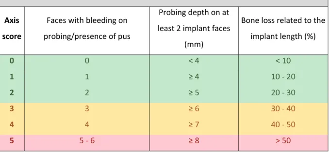
The method of Ainamo et al. (15) cited by Froum and Rosen in 2012 (11) served as a reference. Bleeding on probing can be easily evaluated by indicating the presence or absence of bleeding after probing with a probe on the 6 faces of the implant. The presence or absence of bleeding on probing is determined by the occurrence of bleeding within 15 seconds following a light probing. It is realized on the six sides of the implant: vestibular, vestibular, disto-vestibular, mesio-lingual/mesio-palatal, lingual/palatal, disto-lingual/disto-palatal, measured after 15 seconds and expressed in number of affected implant faces (0 to 6) (Table 1).
Pocket depth
Considering that peri-implant probing measurements are more sensitive to variations in force than the sounding of periodontal pockets, it is recommended to apply a sample pressure equal to 0.25 N (18). The around implant soft clinical probing, recommended to determine the pocket depth for this classification, was found to be reproducible to ± 1 mm in more than 95% of cases (19). In order to have reproducibility, a probing force of 0.25 N is applied using a graduated periodontal probe. This probing is carried out on at least two faces of the implant and is expressed in millimeters (Table 1).
Bone loss
Bone loss is evaluated from retro alveolar intraoral radiography (assessment of the mesial and distal crater), it is expressed as a percentage of bone resorption relative to the length of the implant (Table 1). One possibility is the use of 3D imaging with in particular the Cone Beam which would allow a volume measurement of the bone crater. This device is more and more often present in dental offices and offers a high precision for a lower quantity of x-ray compared to a scanner.
Antecedents of Periodontal Diseases
The score is calculated according to the periodontal status (healthy / treated / pathological) and the type or stage of the periodontal disease (Table 2).
Glycemic Status
In the evaluation model proposed by Chandra (12), the selected parameter is fasting glycaemia. It is expressed in mg/dl. Thus, according to the diagnostic reference values established by the
American Academy of Periodontology (116), a glycaemia value of less than 110 mg/dl implies a low periodontal risk and a value greater than 126 mg/dl, a high risk. Any value between these two values reflects a moderate risk. However, glycaemia provides only a snapshot of glycemic status, while glycated hemoglobin A1c (HbA1c) is useful in assessing glycemic control over a longer period of time (approximately two to three months). It is therefore a more interesting constant ; It is a marker of the risk of long-term complications of diabetes (Table 2).
Tobacco smoking
Referring to earlier models such as Renvert and Persson, the number of cigarette packets per year served as a reference for the study (117). Considering that the number of cigarettes per packet varies between countries, this can be a confounding factor (118). Like Chandra's evaluation model (12), this criterion is now measured in cigarettes per day, in order to take this possibility into account (Table 2).
Oral hygiene
Hygiene is measured by the presence or absence of bacterial subgingival plaque on the 4 faces of the implant: mesial, distal, vestibular and palatal or lingual. It is expressed in number of implant surfaces affected (0 to 4); for implant-supported prostheses (Brånemark prostheses, implant-supported bridges), a further assessment of the accessibility to hygiene is proposed (yes/no): “yes” if the patient can brush in the implanted region using a toothbrush, little brush or dental floss, “no” in case of limited access (linked to the emergence profile in particular) and / or disability of the patient (Table 2).
Excessive cement
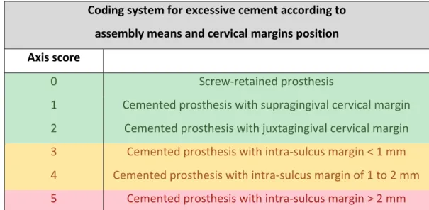
Considering the feature of radiographic evaluation in the detection of excessive cement, the calculation of this parameter is essentially clinical. The exact amount of residual cement is impossible to measure accurately, therefore the correlation between burying the cervical boundary of a sealed crown and the amount of residual cement is used. Indeed, Linkevicius has demonstrated that the more this limit is below the gingiva, the greater is the quantity of cement in excess (29). Therefore, the following parameters have been chosen: the determination of the implant abutment / prosthesis assembly means (screwed prosthesis or a sealed prosthesis); in the case of a sealed prosthesis, the measurement of the relative position of the cervical limit with respect to the marginal gingiva (Table 3).
The developed model
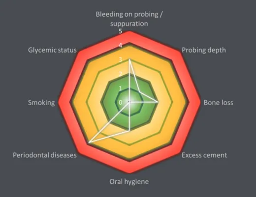
From a graphical point of view, the peri-implantitis risk analysis model is similar to Chandra's model of periodontitis evaluation (12). It generates a functional diagram which, according to the limits of the polygon obtained, allows to classify a patient into 3 categories: low, moderate or high risk. Indeed, the risk is evaluated by means of a color scale ranging from 0 to 3 (Figure 1).
Low risk of peri-implantitis
A peri-implantitis risk is considered to be low if all the parameters are in the low-risk area or if a maximum of two parameters are in the moderate-risk zone and the high-risk area (Figure 2). For example, the patient with low peri-implantitis risk presented 3 out of 6 implant faces with bleeding on probing, a pocket depth < 4 mm on at least 2 faces of the implant, a bone resorption < 30% of implant length (screwed prosthesis), 2 implant faces with plaque, severe chronic periodontitis, and non-smoker and non-diabetic.
Moderate risk of peri-implantitis
A risk of peri-implantitis is considered to be moderate if at least three parameters are in the moderate risk zone and no more than one parameter is in the high-risk area (Figure 3). For example, the patient at moderate risk of peri-implantitis had 3 implant faces on 6 with bleeding on probing, a pocket depth ≥ 6 mm on at least 2 faces of the implant, bone resorption > 30 % of implant length, sealed prosthesis with limit bury under the gingiva < 1 mm, 2 implant faces with plaque, chronic periodontitis treated in 2008 and non-smoker and non-diabetic.
High peri-implantitis risk
A risk of peri-implantitis is considered high if at least 2 parameters are in the high-risk area (Figure 4). For example, the patient at high peri-implantitis risk presented suppuration on all implant surfaces, a pocket depth ≥ 8 mm on at least 2 faces of the implant, bone resorption > 50% of implant length, a screwed prosthesis with intra-sulcus margin of 1 to 2 mm, dental plaque on 1 implant surface, severe chronic periodontitis, was non-smoker and diabetic with HbA1c > 8%.
Evaluation of the severity analysis model
Twenty-eight patients diagnosed with peri-implantitis in at least one implant and 15 patients without peri-implantitis were randomly selected from the odontology department at “La Timone” Hospital in Marseille, France. For each of them, an informed consent form and a questionnaire were obtained. This study is a prerequisite for the approval of the ethics committee of the University Hospital Institute (IHU) in Marseille under number 2016-011. A thorough clinical examination was performed and a retro-alveolar intraoral radiography of the implant(s) with peri-implantitis was also performed. In addition, patients with peri-implantitis were subjected to a 3D radiography of the Cone Bean CBCT type in order to complete the radiological evaluation. In order to avoid errors due to observer variability, all clinical and radiographic examinations were carried out by two previously trained dental surgeons. The parameters recorded were: the number of implant faces with bleeding and / or suppuration on probing, the pocket depth on at least two faces of the implant, the bone loss relative to the implant length evaluated on dental x-rays, the number of implant surfaces with plaque, the parameters required for the determination of excessive cement (screwed or sealed prosthesis, bury limit of sealed prosthesis), periodontal status, diabetes and tobacco consumption. With Microsoft Excel® software, each of these parameters has been plotted on a radar chart. The
proposed model could thus be applied to the cohort of patients with peri-implantitis and controls. The results obtained made it possible to evaluate the risk specific to each patient presenting with peri-implantitis.
Results
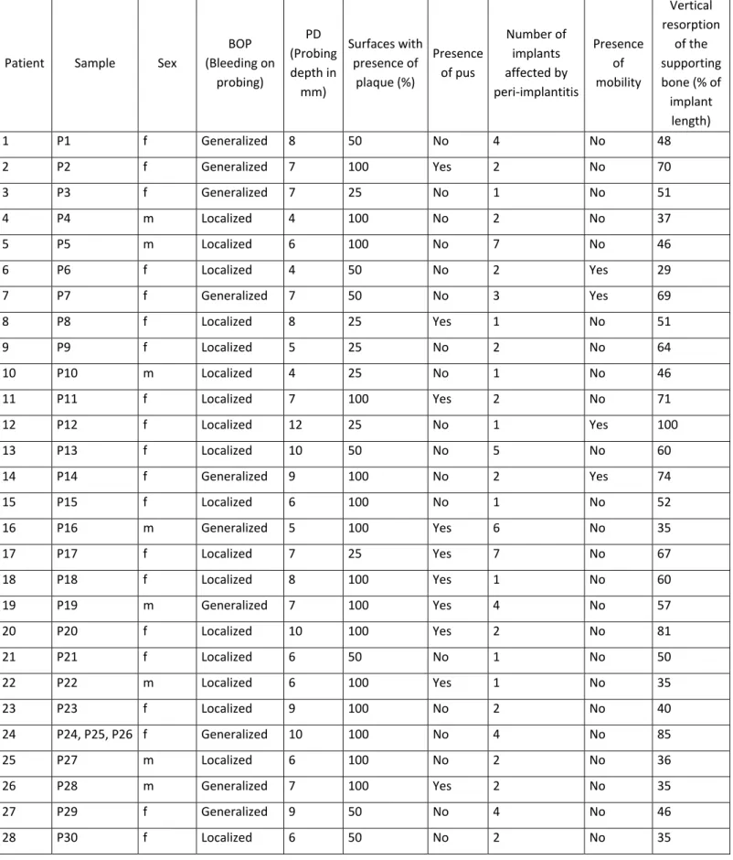
Sixteen male and twenty-seven female patients aged 35-86 with an average age of 62.7 years were enrolled in the study: 28 patients with implantitis and 15 patients without peri-implantitis. All patients received implant therapy. Three peri-implantitis cases were found in the same patient. In total, in individuals with peri-implantitis, 10 had generalized bleeding and 18 had localized bleeding. Pocket depths ranged from 4 to 12 mm. The number of implants affected by the peri-implantitis varied from 1 to 7. In 4 cases of peri-implantitis, the presence of pus was observed. Vertical resorption of the carrier bone (% of implant length) ranged from 29 to 100% (100% in only one case) (Table 4). In addition, 4 individuals were smokers and two other patients were former smokers. One patient was confirmed diabetic. Finally, 22 individuals had active chronic periodontitis, including a debilitating form, four moderate and 17 severe forms. Of the remaining six patients, 3 had a history of treated and stabilized chronic periodontitis. The last 3 showed no signs of periodontal disease.
Of the 28 patients with peri-implantitis, 25 had one (or several) peri-implantitis(s) associated with an implant-supported crown or implant-supported bridge. Thirteen prosthetic elements were assembled by sealing, and twelve screwed. The remaining three patients had complete "Bridge Brånemark" prostheses.
In the proposed evaluation model, 2 cases with low peri-implantitis risk were identified, while 10 moderate-risk and 16 high-risk cases were identified. Approximately 57.1% of cases are in the high-risk peri-implantitis category while only 7.2% of cases are in the low-risk category. The remaining 35.7% correspond to patients with moderate peri-implantitis risk (Table 5). Among the control patients, 7 presented with localized bleeding, 2 with generalized hemorrhage and 6 patients showed no bleeding. Pocket depths ranged from 1 to 3 mm with the exception of one patient with a pocket which measured 4 mm. The presence of bacterial plaque was observed in 7 patients on 25% of the surfaces of the implants. No pus was found in any case.
Discussion
This study was conducted to develop a risk assessment of peri-implantitis, the proposed model is based on an already published effective model evaluating periodontal risk (12). This previous model has proved its usefulness and allowed to evaluate precisely the periodontitis. The model developed was tested on 28 patients with a peri-implantitis and 15 patients with no peri-implantitis. This work allowed to classify patients with peri-implantitis in three categories, each corresponding to a level of risk of evolution of peri-implantitis (low: 2/28 (7.2%), moderate: 10/28 (35,7%), high: 16/28 (57.1%)) All 15 control patients were classified in the low risk area, which validates the model developed.
The obtained score provides quantitative information to the clinicians, while the polygon from the functional diagram adds a qualitative component to the diagnosis. The advantage of the proposed model is therefore double: it is a tool to aid diagnosis while standardizing individual care. After reviewing the literature, eight parameters were selected to be part of the proposed evaluation model; however, several criteria, notably of a clinical nature, were not included. Among them, occlusion and the amount of keratinized mucosa must be mentioned. Concerning the occlusal factor, a biomechanical overload at the bone-implant interface could be the cause, or at least contribute to a marginal bone loss according to the hypothesis of occlusal surcharge. Some recent studies consider that occlusal load / occlusion could be a contributing factor to peri-implantitis (119, 120) But the question remains open and this is the reason for not including this parameter into this model. However, although evidence of the impact of occlusal surcharge on peri-implantitis is lacking, an assessment of patient occlusion during maintenance visits seems desirable (121, 122).
The presence of keratinized gingiva and its required level of thickness in the peri-implant region has little influence as far as proper oral hygiene is maintained. Contrary to the keratinized mucosa which when absent can lead to difficulty in accessing and / or a sensitivity to brushing which impairs the control of the dental plaque and leads to important tissue lesions. For cases with a keratinized gingiva defect in the proximity of the implant, the study concludes that the lack / absence of tissue does not necessarily negatively affect the health of the peri-implant tissues. In the event that the absence of keratinized gingiva would interfere with the control of the elimination of the dental plaque, this proves to be an indirect impact parameter. According to the proposed model, it makes more sense to include a direct assessment of oral hygiene, rather than a clinical criterion influencing it. Despite the fact that this parameter is not included in the evaluation model, the question of the importance of keratinized mucosa at the
peri-implant level persists and this factor should be discussed in future studies on the incidence of peri-implant diseases.
The genotype and its association with tobacco consumption were not included in this model. According to the literature, the polymorphism of the IL-1 gene would have an impact on the peri-implantitis, especially in the case of heavy smokers. Feloutzis et al. (123) reported significant differences in alveolar bone loss between non-smokers and heavy smokers in the "IL-1 positive genotype" group and none in the "IL-1 negative genotype" group. Gruica (124) showed that heavy smokers with positive IL-1A and IL-1B genotypes had a higher risk of developing inflammatory complications and increased peri-implantous marginal bone resorption compared to non-smokers of the same genotype. A latest study by Laine et al. (125) found an association between the polymorphism of the IL-1RN gene and peri-implantitis, particularly in smokers. The proposed evaluation model is based on a simple, rapid and reproducible measurement of each parameter, while remaining accessible to the clinician in his everyday practice. Given the multiple limitations associated with a genotype study, it was decided not to include this criterion into the evaluation model.
It is possible to compare the proposed evaluation model with the work of Froum and Rosen (11) on a new classification of peri-implantitis. Froum classifies the severity of peri-implantitis based on three clinical criteria: bleeding and / or probing suppuration, pocket depth, and peri-implant bone resorption level. The objective of his work is to propose a standardized classification that can be used for communication between researchers and clinicians, allowing a better comparison of the results and calibration of the studies. This classification is limited because it defines an implant status by only three criteria whereas our proposed model allows us to define a risk of progression of the peri-implantitis basing on eight parameters calibrated between them. While Forum measures the severity of the pathology at a specific time, our proposed evaluation model assesses its susceptibility to evolution. The comparison of the results of this study with that of Froum and Rosen makes it possible to demonstrate a link between the severity of a peri-implantitis and its susceptibility to aggravation: the more severe the peri-implantitis is, the greater is the risk of progression. On the contrary, the earlier the diagnosis of peri-implantitis is established, the less likely it is to develop. This correlation confirms the non-linear evolution of peri-implant pathologies, a fortiori if aggravating factors are added (diabetes, tobacco consumption, periodontal disease, residual cement, poor oral hygiene) as shown by the qualitative analysis allowed by the proposed functional diagram (Figure 1).
The results of this study highlight one singularity: the low percentage of patients with low peri-implantitis risk 2/28 (7,2%). This reflects the difficulty in diagnosing early forms, which are often asymptomatic with little or no clinical manifestations and are most often the object of fortuitous discoveries. Moreover, the therapeutics is all the more effective as the management of the peri-implantitis is precocious (25). As Serino and Turri demonstrated in their study (126), the proportion of unhealthy implants returned healthy two years after surgical treatment was higher for implants with a smaller initial bone crater (2-4 mm) compared to implants with initial bone loss ≥ 5 mm (respectively 74% and 40%).
From early diagnosis to regular maintenance, to the rapidity of treatments, prevention has an essential role in the management of peri-implantitis and in reducing their occurrence (9). It is also important to underline the multifactorial aspect of this pathology, hence the importance of an overall understanding of the parameters influencing the level of peri-implantitis risk. The ultimate aim is to categorize each patient and to adapt the therapy individually according to the level of risk of each one.
In order to ensure that this work can serve all students, practitioners and research community the diagnostic tool is available at the following address: http://diagnostic-tool.pagesperso-orange.fr/.
The calculation rules are the same as those described in this article. This online availability is part of the purpose of screening and early care to enhance the outcome of peri-implantitis treatment.
Table and figure references
Table 1: Coding system for bleeding on probing and/or suppuration, probing depth and bone
loss Coding system for bleeding on probing / presence of pus, probing depth and bone loss Axis score Faces with bleeding on probing/presence of pus Probing depth on at least 2 implant faces (mm) Bone loss related to the implant length (%) 0 0 < 4 < 10 1 1 ≥ 4 10 ‐ 20 2 2 ≥ 5 20 ‐ 30 3 3 ≥ 6 30 ‐ 40 4 4 ≥ 7 40 ‐ 50 5 5 ‐ 6 ≥ 8 > 50
Table 2: Coding system for periodontal diseases, glycemic status, smoking and oral hygiene
Coding system for periodontal diseases, glycemic status, smoking and oral hygiene Axis Score Periodontal status HbA1c level (%) Smoking (cigarettes/day) No. of implant faces with presence of plaque 0 Healthy periodontium ≤ 6 Non‐smoker 0 1 Treated periodontitis 6,1 ‐ 7 Former‐smoker 1 2 Slight chronic periodontitis 7,1 ‐ 8 < 10 2 3 Moderate chronic periodontitis 8,1 ‐ 9 10 ‐ 19 3 4 Severe chronic periodontitis 9,1 ‐ 10 20 4 5 Aggressive periodontitis > 10 > 20 No accessibility to oral hygiene
Table 3: Coding system for excessive cement according to assembly means and cervical margins position. Coding system for excessive cement according to assembly means and cervical margins position Axis score 0 Screw‐retained prosthesis 1 Cemented prosthesis with supragingival cervical margin 2 Cemented prosthesis with juxtagingival cervical margin 3 Cemented prosthesis with intra‐sulcus margin < 1 mm 4 Cemented prosthesis with intra‐sulcus margin of 1 to 2 mm 5 Cemented prosthesis with intra‐sulcus margin > 2 mm
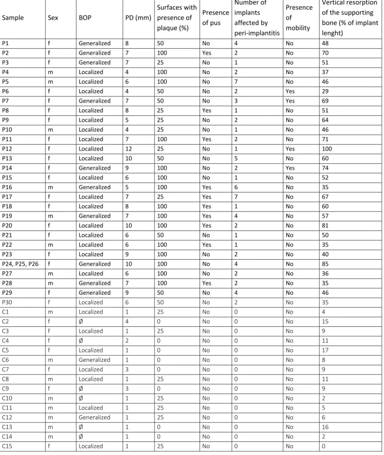
Table 4: Clinical data collection of pathological implants (P) and controls (C): sex, with
bleeding on probing (BOP), probing depth (PD), surfaces with presence of plaque (%), presence of pus, number of implants affected by peri-implantitis, mobility, vertical resorption of the supporting bone (% of implant length).
Sample Sex BOP PD (mm)
Surfaces with presence of plaque (%) Presence of pus Number of implants affected by peri‐implantitis Presence of mobility Vertical resorption of the supporting bone (% of implant lenght)
P1 f Generalized 8 50 No 4 No 48 P2 f Generalized 7 100 Yes 2 No 70 P3 f Generalized 7 25 No 1 No 51 P4 m Localized 4 100 No 2 No 37 P5 m Localized 6 100 No 7 No 46 P6 f Localized 4 50 No 2 Yes 29 P7 f Generalized 7 50 No 3 Yes 69 P8 f Localized 8 25 Yes 1 No 51 P9 f Localized 5 25 No 2 No 64 P10 m Localized 4 25 No 1 No 46 P11 f Localized 7 100 Yes 2 No 71 P12 f Localized 12 25 No 1 Yes 100 P13 f Localized 10 50 No 5 No 60 P14 f Generalized 9 100 No 2 Yes 74 P15 f Localized 6 100 No 1 No 52 P16 m Generalized 5 100 Yes 6 No 35 P17 f Localized 7 25 Yes 7 No 67 P18 f Localized 8 100 Yes 1 No 60 P19 m Generalized 7 100 Yes 4 No 57 P20 f Localized 10 100 Yes 2 No 81 P21 f Localized 6 50 No 1 No 50 P22 m Localized 6 100 Yes 1 No 35 P23 f Localized 9 100 No 2 No 40 P24, P25, P26 f Generalized 10 100 No 4 No 85 P27 m Localized 6 100 No 2 No 36 P28 m Generalized 7 100 Yes 2 No 35 P29 f Generalized 9 50 No 4 No 46 P30 f Localized 6 50 No 2 No 35 C1 m Localized 1 25 No 0 No 4
C2 f Ø 4 0 No 0 No 15
C3 f Localized 1 25 No 0 No 9
C4 f Ø 2 0 No 0 No 11
C5 f Localized 1 0 No 0 No 17 C6 m Generalized 1 0 No 0 No 8 C7 f Localized 3 0 No 0 No 9 C8 m Localized 1 25 No 0 No 11
C9 f Ø 3 0 No 0 No 9
C10 m Ø 1 25 No 0 No 2
C11 m Localized 1 25 No 0 No 5 C12 m Generalized 1 25 No 0 No 6
C13 m Ø 1 0 No 0 No 16
C14 m Ø 1 0 No 0 No 2
Table 5. Distribution of low, moderate and high-risk cases according to the proposed
assessment model.
Distribution of low, moderate and high-risk cases according to the proposed model
Risk level
Total
Low risk Moderate risk High risk
Figure 1: Proposed risk diagram with the clinical criteria, radiographic measures and risk
Figure 2: Patient with low peri-implantitis risk: 3 out of 6 implant faces with bleeding on
probing, a probing depth < 4 mm on at least 2 faces of the implant, a bone resorption < 30% of the implant length (screwed prosthesis), 2 implant faces with plaque, severe chronic periodontitis, and non-smoker and non-diabetic.
Figure 3: Patient with moderate peri-implantitis risk: 3 implant faces on 6 with bleeding on
probing, a probing depth ≥ 6 mm on at least 2 faces of the implant, bone resorption > 30 % implant length, sealed prosthesis with intra-sulcus margin < 1 mm, 2 implant faces with plaque, chronic periodontitis treated in 2008, non-smoker and non-diabetic.
Figure 4: Patient with high peri-implantitis risk: suppuration on all implant surfaces, a probing
depth ≥ 8 mm on at least 2 faces of the implant, bone resorption > 50% implant length, a screwed prosthesis with intra-sulcus margin of 1 to 2 mm, dental plaque on 1 implant surface, severe chronic periodontitis, non-smoker and diabetic with HbA1c > 8%.
References
(1) Lindhe J, Meyle J. Peri-implant diseases: Consensus Report of the Sixth European Workshop on Periodontology. J Clin Periodontol 2008;35(8 Suppl):282-285.
(2) Mombelli A, Muller N, Cionca N. The epidemiology of peri-implantitis. Clin Oral Implants Res 2012;23 Suppl 6:67-76.
(3) Albrektsson T, Buser D, Sennerby L. Crestal bone loss and oral implants. Clin Impl Dent Rel Res 2012;14:783-791.
(4) Rokn A, Aslroosta H, Akbari S, Najafi H, Zayeri F, Hashemi K. Prevalence of peri-implantitis in patients not participating in well-designed supportive periodontal treatments: a cross-sectional study. Clin Oral Implants Res 2017 28(3):314-319.
(5) Serino G, Strom C. Peri-implantitis in partially edentulous patients: association with inadequate plaque control. Clin Oral Implants Res 2009;20:169-174.
(6) Passoni BB, Dalago HR, Schuldt Filho G, Oliveira de Souza JG, Benfatti CA, Magini Rde S, et al. Does the number of implants have any relation with peri-implant disease? J Appl Oral Sci: revista FOB. 2014;22:403-408.
(7) Sousa V, Mardas N, Farias B, Petrie A, Needleman I, Spratt D, et al. A systematic review of implant outcomes in treated periodontitis patients. Clin Oral Implants Res 2015. (8) Clementini M, Rossetti PH, Penarrocha D, Micarelli C, Bonachela WC, Canullo L.
Systemic risk factors for peri-implant bone loss: a systematic review and meta-analysis. Int J Oral Maxillofac Surg 2014;43:323-334.
(9) Nguyen-Hieu T, Borghetti A, Aboudharam G. Peri-implantitis: from diagnosis to therapeutics. J Investig Clin Dent. 2012;3:79-94.
(10) Koka S. Osseoseparation and peri-implantitis: what's in a name? Int J Prosthodont 2015;28:7.
(11) Froum SJ, Rosen PS. A proposed classification for peri-implantitis. Int J Periodont Restor Dent 2012;32:533-540.
(12) Chandra RV. Evaluation of a novel periodontal risk assessment model in patients presenting for dental care. Oral Health Prev Dent 2007;5:39-48.
(13) Lang NP, Tonetti MS. Periodontal risk assessment (PRA) for patients in supportive periodontal therapy (SPT). Oral Health Prev Dent 2003;1:7-16.
(14) Mombelli A, Lang NP. The diagnosis and treatment of peri-implantitis. Periodontology 2000. 1998;17:63-76.
(15) Ainamo J, Bay I. Problems and proposals for recording gingivitis and plaque. Int Dent J. 1975;25:229-235.
(16) Mombelli A, Graf H. Depth-force-patterns in periodontal probing. J Clin Periodont 1986;13:126-130.
(17) Mombelli A, Muhle T, Bragger U, Lang NP, Burgin WB. Comparison of periodontal and peri-implant probing by depth-force pattern analysis. Clin Oral Implants Res 1997;8:448-454.
(18) Lang NP, Wetzel AC, Stich H, Caffesse RG. Histologic probe penetration in healthy and inflamed peri-implant tissues. Clin Oral Implants Res 1994;5:191-201.
(19) Khoury F, Buchmann R. Surgical therapy of peri-implant disease: a 3-year follow-up study of cases treated with 3 different techniques of bone regeneration. J Periodontol 2001;72:1498-1508.
(20) Fransson C, Lekholm U, Jemt T, Berglundh T. Prevalence of subjects with progressive bone loss at implants. Clin Oral Implants Res 2005;16:440-446.
(21) Roos-Jansaker AM, Lindahl C, Renvert H, Renvert S. Nine- to fourteen-year follow-up of implant treatment. Part II: presence of peri-implant lesions. J Clin Periodont 2006;33:290-295.
(22) Charalampakis G, Leonhardt A, Rabe P, Dahlen G. Clinical and microbiological characteristics of peri-implantitis cases: a retrospective multicentre study. Clin Oral Implants Res 2012;23:1045-1054.
(23) Albrektsson T, Zarb G, Worthington P, Eriksson AR. The long-term efficacy of currently used dental implants: a review and proposed criteria of success. Int J Oral Maxillofac Impl 1986;1:11-25.
(24) Roos-Jansaker AM, Lindahl C, Renvert H, Renvert S. Nine- to fourteen-year follow-up of implant treatment. Part I: implant loss and associations to various factors. J Clin Periodont 2006;33:283-289.
(25) Fransson C, Tomasi C, Pikner SS, Grondahl K, Wennstrom JL, Leyland AH, et al. Severity and pattern of peri-implantitis-associated bone loss. J Clin Periodont 2010;37:442-8.
(26) Merheb J, Quirynen M, Teughels W. Critical buccal bone dimensions along implants. Periodontology 2000. 2014;66:97-105.
(27) Grunder U, Gracis S, Capelli M. Influence of the 3-D bone-to-implant relationship on esthetics. Int J Periodont Restor Dent 2005;25:113-119.
(28) Linkevicius T, Puisys A, Vindasiute E, Linkeviciene L, Apse P. Does residual cement around implant-supported restorations cause peri-implant disease? A retrospective case analysis. Clin Oral Implants Res 2013;24:1179-1184.