See discussions, stats, and author profiles for this publication at: https://www.researchgate.net/publication/271225388
Longitudinal gray matter contraction in three variants of primary progressive
aphasia: A tenser-based morphometry study
Article in NeuroImage: Clinical · January 2015
DOI: 10.1016/j.nicl.2015.01.011 CITATIONS 31 READS 129 10 authors, including:
Some of the authors of this publication are also working on these related projects:
Vos Pouces pour la Science (thumbs4science)View project
Technology usage and development in different forms of dementia.View project Simona Brambati Université de Montréal 84PUBLICATIONS 3,334CITATIONS SEE PROFILE Serena Amici 27PUBLICATIONS 793CITATIONS SEE PROFILE Zachary A Miller
University of California, San Francisco
82PUBLICATIONS 1,088CITATIONS
SEE PROFILE
Nina Dronkers
VA Northern California Health Care System & University of California, Davis
149PUBLICATIONS 13,063CITATIONS
UNCORRECTED PR
OOF
1Q1
Longitudinal gray matter contraction in three variants of primary
2
progressive aphasia: A tenser-based morphometry study
3Q2
Simona Maria Brambati
e,1, Serena Amici
a,1, Caroline A. Racine
f, John Neuhaus
d, Zachary Miller
a, Jenny Ogar
a,b,c,
4
Nina Dronkers
a,b,c, Bruce L. Miller
a, Howard Rosen
a, Maria Luisa Gorno-Tempini
a,*5 aDepartment of Neurology, Memory and Aging Center, University of California, San Francisco, San Francisco, CA, USA
6 bDepartment of Veteran3s Affairs, Martinez, CA, USA
7 cUniversity of California, Davis, CA, USA
8 dDepartment of Biostatistics, University of California, San Francisco, San Francisco, CA, USA
9 eCentre de Recherche de l3Institut Universitaire de Gériatrie de Montréal, Montréal, QC, Canada 10 fDepartment of Neurological Surgery, University of California, San Francisco, San Francisco, CA, USA
a b s t r a c t
1 1
a r t i c l e
i n f o
12 Article history:
13 Received 1 July 2014
14 Received in revised form 14 January 2015
15 Accepted 17 January 2015
16 Available online xxxx
17
18 The present study investigated the pattern of longitudinal changes in cognition and anatomy in three variants of
19 primary progressive aphasia (PPA). Eight patients with the non-fluent variant of PPA (nfvPPA), 13 patients with
20 the semantic variant (svPPA), seven patients with the logopenic variant (lvPPA), and 29 age-matched,
neurolog-21 ically healthy controls were included in the study. All participants underwent longitudinal MRI,
neuropsycholog-22 ical and language testing at baseline and at a 1-year follow-up. Tenser-based morphometry (TBM) was applied to
23 T1-weighted MRI images in order to map the progression of gray and white matter atrophy over a 1-year period.
24 Results showed that each patient group was characterized by a specific pattern of cognitive and anatomical
25 changes. Specifically, nfvPPA patients showed gray matter atrophy progression in the left frontal and subcortical
26 areas as well as a decline in motor speech and executive functions; svPPA patients presented atrophy progression
27 in the medial and lateral temporal lobe and decline in semantic memory abilities; and lvPPA patients showed
28 atrophy progression in lateral/posterior temporal and medial parietal regions with a decline in memory,
sen-29 tence repetition and calculations. In addition, in all three variants, the white matter fibers underlying the
30 abovementioned cortical areas underwent significant volume contraction over a 1-year period.
31 Overall, these results indicate that the three PPA variants present distinct patterns of neuroanatomical
contrac-32 tion, which reflect their clinical and cognitive progression.
33 © 2015 Published by Elsevier Inc. This is an open access article under the CC BY-NC-ND license
34 (http://creativecommons.org/licenses/by-nc-nd/4.0/). 35 36 37 38 39 1. Introduction
40 Primary progressive aphasia (PPA) is a syndrome characterized by 41 isolated speech and language symptoms for the first 2 years of the 42 disease (Mesulam, 2003). Three main clinical variants with specific fea-43 tures at presentation have been described: the nonfluent/agrammatic 44 variant of PPA (nfvPPA), semantic variant of PPA (svPPA), and the 45 logopenic variant of PPA (lvPPA) (Gorno-Tempini et al., 2011). Different 46 linguistic features, patterns of atrophy and underlying pathology char-47 acterize each variant (Josephs et al., 2008).
48 nfvPPA is characterized by effortful speech, agrammatism in produc-49 tion and apraxia of speech, and has been associated with gray matter 50 atrophy in the left premotor, anterior insula and inferior frontal regions
51 (Gorno-Tempini et al., 2004b;Josephs et al., 2006;Wilson et al., 2009,
52
2011), as well as severe white matter changes in the dorsal language
net-53
work (superior longitudinal fasciculus and its components) (Galantucci
54
et al., 2011). Some reports suggest that as nfvPPA progresses, patients
55
often develop Parkinsonism and other clinical features suggestive
56
of either a corticobasal syndrome or progressive supranuclear palsy
57
(Gorno-Tempini et al., 2004c; Josephs et al., 2005,2006). nfvPPA
58
patients may become functionally mute early in the disease, while
59
other language functions are still relatively spared (Gorno-Tempini
60
et al., 2004b,2006). Consistently, nfvPPA has often been found to have
61
tau-positive pathology at autopsy (Josephs et al., 2006).
62
svPPA presents with anomia and loss of semantic memory, as well as
63
bilateral atrophy in the anterior temporal lobes (Gorno-Tempini et al.,
64
2004b;Hodges et al., 1992;Mummery et al., 2000;Rosen et al., 2002)
65
and significant involvement of the white matter fiber bundles of the
66
uncinate fasciculus and the inferior longitudinal fasciculus bilaterally
67
(Galantucci et al., 2011). Over time, in addition to the progressive
68
semantic deficits, patients also develop behavioral symptoms (Neary
69
et al., 1998;Seeley et al., 2005) and show progression of atrophy in
NeuroImage: Clinical xxx (2015) xxx–xxx
* Corresponding author at: UCSF Memory & Aging Center, Sandler Neurosciences Center, Suite 190, 675 Nelson Rising Lane, San Francisco, CA, USA. Tel.: +1 415 476 0670.
1First co-authorship.
YNICL-00427; No. of pages: 11; 4C:
http://dx.doi.org/10.1016/j.nicl.2015.01.011
2213-1582/© 2015 Published by Elsevier Inc. This is an open access article under the CC BY-NC-ND license (http://creativecommons.org/licenses/by-nc-nd/4.0/).
Contents lists available atScienceDirect
NeuroImage: Clinical
UNCORRECTED PR
OOF
70 the initially less-affected anterior temporal lobe and orbitofrontal71 regions (Brambati et al., 2009c). From a pathological point of view, 72 approximately 75% of cases have features conforming to type C of 73 the TDP-43 proteinopathies (Cairns et al., 2007; Grossman, 2010;
74 Mackenzie et al., 2011;Sampathu et al., 2006). Only a minority of
75 cases have been associated with Pick3s disease and Alzheimer3s disease 76 pathology (Davies et al., 2005;Grossman, 2010;Hodges et al., 2010;
77 Mesulam et al., 2008).
78 lvPPA (Gorno-Tempini et al., 2004b,2011), presents with word 79 finding pauses, poor repetition and comprehension of sentences, 80 and has been associated with left temporo-parietal atrophy (
Gorno-81 Tempini et al., 2004b;Wilson et al., 2009,2011) and white matter
dam-82 age, mainly in the temporoparietal component of the left superior longi-83 tudinal fasciculus and in the left arcuate fasciculus (Galantucci et al., 84 2011). Over time, lvPPA shows a generalized cognitive decline including 85 language functions (naming, sentence repetition and comprehension), 86 attention, memory recall, and visuo-spatial abilities (Leyton et al.,
87 2013;Rohrer et al., 2013). Anatomically, disease progression is
associat-88 ed with a progression of atrophy in the left temporal, parietal, frontal 89 and caudate areas, and in the right posterior cingulate/precuneus
90 (Rohrer et al., 2013). Converging evidence indicates that Alzheimer3s
91 disease pathology with an atypical presentation may be responsible 92 for lvPPA (Josephs et al., 2008;Mesulam et al., 2008;Rabinovici et al.,
93 2008b;Rohrer et al., 2012,2013).
94 PPA syndromes can be quite heterogeneous in terms of disease dura-95 tion and symptom progression. A better understanding of anatomical 96 disease progression could significantly help predict the time course of 97 the disease, the symptoms that may arise in these patients and could 98 also have a major impact on caregiver education and support. Further-99 more, tracking the disease over time could shed light on the possible 100 mechanisms involved in the spreading of the disease. In this frame-101 work, the characterization of the pattern of atrophy progression rep-102 resents a major challenge in the study of PPA patients. To date, only a 103 few studies have investigated the progression of gray matter atrophy 104 over time in a single variant of PPA, such as svPPA (Brambati et al.,
105 2009c;Chan et al., 2001b;Whitwell et al., 2004) and lvPPA (Rohrer
106 et al., 2013). However, none of these studies have investigated both
107 GM and WM tissue contraction over time in the three clinical variants 108 of PPA.
109 Neuroimaging MRI techniques, such as tenser-based morphometry 110 (TBM), have been developed to allow for objective and automated map-111 ping of tissue loss over time (Chan et al., 2001a;Fox et al., 2000,2001;
112 Fox and Freeborough, 1997;Freeborough et al., 1996;Kipps et al.,
113 2005;Leow et al., 2006;Studholme et al., 2001). TBM as implemented
114 in Statistical Parametric Mapping (SPM), has been successfully applied 115 to identify areas of gray matter contraction in neurodegenerative disor-116 ders such as pre-symptomatic carriers for Huntington3s disease gene 117 mutation (Kipps et al., 2005), frontotemporal dementia (Brambati
118 et al., 2007), svPPA (Brambati et al., 2009c), and lvPPA (Rohrer et al.,
119 2013). In the present study we used TBM as implemented in SPM in 120 order to map the gray and white matter atrophy progression over 121 1 year following diagnosis in 28 PPA patients compared to 29 healthy 122 age-matched controls.
123 2. Methods
124 2.1. Subjects
125 Fifty-seven subjects participated in the neuroimaging study: 28 PPA 126 (8 nfvPPA, mean age 67.9 ± 10.4; 13 svPPA, mean age 62.6 ± 6.4; 7 127 lvPPA, mean age 64.3 ± 7.2) and 29 age-matched neurologically-128 normal individuals (mean age 65.6 ± 7.5) recruited from the Memory 129 and Aging Center (MAC) at the University of California, San Francisco. 130 At the time of enrollment in the study, all research participants 131 underwent a detailed clinical evaluation including a thorough history,
132
neurological examination, cognitive and neuropsychiatric evaluation.
133
The results of the evaluation were reviewed by a multidisciplinary
134
team in order to formulate a consensus diagnosis. A diagnosis of PPA
135
required progressive deterioration of speech and/or language functions,
136
and that deficits be largely restricted to speech and/or language for a
137
period of at least 2 years (Mesulam, 1982,2003). Patients were
diag-138
nosed with non-fluent, semantic or logopenic variants of PPA based on
139
recent guidelines (Gorno-Tempini et al., 2011).
140
Subjects who had had two structural MRIs 1 year apart (the first at
141
the time of diagnosis and the second at 1-year follow-up, mean time
142
interval 12.7 ± 3.3 months) were included in the study. All participants
143
signed a written informed consent that had been approved by the local
144
Committee on Human Research.
145
2.2. Neuropsychological testing: cognitive and language evaluation
146
All participants received a 1-hour standardized neuropsychological
147
assessment as part of either a research or clinical visit. The specific
148
methods regarding neuropsychological testing at the MAC have been
149
described in previous studies (Brambati et al., 2007,2009c;
Gorno-150
Tempini et al., 2004a).Briefly, tests assessed multiple cognitive domains Q3
151
and included the MMSE (Folstein et al., 1975), the CVLT-Short Form
152
(total number of words recalled over 4 learning trials, 30-s free recall,
153
10-minute free recall, and recognition), the Modified Rey–Osterrieth
154
Figure (copy performance, 10-minute recall), the Modified Trail Making
155
Test (Total Time, number of correct lines), the DKEFS Design Fluency
156
Filled Dots Condition (number of correct designs), the Stroop
Interfer-157
ence (number correct), calculations, praxis, lexical Fluency (number
158
of ‘D’ words in 60″), semantic Fluency (number of Animals in 60″),
15-159
Item Boston Naming Test (BNT), Sentence Repetition, and Clinician
160
Rating of Language Symptoms (melody, phrase length, grammar,
161
paraphasic errors, word-finding difficulties, comprehension; 0 =
162
maximal impairment, 4 = normal).
163
The patients also underwent a comprehensive language evaluation
164
as part of an ongoing research study. The language battery has been
165
described in detail in a previous study (Gorno-Tempini et al., 2004b)
166
and included the Western Aphasia Battery (WAB) (Kertesz, 1980)
167
with the following subtests: Spontaneous Speech (Information content
168
and Fluency), Yes/No Comprehension, Auditory Word Recognition Total
169
Score, Sequential Commands Total Score, Repetition Total Score.
Addi-170
tional measures included the Motor Speech Evaluation (MSE) Apraxia
171
of Speech Rating, the MSE Dysarthria Rating (Mack et al., 1992), and
172
the total score for 11 of the CYCLE-R syntactic comprehension subtests
173
(Curtiss and Yamada, 1988).
174
2.2.1. Longitudinal changes in cognitive and language profiles: data
175
analysis
176
All neuropsychological and language variables were examined for
177
normality and most variables were found to be non-normally
dis-178
tributed. Given the non-normal distributions and relatively small
179
samples sizes, we elected to use nonparametric tests to examine
differ-180
ences between the performance at the time of diagnosis and follow-up.
181
In addition, due to small sample sizes we chose to examine patterns of
182
change within each group, rather than group by time interactions.
183
Thus, for each dependent variable, we used a Wilcoxon test to compare
184
performance at the time of diagnosis and at 1-year follow-up within
185
each diagnostic group. Original means and standard deviations can be
186
found inTables 1 and 2, along with notations of significant findings.
187
To perform the statistical analysis SPSS, STATA, and R software were
188 used (Team, 2008). 189 2.3. Imaging 190 2.3.1. Image acquisition 191
The brain structural MRI scans at the time of diagnosis (baseline
192
UNCORRECTED PR
OOF
193 VISION system (Siemens Inc., Iselin, NJ). A volumetric magnetizationpre-194 pared rapid gradient echo (MP-RAGE) MRI was used to obtain a 195 T1-weighted image of the entire brain, using acquisition parameters 196 described elsewhere (Gorno-Tempini et al., 2004b).
197 2.3.2. Tensor based morphometry pre-processing
198 Pre-processing TBM procedures are described in detail in previous 199 articles (Brambati et al., 2007,2009c;Kipps et al., 2005). Briefly, we 200 applied a bias correction to the follow-up T1-weighted scan previously 201 co-registered with the baseline image. As a result of this procedure, a 202 version of the follow-up image that had the same bias as that of the 203 baseline image was obtained. Using a high dimensional intra-subject 204 deformation based on the ‘Deformation tool’ of SPM2, we warped 205 the follow-up image to match the image at the time of diagnosis
206 (Ashburner and Friston, 2000). This approach minimizes the mean
207 squared difference between the images. A regularization step was also 208 included in the function, which kept the deformations smooth, and 209 enforced a one-to-one mapping. The tradeoff between the mean 210 squared difference among the images and the smoothness of the defor-211 mations was defined by a regularization parameter, which was set to 212Q4 four. Based on previous studies (Brambati et al., 2007,2009a;Kipps
213 et al., 2005), eight iterations of the algorithm were considered sufficient
214 to model the deformations that were likely to occur within a subject 215 over time. The amount of volume change was quantified by taking the
216
determinant of the gradient of deformation at a single-voxel level
217
(Jacobian determinants). The following formula was applied to the
218
segmented gray matter image that was obtained from the first scan
219
(Ashburner and Friston, 2003) and the Jacobian determinant map:
220
(Jacobian value-1) gray (or white) matter segments of the scan at
221
the time of diagnosis. The resulting product image represented a
mea-222
sure of the specific gray matter volume change between the first and
223
second scan. A study-specific template and a-priori images were created
224
by averaging each subject3s baseline and follow-up T1-weighted images
225
after normalization and segmentation using the Montreal Neurological
226
Institute (MNI) brain and a-priori images provided with SPM. The
nor-227
malization parameters were estimated by matching the customized
228
gray matter template with the segmented gray matter image from the
229
baseline scan. The normalization parameters were then applied to the
230
product image (Ashburner and Friston, 1999). Normalized images
231
were smoothed using a 12 mm isotropic Gaussian kernel, consistent
232
with previous studies using the same methodological approach in
neu-233
rodegenerative diseases (Brambati et al., 2007,2009a;Kipps et al., Q5
234
2005).
235
2.3.3. Tenser-based morphometry statistical analysis
236
The pattern of gray and white matter progression in different
vari-237
ants of PPA was assessed using ‘condition and covariates’ statistical
238
model, entering sex, age and total intracranial volume at the time of
t1
:1 Table 1
t1
:2 Demographics and neuropsychological screening results in each of the three PPA variants at the time of diagnosis (baseline) and at 1 year follow-up.
t1
:3 nfvPPA (N = 8) svPPA (N = 13) lvPPA (N = 7)
t1
:4 Baseline Follow-up Baseline Follow-up Baseline Follow-up
t1 :5 Demographics t1 :6 Age 67.9 ± 10.4 62.6 ± 6.4 64.3 ± 7.2 t1 :7 Education 16.1 ± 3.0 16.6 ± 2.7 17.4 ± 3.5 t1 :8 Gender (M/F) 2/6 9/4 5/2 t1 :9 Handedness (R/L) 8/0 13/0 6/1 t1 :10 Global function t1 :11 MMSE (30) 27.9 ± 1.7 23.1 ± 7.5 23.3 ± 6.3 17.5 ± 8.8 a 21.6 ± 5.0 16.9 ± 6.8a t1 :12 Memory t1 :13 CVLT total learning (36) 25.1 ± 6.4 23.4 ± 9.7 13.2 ± 5.2 8.9 ± 6.2 a 14.2 ± 4.8 6.8 ± 9.0b t1 :14 CVLT 30 s recall (9) 7.1 ± 1.9 6.5 ± 3.0 2.5 ± 2.0 1.1 ± 1.8 a 3.0 ± 2.4 1.8 ± 3.0b t1 :15 CVLT 10 minute recall (9) 7.1 ± 2.1 5.8 ± 4.1 1.6 ± 2.1 0.7 ± 1.7 2.2 ± 1.9 1.0 ± 2.2 b t1
:16 Modified Rey 10 minute recall (17) 10.5 ± 4.0 8.5 ± 5.9 7.5 ± 4.3 8.4 ± 5.6 8.7 ± 4.0 8.0 ± 5.6
t1
:17 Language
t1
:18 Boston naming test (15) 12.8 ± 2.1 12.5 ± 1.8 4.0 ± 3.4 2.9 ± 2.7 10.4 ± 3.3 7.9 ± 2.9
b t1 :19 D word fluency 5.9 ± 3.9 6.3 ± 4.2 7.2 ± 4.3 5.9 ± 3.5 8.9 ± 4.9 5.4 ± 6.1 t1 :20 Animal fluency 11.9 ± 5.1 8.1 ± 5.5 a 6.1 ± 2.7 4.7 ± 2.4 8.9 ± 4.6 6.1 ± 6.9 t1 :21 Sentence repetition (3) 2.3 ± 1.4 2.1 ± 1.2 2.4 ± 1.0 2.3 ± 0.9 1.2 ± 1.0 1.3 ± 0.8 t1
:22 Examiner3s rating: melodic (4) 2.3 ± 1.4 1.4 ± 1.3 3.9 ± 0.4 3.9 ± 0.3 3.3 ± 0.5 2.0 ± 0.8
b
t1
:23 Examiner3s rating: phrase length (4) 2.6 ± 1.1 2.1 ± 1.5 3.8 ± 0.5 3.4 ± 1.0 3.0 ± 0.8 1.8 ± 0.5
b
t1
:24 Examiner3s rating: grammar (4) 2.9 ± 0.8 2.4 ± 1.5 3.4 ± 1.0 3.1 ± 1.3 3.3 ± 0.8 1.8 ± 1.0
b
t1
:25 Examiner3s rating: paraphasic errors (4) 2.5 ± 1.7 2.6 ± 1.8 3.0 ± 0.7 2.4 ± 1.1 2.0 ± 0.8 1.5 ± 1.3
t1
:26 Examiner3s rating: word finding (4) 2.3 ± 1.2 2.7 ± 1.4 2.4 ± 1.3 2.2 ± 1.2 2.8 ± 1.0 1.0 ± 0.8
b
t1
:27 Examiner3s rating: comprehension (4) 3.6 ± 0.5 3.3 ± 0.8 2.7 ± 1.3 1.9 ± 1.4
a 3.0 ± 0.8 2.0 ± 0.0b
t1
:28 Executive function
t1
:29 Modified trails Time (120q) 60.4 ± 39.3 82.4 ± 45.3
b 65.4 ± 31.2 77.3 ± 41.7 91.3 ± 45.4 91.7 ± 49.1
t1
:30 Modified trails # correct (14) 9.1 ± 6.7 8.1 ± 6.4 12.7 ± 3.8 11.9 ± 3.6 12.7 ± 2.3 7.3 ± 6.1
t1
:31 Stroop interference # correct 25.8 ± 12.3 22.0 ± 8.4 27.3 ± 15.4 32.1 ± 16.6 13.8 ± 7.7 6.5 ± 7.9
b
t1
:32 Design fluency # correct 7.5 ± 3.3 5.3 ± 2.3
a 7.8 ± 3.5 6.4 ± 3.4 6.3 ± 3.1 4.0 ± 1.7
t1
:33 Digits backward (span) 3.0 ± 1.6 3.0 ± 1.7 4.7 ± 1.4 4.8 ± 0.7 2.7 ± 1.0 1.7 ± 1.4
t1
:34 Other
t1
:35 Modified Rey figure copy (17) 15.0 ± 1.3 11.6 ± 5.4
b 16.4 ± 0.9 16.5 ± 0.8 13.8 ± 3.8 13.0 ± 6.4 t1 :36 Calculations (5) 4.6 ± 1.1 3.6 ± 1.3 a 4.6 ± 0.8 4.6 ± 0.5 2.9 ± 1.6 2.3 ± 1.3 t1 :37 Praxis (14) 10.1 ± 3.8 10.4 ± 2.9 12.7 ± 2.6 10.6 ± 2.6 b 12.8 ± 1.9 7.8 ± 4.0b t1
:38 Abbreviations: nfvPPA = non-fluent variant PPA, svPPA = semantic variant PPA, lvPPA = logopenic variant PPA, MMSE = Mini-Mental State Examination, CVLT = California Verbal
t1 :39 Learning Test. t1 :40 aT1 vs. T2 comparison significant at p b 0.05. t1 :41 b T1 vs. T2 comparison a trend at p b 0.10.
UNCORRECTED PR
OOF
239 the first scan as confounding variables. Regionally specific differences in240 gray matter volumes were assessed using the general linear model
241 (Friston et al., 1994) and the significance of each effect was determined
242 using the theory of Gaussian fields (Friston et al., 1996). Specific statis-243 tical analyses were performed to map the progression of both gray and 244 white matter volume change in the three PPA variants compared to con-245 trols over the 1-year period. We accepted a level of significance of 246 p b 0.001, uncorrected at whole-brain level. The anatomical localization 247 of the gray matter voxels was assigned using with the Duvernoy Human 248 Brain Atlas (Duvernoy, 1999, 1998), while Mori3s Atlas of Human White 249 Matter was used to identify the location of the white matter voxels
250 (Mori et al., 2005).
251 3. Results
252 3.1. Language and cognitive profile at the time of diagnosis
253 There were no significant effects of diagnosis on age or gender at the 254 time of diagnosis. Thus, these variables were not included as covariates 255 in any of the following analyses.
256Q6 As per classification criteria, language evaluation showed that
257 1) nfvPPA cases manifested impairments in speech production 258 (Spontaneous Speech Fluency, MSE Ratings of Apraxia of Speech & 259 Dysarthria), altered grammar, and decreased phonemic fluency, proba-260 bly due to apraxia of speech and/or dysarthria, 2) svPPA had defective 261 word recognition, comprehension skills, naming, and verbal fluency 262 (semantic N phonemic) in the context of relatively spared visual–spatial 263 and executive abilities, and 3) lvPPA showed difficulties on tasks 264 of naming, sentence repetition, sequential commands, and syntax 265 comprehension.
266 Nonetheless, the cognitive assessment revealed that 1) nfvPPA 267 patients had mild executive dysfunction (e.g., trails, design fluency) in 268 the context of relatively spared global abilities, including memory and 269 visual-spatial function, 2) svPPA had relative deficits in memory, and 270 3) lvPPA was associated with more global difficulties relative to nfvPPA 271 or svPPA, including decreased memory, executive function (e.g., Stroop 272 interference, digits backward span), figure copy, and calculations.
273 3.2. Longitudinal changes in cognitive and language profile
274 3.2.1. Non-fluent/agrammatic variant PPA
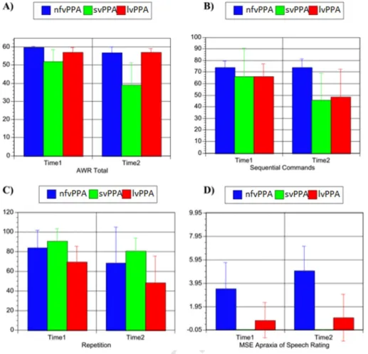
275 At follow-up, nfvPPA was associated with a significant decline 276 (p b 0.05) in WAB Information, WAB Fluency, Auditory Word Recogni-277 tion, MSE Apraxia of Speech rating, syntax comprehension, animal flu-278 ency, calculations, and design fluency (seeTable 2andFig. 1). There
279
were trends (p b 0.10) for a decline in constructional praxis (modified
280
Rey copy) and set-shifting (modified trails). Although not significant,
281
longitudinal decline was also observed in the clinician ratings of melody,
282
phrase length, and grammar. The areas of relative strengths for nfvPPA
283
were comprehension tasks (Yes/No Comprehension, Auditory Word
284
Recognition, Sequential Commands), global cognition (MMSE), verbal
285
(CVLT) and visual memory (Modified Rey-copy), naming (BNT), and
cli-286
nician rating of comprehension skills.
287
3.2.2. Semantic variant PPA
288
On language tests, svPPA was associated with significant decline
289
(p b 0.05) in Information Content, Yes–No Comprehension, Auditory
290
Word Recognition, and Sequential Commands (seeTable 2andFig. 1).
291
At a more general cognitive level, significant decline was also observed
292
in global cognition (MMSE), verbal memory (CVLT total learning and
293
30-s recall), and clinician rating of comprehension skills. There was
294
a trend (p b 0.10) for a decline in praxis. Unfortunately, many of the
295
svPPA scores were at floor level at the time of diagnosis, which made
296
it difficult to detect any longitudinal decline. Visual–spatial and
execu-297
tive skills remained areas of relative strength.
298
3.2.3. Logopenic variant PPA
299
At follow-up, lvPPA was associated with significant decline (p b 0.05)
300
in spontaneous speech Information Content, Sequential Commands,
301
sentence repetition (seeTable 2andFig. 1) and on the MMSE.
Addition-302
ally, there were trends (p b 0.10) for decline in verbal memory
(learn-303
ing), naming, Stroop interference, praxis, and poorer clinician ratings
304
of melody, phrase length, grammar, and word-finding. Although not
sig-305
nificant, additional numerical decline was observed in verbal fluency,
306
design fluency, digits backward, and calculations. Of note, the amount
307
of change in each lvPPA patient3s longitudinal profile appeared to be
308
quite variable relative to svPPA and nfvPPA, which likely made it more
309
difficult to see consistent and significant change when looking at the
310
longitudinal lvPPA data at the group level. Overall, these findings
sug-311
gest that lvPPA patients tend to show relatively more diffuse cognitive
312
impairment in comparison to the other two PPA variants.
313
In summary, although many of the longitudinal analyses within each
314
diagnostic group were not statistically significant, the general pattern of
315
results was as expected. Specifically, nfvPPA showed progression in
316
speech fluency and executive dysfunction, while svPPA showed decline
317
in MMSE, memory and comprehension. lvPPA was more variable, but as
318
a group showed decline in repetition, fluency, comprehension, and the
319
MMSE, with trends for additional decline in memory, naming, praxis,
320
and executive function.
t2
:1 Table 2
t2
:2 Language battery results in each of the three PPA variants at baseline and 1-year follow-up.
t2
:3 nfvPPA svPPA lvPPA
t2
:4 Baseline Follow-up Baseline Follow-up Baseline Follow-up
t2
:5 WAB Information Content (10) 8.0 ± 3.0 5.9 ± 3.8
a 8.7 ± 0.8 7.5 ± 2.1a 8.3 ± 1.9 7.2 ± 1.8a
t2
:6 WAB Fluency (10) 7.3 ± 3.2 4.8 ± 4.1
a 8.7 ± 1.2 8.5 ± 1.5 7.7 ± 1.8 5.5 ± 3.1b
t2
:7 WAB Spontaneous Speech Total (20) 15.3 ± 5.8 10.6 ± 7.7
a 17.4 ± 1.6 16.0 ± 3.1 16.0 ± 3.5 12.7 ± 4.8a
t2
:8 WAB Yes–No Comprehension (60) 57.4 ± 4.1 57.6 ± 3.4 57.0 ± 4.6 49.1 ± 12.2
a 57.8 ± 4.5 48.0 ± 24.0
t2
:9 WAB Auditory Word Recognition (60) 59.8 ± 0.7 56.9 ± 3.3
a 52.8 ± 6.4 40.0 ± 12.8a 57.0 ± 2.9 57.0 ± 2.2 t2 :10 Sequential Commands (80) 74.0 ± 5.3 73.9 ± 7.6 75.43 ± 8.0 50.3 ± 24.2 a 66.0 ± 11.3 48.4 ± 24.2a t2 :11 Repetition (100) 83.7 ± 17.8 68.0 ± 36.6 b 90.6 ± 13.2 82.3 ± 13.9b 69.0 ± 16.1 58.8 ± 26.8a t2
:12 MSE apraxia of speech
t2
:13 (7 = max deficit, 0 = normal)
3.5 ± 2.2 5.0 ± 2.1a 0.0 ± 0.0 0.0 ± 0.0 0.8 ± 1.5 1.0 ± 2.0
t2
:14 MSE dysarthria rating
t2
:15 (7 = max deficit, 0 = normal)
2.6 ± 2.7 1.6 ± 2.5b 0.0 ± 0.0 0.0 ± 0.0 0.0 ± 0.0 0.0 ± 0.0
t2
:16 CYCLE-R syntax comprehension (55) 49.6 ± 4.3 45.3 ± 7.9
a 54.0 ± 1.1 45.0 ± 10.8b 41.7 ± 7.2 35.0 ± 4.4
t2
:17 Abbreviations: nfvPPA = non-fluent variant PPA, svPPA = semantic variant PPA, lvPPA = logopenic variant PPA; WAB = Western Aphasia Battery, MSE = Motor Speech Evaluation,
t2
:18 CYCLE-R = Curtiss–Yamada Comprehensive Language Evaluation-Receptive.
t2 :19 aT1 vs. T2 comparison significant at p b 0.05. t2 :20 b T1 vs. T2 comparison a trend at pb0.10.
UNCORRECTED PR
OOF
321 3.3. Imaging
322 3.3.1. Gray and white matter atrophy progression
323 3.3.1.1. Gray matter. The TBM analysis of the gray matter showed a dis-324 tinct pattern of progressive atrophy in each of the three PPA variants
325 (seeTable 3andFig. 2).
326 3.3.1.1.1. nfvPPA vs. controls. In nfvPPA, over 1 year from initial diag-327 nosis, significant gray matter (GM) contraction was found in the left 328 frontal lobe, and more specifically in the inferior frontal gyrus, pars 329 triangularis (45), rolandic operculum (6) and precentral gyrus (6). 330 Within the temporal lobe, significant GM contraction was observed in 331 the anterior portion of the fusiform gyrus (20). Regions of progressive 332 GM contraction were observed in some subcortical structures such as 333 the bilateral hippocampus/amygdala, right thalamus, and bilateral cere-334 bellum (p b 0.05, FWE-corrected).
335 3.3.1.1.2. svPPA vs. controls. Patients with svPPA showed significant 336 GM contraction over time bilaterally in the anterior temporal lobes 337 including the superior (22), middle (21) and inferior (20) temporal gyri, 338 fusiform gyrus (20/37), temporal pole, and parahippocampal gyrus. 339 Regions of GM contraction over time were also observed in the basal 340 ganglia (left bilateral putamen, left pallidum, and right thalamus) and 341 in the left frontal lobe including medial orbital gyrus (25), superior me-342 dial frontal gyrus (32), anterior cingulate (32), and bilateral insula 343 (p b 0.05, FWE-corrected). When we lowered the threshold to a 344 less conservative one of p b 0.001 uncorrected, further areas of GM con-345 traction were observed in the right inferior frontal gyrus, pars opercularis 346 (44), supramarginal gyrus (40), angular gyrus (39), and superior frontal 347 gyrus (9).
348 3.3.1.1.3. lvPPA vs. controls. No significant regions of GM contraction 349 over 1-year period following diagnosis were observed in the lvPPA
350
group at the pre-established threshold of p b 0.05 FWE-corrected,
prob-351
ably due to the small sample size. For exploratory analysis, we lowered
352
the level of significance to a more permissive threshold of p b 0.001
353
uncorrected in the regions that have shown progressive GM contraction
354
over time in a previous study of our group (Rohrer et al., 2013). Within
355
our regions of interest, lvPPA showed gray matter contraction bilaterally
356
in the anterior portion of the left superior temporal gyrus, and in left
357
inferior temporal and fusiform gyri (0.001 uncorrected). Due to our
358
hypothesis regarding Alzheimer3s disease as a frequent underlying
359
pathology in lvPPA (Rabinovici et al., 2007,2008a), an exploratory
anal-360
ysis was performed to examine whether lvPPA patients showed gray
361
matter contraction in the hippocampus. Significant changes in GM
vol-362
ume over 1 year following diagnosis were observed in left hippocampus
363
at a threshold of p b 0.005 uncorrected.
364
Overall, these results suggest differential patterns of longitudinal
365
gray matter contraction over 1 year following diagnosis in the three
366
PPA variants. Specifically, nfvPPA showed progressive GM volume
con-367
traction mainly in left prefrontal regions, svPPA in bilateral temporal
368
and insular cortex, and the basal ganglia, while lvPPA showed
contrac-369
tion mainly in left temporal regions and hippocampus.
370
3.3.1.2. White matter. The white matter analysis revealed differential
371
patterns of white matter contraction over a 1-year period in the three
372
diagnostic groups (seeTable 4andFig. 2).
373
3.3.1.2.1. nfvPPA vs. controls. nfvPPA had a greater progression of
374
atrophy in the left superior region of the corona radiata (p b 0.05,
375
FWE-corrected). Other areas associated with greater gray matter
con-376
traction were the superior longitudinal fasciculi bilaterally, the right
an-377
terior portion of the corpus callosum, the right middle cerebellar
378
peduncle and the left corticospianal/corticobulbur tract at the midbrain
379
level (p b 0.001, uncorrected).
UNCORRECTED PR
OOF
380 3.3.1.2.2. svPPA vs. controls. svPPA was associated with a progressive 381 reduction of the white matter underlying the temporal lobe (left inferior 382 fronto-occipital fasciculus, uncinate fasciculus, and bilateral inferior lon-383 gitudinal fasciculus; p b 0.05, FWE-corrected). In addition, svPPA showed 384 white matter contraction in the left corpus callosum (genu, body and 385 splenium), bilateral anterior thalamic projection, right corticospinal 386 tract, bilateral superior longitudinal fasciculi (p b 0.05, FWE-corrected). 387 Other areas associated with white matter progression were right inferior 388 cerebellar peduncle and left superior longitudinal fasciculus (p b 0.001, 389 uncorrected).
390 3.3.1.2.3. lvPPA vs controls. lvPPA showed greater white matter pro-391 gression in the right superior longitudinal fasciculi and left posterior 392 cingulate (p b 0.05, FWE-corrected). At a lower level of significance
393
(p b 0.001, uncorrected) also left inferior longitudinal/inferior
front-394
occipital fasciculus was associated with white matter contraction.
395
Overall, these results suggest differential patterns of longitudinal
396
WM contraction over 1 year following diagnosis in the three PPA
vari-397
ants. Specifically, nfvPPA showed progressive WM volume contraction
398
bilaterally in the frontal lobes, svPPA in the temporal lobes, while
399
lvPPA in WM regions in correspondence to temporo-parietal regions.
400
4. Discussion
401
In the present study, we used TBM to track the progression of brain
402
tissue contraction over 1 year following the diagnosis in the three
clin-403
ical variants of PPA (Gorno-Tempini et al., 2011), including eight
404
patients with nfvPPA, 13 with svPPA and seven with lvPPA. The current
405
study showed that the three variants have distinct and only partially
406
overlapping patterns of gray and white matter atrophy progression.
407
More specifically, the results revealed a pattern of GM atrophy
progres-408
sion within the brain regions that are generally first targeted by each
409
variant, i.e., the left prefrontal cortex and subcortical regions in nfvPPA,
410
the anterior temporal lobes in svPPA and the posterior middle and
supe-411
rior temporal gyrus in the lvPPA. Moreover, GM contraction spreads
412
over time towards nonadjacent regions such as the insular cortex and
413
the basal ganglia in svPPA and in the left inferior temporal regions and
414
the hippocampus in lvPPA. In all three variants, the white matter fibers
415
underlying the abovementioned cortical areas underwent significant
416
volume reduction in 1 year. In the following paragraphs, we describe
417
the gray and white matter progression together with their clinical
418
implications for each of the three PPA variants.
419
4.1. Nonfluent variant PPA (nfvPPA)
420
nfvPPA cases showed greater gray matter contraction in left frontal
421
and subcortical regions. White matter progression was seen in left
coro-422
na radiata, underlying the motor cortex and the supplementary motor
423
area, which are fibers that connect frontal areas with subcortical nuclei
424
and the spinal cord (Mori et al., 2005). These results extend
cross-425
sectional imaging studies that found the left posterior frontal cortical
426
and subcortical regions to be the most affected area in nfvPPA (
Gorno-427
Tempini et al., 2004b;Josephs et al., 2006;Nestor et al., 2003). The
428
brain regions that showed significant progression in nfvPPA represent
429
a large network involved in speech production (Hickok, 2009;Price,
430
2010), grammar comprehension (Amici et al., 2007;Caplan, 1992;
431
Wilson et al., 2010b,2011,2012b) and working memory (Amici et al., Q7
432
2007;Jonides et al., 1993,1998;Wilson et al., 2010a). Nonetheless,
sig-433
nificant progression of WM volume loss was observed in dorsal
lan-434
guage tracts that have been shown to connect brain regions that are
435
critically involved in syntactic processing (Wilson et al., 2011). These
436
anatomical changes likely contribute to the observed decreases in
gram-437
mar, phrase length and melody of speech, and to the decline in
execu-438
tive skills. Our findings are also in line with previous
pathologically-439
confirmed case observations. In fact, atrophy of the perirolandic region,
440
middle and inferior frontal gyrus, thalamus and bulbar brainstem
441
involvement has been found in nfvPPA pathologically confirmed
442
cases (Josephs et al., 2006). These regions are also most involved in
443
4R tauopathies such as progressive supranuclear palsy (Boxer et al.,
444
2006;Brenneis et al., 2004;Cordato et al., 2005;Padovani et al., 2006)
445
and cortico-basal degeneration (Boxer et al., 2006), which is the most
446
common pathology associated with nfvPPA (Josephs et al., 2006).
447
4.2. Semantic variant PPA (svPPA)
448
svPPA patients demonstrated longitudinal gray matter contraction
449
in several regions of the lateral and medial temporal lobes, insula and
450
ventromedial cortex. Main areas of WM contraction over time were
451
observed bilaterally in the inferior longitudinal fasciculus connecting
452
occipital and anterior temporal regions, and in the uncinate fasciculus
t3
:1 Table 3
t3
:2 Voxel of significant gray matter contraction over 1 year in each of the three PPA variants t3 :3 vs. controls. t3 :4 Region (BA) H x y z T Z t3 :5 nfvPPA vs CTRL t3
:6 Inferior frontal gyrus, pars triangularis (45) L −47 17 8 7.2 6.0
t3
:7 Rolandic operculum (6) L −51 0 11 8.1 6.4
t3
:8 Precentral gyrus (6) L −42 8 41 7.8 6.3
t3
:9 Fusiform gyrus, anterior portion (20) L −36 −5 −29 7.0 5.8
t3 :10 Hippocampus/amygdala L −19 −4 −11 6.4 5.4 t3 :11 L −23 −31 4 7.6 6.1 t3 :12 R 21 −36 7 6.7 5.6 t3 :13 Thalamus R 13 −22 16 6.7 5.6 t3 :14 Cerebellum R 2 −43 −43 5.9 5.1 t3 :15 L −9 −62 −41 3.7 3.5* t3 :16 svPPA vs CTRL t3
:17 Superior temporal gyrus (22) L −46 −10 −1 6.4 5.4
t3
:18 L −34 6 −27 9.3 7.0
t3
:19 Middle temporal gyrus, anterior portion (21) L −61 −27 −15 6.1 5.3
t3
:20 R 55 −2 −20 7.4 6.1
t3
:21 Inferior temporal gyrus, anterior portion (20) L −49 9 −43 6.6 5.6
t3 :22 L −58 −12 −24 6.4 5.5 t3 :23 R 39 2 −37 10.2 7.4 t3 :24 Fusiform gyrus (20/37) L −34 −39 −23 11.1 7.8 t3 :25 R 27 17 −43 7.8 6.3 t3 :26 Temporal Pole (20/38) R 51 9 −14 7.3 6.0 t3 :27 R 45 23 −31 6.0 5.2 t3 :28 R 31 19 −42 7.7 6.2 t3 :29 Parahippocampal gyrus L −28 −13 −28 10.3 7.5 t3 :30 L −20 −41 −7 7.1 5.9 t3 :31 R 27 −24 −26 9.2 6.9 t3 :32 R 17 −4 −24 9.8 7.3 t3 :33 Putamen L −17 15 3 8.5 6.6 t3 :34 R 19 15 7 7.2 5.9 t3 :35 Pallidum L −15 1 −5 6.6 5.5 t3 :36 Thalamus R 12 −8 6 6.9 5.7 t3 :37 R 9 −3 −1 6.4 5.4 t3 :38 Insula (13) L −33 15 −11 8.1 6.4 t3 :39 R 45 11 −11 6.4 5.4 t3
:40 Medial orbital gyrus (25) L −11 14 −14 10.3 7.5
t3
:41 Superior medial frontal gyrus (32) L −7 41 31 6.2 5.3
t3
:42 Anterior cingulate (32) L −5 49 8 6.6 5.5
t3
:43 Inferior frontal gyrus, pars opercolaris (44) R 57 13 20 5.2 4.6
* t3 :44 Supramarginal gyrus (40) L −58 −33 39 4.5 4.1* t3 :45 Angular gyrus (39) L −41 −70 45 3.9 3.6 * t3
:46 Superior frontal gyrus (9) R 19 46 36 5.0 4.5
* t3 :47 L −18 46 35 3.8 3.6 * t3 :48 lvPPA vs CTRL t3
:49 Superior temporal gyrus, anterior portion (20) R 51 6 −9 3.6 3.4
*
t3
:50 L −33 8 −27 3.5 3.3
*
t3
:51 Inferior temporal gyrus (20) L −44 −38 −23 4.8 4.4*
t3 :52 Fusiform gyrus (37) L −35 −37 −23 4.6 4.1 * t3 :53 Hippocampus L −23 −1 −19 3.4 3.3§ t3
:54 nfvPPA = non-fluent variant of primary progressive aphasia, svPPA = semantic variant of t3
:55 primary progressive aphasia, lvPPA = logopenic variant of primary progressive aphasia, t3
:56 CTRL = age- and sex-matched healthy control.
t3 :57
* p b 0.001 uncorrected for multiple comparison.
t3 :58
UNCORRECTED PR
OOF
453 connecting the anterior temporal regions with the frontal lobe. Both of 454 these GM and WM brain structures have been shown to be affected in 455 svPPA patients even at early stages of the disease (Agosta et al., 2010;
456 Borroni et al., 2007;Gorno-Tempini et al., 2004b;Mummery et al.,
457 2000;Rosen et al., 2002). Overall, our findings seem to indicate that
458 the regions usually atrophic in svPPA patients become more atrophic 459 after 1 year and the atrophy spread both medially and posteriorly 460 within the temporal lobe together with the white matter bundles that 461 connect the temporal lobe with the frontal and occipital cortex. These 462 results are consistent with previous DTI (Agosta et al., 2010;Borroni
463 et al., 2007), fluid registration (Whitwell et al., 2004) and TBM findings
464 (Brambati et al., 2009c).
465 The areas that underwent major atrophic changes in svPPA, ven-466 tral and lateral temporal lobes, are part of a network involved in 467 semantic memory (Butler et al., 2009;Mummery et al., 1999;Williams
468 et al., 2005), exception word reading (Brambati et al., 2009b;Wilson
469 et al., 2012a), identification of visual attributes (D3Esposito et al.,
470 1997;Vandenbulcke et al., 2006), famous faces (Brambati et al.,
471 2010;Gesierich et al., 2012;Gorno-Tempini et al., 1998;Kanwisher
472 et al., 1997), buildings and landscapes (Epstein and Kanwisher, 1998).
473 The progressive volume contraction in these areas is clinically asso-474 ciated with worsened single word comprehension abilities. 475 Other regions that showed greater progression were the insula, the 476 ventromedial frontal and anterior cingulate, which have been associ-477 ated with behavioral symptoms, emotional processing, mood regula-478 tion, and eating behavior (Rosen et al., 2005;Williams et al., 2005;
479 Woolley et al., 2007) and less associated with executive function deficits
480 (Possin et al., 2009). The medial frontal atrophy is likely associated with
481 the development of behavioral and social dysfunction observed in this 482 population (Seeley et al., 2005,2008;Seeley, 2010).
483 Our longitudinal findings seem to provide critical support to the 484 recently proposed pathophysiological model of svPPA progression
485 (Fletcher and Warren, 2011). According to this model, the anterior
tem-486 poral lobe would be vulnerable to the neurodegenerative effects of TDP-487 43-C (Mackenzie et al., 2006;Mesulam et al., 2014;Rohrer et al., 2010;
488
Whitwell et al., 2010). However, the development of the semantic PPA
489
manifestations would arise from disintegration of a distributed neural
490
network with specific intrinsic anatomical and functional connectivity
491
with the ATL (Fletcher and Warren, 2011). Consistently with this
492
hypothesis, Guo and colleagues, report that svPPA patients showed
493
reduced intrinsic connectivity throughout a distributed set of regions
494
connected with the anterior temporal lobe in healthy controls (Guo
495
et al., 2013). Nonetheless, the same authors reported that scores on
496
semantic tasks correlated with physiological deficits outside the anterior
497
temporal lobe, suggesting that the severity of the semantic impairments
498
in svPPA is associated with the spread of the disease to cortical areas
499
connected to the ATL (Guo et al., 2013). Interestingly, the set of regions
500
whose physiological deficits correlate with the severity of the
impair-501
ments in the semantic representations of emotions in the study by
502
Guo and colleagues (i.e. insula, anterior cingulated, medial frontal
cor-503
tex), also present with progressive volume contraction over a 1-year
504
period in the present study.
505
Different possible candidates have been identified as potentially
506
responsible for the spreading of the disease from the ATL to connected
507
brain regions, including axonal degeneration, transynaptic spreading
508
of abnormal protein, or abnormal folding in tau molecules induced by
509
nearby folded tau (Bartz et al., 2002;Frost et al., 2009;Salehi et al.,
510
2009) (seeFletcher and Warren, 2011for a review). However, the
pre-511
cise molecular mechanisms leading to large-network distraction in
512
svPPA still remain largely unknown.
513
4.3. Logopenic variant PPA (lvPPA)
514
lvPPA showed main GM volume reduction over 1 year following
515
diagnosis in the left temporal lobe and hippocampus. White matter
516
progression was present in the right superior longitudinal fasciculus
517
and left posterior cingulum. The superior longitudinal fasciculus is a
518
large bundle that connects perisylvian frontal, parietal and temporal
519
cortex (Catani and Mesulam, 2008;Dejerine, 1895;Duffau, 2008;
520
Petrides and Pandya, 1984). The arcuate fasciculus is part of the superior
Fig. 2. Transverse, coronal and sagittal slices of the main peak of gray matter (shown in red) and white matter (shown in yellow) contraction in nfvPPA (first row), svPPA (second row), and lvPPA (third row) versus controls. The results are superimposed on a section of the study-specific template. The x, y, and z values reported in the figure represent the position of slices within the Montreal Neurologic Institute (MNI) stereotaxic space. The threshold is set at p b 0.001 uncorrected for display purpose.
UNCORRECTED PR
OOF
521 longitudinal fasciculus and is typically damaged in vascular conduction 522 aphasia (Catani et al., 2005;Geschwind, 1965;Hickok and Poeppel, 523 2004), which shares the repetition deficits of lvPPA (Geschwind, 1965;
524 Gorno-Tempini et al., 2004b). Posterior cingulum white matter fibers
525 connect the anterior thalamus, anterior cingulate cortex, temporal 526 lobe, and hippocampus and are atrophic in both MCI and AD patients
527 (Damoiseaux et al., 2009). The brain regions showing progressive tissue
528 loss in our study do not represent the areas usually reported to be atro-529 phic at the beginning of the disease in the cross-sectional study (
Gorno-530Q8 Tempini et al., 2008,2004b;Rohrer et al.), i.e. the left posterior superior
531 temporal and inferior parietal area. During the 1-year interval following 532 diagnosis, the atrophy spread anterior-inferiorly and medially in the 533 temporal lobe together with the underlying white matter connections 534 (e.g., posterior cingulate). These results are consistent with a previous 535 longitudinal study of our group revealing very similar results (Rohrer
536 et al., 2013).
537 In lvPPA, the areas more affected at follow-up represent a large net-538 work involved in episodic (Buckner et al., 1998;Desgranges et al., 1998;
539 Henson et al., 1999;Rajah and McIntosh, 2008) and semantic memory.
540
In accordance with these anatomical findings, the lvPPA patients
mani-541
fested a decline in calculations, verbal and visual memory at follow-up
542
of both neuropsychological and language assessments. The
neuropsy-543
chological results are consistent with previous reports indicating that
544
the progression of the disease is characterized by the appearance of
ver-545
bal memory and calculation symptoms together with the worsening of
546
anomia, repetition and fluency deficits (Roher et al., 2013). This would
547
probably explain why the lvPPA group appears to have the greatest
de-548
cline over the 12 month period. Nonetheless the areas showing
progres-549
sive GM contraction over time in lvPPA belong to the network of regions
550
usually damaged in Alzheimer’s disease and its preclinical phases, i.e.
551
mild cognitive impairment (Brambati et al., 2009a;Fox et al., 2001;
552
Killiany et al., 2000;Minoshima et al., 1997;Seeley et al., 2009). Finally,
553
increased rate of atrophy in individuals at risk for familial AD was found
554
classically in the medial temporal lobe but also inferolateral temporal
555
lobe (Fox and Rossor, 1999). This impressive neuroanatomical overlap
556
between lvPPA and AD cases are consistent with the evidence that
557
lvPPA and AD share the same underlying pathology (Josephs et al.,
558
2008;Mesulam et al., 2008;Rabinovici et al., 2007,2008a;Rohrer
559
et al., 2012). Also the progression of gray matter in the hippocampus
560
(albeit at a lower level of significance), is potentially consistent
561
with underlying AD pathology. A possible explanation for slower
pro-562
gression in this area could be that in “atypical AD” cases presenting as
563
fluent or non-fluent PPA, the medial temporal lobe is minimally affected
564
(Galton et al., 2000).
565
4.4. Limitations
566
Future studies involving a greater number of patients matched by
567
time of onset of symptoms and including a longer follow-up are
neces-568
sary to further clarify the cognitive and anatomical progression of the
569
disease in the three clinical variants of the disease. In particular, larger
570
samples will allow us to run direct correlation analyses to test the
asso-571
ciation between the anatomical changes and the worsening of clinical
572
symptoms over time and to compare the rate of cognitive and language
573
decline among PPA variant subtypes. However, our sample size was
574
comparable with previous anatomical studies in this patient population
575
(Mandelli et al., 2014;Rogalski et al., 2014) and was a practical
conse-576
quence of the rareness of the disease.
577
From a methodological point of view, a replication of the results and
578
of the analysis of the longitudinal data using the DARTEL approach could
579
be useful to validate the present findings.
580
Nonetheless, it must be noted that the white matter contraction
581
pattern observed in our study sometimes involves regions adjacent to
582
the ventricles, which raises the question of whether this result can be
583
an effect of ventricle enlargement. It seems unlikely that this bias
584
could entirely explain the present result given that different regions of
585
WM contraction have been implicated in different PPA variants and
586
that the total intracranial volume at the time of the first scan has been
587
included as a covariate in the statistical model. However future studies
588
that will specifically address this question could certainly elucidate the
589
relationship between ventricle enlargement and longitudinal changes
590
in WM tissue.
591
4.5. Conclusions
592
In the present study, we showed that nfvPPA, svPPA and lvPPA have
593
different longitudinal patterns of neuroanatomical contraction that are
594
related to their clinical and cognitive progression. nfvPPA progresses
595
in areas involved in speech production and agrammatism; svPPA in
596
areas associated to semantic memory, emotion processing and
behav-597
ior, lvPPA in regions supporting repetition, episodic and semantic
mem-598
ory and attention. These findings can be crucial to develop intervention
599
strategies, such as speech rehabilitation therapies that are tailored
600
to patients3 progression profiles, and to correctly inform families and
t4
:1 Table 4
t4
:2 Voxels of significant white matter contraction over 1 year in the three PPA variants.
t4
:3 Region H x y z T Z
t4
:4 nfvPPA vs CTRL
t4
:5 Superior corona radiata L −12 −10 37 6.4 5.4
t4
:6 R 14 −7 31 5.6 5.0*
t4
:7 Superior longitudinal fasciculus R 47 −5 37 3.7 3.4*
t4 :8 R 67 −20 −1 3.3 3.3* t4 :9 L −43 −10 41 5.3 4.7 * t4 :10 L −30 −19 25 4.5 4.1* t4 :11 L −49 15 3 3.7 3.5* t4
:12 Anterior corpus callosum R 13 17 18 5.3 4.7*
t4
:13 Middle cerebellar peduncle L 14 −33 −33 4.0 3.8*
t4
:14 Corticospinal tract (midbrain level) R −14 −23 −14 4.7 4.2*
t4
:15 svPPA vs CTRL
t4
:16 Inferior longitudinal fasciculus L −27 −7 −17 8.3 6.5
t4 :17 L −23 −9 −15 6.6 5.6 t4 :18 L −37 −43 −9 7.9 6.3 t4 :19 L −44 −23 −19 8.0 6.3 t4 :20 R 39 1 −39 6.2 5.3 t4 :21 L −21 −13 −17 6.3 5.3 t4
:22 Anterior thalamic projection R 15 −5 22 8.0 6.4
t4 :23 L −13 3 19 6.1 5.2 t4 :24 Uncinate fasciculus L −31 1 −23 7.9 6.3 t4 :25 L −29 −9 −15 7.8 6.3 t4
:26 Superior longitudinal fasciculus R 33 −37 13 5.9 5.1
t4 :27 L −13 −1 21 6.3 5.3 t4 :28 L −13 3 19 6.0 5.2 t4 :29 L −13 7 17 5.9 5.1 t4 :30 Corticospinal tract R 15 −5 22 7.7 6.2 t4
:31 Splenium of corpus callosum L −8 −34 17 8.6 6.7
t4
:32 Body of corpus callosum L −11 −5 25 6.7 5.5
t4 :33 L −9 1 25 6.0 5.2 t4 :34 L −9 11 19 6.0 5.2 t4 :35 L −9 7 21 5.9 5.1 t4
:36 Genu of corpus callosum L −11 15 17 6.1 5.2
t4
:37 L −15 23 11 6.1 5.2
t4
:38 L −14 21 15 6.0 5.2
t4
:39 Inferior cerebellar peduncle R 11 −44 −43 4.0 3.7*
t4
:40 lvPPA vs CTRL
t4
:41 Superior longitudinal fasciculus R 25 −44 25 6.5 5.5
t4 :42 R 24 −28 31 6.1 5.3 t4 :43 −22 −33 33 4.6 4.2* t4 :44 Posterior cingulum L −18 −52 26 6,1 5.2 t4 :45 L −20 −55 23 6.1 5.2 t4 :46 R 7 −31 7 5.6 4.9* t4
:47 Inferior longitudinal fasciculus L −34 −47 −8 4.2 3.9*
t4
:48 nfvPPA = non-fluent variant of primary progressive aphasia, svPPA = semantic variant of t4
:49 primary progressive aphasia, lvPPA = logopenic variant of primary progressive aphasia, t4
:50 CTRL = age- and sex-matched healthy control.
t4 :51
UNCORRECTED PR
OOF
601 caregivers of the challenges they will eventually need to face with the602 patients. The proposed approach could be extremely useful to test the 603 efficacy of intervention strategies aimed at slowing down the progres-604 sion of the disease in these patients.
605 Uncited references
606 No citations were found for the following references:Dronkers
607 (1996);Dronkers et al. (2000).
608 Acknowledgment
609 This work was supported by the National Institute of Neurological 610 Diseases and Stroke (R01 NS50915), the National Institute on Aging 611 (P01 AG019724 and P50 AG-03-006), the California Department of 612 Health Services (DHS 04-35516), U.S. Department of Veterans Affairs, 613Q9 Office of Research and Development, Rehabilitation R&D Program, a
614 Network Grant to the Hillblom Foundation and the UCSF General Clini-615 cal Research Center (M01 RR00079).
616 References
617 Agosta, F., Henry, R.G., Migliaccio, R., Neuhaus, J., Miller, B.L., Dronkers, N.F., Brambati,
618 S.M., Filippi, M., Ogar, J.M., Wilson, S.M., Gorno-Tempini, M.L., 2010. Language
net-619 works in semantic dementia. Brain 133 (1), 286–299.http://dx.doi.org/10.1093/
620 brain/awp23319759202.
621 Amici, S., Brambati, S.M., Wilkins, D.P., Ogar, J., Dronkers, N.L., Miller, B.L., Gorno-Tempini,
622 M.L., 2007. Anatomical correlates of sentence comprehension and verbal working
623 memory in neurodegenerative disease. J. Neurosci. 27 (23), 6282–6290.http://dx.
624 doi.org/10.1523/JNEUROSCI.1331-07.200717554002.
625 Ashburner, J., Friston, K., 2003. Image segmentation. In: Frackowiack, R., Finston, K., Frith, 626 C., Dolan, R., Friston, K., Price, C., Zeki, S., Ashburner, J., Penny, W. (Eds.), Human Brain
627 Function. Academic Press.
628 Ashburner, J., Friston, K.J., 1999. Nonlinear spatial normalization using basis functions.
629 Hum. Brain Mapp. 7 (4), 254–26610408769.
630 Ashburner, J., Friston, K.J., 2000. Voxel-based morphometry — the methods. Neuroimage
631 11 (6 1), 805–821.http://dx.doi.org/10.1006/nimg.2000.058210860804.
632 Bartz, J.C., Kincaid, A.E., Bessen, R.A., 2002. Retrograde transport of transmissible mink
en-633 cephalopathy within descending motor tracts. J. Virol. 76 (11), 5759–5768.http://dx.
634 doi.org/10.1128/JVI.76.11.5759-5768.200211992004.
635 Borroni, B., Brambati, S.M., Agosti, C., Gipponi, S., Bellelli, G., Gasparotti, R., Garibotto, V., Di
636 Luca, M., Scifo, P., Perani, D., Padovani, A., 2007. Evidence of white matter changes on
637 diffusion tensor imaging in frontotemporal dementia. Arch. Neurol. 64 (2), 246–251.
638 http://dx.doi.org/10.1001/archneur.64.2.24617296841.
639 Boxer, A.L., Geschwind, M.D., Belfor, N., Gorno-Tempini, M.L., Schauer, G.F., Miller, B.L.,
640 Weiner, M.W., Rosen, H.J., 2006. Patterns of brain atrophy that differentiate
641 corticobasal degeneration syndrome from progressive supranuclear palsy. Arch.
642 Neurol. 63 (1), 81–86.http://dx.doi.org/10.1001/archneur.63.1.8116401739.
643 Brambati, S.M., Belleville, S., Kergoat, M.J., Chayer, C., Gauthier, S., Joubert, S., 2009a.
644 Single- and multiple-domain amnestic mild cognitive impairment: two sides of the
645 same coin? Dement. Geriatr. Cogn. Disord. 28 (6), 541–549.http://dx.doi.org/10.
646 1159/00025524020016185.
647 Brambati, S.M., Benoit, S., Monetta, L., Belleville, S., Joubert, S., 2010. The role of the left
an-648 terior temporal lobe in the semantic processing of famous faces. Neuroimage 53 (2),
649 674–681.http://dx.doi.org/10.1016/j.neuroimage.2010.06.04520600979.
650 Brambati, S.M., Ogar, J., Neuhaus, J., Miller, B.L., Gorno-Tempini, M.L., 2009b. Reading
dis-651 orders in primary progressive aphasia: a behavioral and neuroimaging study.
652 Neuropsychologia 47 (8–9), 1893–1900. http://dx.doi.org/10.1016/j.
653 neuropsychologia.2009.02.03319428421.
654 Brambati, S.M., Rankin, K.P., Narvid, J., Seeley, W.W., Dean, D., Rosen, H.J., Miller, B.L.,
655 Ashburner, J., Gorno-Tempini, M.L., 2009c. Atrophy progression in semantic dementia
656 with asymmetric temporal involvement: a tenser-based morphometry study.
657 Neurobiol. Aging 30 (1), 103–111.http://dx.doi.org/10.1016/j.neurobiolaging.
658 2007.05.01417604879.
659 Brambati, S.M., Renda, N.C., Rankin, K.P., Rosen, H.J., Seeley, W.W., Ashburner, J., Weiner,
660 M.W., Miller, B.L., Gorno-Tempini, M.L., 2007. A tensor based morphometry study of
661 longitudinal gray matter contraction in FTD. Neuroimage 35 (3), 998–1003.http://
662 dx.doi.org/10.1016/j.neuroimage.2007.01.02817350290.
663 Brenneis, C., Seppi, K., Schocke, M., Benke, T., Wenning, G.K., Poewe, W., 2004. Voxel based
664 morphometry reveals a distinct pattern of frontal atrophy in progressive
665 supranuclear palsy. J. Neurol. Neurosurg. Psychiatry 75 (2), 246–24914742598.
666 Buckner, R.L., Koutstaal, W., Schacter, D.L., Dale, A.M., Rotte, M., Rosen, B.R., 1998.
667 Functional-anatomic study of episodic retrieval. II. Selective averaging of
event-668 related fMRI trials to test the retrieval success hypothesis. Neuroimage 7 (3),
669 163–175.http://dx.doi.org/10.1006/nimg.1998.03289597658.
670 Butler, C.R., Brambati, S.M., Miller, B.L., Gorno-Tempini, M.L., 2009. The neural
corre-671 lates of verbal and nonverbal semantic processing deficits in neurodegenerative
672 disease. Cogn. Behav. Neurol. 22 (2), 73–80.http://dx.doi.org/10.1097/WNN.
673 0b013e318197925d19506422.
674 Cairns, N.J., Bigio, E.H., Mackenzie, I.R., Neumann, M., Lee, V.M., Hatanpaa, K.J., White 3rd,
675 C.L., Schneider, J.A., Grinberg, L.T., Halliday, G., Duyckaerts, C., Lowe, J.S., Holm, I.E.,
676 Tolnay, M., Okamoto, K., Yokoo, H., Murayama, S., Woulfe, J., Munoz, D.G., Dickson,
677 D.W., Ince, P.G., Trojanowski, J.Q., Mann, D.M., 2007. Neuropathologic diagnostic and
678 nosologic criteria for frontotemporal lobar degeneration: consensus of the Consortium
679 for Frontotemporal Lobar Degeneration. Acta Neuropathol. 114 (1), 5–22.http://dx.
680 doi.org/10.1007/s00401-007-0237-217579875.
681 Caplan, D., 1992. Language: Structure, Processing, and Disorders. MIT Press, Cambridge,
682 MA.
683 Catani, M., Jones, D.K., ffytche, D.H., 2005. Perisylvian language networks of the human
684 brain. Ann. Neurol. 57 (1), 8–16.http://dx.doi.org/10.1002/ana.2031915597383.
685 Catani, M., Mesulam, M., 2008. The arcuate fasciculus and the disconnection theme in
lan-686 guage and aphasia: history and current state. Cortex 44 (8), 953–961.http://dx.doi.
687 org/10.1016/j.cortex.2008.04.00218614162.
688 Chan, D., Fox, N.C., Scahill, R.I., Crum, W.R., Whitwell, J.L., Leschziner, G., Rossor, A.M.,
689 Stevens, J.M., Cipolotti, L., Rossor, M.N., 2001b. Patterns of temporal lobe atrophy in
690 semantic dementia and Alzheimer3s disease. Ann. Neurol. 49 (4), 433–44211310620.
691 Chan, D., Fox, N.C., Scahill, R.I., Crum, W.R., Whitwell, J.L., Leschziner, G., Rossor, A.M.,
692 Stevens, J.M., Cipolotti, L., Rossor, M.N., et al., 2001a. Patterns of temporal lobe atrophy
693 in semantic dementia and Alzheimer3s disease. Ann. Neurol. 49 (4), 433–44211310620.
694 Cordato, N.J., Duggins, A.J., Halliday, G.M., Morris, J.G., Pantelis, C., 2005. Clinical deficits
695 correlate with regional cerebral atrophy in progressive supranuclear palsy. Brain
696
128 (6), 1259–1266.http://dx.doi.org/10.1093/brain/awh50815843423.
697 Curtiss, S., Yamada, J., 1988. Curtiss–Yamada Comprehensive Language Evaluation
698 Unpublished test.
699 D’Esposito, M., Detre, J.A., Aguirre, G.K., Stallcup, M., Alsop, D.C., Tippet, L.J., Farah, M.J.,
700 1997. A functional MRI study of mental image generation. Neuropsychologia 35 (5),
701
725–730.http://dx.doi.org/10.1016/S0028-3932(96)00121-29153035.
702 Damoiseaux, J.S., Smith, S.M., Witter, M.P., Sanz-Arigita, E.J., Barkhof, F., Scheltens, P.,
703 Stam, C.J., Zarei, M., Rombouts, S.A., 2009. White matter tract integrity in aging and
704
Alzheimer3s disease. Hum. Brain Mapp. 30 (4), 1051–1059.http://dx.doi.org/10.
705 1002/hbm.2056318412132.
706 Davies, R.R., Hodges, J.R., Kril, J.J., Patterson, K., Halliday, G.M., Xuereb, J.H., 2005. The
path-707 ological basis of semantic dementia. Brain 128 (9), 1984–1995.http://dx.doi.org/10.
708 1093/brain/awh58216000337.
709 Dejerine, J., 1895. Anatomie des centres nerveux. L3année psychologique, Paris,
710 pp. 559–565.
711 Desgranges, B., Baron, J.C., Eustache, F., 1998. The functional neuroanatomy of episodic
712 memory: the role of the frontal lobes, the hippocampal formation, and other areas.
713
Neuroimage 8 (2), 198–213.http://dx.doi.org/10.1006/nimg.1998.03599740762.
714Q10
Dronkers, N.F., 1996. A new brain region for coordinating speech articulation. Nature 384 715
(6605), 159–161.http://dx.doi.org/10.1038/384159a08906789.
716Q11
Dronkers, N.F., Redfern, B.B., Knight, R.T., 2000. The neural architecture of language disor-717 ders. In: Gazzaniga, M.S. (Ed.), The New Cognitive Neurosciences. MIT Press,
Cam-718 bridge, MA, pp. 949–958.
719 Duffau, H., 2008. The anatomo-functional connectivity of language revisited. New insights
720 provided by electrostimulation and tractography. Neuropsychologia 46 (4), 927–934.
721 http://dx.doi.org/10.1016/j.neuropsychologia.2007.10.02518093622.
722 Duvernoy, H., 1999. The Human Brain: Surface, Three-dimensional Sectional anatomy
723 With MRI, and Vascularization. Springer Science & Business Media.
724 Epstein, R., Kanwisher, N., 1998. A cortical representation of the local visual environment.
725 Nature 392 (6676), 598–601.http://dx.doi.org/10.1038/33402.
726 Fletcher, P.D., Warren, J.D., 2011. Semantic dementia: a specific network-opathy. J. Mol.
727
Neurosci. 45 (3), 629–636.http://dx.doi.org/10.1007/s12031-011-9586-321710360.
728 Folstein, M.F., Folstein, S.E., McHugh, P.R., 1975. “Mini-mental state”. A practical method
729 for grading the cognitive state of patients for the clinician. J. Psychiatr. Res. 12 (3),
730
189–198.http://dx.doi.org/10.1016/0022-3956(75)90026-61202204.
731 Fox, N.C., Cousens, S., Scahill, R., Harvey, R.J., Rossor, M.N., 2000. Using serial registered
732 brain magnetic resonance imaging to measure disease progression in Alzheimer
733 disease: power calculations and estimates of sample size to detect treatment
734
effects. Arch. Neurol. 57 (3), 339–344.http://dx.doi.org/10.1001/archneur.57.3.
735 33910714659.
736 Fox, N.C., Crum, W.R., Scahill, R.I., Stevens, J.M., Janssen, J.C., Rossor, M.N., 2001. Imaging of
737 onset and progression of Alzheimer3s disease with voxel-compression mapping of
738
serial magnetic resonance images. Lancet 358 (9277), 201–205.http://dx.doi.org/
739 10.1016/S0140-6736(01)05408-311476837.
740 Fox, N.C., Freeborough, P.A., 1997. Brain atrophy progression measured from registered
741 serial MRI: validation and application to Alzheimer3s disease. J. Magn. Reson. Imaging
742
7 (6), 1069–1075.http://dx.doi.org/10.1002/jmri.18800706209400851.
743 Fox, N.C., Rossor, M.N., 1999. Diagnosis of early Alzheimer3s disease. Rev. Neurol. (Paris)
744 155, S33–S37.
745 Freeborough, P.A., Woods, R.P., Fox, N.C., 1996. Accurate registration of serial 3D MR brain
746 images and its application to visualizing change in neurodegenerative disorders.
747
J. Comput. Assist. Tomogr. 20 (6), 1012–1022.
http://dx.doi.org/10.1097/00004728-748 199611000-000308933812.
749 Friston, K.J., Holmes, A., Poline, J.-B., Price, C.J., Frith, C.D., 1996. Detecting activations in
750 PET and fMRI: levels of inference and power. Neuroimage 4 (3 Pt 1), 223–235.
751 http://dx.doi.org/10.1006/nimg.1996.00749345513.
752 Friston, K.J., Holmes, A.P., Worsley, K.J., Poline, J.-P., Frith, C.D., Frackowiak, R.S.J., 1994.
753 Statistical parametric maps in functional imaging: a general linear approach. Hum.
754
Brain Mapp. 2 (4), 189–210.http://dx.doi.org/10.1002/hbm.460020402.
755 Frost, B., Ollesch, J., Wille, H., Diamond, M.I., 2009. Conformational diversity of wild-type
756 Tau fibrils specified by templated conformation change. J. Biol. Chem. 284 (6),
757
3546–3551.http://dx.doi.org/10.1074/jbc.M80562720019010781.
758 Galantucci, S., Tartaglia, M.C., Wilson, S.M., Henry, M.L., Filippi, M., Agosta, F., Dronkers, N.F.,
759 Henry, R.G., Ogar, J.M., Miller, B.L., Gorno-Tempini, M.L., 2011. White matter damage in
![L'IMMIGRATION ET L'EUROPE [Immigration and Europe]](data:image/gif;base64,R0lGODlhAQABAIAAAP///wAAACH5BAEAAAAALAAAAAABAAEAAAICRAEAOw==)