A
SSESSMENT OF PAIN PERCEPTION IN PATIENTS WITH
DISORDERS OF CONSCIOUSNESS
:
A BEHAVIORAL AND
NEUROPHYSIOLOGICAL BEDSIDE APPROACH
Nicolas LEJEUNE
UCLouvain - ULiège
Sous la direction des Professeurs
André MOURAUX et Steven LAUREYS
Thèse présentée en vue de l’obtention du grade de
Docteur en Sciences Médicales
2
Cover photography
© Philippe Braquenier
A
SSESSMENT COMMITTEES
UPERVISORSProf. André Mouraux (UCLouvain)
Prof. Steven Laureys (ULiège)
J
URY MEMBERSProf. Emmanuel Hermans (President of the jury - UCLouvain)
Prof. Marie-Elisabeth Faymonville (Secretary – ULiège)
Prof. Marzia De Lucia (Université de Lausanne)
Prof. Andrea Truini (Sapienza Università di Roma)
Dr. Camille Chatelle (UCLouvain)
CONTENTS
CONTENTS ... 5 ACKNOWLEDGMENTS ... 7 ABSTRACT ... 11 RÉSUMÉ ... 13 GLOSSARY ... 15 CHAPTER 1. INTRODUCTION ... 19 1.1.DISORDERS OF CONSCIOUSNESS... 20 1.1.1. Clinical entities ... 201.1.2. Assessment of residual awareness ... 24
1.1.3. Assessment of pain perception ... 32
1.2.PAIN, NOCICEPTION AND THE SOMATOSENSORY SYSTEM ... 34
1.2.1. Anatomy and physiology ... 34
1.2.2. Probing the somatosensory pathways... 42
CHAPTER 2. AIMS OF THE THESIS ... 57
CHAPTER 3. BEHAVIORAL ASSESSMENT ... 61
3.1BACKGROUND:THE NOCICEPTION COMA SCALE. ... 62
3.1.1. Psychometric properties ... 63
3.1.2. Neural correlates ... 65
3.1.3. Applicability in a clinical setting ... 65
3.1.4. Limitations ... 67
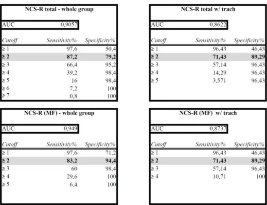
3.2. THE NCS-R TO IDENTIFY PATIENTS WITH PRESERVED NEURAL BASIS FOR PAIN EXPERIENCE ... 68
3.2.1. Material and methods ... 69
3.2.2. Results... 70
3.2.3. Discussion ... 75
3.3.THE NCS-R IN PATIENTS WITH A TRACHEOSTOMY ... 80
3.3.1. Material and methods ... 80
3.3.2. Results... 81
3.3.3. Discussion ... 84
6
CHAPTER 4. NEUROPHYSIOLOGICAL ASSESSMENT... 91
4.1.BACKGROUND:EEG RESPONSES TO NOCICEPTIVE INPUTS IN DOC ... 92
4.1.1. Recording of laser-evoked potentials ... 92
4.1.2. Limitations ... 94
4.2.MULTIMODAL ASSESSMENT OF SOMATOSENSORY PATHWAYS IN PATIENTS WITH DOC 97 4.2.1. Material and methods ... 99
4.2.2. Results... 101
4.2.3. Discussion ... 109
4.3. ELICITING ROBUST BRAIN RESPONSES TO NOXIOUS STIMULI USING A HIGH-SPEED HEATING THERMODE ... 111
4.3.1. Part 1 - Brain responses to laser and contact heat stimuli... 112
4.3.2. Part 2 - Perception-matched laser and contact heat stimuli ... 119
4.3.3. Part 3 - Comparing brain responses elicited by perception-matched stimuli ... 120
4.3.4. Discussion ... 123
4.4.BRAIN RESPONSES TO PERIODIC THERMAL STIMULATIONS IN HEALTHY SUBJECTS ... 124
4.4.1. Material and methods ... 126
4.4.2. Results... 131
4.4.3. Discussion ... 138
4.5.CONCLUSION ... 142
CHAPTER 5. FUTURE PERSPECTIVES AND CONCLUDING REMARKS ... 147
REFERENCES... 157
APPENDIX A. SUPPLEMENTARY MATERIAL ... 185
ACKNOWLEDGMENTS
Ce manuscrit est le fruit de longues années de réflexion, d’apprentissage, de travail, de collaborations. Ces quatre années furent marquées par des moments exaltants mais aussi des moments de doute, où remise en question et résilience sont maîtres-mots. Je souhaiterais remercier toutes les personnes qui ont contribué, de près ou de loin, à cette entreprise passionnante que fût l’accomplissement de cette thèse.
Tout d’abord, je tiens à remercier mes promoteurs, les Professeurs André Mouraux et Steven Laureys, qui m’ont accordé leur confiance dès l’instant où nous nous sommes rencontrés, tandis que je leur soumettais mon projet de thèse. Merci à vous deux pour le soutien, merci de m’avoir fait découvrir le monde passionnant de la recherche. C’était un rêve d’enfant que j’avais laissé de côté au bénéfice d’une pratique neurologique que j’estime aujourd’hui indissociable de la recherche. Merci André pour tes précieux conseils, ta disponibilité, ta pédagogie, le partage de tes connaissances et ta capacité de réassurance. Merci Steven pour ton soutien sans faille, ton enthousiasme, ton optimisme, ton leadership inspirant et cette capacité que tu as à inciter ton équipe à croire en ses rêves. Je tiens également à remercier les membres de mon comité d’accompagnement et membres du jury de thèse, les Professeurs Emmanuel Hermans (Président), Marie-Elisabeth Faymonville et la Docteure Camille Chatelle. Merci à vous de m’avoir guidé au long de ces quatre années et merci pour les échanges toujours constructifs lors de mes comités d’accompagnement. I want also to thank Professors Marzia De Lucia and Andrea Truini for agreeing to be part of my external jury and to be present for my private defense. Je tiens également à remercier la Professeure Anne Jeanjean. C’est avec elle que j’ai fait mes premiers pas de stagiaire en neurologie en 2008, mes premiers pas d’assistant en neurologie en 2010 et a fait partie de mon comité d’accompagnement dès le début de ma thèse en 2015. Malgré la maladie, elle a tenu ses engagements jusqu’à mon comité d’écriture en septembre 2019 avant de malheureusement nous quitter il y a peu. Je tenais donc à lui rendre hommage pour la source d’inspiration qu’elle a été pour le clinicien-chercheur que je suis aujourd’hui.
8
Combiner pratique clinique et recherche est une merveilleuse opportunité. Cela m’a permis de m’inspirer des besoins de mes patients pour dessiner les axes de mes recherches.
Dans ce cadre, je ne peux que remercier l’ensemble de l’équipe du C2 du CHN William Lennox et de l’unité UCAre pour tout le travail effectué au quotidien auprès de nos patients. Merci tout particulièrement au Docteur Bruno Pierson et David Dikenstein. Merci Bruno et David pour votre enthousiasme à me suivre dans cette dynamique d’excellence tant clinique que scientifique, toujours au bénéfice de nos patients. Merci également au Docteur Anne Frédérick, directrice médicale du CHN ; Anne, merci de m’avoir accordé ta confiance depuis presque 8 ans, de m’avoir soutenu tant dans mes projets de thèse que dans les projets de cette unité de revalidation neurologique et d’éveil de coma que tu m’as confiée.
Pour assumer ces différentes responsabilités, il aura fallu être un peu acrobate également. En effet, non content de cumuler deux mi-temps, j’ai également dû diviser mon mi-temps de recherche entre Bruxelles et Liège. Cela a demandé une bonne dose d’organisation, mais également de sacrifices car il n’est pas aisé de s’intégrer dans deux groupes de recherche avec si peu de temps de présence. Merci à tous pour votre accueil et votre aide. Merci à toute l’équipe Nocions, et particulièrement à Dounia, Eva, Julien et Arthur pour votre collaboration, aide et disponibilité. Merci également à l’ensemble de l’équipe Coma pour ces belles années, ces collaborations, ces séances de team buildings et ces chouettes congrès. Merci à Jitka, Yorgos, Marie-Michèle et Leandro pour leur aide dans la relecture de ce travail. Je tiens également à adresser un remerciement plus particulier à Aurore et Camille. Merci à toutes les deux pour votre soutien, votre aide précieuse tout au long de mon parcours ; mais surtout merci de m’avoir inspiré, de m’avoir, peut-être sans le savoir, tellement motivé à entreprendre cette thèse. Vous avez été et êtes des modèles pour moi. Je crois que je peux dire que sans vous avoir rencontrées, je ne serais sans doute pas là où je suis aujourd’hui.
9 Je souhaiterais également remercier les patients ainsi que leurs familles qui nous accordent leur confiance dans ces moments tragiques de leur vie. Vous êtes notre principale source d’inspiration et de motivation.
Je souhaiterais remercier ma famille et mes amis. Merci à mes parents de m’avoir donné l’envie d’être curieux, de m’avoir incité à me questionner sur tout et rien à la fois, de m’avoir poussé à grandir intellectuellement. Merci également à mes amis sans qui je ne serais pas non plus la personne que je suis, et ces remerciements vont tout particulièrement aux membres du CDLF et leurs compagnes. Vous le savez déjà, vous faites partie de ma famille. Même si ces dernières années et certains de mes sacrifices nous ont un peu éloigné, je sais que je peux compter sur votre soutien en toute circonstance. Savoir que vous êtes présent pour les bons moments mais surtout pour les coups durs m’a souvent aidé à avancer. Merci également à Jules pour son soutien sans faille.
Et enfin, merci à celle qui me supporte au quotidien. Madisson, merci pour ton soutien dans les moments de découragement, merci pour ce rôle difficile et ingrat que tu as tenu durant de nombreux mois. Ta patience a été mise à rude épreuve ! Merci pour ton coaching et ton organisation, sans cela, je ne sais pas si j’aurais pu terminer en temps et en heure ! Merci pour les sacrifices que tu fais et d’avoir toléré ceux que je nous ai imposé. Merci d’être celle que tu es, de me comprendre si bien. Je me sens beaucoup moins bizarre depuis que je peux être bizarre avec toi. Alors, parce que je ne te l’ai sûrement pas assez dit et ne te le dirai jamais assez, encore merci…
ABSTRACT
ncreased survival following a severe brain injury has led, in the last decades, to a dramatic increase of patients with a prolonged disorder of consciousness (DoC). The study of these patients faces today a paradigm shift, coming from the assessment of residual consciousness to that of residual specific cognitive processes. Pain is a subjective and, therefore, probably conscious experience. Regarding its close relationship with survival and consciousness, assessing residual ability to perceive pain is of major importance. Presenting both an impaired state of consciousness and a threat for their survival, patients with DoC constitute a pathological model of choice.
This thesis addresses several issues related to the assessment of pain perception in those non-communicative patients. Seminal studies on the topic used costly, hardly available and impractical neuroimaging paradigms. We here demonstrate that an approach based on behavioral and neurophysiological assessment (i.e., by means of electroencephalography [EEG]) complement well each other, is easily implementable in clinical practice and usable at bedside. Behavioral assessments, based on the Nociception Coma Scale – Revised (NCS-R) are thought to be useful for determining whether a patient is experiencing pain at a given moment, whereas EEG would help to determine whether the brain of the patient can process such a conscious experience of pain.
Behavioral studies of this thesis allowed to improve the understanding of the NCS-R scores and its applicability in a clinical context (e.g., in patients with a tracheostomy). Combined with an analysis of brain resting metabolism, they also allowed to identify patients with preserved neural basis for pain experience, which is the preservation of, at least, the left insula and the anterior cingulate cortex. Neurophysiological studies of this thesis demonstrate the possibility to identify brain
12
responses to selective somatosensory stimuli in some patients in a minimally conscious state. Unfortunately, brain responses displayed a very low signal-to-noise ratio. This highlighted the need of a dedicated approach for those patients. Hence, we developed and assessed several methods of stimulus presentation and validated an experimental setup which is easily transportable and usable at bedside.
The presented approach gives us, at the same time, the unique opportunity to look closely at each patient and to open up new perspectives. The strength of the relationship between pain and consciousness is such that it is required, to disentangle them, to go to the frontiers of consciousness; only in this way we will understand how pain experience emerges from the brain. Not only for science and knowledge, but also for ethical and clinical implications of these discoveries on the care of such highly vulnerable patients.
RÉSUMÉ
u cours des dernières décennies, l’augmentation de la survie à la suite d’une lésion cérébrale sévère a mené à une augmentation spectaculaire du nombre de patients présentant de manière prolongée un état de conscience altérée (ECA). Aujourd’hui, l'étude de ces patients est confrontée à un changement de paradigme, passant de la recherche d’une conscience résiduelle à celle de processus cognitifs spécifiques préservés. La douleur, en tant qu’expérience subjective, et dès lors, probablement consciente, en fait partie. Étant donné les relations étroites à la fois entre douleur et conscience ainsi qu’entre douleur et survie, évaluer la capacité du cerveau à expérimenter consciemment la douleur est d’importance capitale. Présentant à la fois une altération de conscience et un état neurologique critique mettant en péril leur survie, les patients ECA constituent un modèle pathologique de choix dans cette optique.
Cette thèse aborde plusieurs questions liées à l'évaluation de la perception de la douleur chez ces patients non communicants. Les études princeps sur le sujet utilisaient des paradigmes de neuroimagerie coûteux, difficilement disponibles et peu pratiques en routine clinique. Nous démontrons ici que la complémentarité des évaluations comportementales et électroencéphalographiques (EEG) est probablement nécessaire mais également aisément implémentable en pratique clinique, au chevet du patient. Les évaluations comportementales, basées sur la Nociception Coma Scale – Revised (NCS-R), devraient permettre de déterminer si un patient ressent la douleur à un moment précis, tandis que l'approche EEG devrait aider à déterminer si le cerveau du patient a la capacité de traiter la perception douloureuse de manière consciente.
Les études comportementales de cette thèse ont permis d'améliorer la compréhension des scores obtenus à la NCS-R ainsi que son applicabilité dans un contexte clinique (par exemple, chez des patients porteurs d’une trachéostomie). Associées à une analyse du métabolisme au repos du cerveau, elles permettent
14
d’identifier les patients présentant les bases neurales pour expérimenter consciemment la douleur, qui sont la préservation de l'insula gauche et du cortex cingulaire antérieur. Les études neurophysiologiques de cette thèse ont permis d'identifier des réponses cérébrales à des stimuli somatosensoriels sélectifs chez certains patients en état de conscience minimale. Malheureusement, ces réponses cérébrales affichaient un très faible rapport signal-sur-bruit. Cela a permis de mettre en évidence la nécessité d'une approche spécifique pour ces patients. Dès lors, nous avons développé et évalué différentes méthodes de présentation de stimuli et validé une configuration expérimentale facilement transportable et utilisable au chevet des patients.
L'approche présentée donne à la fois l'occasion unique d’être au plus proche du patient tout en ouvrant de nouvelles perspectives. La force de la relation entre la douleur et la conscience est telle qu'il faut, pour les démêler, aller aux frontières de la conscience ; ce n'est qu'ainsi que nous comprendrons comment l'expérience de la douleur émerge du cerveau. Non seulement pour la science, mais aussi pour les implications éthiques et cliniques que ces découvertes ont et auront sur les soins de ces patients hautement vulnérables.
GLOSSARY
ACC Anterior Cingulate Cortex. A brain area located on the medial part of the
frontal lobe, involved in the pain experience
AMH Mechano- and heat-sensitive Aδ-fiber nociceptors. Nociceptors whose
activation generate the sensation of first pain. The signal is conveyed to the brain through thinly myelinated Aδ-fibers.
CEPs Cool-evoked potentials. ERPs elicited by the activation of cool-sensitive afferents.
CHEPs Contact-Heat Evoked Potentials. ERPs elicited by the activation of nociceptors through a heating contact thermode.
CMH Mechano- and heat-sensitive C-fiber nociceptors. Nociceptors whose
activation generate the sensation of second pain. The signal is conveyed to the brain through unmyelinated C-fibers.
CRS – R Coma Recovery Scale – Revised. A clinical standardized evaluation of
the level of consciousness.
DCML Dorsal Column Medial Lemniscus. A sensory pathway conveying the
sensation for tactile object recognition, light touch, and proprioception to the brain.
DoC Disorders of Consciousness. This term refers to clinical entities of
pathological altered consciousness and includes coma,
vegetative/unresponsive wakefulness syndrome and minimally conscious state.
EEG Electroencephalography. A method to record the electrical activity
arising from the brain.
ERPs Event - Related Potentials. Modification of the ongoing electrical activity of the brain, usually in response to a transient sensory event.
16
eMCS Emergence of Minimally Conscious State. A clinical entity wherein patients show a reliable communication and/or a functional use of objects. They are therefore not considered to be in a DoC anymore.
GFP Global Field Power. Quantifies the amount of activity at each time point
in the field considering the data from all recording electrodes simultaneously resulting in a reference-independent descriptor of the potential field.
Harmonic A harmonic is a signal whose frequency is an integral multiple of the fundamental frequency (i.e., the frequency of a given stimulus).
LEPs Laser-Evoked Potentials. ERPs elicited by the activation of
thermonociceptors by the use of a laser device.
MCS Minimally Conscious State. A clinical entity wherein patients can be awake, display fluctuating signs of consciousness but without being able to functionally communicate.
MCS- MCS minus. MCS patients without observed language-related behaviors.
MCS+ MCS plus. MCS patients with residual language abilities
MCS* Non-behavioral MCS; also known as Cognitive Motor Dissociation
(CMD). A clinical entity wherein patients do not exhibit any sign of consciousness at bedside while showing brain activity compatible with the presence of residual awareness or cognitive abilities.
MRI Magnetic Resonance Imaging. This technique allows to analyze the
structure of the brain or its activity (functional MRI [fMRI]).
NCS – R Nociception Coma Scale – Revised. A clinical standardized scale to assess nociception in patients with a disorder of consciousness.
Nociception Refers to the neural process of encoding noxious stimuli. Pain sensation is not necessarily implied.
PET Positron Emission Tomography. A functional imaging technique
17 Salience The salience of a stimulus refers to its quality, in an environment, to
attract someone’s attention.
S1 Primary somatosensory cortex.
SII Secondary somatosensory cortex.
SC Slowly-adapting CMH.
SNR Signal-to-Noise Ratio. On the electroencephalographic recording,
describes the ratio between the amplitude of the signal of interest and the amplitude of the background noise, unrelated to the signal of interest.
SS-EPs Steady-State Evoked Potentials. Also known as Steady-State Responses
(SSRs). SS-EPs reflect a sustained cortical response induced by the long-lasting periodic repetition of a sensory stimulus; unlike ERPs, SS-EPs are best identified in the frequency domain, as peaks appearing at the frequency of the stimulus and/or at harmonics of that frequency.
SSRs See SS-EPs.
STT Spino-Thalamic Tract. A sensory pathway conveying noxious, thermal
and visceral information to the brain.
SUV Standardized Uptake Value. The ratio between the imaged radioactive
concentration (using PET) of a tracer and its injected concentration in the whole body.
TBI Traumatic Brain Injury.
TCSII A micro Peltier-based contact thermode able to generate very steep cooling/heating ramps of up to 300 °C/s.
UWS Unresponsive Wakefulness Syndrome; also known as Vegetative State
(VS). A clinical entity wherein patients can be awake but without any awareness of themselves and their surroundings.
CHAPTER 1. INTRODUCTION
“When your day is long and the night is yours alone, When you’re sure you’ve had enough of this life,
Well, hang on. Don’t let yourself go. Everybody cries and everybody hurts…sometimes.”
- R.E.M. he improvement of intensive care techniques, especially the accession to modern resuscitation techniques in the 1950s, has led to a dramatic increase in the number of patients with a prolonged disorder of consciousness (DoC). This raises numerous ethical, socio-economical and quality of life issues. Pain, one of the most important determinant of quality of life (Katz, 2002), is a subjective, and therefore, probably conscious experience. Unfortunately, given their state of impaired consciousness, patients with DoC are unable to communicate such a subjective experience. These patients, all along their care pathway, face a high rate of potentially painful medical complications (Ganesh et al., 2013; Whyte et al., 2013). Knowing their said great vulnerability, a crucial aspect in evaluating and improving the quality of life of these patients is to determine whether they have the ability to perceive pain.
The nature of the relationship between pain and consciousness remains barely known to date. Exploring the ability to process pain in patients with DoC is interesting both from a clinical and neuroscientist perspective: to understand a patient’s ability to experience pain at an individual level and, ideally, at a given time point; and to question the link between consciousness and pain experience through a pathological model where those physiologically linked cognitive experiences could somehow be dissociated from each other.
This first chapter aimed at giving the necessary background to understand Chapters 3 and 4, the core chapters of this thesis. I will first go through the spectrum
20
of the pathological altered states of consciousness and the means to assess residual consciousness. Afterwards, I will review the neurophysiology of pain as well as the means to elicit brain responses following the activation of somatosensory pathways.
1.1. DISORDERS OF CONSCIOUSNESS
Consciousness can be defined using two main components, arousal and awareness (Zeman, 2001). Arousal refers to the level of alertness or vigilance and involves the activity of the brainstem reticular formation, hypothalamus, and basal forebrain. Awareness refers to the contents of consciousness and is related to the activity of a widespread set of frontoparietal associative areas (Vanhaudenhuyse, Demertzi, et al., 2010). In healthy people, awareness and arousal are usually correlated, in the sense that the less aroused we get the less aware of our surroundings and ourselves we become (Steven Laureys, 2005). However, that assumption does not remain true in the case of patients with a prolonged DoC, for whom a dissociation between arousal and awareness components occur.
1.1.1.
C
LINICAL ENTITIESIn this section will be described the clinical entities to which the term “Disorders of Consciousness” refers, but also those which resemble them to avoid any misunderstanding in the further reading.
A
-
B
RAIN DEATHBrain death is caused by an acute brain injury leading to an irreversible and complete absence of electric (i.e., iso-electric electroencephalogram) and metabolic activity in the brain and the brainstem (Figure 1.1) (Steven Laureys et al., 2004), in the absence of any confounding factor such as hypothermia or severe electrolytic disturbance (Wijdicks et al., 2010). Clinically, a patient in brain death will show an absence of motor response and an absence of brainstem reflexes, including spontaneous apnea. Brain dead patients are not able to maintain their autonomic functions by themselves and require the use of external technical support (such as mechanical ventilation). Stopping these technical
21 supports does not lead to death as the patient is already dead; it only signs the end of the functional preservation of the other organs, that could have eventually been used for organ transplantation.
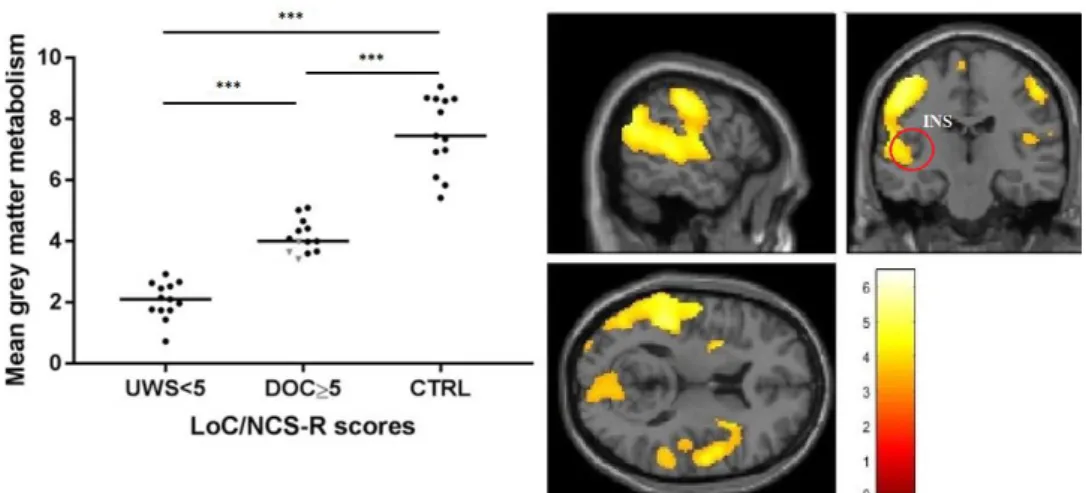
Figure 1.1. Cerebral resting brain metabolism acquired with 18Fluorodeoxyglucose – positron emission tomography (FDG-PET) in (a) healthy control, patients with (b) brain death and (c) unresponsive wakefulness syndrome (UWS) or vegetative state (VS). In brain death, we can observe an “empty skull sign” whereas in UWS/VS, the cerebral metabolism is highly and globally decreased but not absent. The color scale shows the amount of glucose metabolized per 100 g of brain tissue per minute. Adapted from (Steven Laureys, 2005)
B
-
C
OMAComa is an acute, transient, but complete state of unresponsiveness lasting for more than one hour. Due to a complete failure of the arousal system, this unresponsiveness is accompanied by an absence of eye opening, even when the patients are intensively stimulated (Jennett & Plum, 1972). The patient has no awareness of self and surrounding while some reflexive movements can be observed (Posner et al., 2007). Autonomic function regulation is compromised and often requires a respiratory assistance. Transient by definition, coma rarely lasts longer than two to five weeks (Steven Laureys, 2007). Afterwards, if they survive this acute stage, patients will either recover or awaken to remain for an undetermined amount of time (days to years) in a
22
state of unresponsive wakefulness, called the vegetative state or the unresponsive wakefulness syndrome.
C
-
U
NRESPONSIVEW
AKEFULNESSS
YNDROMEUnresponsive Wakefulness Syndrome (UWS) is somehow the European counterpart of the Vegetative State (VS), which remains much present in the American literature (Steven Laureys et al., 2010). Patients in a UWS/VS display three main features: (1) “sleep-wake-like cycles” consisting in periods of eye opening and closing (which is inconsistent with a diagnosis of coma); (2) at least a partial preservation of brainstem autonomic functions; and (3) the absence of awareness of self and surrounding, showing only reflexive movements (Monti, Laureys, et al., 2010). Reflexive movements can be, for instance, non-purposeful limb movements, a startle reflex to auditory or visual salient stimuli, orofacial chewing movements, eye movements without fixation. Accurate diagnosis of UWS patients is of capital importance as this diagnosis is associated with a less favorable outcome (Giacino et al., 2018), but also because the presence of consciousness will modify the rehabilitation as well as the interactions with these patients. If a patient does not regain any sign of consciousness after three months (from anoxic etiology) or twelve months (from traumatic injury), the patient is called to be in a “chronic UWS/VS”, a term that should be followed by the duration of the DoC, because evidence supports that the likelihood of recovery decreases with the duration of unresponsiveness (Giacino et al., 2018)
D
-
M
INIMALLYC
ONSCIOUSS
TATEPatients in Minimally Conscious State (MCS) can show discernible, oriented, purposeful but fluctuating cognitively-mediated behaviors. Among behaviors associated with a diagnosis of MCS, fixation, visual pursuit and reproducible movement to command are each observed in more than 50% of patients. These items are also the most frequently observed when the patients only show one sign of consciousness (Wannez et al., 2018). The most common initial signs identified in MCS patients are
23 visual pursuit (41%), reproducible command-following (25%) and automatic movements (24%) (Martens et al., 2019).
Recently, a subcategorization of MCS patients was proposed regarding the presence (MCS+; MCS plus) or the absence (MCS-; MCS minus) of language-related behaviors (Bruno et al., 2012). MCS+ could be somehow defined as “MCS with residual language abilities” while MCS- could reflect either a lower level of consciousness and/or the presence of more severe aphasia in MCS patients (Aubinet et al., 2018). Therefore, MCS- patients cannot be defined per se as less conscious than MCS+ patients. This statement is of importance as it is suspected that the presence of aphasia leads to misdiagnosis of patients regarding their level of consciousness (Schnakers et al., 2015).
Misdiagnosis remains a huge problem in the field of DoC. A misdiagnosis rate of 41% has been reported, meaning that 41% of patients diagnosed as UWS by their caregivers were actually identified as MCS when using a standardized and validated behavioral scale (i.e., the Coma Recovery Scale – Revised; see next section) (Schnakers et al., 2009). There are several reasons to such a high misdiagnosis rate. It can be related to the (lack of) experience of the examiner (or caregivers) or the absence of use of systematic tools to assess residual awareness. Even with experienced examiners, there still remain misdiagnosed patients as demonstrated by the use of paraclinical exams (Stender et al., 2014). Indeed, the evaluation can be limited by the patients’ disabilities (such as severe motor impairment, aphasia, etc.) preventing them to fully participate in the evaluation. This specific subset of patients that do not exhibit any sign of overt consciousness at bedside while showing brain activity compatible with the presence of residual cognition or awareness are labeled as “non-behavioral MCS” (MCS*) or as having a Cognitive Motor Dissociation (CMD) (Gosseries et al., 2014; Owen et al., 2006; Schiff, 2015).
E
-
E
MERGENCE FROM THEM
INIMALLYC
ONSCIOUSS
TATEThe emergence from a MCS (eMCS) is defined by the appearance of a reliable and accurate functional communication and/or functional use of at least two different objects (Giacino et al., 2002). This state, also referred to as “confusional state” or
24
“post-traumatic amnesia”, consists of a largely heterogeneous group of patients. Those patients can exhibit severe attentional deficit, disinhibition, labile behavior, hypokinetism or agitation. Therefore, they are often non-cooperative to rehabilitation therapy or systematic assessment of their impairments. Sometimes, the recovery of functional communication and object use does not occur simultaneously (Kalmar & Giacino, 2005; Taylor et al., 2007). When functional use of object recovers before functional communication, patients do not have the ability to report successfully subjective perception.
F
-
L
OCKED-
INS
YNDROMELocked-in syndrome (LIS) patients do not suffer from impaired consciousness and this entity cannot be included in the DoC spectrum. This syndrome, usually caused by a stroke of the basilar artery, results in an ischemia of the anterior part of the pons, causing anarthria and quadriplegia (including head and neck), but usually sparing the vertical oculomotor movement. As the lesion is limited to the brainstem, cognitive impairment is very limited but the ability to communicate is lost by the absence of motor control. Since these patients look like patients with DoC, a delayed diagnosis is not unusual. Diagnosis is of extreme importance as those patients are fully conscious and can be taught to communicate using eye movements.
1.1.2.
A
SSESSMENT OF RESIDUAL AWARENESSAlthough behavioral evaluation at bedside remains the gold-standard for assessing residual awareness in patients with DoC (Giacino et al., 2018), its use shows a significant remaining rate of misdiagnosis when confronted to paraclinical examination (Stender et al., 2014). Therefore, behavioral and paraclinical evaluations should be integrated. In this section, I will give an overview of the behavioral and paraclinical modalities available for the assessment of residual awareness in patients with DoC.
25
A
–
C
LINICAL BEHAVIORAL EVALUATIONThe Coma Recovery Scale – Revised (CRS-R; Table 1.1) (Giacino et al., 2004) was designed to allow a differential diagnosis between the different DoC entities, from coma to eMCS, and more specifically between UWS and MCS. The CRS-R consists of 23 hierarchically organized items in six subscales addressing (1) visual, (2) motor, (3) auditory, (4) oromotor/verbal, (5) communication and (6) arousal functions. The lowest item on each subscale represents reflexive activity while the highest item represents cognitively-mediated behaviors (Giacino et al., 2004). Because the CRS-R is not linear, the level of consciousness is imperfectly reflected by the total score (i.e., a UWS patient could have a higher score than an MCS patient, because the diagnosis of consciousness depends on the diagnosis associated with the highest-order behavior observed). Recent recommendations state that, in order to reduce the rate of misdiagnosis, patients should be serially assessed (Giacino et al., 2018) and at least five CRS-R should be performed within a short time interval (e.g., two weeks) (Wannez et al., 2017). Unfortunately, the complete protocol (30-45 minutes) of the CRS-R is not always suitable in clinical practice given time constraints. However, it was shown recently that limiting the CRS-R assessment to the five most frequently observed items in MCS patients (i.e., visual fixation, visual pursuit, reproducible movement to command, automatic motor response and localization to noxious stimulation) would allow to detect 99% of the patients diagnosed MCS at a behavioral level (Wannez et al., 2018). However, if the CRS-R is considered to date as the gold-standard for behavioral assessment of patients with DoC, we have to be aware that due to a lack of a diagnostic ground truth, criterion validity and diagnostic value (i.e., the scale’s ability to establish an accurate diagnosis compared with the true diagnosis as measured by a reference standard) cannot be determined for any available scoring system (Seel et al., 2010).
26
Table 1.1. The Coma Recovery Scale – Revised
4 Consistent movement to command MCS + 3 Reproducible movement to command MCS + 2 Localization to sound
1 Auditory startle 0 None
5 Object recognition MCS -4 Object localization : reaching MCS
-3 Visual Pursuit MCS
-2 Fixation (*) MCS
-1 Visual startle 0 None
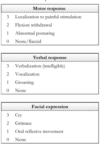
6 Functional object use eMCS 5 Automatic motor response MCS -4 Object manipulation MCS -3 Localization to noxious stimuli MCS -2 Flexion withdrawal
1 Abnormal posturing 0 None/Flaccid
3 Intelligible verbalization MCS + 2 Vocalization/Oral movement
1 Oral reflexive movement 0 None
2 Functional : accurate eMCS 1 Non-functional : intentional MCS+ 0 None
3 Attention
2 Eye opening without stimulation 1 Eye opening with stimulation 0 Unarousable
(*) Denotes MCS - except for anoxic etiologies Denotes MCS
Denotes MCS + Denotes eMCS
Arousal scale Auditory function scale
Visual function scale
Motor function scale
Oromotor/verbal function scale
27
B
–
N
EUROIMAGING TECHNIQUESFrom a neuroscientific point of view, neuroimaging techniques are important means to study the neural correlates of consciousness, that is defined as the minimal neural mechanisms jointly sufficient for any one conscious experience (Crick & Koch, 1990). These are also interesting means to probe the residual awareness in patients with DoC. Neuroimaging can contribute to reduce misdiagnosis rate in these patients by identifying, or eventually quantifying their residual consciousness (Bodart et al., 2017; Stender et al., 2014, 2015).
In this section, we will discuss neuroimaging techniques by describing the most commonly used paradigms and the most commonly used techniques: (functional) magnetic resonance imaging (MRI), positron emission tomography (PET) and electroencephalography (EEG).
1. R
ESTING,
PASSIVE AND ACTIVE PARADIGMSResting paradigms are paradigms in which patients are asked to mind-wander, without
performing any specific task, allowing to detect spontaneous brain activity without the influence of any internal or external stimulation. Resting protocols are suitable means to study patients with impaired communication, such as patients with DoC. Most common techniques using resting paradigms are functional MRI (e.g., Soddu et al., 2011), PET (e.g., Steven Laureys et al., 2004) and EEG (e.g., Chennu et al., 2017).
Passive paradigms rely on the analysis of brain activity in response to external stimuli
(e.g., somatosensory, visual, auditory), in the absence of any instruction to the patient. For instance, using functional MRI, it is possible to identify brain responses that are specific to the DoC patient’s preferred music (Heine et al., 2017). In EEG, classical passive paradigms are constituted by the recording of time-locked brain responses to external stimuli, such as the patient’s own-name (S. Laureys et al., 2004; Wang et al., 2015). However, one main limitation of all those passive paradigms is the heterogeneity of the widespread lesions of patients with DoC, potentially inhibiting the detection of those external stimuli (e.g., aphasia, blindness…). Another kind of passive paradigm
28
overcome this limitation: using the EEG, it is possible to analyze the modulation of brain activity in response to an external magnetic stimulus (combination of transcranial magnetic stimulation with EEG [TMS-EEG]). After stimulus onset, the complexity of the response is computed to obtain an index, the “Perturbational Complexity Index”, a measurement of residual consciousness (Bodart et al., 2017).
Active paradigms require the subject to perform a specific task on request and are
based on the analysis of willful modulation of brain activity in response to a command, for instance, using mental imagery tasks. Active paradigms suffer from the severe limitations inherent to the clinical state of patients with DoC, which may suffer from limited working memory or attention span. These techniques are resource costly for the patient, but when these paradigms work, they can eventually allow functional (not meaning easy) communication with the patient, as it was demonstrated with the famous “tennis playing motor imagery paradigm” (Bodien et al., 2017; Monti, Vanhaudenhuyse, et al., 2010; Owen et al., 2006). Motor imagery is also widely used in brain-computer interfaces using EEG (EEG-BCI) (Luauté et al., 2015), but other tasks can be used to elicit willful modulation of the EEG-responses, such as focusing on a lateralized somatosensory stimulus (Annen et al., 2018). Those methods, while resulting in a low rate of success are of major importance at an individual level, especially in patients not showing any sign of consciousness at bedside. Of course, when performing active paradigms, one should keep in mind that the absence of responsiveness does not equal to the absence of residual awareness (Sanders et al., 2012).
2. P
OSITRONE
MISSIONT
OMOGRAPHYPositron Emission Tomography is a neuroimaging technique based on the identification and the quantification of the uptake of a radioactive tracer. The most commonly used tracer is the fluorodeoxyglucose (abbreviated 18F-FDG, or simply
FDG), a glucose analog therefore taken-up by high-glucose-using cells, as neurons. The uptake of FDG during acquisition reflects the presence of active neurons.
In the field of DoC, the first FDG-PET was reported in 1987 and showed in a VS patient a decrease in global cerebral glucose uptake of 40-50% (Levy et al., 1987). Since
29 then, these results were reproduced in several studies (DeVolder et al., 1990; S. Laureys et al., 1999; Tommasino et al., 1995). However, global metabolism is not the only correlate of the level of consciousness. Indeed, at a regional level, it has been consistently shown that medial posterior cortex (encompassing the precuneus and adjacent posterior cingulate cortex) was more preserved in healthy subject and MCS patients than in UWS patients (Steven Laureys et al., 2004), potentially signing the importance of this area in the generation of consciousness. Furthermore, it was also demonstrated that the hypometabolism was more widespread and prominent in MCS patients when compared to eMCS patients (Nakayama et al., 2006). These studies allowed to identify a large fronto-parietal network whose functional integrity could be identified by machine-learning classifiers, allowing to calculate the probability for a patient to be conscious or not (Phillips et al., 2011). Unfortunately, those classifiers are not sufficiently powerful to classify correctly patients across the entire spectrum of the DoC. Therefore, to date, no standardized tool is available to classify patients according to their DoC. This classification still relies on the visual analysis by experienced researchers of images with the FDG Standardized Uptake Values (SUV). Despite this limitation, FDG-PET was recognized as the best tool allowing to disentangle UWS from MCS patients (Stender et al., 2014)
3. M
AGNETICR
ESONANCEI
MAGINGThe study of consciousness owes a lot to MRI. Thanks to functional MRI (fMRI), it was shown that consciousness can be divided into two anatomical networks: (1) the large fronto-parietal lateral “external awareness network”, devoted to the consciousness of the external world through the different senses (e.g., visual, auditory, somatosensory inputs); and (2) the mesial “internal awareness network” or “default mode network” (DMN), encompassing posterior cingulate/precuneus, anterior cingulate cortex and temporo-parietal junctions, which is more involved in self-related processes (such as mind-wandering, stimulus independent thoughts or self-related thoughts (Buckner et al., 2008; Fox et al., 2005; Mason et al., 2007)). Interestingly, those two networks are anti-correlated in healthy subjects, meaning that
30
when one is active, the other is not (Vanhaudenhuyse, Demertzi, et al., 2010). This anticorrelation is suspected to be linked to the recovery of consciousness since it has been demonstrated that patients who emerged from MCS showed a partial preservation of between-network anticorrelations, unlike patients remaining in a DoC that showed pathological between-network correlations (Di Perri et al., 2016).
From a technical and practical point of view, MRI has the double advantage to perform analysis on both brain structure and function into a single exam session and to display a high spatial resolution. Unfortunately, being able to record high-resolution images requires that the patients remain perfectly still, which is hardly the case in patients with DoC. Sedation of the patient increases the quality of acquisition, which is of particular interest for structural MRI. Unfortunately, it will inevitably affect the acquisition of fMRI data, which is an indirect measure of the blood flow, related to the regional consumption of blood oxygen. With sedation, the blood supply decreases inevitably in consciousness networks, affecting the acquired signal, regardless of the type of paradigm used (resting, passive or active). To avoid poor quality data acquisition due to the clinical condition of patients with DoC, it is necessary to adopt fast acquisition paradigms such as resting state fMRI. Passive or active paradigms (e.g., such as required for the study of pain) are much more challenging to achieve. Resting-state fMRI studies have detected reduced connectivity in the DMN of patients with DoC as compared to healthy subjects, mostly affecting the precuneus, a brain area considered to be a critical hub within this network (Cavanna, 2007; Vanhaudenhuyse, Noirhomme, et al., 2010). Passive or active paradigms, while technically challenging, are critical means to detect residual awareness in patients with DoC. In a famous cases series, researchers succeeded establishing a functional communication with patients, including patients evaluated UWS at bedside. For this active paradigm, the patients were asked to “imagine playing tennis” for responding “yes” and to “imagine walking into their house” for responding “no” to biographical questions (Monti, Vanhaudenhuyse, et al., 2010; Owen et al., 2006). Unfortunately, fMRI-based communication, because of the need of an MRI
31 scanner, is impractical and does not meet the needs of patients who would benefit much more from a bedside assessment.
4. E
LECTROENCEPHALOGRAPHYElectroencephalography is based on the recording of the electrical activity of the brain. This technique displays several advantages: portable, cost-effective and usable at patient’s bedside, it can be easily used with any kind of paradigm (resting, passive or active). In this section, I will not discuss active paradigms since they are mainly used to attempt functional communication through a Brain-Computer Interface (BCI), which is out of the scope of this thesis.
Resting EEG paradigms has been used to assess the level of consciousness. It was shown that, in comparison to controls, UWS (but not MCS) patients show higher delta (brain activity in the 1-4 Hz range) and theta activity (4-8 Hz), while alpha activity (8-12 Hz) was strongly decreased in both UWS and MCS groups (Lechinger et al., 2013). Using a high-density EEG and applying network analysis tools, connectivity measures allow to identify specific spectral signatures of reorganized brain networks in patients with DoC. For instance, using different network metrics (e.g., such as alpha band connectivity), it seems possible to differentiate patients’ level of consciousness across the whole spectrum of DoC (Chennu et al., 2014, 2017).
EEG-based passive paradigms allow to record brain responses to external stimuli, as mentioned in the preceding section. For instance, brain responses to stimuli or odd stimuli within a sequence (e.g., the subject own name or to deviant tones [mismatch negativity]) can be assessed (Fischer et al., 2008, 2010). Brain responses can also be elicited and analyzed following violations of auditory regularities in an oddball local-global paradigm (Bekinschtein et al., 2009; Faugeras et al., 2012). The main objectives of those paradigms are either to provide some prognostication of recovery or to differentiate UWS from MCS patients (Kotchoubey, 2017). An interesting and promising application of EEG-based passive paradigm using TMS-EEG has already been mentioned previously.
32
1.1.3.
A
SSESSMENT OF PAIN PERCEPTIONNociception and pain are usually closely related to each other. However, pain can occur without the activation of nociceptors (Flor et al., 2006), while the activation of nociceptors can trigger reflex somatic and autonomic responses without necessarily generating a conscious experience of pain (Robert K. Hofbauer et al., 2004). As pain is a subjective cognitive process, emerging from cortical processing (R. D. Treede et al., 1999), and therefore probably constitutes a conscious experience, this dissociation between nociception and pain might occur in a state of impaired consciousness. Hence, the study of patients with DoC provides a unique opportunity to investigate the relationship between pain and consciousness.
Seminal studies on the topic used passive paradigm in neuroimagery, such as the recording of brain responses to non-specific noxious stimuli (i.e., electrical stimuli) using PET or fMRI. In two seminal studies on UWS patients, Laureys et al. (2002) and Kassubek et al. (2003) found slightly divergent activation patterns in these patients. In the first study, the activation pattern was limited to the brainstem and the primary somatosensory cortex (S1) without functional connection to other brain areas known to play a role in pain experience, suggesting that, in these patients, the painful electrical stimuli did not elicit a conscious percept. In the latter study, the activation of S1 was accompanied with an activation of the secondary somatosensory cortex (S2), the posterior insula and the anterior cingulate cortex (ACC), i.e., brain regions that are consistently activated during pain experience in healthy individuals. These contrasting results might eventually be explained by the population of the study. Indeed, at that time, the diagnosis of MCS was not well known and patients were indifferently described as being in a Persistent Vegetative State (PVS) while some of them might have exhibited signs of MCS. Hence, Boly et al. (2008) replicated these studies, including three well-defined group of UWS, MCS patients and healthy controls. In MCS and in healthy controls, the electrical stimuli elicited a similar pattern of brain activity, including the thalamus, S1, S2, the insula and the ACC, while UWS patients displayed a reduced activity in these areas. These results are compatible with the notion that functional brain
33 imaging could be able to distinguish different groups of patients with disorders of consciousness, having differing abilities to experience pain (André Mouraux, 2015).
Using fMRI, Markl et al. (2013) compared brain responses to electrical stimuli in a group of well-selected UWS patients (according to behavioral assessment using the CRS-R) to age-matched healthy controls. They observed an activation in different regions of interest, but in a low rate. S1 was activated in only 3/30, S2 in 5/30 and/or ACC in 5/30 patients. Noteworthily, significant activation was observed more frequently in healthy controls, but not systematically (e.g., activation of the ACC in 6/15 healthy controls).
An important and common drawback of those studies is the fact that they all rely on electrical stimuli which may not have been of a sufficient intensity to reliably activate nociceptive afferents. Moreover, these stimuli are neither specific for nociception nor for the spino-thalamic tract (STT), making impossible to isolate the contribution of the medial lemniscus pathway to the observed responses. Nevertheless, the finding that noxious stimulation elicited widespread cortical activity in this group of patients suggests, at least for some of them, a residual ability to experience pain. Most importantly, the different patterns of subcortical and cortical activation in different groups of patients with DoC suggests that functional brain imaging might be useful to identify patients more or less likely to perceive pain following nociceptive stimulation. Unfortunately, the accessibility, the availability and the cost of functional neuroimagery, as well as the technical difficulties encountered with those patients (from acquisition to data analysis) make the use of these techniques very challenging.
In the last years, a few studies tried to investigate the question of pain perception in patients with DoC using EEG methods. These findings will be described in detail in the introduction of Chapter 4, dedicated to the neurophysiological assessment of pain in those patients.
34
1.2. PAIN, NOCICEPTION AND THE SOMATOSENSORY SYSTEM
Nociception is not synonym of pain. Although intimately related, they are two distinct physiological phenomena. Nociception could be defined as a sensory modality whose aim would be to signal a potential threat to the body’s integrity and trigger appropriate defensive reactions vital for survival (Legrain et al., 2011). Pain refers to a conscious percept and is defined as a “distressing experience associated with actual or potential
tissue damage with sensory, emotional, cognitive and social components“ (Williams & Craig, 2016).
Interestingly, in its definition of pain, the International Association for the Study of Pain specifies that “the inability to communicate verbally does not negate the possibility that an
individual is experiencing pain and is in need of appropriate pain-relieving treatment. Pain is always subjective” (Merskey, 1994). In this section, I will first review the anatomy and physiology
of the somatosensory and nociceptive pathways; then, I will review the different means available to probe these pathways.
1.2.1.
A
NATOMY AND PHYSIOLOGYThe somatosensory system consists of two major pathways: the dorsal column– medial lemniscus (DCML) and the spinothalamic tract (STT) (Figure 1.2). The DCML subserves mechanoreception (tactile object recognition, localization of skin contact, detection of vibration and texture) and proprioception (joint position, movement and force). The STT subserves thermoreception (coolness and warmth), nociception (impending tissue damage and pain) and visceroception (Cruccu et al., 2008).
A
-
S
OMATOSENSORY RECEPTORS AND AFFERENT FIBERS1. T
HE SPINO-
THALAMIC TRACTThe spino-thalamic tract system is the part of the somatosensory system that conveys nociceptive and thermal inputs from the periphery (comprising thermoreceptors, nociceptors and nerve fibers) to the brain.
35
Figure 1.2. Schematic drawing of the somatosensory system. The somatosensory system consists of two major pathways. The DCML (black lines) subserves mechanoreception and proprioception. The STT (red lines) subserves nociception, thermoreception and visceroception. From (R.-D. Treede, 2007).
Nociceptors constitute a specific class of sensory receptors that are activated by stimuli considered as a potential threat for the body (Burgess & Perl, 1967). Their activation is more related to the intensity of the stimulus (i.e., the stimulus should be considered as noxious) than to its modality (mechanical, thermal or chemical stimuli). For this reason, they are considered as “polymodal receptors”. Nociceptors are free nerve endings receptors, located in the skin (in the epidermis and dermal-epidermal junction (Novotny & Gommert-Novotny, 1988; Reilly et al., 1997)), bone, joint capsule, tendon, muscle, and many visceral organs. When nociceptors are activated, the signal is conveyed through thinly-myelinated or unmyelinated nerve fibers: Aδ- and C-fibers, respectively. Aδ- fibers, because of their myelination, are able to convey the signal with a velocity of 4 - 36 m/s, while unmyelinated C-fibers have a much lower conduction velocity of 0.4 - 2 m/s (Gardner & Johnson, 2014). Given the ability of those polymodal
36
receptors to respond to mechanical and heat stimuli, they are called “mechano- and heat-sensitive Aδ-fiber nociceptors” (AMH) and “mechano- and heat-sensitive C-fiber nociceptors” (CMH). Although they are activated by the same kind of stimuli, all of them do not respond in a uniform way.
• AMH receptors respond to mechanical input or thermal stimuli with an activation threshold around 46°C. (R. D. Treede et al., 1995).
o ‘Type 1’ AMH (AMH-1) show a tonic response only peaking
several seconds after stimulus onset.
o ‘Type 2’ AMH (AMH-2), are quickly adapting receptors that respond in a strong but very transient fashion, almost synchronous with stimulus onset.
• CMH receptors have a lower thermal activation threshold, around 41°C (Carmon et al., 1976; Cruccu et al., 2008; Julius & Basbaum, 2001).
o ‘Quick’ CMH (QC) respond vividly with a peak discharge occurring 0.4 second after the onset of the stimulus, and then rapidly adapt (within one second).
o ‘Slow’ CMH (SC) respond more gradually with a peak discharge occurring approximately two seconds after the onset of the stimulus and then tend to maintain a tonic level of activation throughout the whole stimulus period (Meyer & Campbell, 1981; Wooten et al., 2014).
Physiology of cool perception has been less extensively studied. Briefly lowering the skin temperature by a few degrees cannot be considered as nociceptive and, most importantly, the elicited percept is not painful. The main function of cool-sensitive afferents is probably to provide thermal cues for tactile discrimination (e.g., to discriminate two materials such as wood and metals based on their differing thermal conduction properties) (Ho & Jones, 2006). However, skin cooling, even by a few degrees cool will activate the free nerve endings of superficial layers of the skin and mucosae. Cool thermoreceptor neurons exhibit spontaneous electrical activity at resting
37 temperature (33°C), which increases in response to temperature reduction. Conversely, this basal action potential firing is suppressed by mild heating of the receptive field (Brock et al., 2001; Hensel & Zotterman, 1951). After signal transduction occurred, the action potential is conveyed to the brain by a specific set of Aδ- fibers (i.e., cool fibers), through the STT (as noxious heat) without eliciting a painful sensation. Cold nociceptors, whose activation threshold is around 14°C, are innervated by C-fibers (Harrison & Davis, 1999).
Spinothalamic projections travel a short distance within the ipsilateral spinal cord. They then enter the dorsal horn at segmental level, cross the midline and project, as the DCML, to the ventral posterolateral nucleus of the thalamus (VPL). VPL neurons receive nociceptive input and then project to the primary somatosensory cortex, the secondary somatosensory cortex, the insula and the anterior cingulate cortex (Cruccu et al., 2008). However, the STT is not the only ascending pathway; the spinoreticular tract, which is positioned closely to the STT, conveys nociceptive inputs to the reticular formation (and then to the thalamus) and some of its projections play a role in the arousal in response to nociceptive input.
2. T
HE DORSAL COLUMN-
MEDIAL LEMNISCUSDorsal column-medial lemniscus pathway conveys from periphery to the brain the sensation for tactile object recognition, light touch, two-point discrimination, and proprioception.
Unlike for the STT, the peripheral terminals of the DCML are corpuscular nerve endings (and not free nerve endings) in the skin, joint capsule and muscle. Those receptors are sensitive to mechanical stimuli such as vibration, pressure or stretching of the receptor. When those mechanoreceptors are activated, the related-signal will be conveyed through the DCML by large diameters fibers, namely Aα- and Aβ-fibers. Those fibers have a low activation threshold, are highly myelinated and convey the signal with a high velocity of 72-120 m/s and 36-72 m/s, respectively (Gardner & Johnson, 2014). Large myelinated fibers do not convey nociceptive input, but can influence the spinal transmission of nociceptive stimuli, as exemplified by the gate
38
control theory of pain. In a few words, this theory explains the mechanism by which the perception of nociceptive input can be modulated by the concurrent activation of Aβ-fibers through the activation of inhibitory spinal interneurons (R. Melzack & Wall, 1965).
From periphery to the spinal cord, the DCML pathway is completely segregated from the STT. However, they both project onto the VPL of the thalamus and they probably share pathways to the cortical areas as nociceptive inputs do not appear to project onto a cortical area exclusively devoted to receiving and processing nociceptive inputs (G. D. Iannetti & Mouraux, 2010; R.-D. Treede et al., 2003).
B
-
C
ORTICAL REPRESENTATION OF NOCICEPTION AND PAINNociceptive inputs are conveyed to the brain through multiple ascending pathways, with several thalamic relays, roughly divided in a medial and a lateral system, according to the localization of the relay (J. Brooks & Tracey, 2005; Willis, 1985). While other sensory modalities have a primary sensory cortex to receive and process their related sensory inputs (e.g., visual inputs projects on primary visual cortex), nociceptive inputs do not appear to project onto a dedicated cortical area (Andersson & Rydenhag, 1985; G. D. Iannetti & Mouraux, 2010). Instead, nociceptive-specific neurons (i.e., neurons responding exclusively to the activation of nociceptors) appear to be disseminated in cortical structures that are also involved in the processing of non-nociceptive sensory inputs and/or in other higher-order brain functions. Indeed, when a noxious sensory stimulus is applied on the human skin, neuronal activity is elicited in a vast network of brain regions, including S1, S2, the insula, and the ACC (Apkarian et al., 2005; Bushnell & Apkarian, 2006; L. Garcia-Larrea et al., 2003; Peyron et al., 2000; Tracey & Mantyh, 2007).
In this section, I will describe the involvement of those different structures in nociception and pain processing. Unless otherwise specified, the content of this paragraph is devoted to the cortical processing related to transient exteroceptive stimuli, using non-invasive approaches.
39
1. A
N OBLIGATORY INVOLVEMENT OF THE PRIMARY SENSORY CORTEX?
Primary somatosensory cortex involvement in the processing of nociceptive input and the perception of pain remains controversial (Apkarian et al., 2005; Bushnell et al., 1999; Liang et al., 2013; Mountcastle, 2005; Ploner et al., 1999). fMRI studies have shown an activation of contralateral S1 following the application of nociceptive stimuli (Bingel et al., 2004; Bushnell et al., 1999; Hu et al., 2014; Valentini et al., 2012), but this activation is not systematic and could be modulated via a top-down attentional modulation related to the fact that the nociceptive stimulus is likely to attract attention (Jones et al., 1992). To date, there is no evidence that demonstrates unequivocally an obligatory involvement of S1 in extracting the sensory-discriminative dimension of pain perception (i.e., a role in evaluating the intensity, localization and duration of a nociceptive stimulus) (André Mouraux, 2015).
2. E
VIDENT(
BUT NOT SPECIFIC)
INVOLVEMENT OF OPERCULO-
INSULAR CORTEXThe operculo-insular cortex refers to the secondary somatosensory cortex, the posterior and anterior insula. These brain areas are consistently activated bilaterally following a nociceptive stimulus (Bushnell & Apkarian, 2006; L. Garcia-Larrea et al., 2003; Peyron et al., 2000; Tracey & Mantyh, 2007; R. D. Treede et al., 1999). Indeed, a large number of the STT afferents project onto a brain region called the “posterior insula medial operculum” (PIMO). PIMO is described by some authors as being pain specific and could therefore constitute some form of “primary nociceptive cortex” (Craig et al., 2000; L. Garcia-Larrea, 2012). This hypothesis was supported by at least three findings: (1) focal epilepsy originating in this region can elicit (but not systematically) painful seizures (Charlesworth et al., 2009); (2) several case reports suggest that lesions of the insula can alter pain perception (although the opposite has also been reported) (Baier et al., 2014; Feinstein et al., 2016); and (3) the direct electrical stimulation of this area induces pain-related experience. However, this latter finding is far from being systematic (14 out of 43 patients), and when pain was elicited, other sensations were often aroused concomitantly (Ostrowsky et al., 2002).
40
Recently, Evrard (2019) suggested an interesting and novel approach of the functional organization of the insula, that could be described in three axes: (1) the “sensory-motor axis”, receiving e.g., sensory inputs from thalamus and having some control on the motoneuron of the spinal cord, directly affecting the contraction of striate muscles; (2) the “spino-cranial axis”, comprising afferents and efferents from the sympathetic and parasympathetic branches of the autonomic nervous system; and (3) the “cognition-emotion axis”, comprising two subdivisions of the anterior insular cortex, with the dorsal part being associated with cognitive tasks and the ventral part with emotional responses (for complete review, see Evrard, 2019). Hence, the insula is implicated in the processing of a wide range of sensory inputs, and contributes to a large number of cognitive, affective, interoceptive, and homeostatic functions, independently of sensory modality (zu Eulenburg et al., 2013). The insula appears to play an important role in the detection of salience (i.e., the property of a stimulus to stand out relative to neighboring stimuli) (Dowman et al., 2008; Downar et al., 2000), possibly constituting a hub connecting sensory areas to other networks involved in the processing and integration of external and internal information (André Mouraux, 2015).
3. S
IGNIFICANCE OF THE SO-
CALLED“
PAIN MATRIX”
Probably because of the absence of evidence of a primary nociceptive cortex, it has been suggested that the perception of pain emerges from the joint activation of a network of brain structures, encompassing all structures shown to be consistently activated following nociceptive stimulation, in particular, somatosensory areas (S1, S2), the insula and the ACC (Luis Garcia-Larrea & Peyron, 2013; Tracey & Mantyh, 2007). It has been repeatedly demonstrated that, within this so-called “pain matrix”, the magnitude of the brain responses elicited by a transient exteroceptive nociceptive stimulus correlate robustly with the intensity of perceived pain (Coghill et al., 1999; Derbyshire et al., 1997; G. D. Iannetti et al., 2005; Tölle et al., 1999). However, although probably necessary for the perception of pain, this “pain-matrix” is probably not specific for encoding pain experience and there are several arguments against that:
41 o The magnitude of the responses can be modulated by other factors than the intensity of pain perception. Indeed, the response habituates after a few trials without any difference in the elicited percept (i.e., the EEG response decreases without any decrease in the intensity of perception) (G. D. Iannetti et al., 2008). Moreover, the magnitude of those pain-evoked responses can also be differentially modulated in different subregions of this “pain matrix”. For instance, hypnotic suggestion of increased intensity of perceived pain has been shown to increase selectively the response magnitude in somatosensory areas; on the other hand, hypnotic suggestion of increased pain unpleasantness has been shown to selectively increase the response magnitude in the ACC. This also supports the hypothesis that somatosensory cortices might be more implied in the sensory-discriminative aspects of pain, while insula and ACC might be more related to the cognitive-affective dimension of the pain experience (R. K. Hofbauer et al., 2001; Rainville et al., 1997).
o The fact that nociceptive stimuli consistently elicit the same pattern of activation (S1, S2, insula, ACC) does not imply that this pattern of activation is stimulus-specific. Indeed, the “pain matrix” response can be completely dissociated from pain perception (G. D. Iannetti et al., 2008; Lee et al., 2009; A. Mouraux et al., 2003). Furthermore, it has been demonstrated in an elegant set of experiments (A. Mouraux & Iannetti, 2009; André Mouraux, Diukova, et al., 2011) that the bulk of nociceptive ERPs could be explained by multimodal neural activity also contributing to the ERPs elicited by non-nociceptive somatosensory, auditory and visual stimuli. Importantly, all stimuli were presented within a random sequence, using a large and unpredictable inter-stimulus interval, such as to maximize their salience (A. Mouraux & Iannetti, 2009). Using fMRI and the same paradigm, it has also been demonstrated that nociceptive, somatosensory, auditory and visual stimuli elicited spatially indistinguishable responses in the cingulate, the insula and the largest part of S2. Furthermore, a matching pattern of activation was also observed for nociceptive and non-nociceptive somatosensory stimuli in S1 and in a small subregion of S2, whereas the magnitude of these responses was correlated with subjective ratings of stimulus