1 Ti ss ue Engineeri n g The thir d er a of Tissue Engi nee ring: re ve rs ing the inno va tion dri ve rs (DOI: 1 0 .108 9/ te n.TEA .20 19 .00 6 4 ) This paper
has been peer-rev
iew ed an d acc ept ed f o r p u blicat
ion, but has yet t
o und ergo copy editin g and proof c o rr ec ti on. The fi nal published v ersi o n may d if fer from this pr o o f.
The third era of Tissue Engineering: reversing the innovation
drivers
Liesbet Geris, MSc, PhD (corresponding author)
1. Biomechanics Research Unit, GIGA - In silico Medicine, Université de Liège | Quartier Hôpital, Avenue de l’Hôpital 11 (B34) | 4000 Liège | Belgium. e-mail : [email protected]. Tel: +32 16 372690. Fax: +32 16 342543.
2. Prometheus, LRD Division of Skeletal Tissue Engineering, KU Leuven (Belgium) 3. Biomechanics Section, KU Leuven (Belgium)
Ioannis Papantoniou, MSc, PhD
1. Skeletal Biology & Engineering Research Center, KU Leuven | Herestraat 49 (831) | 3000 Leuven | Belgium. E-mail: [email protected] . Tel: +32 16 372743. Fax: +32 16 342543.
2. Prometheus, LRD Division of Skeletal Tissue Engineering, KU Leuven (Belgium)
2 Tissue E n gi neer ing The t h ir d er a of T issue Engineer ing: rev er sing t h e inno va tion dri ve rs (DOI: 1 0 .108 9/ te n.TE A.20 19 .00 6 4 ) This paper h as been peer-revi ew ed a nd a ccepted for pu blicati o n, b u t has y e t to u nderg o c o p yed it in g and proof correcti on . The fi
nal published ver
si
o
n may differ from
this proof.
Abstract
Ever since the introduction of the concept of Tissue Engineering, the field has developed and matured from a hype to a proper scientific discipline. Recently, the field is witnessing a reversal of the innovation drivers as it has started to move from a technology-driven science-focused field towards a patient-driven manufacturing-focused one. This evolution is made possible through innovations at the interface between biology and technology, including robust biological building blocks, precise biomanufacturing technologies, in-depth characterization methods and in silico models. Combining this with novel insights in TE-related regulatory sciences and business strategies, the field is ready to meet the grand challenge of designing, developing and delivering living implants with the accuracy and robustness expected from inanimate implants, leading to sustainable, predictable and vastly superior biological and clinical results.
Impact Statement
In this perspective we discuss the different stages of development the Tissue Engineering (TE) field has gone through in its relatively young history. We discuss how TE is evolving from a technology-driven, science-focused field towards a patient-driven, manufacturing-focused one where patients’ needs are translated into production process requirements, and subsequently into technological and biological innovations needed to meet the regulatory and clinical demands.
3 Tissue E n gi neer ing The t h ir d er a of T issue Engineer ing: rev er sing t h e inno va tion dri ve rs (DOI: 1 0 .108 9/ te n.TE A.20 19 .00 6 4 ) This paper h as been peer-revi ew ed a nd a ccepted for pu blicati o n, b u t has yet t o u nderg o co p yeditin g and proof correc tion . The fi
nal published versi
o
n may differ from
this proof.
Introduction
Undoubtedly, the interdisciplinary field of Tissue Engineering (TE) has made vast progress since it was coined in the mid-eighties, undergoing a dramatic technological but also conceptual evolution. However, to date there is still a lack of a widespread implementation of TE therapeutics in clinics with only four TE products having obtained to date official marketing authorisation in the EU. These are Spherox (CO.DON), Holoclar (Chiesi Farmaceutici), MACI (Vericel) and Chondrocelect (TiGenix), however, the last two products are no longer authorised or have been suspended [1]. This demonstrates that there is still considerable progress to be made before a systematic and consistent pipeline of TE products to the clinic can be established.
A historic perspective
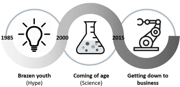
In the relatively young history of TE, roughly three periods can be distinguished (Figure 1).
The first era of TE (~ 1985-2000) is a perfect example of the brazenness that is typical for young and highly innovative fields. As any new technology or field going through the Gartner hype cycle, the first phase was characterised by bold claims and inflated expectations. Large investments were made in start-ups with the aim of making it to the clinics by the turn of the century. However, these promises were too high and the suggested clinical introduction was not realistic. Furthermore, the challenge of creating living implants was ill-defined at the time and the teams approaching the challenge were still deeply embedded in their respective disciplines, be it on the technology or the biology side. The main innovation drivers were technological (mostly related to biomaterials) or biological (mostly related to stem cells) innovations from the respective disciplines. Around the 2000s, many of these start-ups had failed or folded and the field was going through the ‘trough of disillusionment’ (cf. Gartner hype cycle) [2]. However, in academia, the TE field continued its evolution into a distinct scientific field with its own community and jargon. The second era of TE (~2000-2015) can be described as the coming of age of TE as a scientific discipline, finally overcoming some of the major challenges related to its interdisciplinarity. A new generation of researchers appeared,
4 Tissue E n gi neer ing The t h ir d er a of T issue Engineer ing: rev er sing t h e inno va tion dri ve rs (DOI: 1 0 .108 9/ te n.TE A.20 19 .00 6 4 ) This paper h as been peer-revi ew ed a nd a ccepted for pu blicati o n, b u t has y e t to u nderg o c o p yed it in g and proof correcti on . The fi
nal published ver
si
o
n may differ from
this proof.
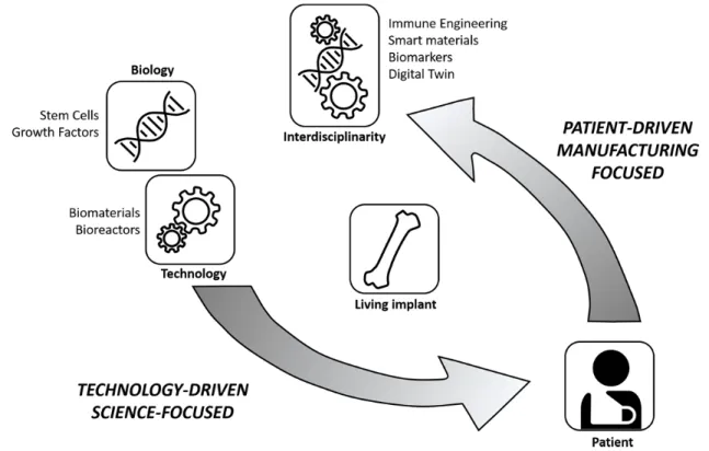
that could be described as the first real tissue engineers, able to cross the lines between the different disciplines. Paradigms were questioned and alternatives such as Developmental Engineering [3,4] were proposed while major breakthroughs in technologies such as the development of the induced pluripotent stem cell (iPS) technology [5], gene editing [6] and bioprinting [7,8] provided, from different angles, unprecedented possibilities. However, this increased quality was not yet translating into a tangible impact on patients’ lives. This growing realisation meant that TE started entering a new phase, one that sees the reversal of the innovation drivers. The innovation drivers are no longer the developments in individual disciplines but rather the needs of the patient (Figure 2). In order to reach the patient, products need to possess a set of quality attributes that ensure product potency while they should be engineered in a robust manufacturing process guaranteeing the quality of the resulting product. For this to happen, innovations are needed in terms of developing robust biological building blocks – tissue structures, precise manufacturing technologies and high-throughput quality control (QC) tools. Recent developments in these areas, discussed in more detail in the following sections, contribute to an increase in the capacity of the entire field to produce potent and clinically relevant healthcare solutions.
Starting from a clear identification of the patients’ needs, bearing in mind the manufacturing requirements, means the grand challenge for the third era of TE is to design, develop and deliver living implants with the accuracy and robustness we expect from inanimate implants, leading to predictable and vastly superior biological and clinical results. In order to tackle this grand challenge, technological and biological advances are required. In the second part of this perspective we will highlight a number of these advances and discuss their role in the global picture of TE.
From technological and biological advances … Developmental Engineering
Developmental engineering (DE) refers to the use of developmental processes as blueprint for the design of TE products. Developmental cascades show a tight regulation and
5 Tissue E n gi neer ing The t h ir d er a of T issue Engineer ing: rev er sing t h e inno va tion dri ve rs (DOI: 1 0 .108 9/ te n.TE A.20 19 .00 6 4 ) This paper h as been peer-revi ew ed a nd a ccepted for pu blicati o n, b u t has yet t o u nderg o co p yeditin g and proof correc tion . The fi
nal published versi
o
n may differ from
this proof.
robustness. In mimicking these processes, the aspiration is to overcome the current lack of quantitative metrics able to capture the degree of phenotypic progression that could forecast final TE product potency. Hence, concepts such as ‘developmental engineering’ [3,4], ‘engineered tissues as organ germs’ [9] and ‘reverse engineering development’ [10] are gaining ground. This allows for a gradual transition from the top-down question of “what is a biomimetic combination of cells-material-growth factor closely mimicking a target tissue” to a bottom-up question of “how can key developmental niches be accurately dissected, designed and precisely biomanufactured at the correct length scales into efficacious TE products”.
Qualitative examples of the DE approach have been successfully provided, for instance in bone tissue engineering, where cartilage intermediate templates have been shown to give rise to bone ossicles through endochondral ossification. The DE approach was also applied successfully using both adult [11,12] and embryonic [13,14] stem cell sources and both scaffold-free and scaffold-based approaches [15,16], demonstrating the robustness of this paradigm. However, to date, it has not yet been demonstrated that TE implants can guide regeneration in vivo, leading to an outcome where contaminating tissue structures are absent (contaminating tissues are tissues unrelated to the regenerative context and not contributing to TE product potency). In addition, engineered TE products possessing a hierarchically complex structure able to perform dual (or more) regenerative tasks upon implantation remain elusive. Scalable production of tissue modules that can guide regenerative events upon implantation is thus an important hallmark for the next generation of TE products.
In-depth Characterisation Technologies – Single cell analysis and molecular characterization
“You are as good an engineer as quality controls allow you to be”. One of the most important ongoing activities is the development of the analytics toolbox of the field. At the top of this, single cell genomics revolutionized the way we view cell populations. It provides a window into developmental cascades and tissue composition at the cell scale. The quantification of the kinetics of cell population transition from one cell type to another
6 Tissue E n gi neer ing The t h ir d er a of T issue Engineer ing: rev er sing t h e inno va tion dri ve rs (DOI: 1 0 .108 9/ te n.TE A.20 19 .00 6 4 ) This paper h as been peer-revi ew ed a nd a ccepted for pu blicati o n, b u t has y e t to u nderg o c o p yed it in g and proof correcti on . The fi
nal published ver
si
o
n may differ from
this proof.
along developmental cascades will provide an unprecedented compass for the design of TE products. However, TE is more than single cells and hence technologies that can provide insight in the molecular composition and architecture of extracellular matrices are equally needed. Quantitative Raman imaging represents a novel label-free method that enables visualization of 3D cell morphology and volumetric quantification of biomolecular structures with submicron-size detail [17]. This provides, amongst others, an excellent tool for deciphering ECM in terms of its molecular properties. For example, for native and engineered cartilage tissue Quantitative Raman imaging allowed a quantitative analysis of the distribution and organization of ECM constituents [18]. In addition, advances in nanoCT imaging allow for the 3D representation of the structural organization of complex tissues. For example, recently mineralized bone, bone marrow vasculature and adipose tissue can be detected simultaneously in 3D, providing insight in the design principles that complex bone TE constructs should possess to capture interactions between these three tissues [19]. Contrast enhanced nanoCT has furthermore produced insights at the nano-scale providing architectural information on tissue complexity [20]. Taken together, the TE field possesses a toolbox that captures ever more accurately the native tissue composition, structure, complexity and organization. These technologies allow deciphering in situ regenerative events and developmental cascades, providing a high-resolution picture of the properties that TE products should possess in order to exhibit regenerative properties. These technologies could therefore lead to precise characterization of the mechanisms of action of future TE products and their corresponding critical quality profile, essential for manufacturing processes described in the following section.
High-precision, scalable biomanufacturing
Biofabrication technologies able to operate with ever-increasing resolution and precision, provide an unprecedented opportunity for building tissues of increased complexity with a single molecule, cell, niche resolution comparable to that encountered in native tissues [21,22]. Examples of this enhancement in biomanufacturing capacity are the development of melt electrowriting [23], stereolithography [24] and laser-assisted technologies [25], all operating below 100 m resolution. In addition, robotic devices have been shown to
7 Tissue E n gi neer ing The t h ir d er a of T issue Engineer ing: rev er sing t h e inno va tion dri ve rs (DOI: 1 0 .108 9/ te n.TE A.20 19 .00 6 4 ) This paper h as been peer-revi ew ed a nd a ccepted for pu blicati o n, b u t has yet t o u nderg o co p yeditin g and proof correc tion . The fi
nal published versi
o
n may differ from
this proof.
possess the capacity to manipulate tissue modules such as single spheroids and positioning them in pre-ordered grids allowing them to fuse [26] or deposit them in printed scaffolds [27]. The ability to reproduce accurately CAD-based designs allows the incorporation of automation principles during production, although to date there are limited online QC methodologies that could validate the comparability between designed’ and ‘as-produced’ products. In addition, technical bottlenecks such as the production of vascularized, multi-centimeter sized implants will require the combined use of the aforementioned technologies now that successful proof of concept has been generated for endochondral bone repair [28,29]. The potential to build larger tissue structures based on robust modules, i.e. following a robust biological paradigm, produced through robust manufacturing processes and possessing a defined set of quality attributes, allows the implementation of Quality by Design in TE manufacturing [30] using the quality characterization methodologies described earlier.
In silico toolbox – digital twins of intracellular gene regulation to bioreactor biology.
As described above, the TE field is evolving into adopting solid biologic paradigms that provide robust biological building blocks, high precision biofabrication technologies that can use these building blocks to construct TE products and unprecedented quality characterization technologies that allow to assess the quality during and after production. Another enabling technology that could play an important role in establishing the systematic pipeline of TE products to the clinics, is in silico modeling. In silico is a term that was coined in analogy to the terms in vitro and in vivo, it is derived from the word silicium being the main component of computer chips and refers to computer modeling and simulation. In line with the Industry 4.0 concept, being the current trend of automation and data exchange in manufacturing technologies, digital twins can be created of every step of the TE production process [31]. Digital twins are digital replicas (computer models) of physical entities (e.g. process steps) that exchange information with their physical counterpart through sensors or historic data and that can be used to unable understanding and optimization of the physical process they represent. Intracellular gene regulatory models [32,33] are capable of reducing the in vitro large-scale screening to optimize culture medium composition. A digital twin of the bioprinting process [34] or the
8 Tissue E n gi neer ing The t h ir d er a of T issue Engineer ing: rev er sing t h e inno va tion dri ve rs (DOI: 1 0 .108 9/ te n.TE A.20 19 .00 6 4 ) This paper h as been peer-revi ew ed a nd a ccepted for pu blicati o n, b u t has y e t to u nderg o c o p yed it in g and proof correcti on . The fi
nal published ver
si
o
n may differ from
this proof.
subsequent maturation process in the bioreactor [35,36] allow for an optimization of these processes with a minimum of trial and error. Verification, validation and uncertainty quantification are key principles in building credibility for these models [37]. These digital twins can be connected in silico to form a digital TE pipeline and identify the crucial process steps and QC checks that guarantee high quality and predictable products. This provides a unique context for the production of in silico designed TE products where the appropriate quality attributes necessary for their functionality could be inbuilt. This way, in silico models guide the way from robust biological building blocks to a high-quality production process.
… to innovation in regulation and business
The biological and technological advances discussed in the previous section are by no means an exhaustive summary - think e.g. about advances in the incorporation of immune engineering requirements in TE products and renewable cell sources such as iPS cells, that have not been included in this perspective but are discussed elsewhere [5,38]. What is striking in the discussed examples is the joined involvement of both engineering and biological disciplines. Addressing the TE grand challenge requires true integration of all disciplines involved, transcending the individual disciplines and establishing TE as a transdisciplinary scientific domain. Simultaneously, it has become quite clear that the innovation is not only needed on the R&D side. TE solutions are inherently more complex and personalized than most pharmaceuticals and hence require tailor-made regulatory assessment, clinical trial design and business approaches.
On the regulatory side, it is important to involve regulatory bodies early on in the design and development of TE products [2]. In order to pass regulatory scrutiny, these TE products should be well-characterised, robust, with consistent efficacy and an
acceptable and controlled positive benefit/risk ratio. As argued in the previous section, enabling technologies can play a key-role in meeting those demands.
With respect to the design of clinical trials for TE products, the one-size-fits-all, large randomized clinical trials are no longer in line with the current scientific, clinical and economic reality. Clinical trial failures, especially in phase III, have huge safety and
9 Tissue E n gi neer ing The t h ir d er a of T issue Engineer ing: rev er sing t h e inno va tion dri ve rs (DOI: 1 0 .108 9/ te n.TE A.20 19 .00 6 4 ) This paper h as been peer-revi ew ed a nd a ccepted for pu blicati o n, b u t has yet t o u nderg o co p yeditin g and proof correc tion . The fi
nal published versi
o
n may differ from
this proof.
commercial/financial consequences [2]. An evolution has started towards more
innovative, efficient and adaptive trial designs [39], built around active participation of patients and patient organisations. Adaptive clinical trials, irrespective of the trial phase, use the results accumulated in the trial to modify the trial’s course in accordance with pre-specified rules [40]. It is furthermore important to de-risk the execution of clinical trials, especially in Phase III, by carefully analysing the risks of the trial execution and by a continuously surveillance of the quality of data being collected [2].
On the business end of the spectrum, innovation is required to cope with the
challenging combination of complexity and limited production volumes [41]. Principles such as design-to-cost [42] will become essential for the manufacturing of high-tech medical products such as TE implants. Design-to-cost is a management strategy designed to achieve an affordable product by targeting the manufacturing cost as an independent design parameter that must be met during product development. This strategy guarantees that at the end of the R&D process, the developed product can be manufactured at a cost that is not prohibitive to its clinical uptake and penetration in the market. The rigorous quality assessment, scalable manufacturing technologies and in silico models described in the previous section, are elements that are crucial for reaching the design-to-cost objectives.
Conclusion
Starting from a clear identification of the patients’ needs and the manufacturing requirements of the corresponding TE solutions, the need for specific biological and technological advances can be identified. The capacity to design and build tissues with predictive performance thanks to these technological and biological advances, will result in a high quality and robust TE production process and resulting product. This will facilitate approval from regulatory bodies while attracting investments by lowering risks associated to market-stage product failure and hence will contribute to a novel viable medical sector able to revolutionize healthcare.
10 Tissue E n gi neer ing The t h ir d er a of T issue Engineer ing: rev er sing t h e inno va tion dri ve rs (DOI: 1 0 .108 9/ te n.TE A.20 19 .00 6 4 ) This paper h as been peer-revi ew ed a nd a ccepted for pu blicati o n, b u t has y e t to u nderg o c o p yed it in g and proof correcti on . The fi
nal published ver
si
o
n may differ from
this proof.
Acknowledgments
The authors gratefully acknowledge support from the Research Foundation Flanders (FWO Vlaanderen; I.P: 12O7916N), the Belgian fund for national research (FNRS; T.0256.16), the Regenerative Medicine Crossing Borders initiative (www.regmedxb.com) powered by EWI-Vlaanderen, and the European Research Council under the European Union's Horizon 2020 framework program ERC/CoG 772418 (L.G.). The funders had no role in study design, data collection and analysis, decision to publish, or preparation of the manuscript. This work is part of Prometheus, the KU Leuven R&D division for skeletal tissue engineering (http://www.kuleuven.be/prometheus).
11 Tissue E n gi neer ing The t h ir d er a of T issue Engineer ing: rev er sing t h e inno va tion dri ve rs (DOI: 1 0 .108 9/ te n.TE A.20 19 .00 6 4 ) This paper h as been peer-revi ew ed a nd a ccepted for pu blicati o n, b u t has yet t o u nderg o co p yeditin g and proof correc tion . The fi
nal published versi
o
n may differ from
this proof.
1. Detela G., Lodge A. EU Regulatory Pathways for ATMPs: Standard, Accelerated and Adaptive Pathways to Marketing Authorisation. Mol Ther – Meth&Clin
development 13, 205-232, 2019.
2. Ronfard V., Vertès A.A., May M.H., Dupraz A., van Dyke M.E., Bayon Y. Evaluating the Past, Present, and Future of Regenerative Medicine: A Global View. Tissue Eng Part B Rev. 23(2):199-210, 2017.
3. Lenas, P., Moos, M., Luyten, F.P. Developmental engineering: a new paradigm for the design and manufacturing of cell-based products. Part II: from genes to networks: tissue engineering from the viewpoint of systems biology and network science. Tissue Eng Part B Rev. 15(4):395-422, 2009.
4. Lenas, P., Moos, M., Luyten, F.P. Developmental engineering: a new paradigm for the design and manufacturing of cell-based products. Part I: from
three-dimensional cell growth to biomimetics of in vivo development. Tissue Eng Part B Rev. 15(4):381-94, 2009.
5. Zhang, F., Citra, F., Wang, D. Prospects of Induced Pluripotent Stem Cell Technology in Regenerative Medicine. Tissue Eng Part B Rev 17(2) , 115, 2011.
6. Grobarczyk B., Franco B., Hanon K., and Malgrange B. Generation of isogenic human iPS cell line precisely corrected by genome editing using the CRISPR/Cas9 system. Stem Cell Rev 11, 774, 2015.
7. Mironov, V., Reis, N., Derby, B. Review: Bioprinting: A Beginning. Tissue Eng 12(4), 631-634.
8. Sears, N., Seshadri, D.R., Dhavalikar, P.S., Cosgriff-Hernandez, E. A Review of Three-Dimensional Printing in Tissue Engineering. Tissue Eng Part B Rev. 22(4):298-310, 2016.
9. Martin I. Engineered Tissues as Customized Organ Germs. Tissue Eng Part A. 20(7-8):1132-3, 2014.
10. Marcucio, R.S., Qin, L., Alsberg, E., Boerckel, J.D. Reverse engineering development: Crosstalk opportunities between developmental biology and tissue engineering. J Orthop Res. 35(11):2356-2368, 2017.
11. Farrell, E., Both, S.K., Odörfer, K.I., Koevoet, W., Kops, N., O'Brien, F.J., Baatenburg de Jong, R.J., Verhaar, J.A., Cuijpers, V., Jansen, J., Erben, R.G., van Osch, G.J.
12 Tissue E n gi neer ing The t h ir d er a of T issue Engineer ing: rev er sing t h e inno va tion dri ve rs (DOI: 1 0 .108 9/ te n.TE A.20 19 .00 6 4 ) This paper h as been peer-revi ew ed a nd a ccepted for pu blicati o n, b u t has y e t to u nderg o c o p yed it in g and proof correcti on . The fi
nal published ver
si
o
n may differ from
this proof.
vivo generation of bone via endochondral ossification by in-vitro chondrogenic priming of adult human and rat mesenchymal stem cells. BMC Musculoskelet Disord. 12:31, 2011.
12. Scotti, C., Piccinini, E., Takizawa, H., Todorov, A., Bourgine, P., Papadimitropoulos, A., Barbero, A., Manz, M.G., Martin, I. Engineering of a functional bone organ through endochondral ossification. Proc Natl Acad Sci U S A. 110(10):3997-4002, 2013.
13. Jukes, J.M., Both, S.K., Leusink, A., Sterk, L.M., van Blitterswijk, C.A., de Boer, J. Endochondral bone tissue engineering using embryonic stem cells. Proc Natl Acad Sci U S A. 105(19):6840-5, 2008.
14. Fernando, W.A., Papantoniou, I., Mendes, L.F., Hall, G.N., Bosmans, K., Tam, W.L., Teixeira, L.M., Moos, M. Jr, Geris, L.*, Luyten, F.P.*. Limb derived cells as a
paradigm for engineering self-assembling skeletal tissues. J Tissue Eng Regen Med. 12(3):794-807, 2018.
15. Matsiko, A., Thompson, E.M., Lloyd-Griffith, C., Cunniffe, G.M., Vinardell, T., Gleeson, J.P., Kelly, D.J., O'Brien, F.J. An endochondral ossification approach to early stage bone repair: Use of tissue-engineered hypertrophic cartilage constructs as primordial templates for weight-bearing bone repair. J Tissue Eng Regen Med. 12(4):e2147-e2150, 2018.
16. Visser, J., Gawlitta, D., Benders, K.E., Toma, S.M., Pouran, B., van Weeren, P.R., Dhert, W.J., Malda, J. Endochondral bone formation in gelatin methacrylamide hydrogel with embedded cartilage-derived matrix particles. Biomaterials. 37:174-82, 2015.
17. Kallepitis, C., Bergholt, M.S., Mazo, M.M., Leonardo, V., Skaalure, S.C., Maynard, S.A., Stevens, M.M., Quantitative volumetric Raman imaging of three dimensional cell cultures. Nature Comm, 8, 14843, 2017.
18. Albro, M.B., Bergholt, M.S., St-Pierre, J.P., Vinals Guitart, A., Zlotnick, H.M., Evita, E.G., Stevens, M.M. Raman spectroscopic imaging for quantification of depth-dependent and local heterogeneities in native and engineered cartilage. NPJ Regen Med. 3:3, 2018.
13 Tissue E n gi neer ing The t h ir d er a of T issue Engineer ing: rev er sing t h e inno va tion dri ve rs (DOI: 1 0 .108 9/ te n.TE A.20 19 .00 6 4 ) This paper h as been peer-revi ew ed a nd a ccepted for pu blicati o n, b u t has yet t o u nderg o co p yeditin g and proof correc tion . The fi
nal published versi
o
n may differ from
this proof.
19. Kerckhofs, G., Stegen, S., Van Gastel, N., Sap, A., Falgayrac, G., Penel, G., Durand, M., Luyten, F., Geris, L., Vandamme, K., Parac-Vogt, T., Carmeliet, G. Simultaneous three-dimensional visualization of mineralized and soft skeletal tissues by a novel microCT contrast agent with polyoxometalate structure. Biomaterials, 159, 1-12, 2018.
20. Busse, M., Müller, M., Kimm, M.A., Ferstl, S., Allner, S., Achterhold, K., Herzen, J., Pfeiffer, F. Three-dimensional virtual histology enabled through cytoplasm-specific X-ray stain for microscopic and nanoscopic computed tomography. PNAS, 115 (10), 2293-2298, 2018.
21. Gladman, S.A., Elisabetta A., Matsumoto, E.A., Nuzzo, R.G., Mahadevan, L., Lewis, J.A., Biomimetic 4D printing. Nature Materials, 15, 413–418, 2016.
22. Moroni, L., Burdick, J.A., Highley, C., Lee S.Y., Morimoto, Y., Takeuchi, S., Yoo, J.J., Biofabrication strategies for 3D in vitro models and regenerative medicine. Nature Rev Mat 3, 21–37, 2018.
23. Dalton, P.D., Melt electrowriting with additive manufacturing principles. Curr Op Biomed Eng 2, 49-57, 2017.
24. Melchels, F.P.W., Feijen, J., Grijpma, D.W., A review on stereolithography and its applications in biomedical engineering. Biomaterials, 31(24), 6121-6130, 2010. 25. Keriquel, V., Oliveira, H., Rémy, M., Ziane, S., Delmond, S., Rousseau, B., et al. In
situ printing of mesenchymal stromal cells, by laser-assisted bioprinting, for in vivo bone regeneration applications. Scientific Reports. 7, 1778, 2017.
26. Moldovan, N. I., Hibino, N. & Nakayama, K. Principles of the Kenzan Method for Robotic Cell Spheroid-Based Three-Dimensional Bioprinting. Tissue Eng. Part B Rev. 23, 237–244, 2017.
27. Mekhileri, N.V., Lim, K.S., Brown, G.C.J., Mutreja, I., Schon, B.S., Hooper, G.J., Woodfield, T.B.F. Automated 3D bioassembly of micro-tissues for biofabrication of hybrid tissue engineered constructs. Biofabrication, 10 (2), 2018.
28. Daly, A.C., Pitacco, P., Nulty, J.; Cunniffe, G.M., Kelly, D.J. 3D printed microchannel networks to direct vascularisation during endochondral bone repair. Biomaterials, 162, 34-46, 2018.
14 Tissue E n gi neer ing The t h ir d er a of T issue Engineer ing: rev er sing t h e inno va tion dri ve rs (DOI: 1 0 .108 9/ te n.TE A.20 19 .00 6 4 ) This paper h as been peer-revi ew ed a nd a ccepted for pu blicati o n, b u t has y e t to u nderg o c o p yed it in g and proof correcti on . The fi
nal published ver
si
o
n may differ from
this proof.
29. Kolesky, D.B., Homan, K.A., Skylar-Scott, M.A., Lewis, J.A. Three-dimensional bioprinting of thick vascularized tissues. PNAS, 113 (12), 3179-3184, 2016. 30. Lipsitz, Y.Y., Timmins, N.E., Zandstra, P.W. Quality cell therapy manufacturing by
design. Nat Biotechnol. 34(4):393-400, 2016.
31. Geris, L., Lambrechts, T., Carlier, A., Papantoniou, I. The future is digital: In silico tissue engineering. Curr Op Biomed Eng, 6, 92-98, 2018
32. Kerkhofs, K., Leijten, J., Bolander, J., Luyten, F.P., Post, J., Geris, L. A qualitative model of the differentiation network in chondrocyte maturation: a holistic view of chondrocyte hypertrophy. PLoS One, 11, e0162052, 2016.
33. Emmert-Streib,F., Dehmer, M., Haibe-Kains, B. Gene regulatory networks and their applications: understanding biological and medical problems in terms of networks. Front Cell Dev Biol. 2: 38, 2014.
34. Gohl, J., Markstedt, K., Mark, A., Håkansson, K., Gatenholm, P., Edelvik F. et al., Simulations of 3D bioprinting: predicting bioprintability of nanofibrillar inks. Biofabrication, 10(3): p. 034105, 2018.
35. Guyot, Y, Papantoniou, I., Luyten, F.P., Geris, L. Coupling curvature-dependent and shear stress-stimulated neotissue growth in dynamic bioreactor cultures: a 3D computational model of a complete scaffold. Biomech Model Mechanobiol. 15(1):169-80, 2016.
36. Mehrian M, Guyot Y, Papantoniou I, Olofsson S, Sonnaert M, Misener R, Geris L. Maximizing neotissue growth kinetics in a perfusion bioreactor: An in silico strategy using model reduction and Bayesian optimization. Biotechnol Bioeng. 115(3):617-629, 2018.
37. ASME V&V40. Assessing Credibility of Computational Modeling through Verification and Validation: Application to Medical Devices. 60p, 2018. ISBN: 9780791872048. 38. Hoffman T., Khademhosseini A., Langer R.S.. Chasing the Paradigm: Clinical
Translation of 25 Years of Tissue Engineering. Tissue Eng Part A. In press, 2019. doi: 10.1089/ten.TEA.2019.0032.
39. Garralda, E., Dienstmann, R., Piris, A., Braña, I., Rodon, J., Tabernero, J. New clinical trial designs in the era of Precision Medicine. Mol Oncol. In press, 2019
15 Tissue E n gi neer ing The t h ir d er a of T issue Engineer ing: rev er sing t h e inno va tion dri ve rs (DOI: 1 0 .108 9/ te n.TE A.20 19 .00 6 4 ) This paper h as been peer-revi ew ed a nd a ccepted for pu blicati o n, b u t has yet t o u nderg o co p yeditin g and proof correc tion . The fi
nal published versi
o
n may differ from
this proof.
40. Pallmann P., Bedding A.W., Choodari-Oskooei B., Dimairo M., Flight L., Hampson L.V., Holmes J., Mander A.P., Odondi L., Sydes M.R., Villar S.S., Wason J.M.S., Weir C.J., Wheeler G.M., Yap C., Jaki T. Adaptive designs in clinical trials: why use them, and how to run and report them. BMC Med. 16(1):29, 2018.
41. National Academies of Sciences, Engineering, and Medicine. 2017. Navigating the manufacturing process and ensuring the quality of regenerative medicine
therapies: Proceedings of a workshop. Washington, DC: The National Academies Press. doi: https://doi.org/10.17226/24913.
42. Grady, Jeffrey O; Requirements Foundation in: System Requirements Analysis System requirements analysis , 2014, p.93-150. DOI: 10.1016/B978-0-12-417107-7.00002-6.
Address correspondence to Liesbet Geris
Biomechanics Research Unit – GIGA in silico medicine
University of Liège Quartier Hôpital Avenue de l’Hôpital 11 (B34) 4000 Liège Belgium e-mail : [email protected]
Author Disclosure Statement
No competing financial interests exist.
16 Tissue Engi ne er ing The t h ir d era o f Ti ss ue Engineeri n g: r eversing t h e inno va ti on dri ver s ( D O I: 1 0 .108 9/ten.TEA.20 19 .00 6 4 ) This paper ha s been peer-review ed a nd accepted for pu blicatio n, but h as y et to underg o c o p yedit in g a n d p ro o f correction. The fi nal pub lished versio n m ay differ f rom t h is proof . Figure captions
Figure 1: The three eras of Tissue Engineering. The years indicated in the figure are rough indications, not related to any particular event.
17 Tissue Engi ne er ing The t h ir d era o f Ti ss ue Engineering : r eversing t h e inno va ti on d ri ver s ( D O I: 1 0 .108 9/ten.TEA.20 19 .00 6 4 ) This paper ha s been peer-review ed a nd accepted for pu blicatio n, but h as y et to underg o c o p yedit in g a n d p ro o f correction. The fi nal pub lished versio n m ay differ f rom t h is proof .
Figure 2: Reversing the innovation drivers. Originally being technology driven and science focused, the TE field is (or should be) increasingly becoming patient driven and
manufacturing focused.