B
OVINE HERPESVIRUS
1-
INDUCED APOPTOSIS
:
PHENOTYPIC
CHARACTERIZATION OF SUSCEPTIBLE PERIPHERAL
-
BLOOD
MONONUCLEAR CELLS
E. Hanon, M. Lambot, S. Hoornaert, J. Lyaku, and P. P. Pastoret
Department of Immunology/Vaccinology, Faculty of Veterinary Medicine, University of Liège, Liège, Belgium
ABSTRACT
Bovine herpesvirus 1 (BHV-1), a member of the Alphaherpesvirinae, induces apoptotic cell death in peripheral blood mononuclear cells (PBMC). To investigate the process by which BHV-1 induces apoptosis, we determined the susceptibility of the three main PBMC subpopulations to BHV-1-induced apoptosis. This study shows that BHV-1 can induce apoptosis individually in T lymphocytes, B lymphocytes and monocytes. This conclusion is based on the following findings: (i) BHV-1 substantially reduces the percentages of viable T and B lymphocytes in PBMCs. (ii) Concomitant detection of cell phenotype and apoptosis indeed showed higher percentages of apoptotic T lymphocytes and B lymphocytes in BHV-1-infected PBMCs than in mock-infected cells. (iii) Each individual PBMC subpopulations (B lymphocytes, T lymphocytes and monocytes) undergo apoptosis when incubated with BHV-1. These data also suggest that BHV-1 does not require the recruitment of one or more individual PBMC subpopulations (e.g. cytotoxic cells) to induce apoptosis. Finally, we observed that BL-3 cells which have been characterized as bovine tumoral B lymphocytes also undergo apoptosis when incubated with BHV-1. Therefore, the use of the BL-3 cell line provides a new experimental model to investigate the apoptotic process induced by BHV-1 in vitro.
Introduction
Bovine herpesvirus 1 (BHV-1), a member of the Alphaherpesvirinae [20], is the aetiological agent of infectious bovine rhinotracheitis, infectious pustular vulvovaginitis, abortion and fatal neonatal systemic infections [26]. In addition to initial respiratory infections, BHV-1 can predispose infected animals, presumably through immunodepression [2, 8], to secondary Pasteurella haemolytica infections leading to severe pneumonia and death [26].
In a variety of acute viral infections, there is increasing evidence that immunodepression is directly associated with the induction of apoptosis in immune cells [16, 19]. Apoptotic cell death is a genetically encoded suicide program which allows for the elimination of cells that have been produced in excess, developed improperly, or sustained genetic damage [23]. We have previously shown that BHV-1, even when inactivated, is able to induce apoptosis in mitogen-stimulated peripheral blood mononuclear cells (PBMCs) [11]. Apoptotic cell death is characterized morphologically by cell shrinkage, apoptotic body formation, condensation of the chromatin [6, 15], and biochemically by fragmentation of DNA into oligonucleosomal DNA fragments [1, 25]. Furthermore, the early stages of apoptosis are associated with the translocation of phosphatidylserine (PS) from the inner side of the plasma membrane to the outer layer [7]. This exposure of PS on the external surface of the cell membrane can be detected by the use of annexin V which is a Ca2+-dependent phospholipid-binding protein [18, 24].
Since the inactivated BHV-1 viral particles are still able to induce apoptosis in PBMC cultures, it has been postulated that the mechanism of induction involves: (i) viral-cellular molecules interactions during the attachment or penetration processes, or (ii) the effect of viral structural protein(s) released into the cell [11]. However, it is still unknown whether BHV-1-induced apoptosis occurs as a consequence of a direct viral particle-cell interaction or requires an indirect recruitment of cytotoxic cells [19, 27, 28]. Furthermore, although it has been already described that interleukin-2-dependent T lymphocytes incubated with BHV-1 undergo a cell death process resembling apoptosis [9], it is currently unknown whether BHV-1-induced apoptosis is restricted to one specific PBMC subpopulation or can occur in all the constituent subpopulations. Therefore, in order to investigate further the process by which BHV-1 induces apoptosis, we determined the susceptibility of the three main PBMC subpopulations to BHV-1- induced apoptosis. Here, we demonstrate that BHV-1-induced apoptosis occurs in T lymphocytes, B lymphocytes and monocytes. The results of the present study demonstrate that BHV-1 does not require the recruitment of one or more individual PBMC subpopulations (e.g. cytotoxic cells) to induce apoptosis. These observations also provide important indications to the possible mechanism(s) involved in the development of immunodepression which occurs in BHV-1-infected cattle.
Materials and methods
VIRUS
The BHV-1 Cooper strain was kindly provided by Dr. J. T. van Oirschot (Lelystad, The Netherlands). The virus was multiplied on Madin Darby Bovine Kidney cells (MDBK) (American type culture collection CCL22) and purified as described by Szilágyi and Cunningham [22] with some minor modifications [14]. Purified virus was resuspended in RPMI 1640 (Gibco, Gent, Belgium) supplemented with 2 mM glutamine (Gibco), 100 IU/ml penicillin and 100 µg/ml streptomycin (Gibco) and stored at −70◦C until use.
MONOCLONAL ANTIBODIES
The following monoclonal antibodies (mAb) were used: mAb CC42 which recognizes the BoCD2 antigen present on CD2 T lymphocytes [5]; mAb 1H4 which is specific for bovine cell surface IgM present on B lymphocytes [13]; mAb 2G1 which defines bovine monocytes/macrophages and polymorphonuclear cells [12]; mAb CC15 which recognizes the WC1 antigen present on bovine -𝛄𝛅 T lymphocytes [5].
CELLS AND CULTURE CONDITIONS
MDBK cells were cultured in minimum essential medium (Gibco) containing 5% fœtal calf serum (FCS) (Gibco). This cell line was maintained free of mycoplasma and of bovine viral diarrhoea virus. Bovine lymphoma B cells (BL-3; American type culture collection CRL 8 037) were cultured in Optimem medium (Gibco) containing 20% FCS, 100 IU/ml penicillin, 0.5 µM amphothericin (Gibco) and 100 µg/ml streptomycin. PBMCs were obtained from 12- to 24-months-old steers serologically negative to BHV-1 as described elsewhere [11]. The enrichment procedure for different cell populations of PBMCs was carried out with paramagnetic beads according to manufacturer’s instructions [17]. The selection of B lymphocytes, CD2 T lymphocytes, WC1 T lymphocytes and monocytes was performed using mAbs 1H4 (1:2 000 diluted ascite), CC42 (1:10 000 diluted ascite), CC15 (1:10 000 diluted ascite) and 2G1 (1:2 000 diluted ascite) respectively (positive selection). We also purified T lymphocytes, B lymphocytes and monocytes by selective depletion of PBMC subpopulations (negative selection). MAbs 1H4 and 2G1 were used to enrich T lymphocytes. MAbs CC42, CC15 and 2G1 were used to enrich B lymphocytes and mAbs CC42, CC15 and 1H4 were used to enrich monocytes. Purified cells and PBMCs were cultured in RPMI 1 640 medium supplemented with 10% FCS, 5 × 10⁻⁵M
2-mercaptoethanol (Gibco), 2 mM glutamine, 100 IU/ml penicillin and 100 µg/ml streptomycin. To induce cellular proliferation, the culture medium was supplemented with 0.5 µM Ionomycin (Sigma, Bornem, Belgium) and 10 nM Phorbol 12, 13-dibutyrate (Sigma) (IONO/PDB). Cells were cultured at a density of 2 × 10⁶ cells/ml.
FLOW CYTOMETRY
Flow cytometric analysis was performed using a Becton-Dickinson fluorescence-activated cell sorter (Facstar Plus), equipped with an argon laser (ILT air cooled with 100 mW excitation lines
at 488 nm). Debris were excluded from the analysis by the conventional scatter gating method. The cells or the nuclei doublets were excluded from analysis by using the pulse processor boards (Becton-Dickinson). Ten-thousand events per sample were collected in a list mode, stored, and analyzed by the Consort 32 system (Becton-Dickinson).
IMMUNOFLUORESCENT CELL SURFACE STAINING
After harvesting, PBMCs were washed twice with PBS and incubated for 30 min with the appropriate predetermined dilution of primary mAb (1H4, CC42, CC15, or 2G1). The cells were washed twice with PBS containing 5% FCS, and further incubated with FITC-conjugated F(ab’)2
goat anti-mouse IgG (H+L chains) (Dako, Gentbrugge, Belgium) for 30 min. After an additional wash with PBS containing 5% FCS, the cells were resuspended in PBS containing5 µ,g/ml propidium iodide (PI; Sigma) and analyzed by flow cytometry for green (FITC) and red (PI) fluorescences.
FITC-ANNEXIN V/PHENOTYPE DOUBLE STAINING
Cellular phenotype was determined by immunofluorescent cell surface staining as described above with some minor modifications. Briefly, cells were incubated with the primary mAb (1H4, CC42, CC15 or 2G1), washed twice and further incubated with PE-conjugated F(ab’)2 goat
anti-mouse IgG (H+L chains) (Dako). After an additional wash with PBS containing 5% FCS, the cells labeled with FITC-Annexin V and PI as described in the Boehringer Mannheim apoptosis detection kit (Annexin-V-FLUOS) (Boehringer Mannheim, Mannheim, Germany). Finally, cells were analyzed by flow cytometry for green (FITC), orange (PE) and red (PI) fluorescences. Dead cells (PI positive cells) were excluded from the analysis.
DETECTION OF DNA FRAGMENTATION
Detection of DNA fragmentation was carried out by the TUNEL procedure. Briefly, cells were washed with PBS containing 10% FCS and fixed in PBS containing 1% paraformaldehyde. The cells were incubated for 15 min at 4◦C, washed, resuspended in 2 ml of ice cold 70% ethanol,
and incubated overnight at −20◦C. Fixed cells were washed twice with PBS and the TUNEL
reaction carried out as described in the apoptosis detection kit (in situ cell death detection kit, fluorescein; Boehringer Mannheim). Cells were then resuspended in PBS and analyzed by flow cytometry for green (FITC) fluorescence.
ELECTRON MICROSCOPY
Electron microscopic analysis was carried out as described by Bielefeldt Ohmann and Bloch [3]. Briefly, the cultured cells were collected, washed twice in PBS and fixed in PBS containing 2.5% glutaraldehyde. The cells were then incubated for 1 h in the presence of 1% osmium tetroxide, dehydrated through a graded series of ethanols, infiltrated and embedded in epon. Sections of 90 nm were collected onto copper grids, double stained with uranyl acetate and lead citrate and examined in a ZEISS 910 electron microscope.
Results
EFFECT OF BHV-1 ON THE VIABILITY OF T LYMPHOCYTES, B LYMPHOCYTES, AND
MONOCYTES
We firstly investigated the effect of BHV-1 on the viability of CD2 T lymphocytes, WC1 T lymphocytes, B lymphocytes, and monocytes. For this assay, PBMCs were mock-infected or infected with BHV-1 (MOI of 10 PFU per cell) and stimulated with IONO/PDB. Cells were harvested at 12, 24 and 36 h processed for immunofluorescent cell surface staining, and analyzed by flow cytometry for green (cellular phenotype) and red (dead cells) fluorescences. Twelve hours after infection, the percentage of T lymphocytes (CD2 and WC1), B lymphocytes, and monocytes were similar between mock- and BHV-1-infected cultures (Fig. 1). Twenty-four and 36 h after infection, BHV-1-infected cultures had decreased percentages of T and B lymphocytes (Fig. 1). Indeed, at 36 h after infection, the percentages of CD2 T lymphocytes, WC1 T lymphocytes and B lymphocytes detected in BHV-1-infected and mock-infected PBMCs were 9.5%, 10.2%, 5.3% and 24.9%, 23.7%, 12%, respectively (Fig. 1). Most probably due to adherence, the 3% of monocytes detected were too few to make any conclusions (Fig. 1). We next determined whether the reduction in the percentage of T and B lymphocytes by BHV-1 is due to induction of apoptotic cell death.
The use of FITC-annexin V to label PS allows the detection of early apoptotic cells and is compatible with the concomitant detection of the cell phenotype [24]. To investigate the ability of BHV-1 to trigger apoptosis in the different PBMC subpopulations, IONO/PDB-stimulated PBMCs were mock-infected or infected with BHV-1 and incubated for 24 h. After harvesting, cells were further processed for FITC-annexin V/phenotype double staining as described here previously. Flow cytometric analysis showed that the percentage of PS expressing cells were higher in BHV-1-infected cultures than in mock-infected cultures. In addition, the percentage of PS expressing T lymphocytes (CD2 and WC1) and B lymphocytes observed in BHV-1 infected PBMCs were higher than in mock-infected cells (Fig. 2). This indicates therefore that, 24 h after infection, BHV-1-infected PBMCs contains early apoptotic T and B lymphocytes.
One or more specific PBMC subpopulation could be required by BHV-1 for the induction of apoptosis in T and B lymphocytes. Therefore, we investigated the effect of BHV-1 on purified PBMC subpopulations.
Figure 1.
Légende de la figure. Percentages of viable CD2 T lymphocytes (A), WC1 T lymphocytes (B), B lymphocytes (C), and monocytes (D) in IONO/PDB-stimulated PBMCs mock-infected or infected with BHV-1 (MOI of 10), and incubated for 12, 24 or 36 h. Each reported value represents the average ± S.D. for triplicate cultures
EFFECT OF BHV-1 ON PURIFIED PBMC SUBPOPULATIONS
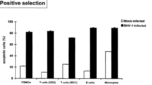
The levels of purity of enriched PBMC subpopulations as assessed by cell surface immunofluorescence were always greater than 80% for negative selection and 90% for positive selection. Purified cells were stimulated with IONO/PDB, mock-infected or infected with BHV-1, and incubated for 36 h. The cells were further processed for quantitative analysis of apoptosis by in situ detection of DNA fragmentation. Flow cytometric analysis showed that the percentages of apoptotic cells in positively purified T lymphocytes (CD2 and WC1) and B
lymphocytes were significantly higher in BHV-1-infected cultures than in mock-infected cultures (Fig. 3). In positively purified monocytes, the percentage of apoptotic cells was also increased by BHV-1 despite the high background observed in mock- infected cells (Fig. 3). The results obtained with negatively purified T lymphocytes, B lymphocytes and monocytes (Fig. 4) were similar to those obtained with positively selected subpopulations (Fig. 3). This indicated that the procedure used to purify the PBMC subpopulations did not influence the ability of BHV-1 to induce apoptosis.
Since negatively or positively selected B lymphocytes are susceptible to BHV- 1-induced apoptosis (Fig. 3 and 4), we also investigated whether BHV-1 could trigger apoptosis in BL-3 cells. These are bovine cells which have been characterized as tumoral B lymphocytes [21]. For this purpose, BL-3 cells were mock- infected or infected with BHV-1 and incubated for 0, 24 and 48 h. The cells were processed for detection of apoptosis by in situ detection of DNA fragmentation. At 24 and 48 h post-infection, BL-3 cells incubated with BHV-1 yielded 15.6% and 52.1% respectively of apoptotic cells. At the same times mock-infected cells yielded 4% and 7% respectively. We also determined, under electron microscopy, the morphological characteristics of mock- and BHV-1-infected BL-3 cells. After 48 h of incubation with BHV-1, a significant proportion of BL-3 cells had membranes-bound apoptotic bodies and distinctive condensation of the chromatin in one or several dark masses (Fig. 5B). These results demonstrated that BHV-1 is also able to induce apoptosis in BL-3 cells.
We finally investigated whether MDBK cells are susceptible to BHV-1- induced apoptosis. For this assay, mock- or BHV-1-infected MDBK cells were incubated with BHV-1 (M.O.I. of 10) for 24 h (until extensive cytopathogen effect occurred). After being harvested, cells were processed for in situ detection of DNA fragmentation and analyzed by flow cytometry. The percentages of cells positive for DNA fragmentation in mock- and BHV-1-infected MDBK cultures were respectively 0.5% and 1%. All together, these results suggest that MDBK cells are less susceptible to BHV-1-induced apoptosis than BL-3 cells and PBMCs.
Figure 3. : Percentages of apoptotic cells in positively purified PBMC subpopulations stimulated with IONO/PDB, mock-infected or infected with BHV-1 (MOI of 10), and incubated for 36 h. Each reported value represents the average ± S.D. for triplicate cultures
Figure 4. : Percentages of apoptotic cells in negatively purified PBMC subpopulations stimulated with IONO/PDB, mock-infected or infected with BHV-1 (MOI of 10), and incubated for 36 h. Each reported value represents the average ± S.D. for triplicate cultures
Figure 5.
Légende de la figure. Electron micrographs of BL-3 cells mock-infected (A) or infected with BHV-1 (B) (MOI of BHV-10), and incubated for 48 h
Discussion
We have previously shown that BHV-1 is able to induce apoptosis in mitogen-stimulated PBMCs [11]. To characterize further the process by which BHV-1 induces apoptosis, we determined the susceptibility of the three main PBMC subpopulations to BHV-1-induced apoptosis. This study shows that BHV-1 can induce apoptosis individually in T lymphocytes, B lymphocytes and monocytes. This conclusion is based on the following findings: (i) BHV-1 substantially reduces the percentages of viable T and B lymphocytes in PBMCs (Fig. 1). (ii) Concomitant detection of cell phenotype and apoptosis indeed showed higher percentages of apoptotic T lymphocytes (CD2 and WC1) and B lymphocytes in BHV-1-infected PBMCs than in mock-infected cells (Fig. 2). (iii) Each individual PBMC subpopulations (B lymphocytes, T lymphocytes and monocytes) are susceptible to BHV-1-induced apoptosis (Figs. 3 and 4).
The ability of BHV-1 to induce apoptosis in each individual PBMC subpopulations provides important indications about the process by which BHV-1 activates the apoptotic pathway. It has been reported that apoptosis can occur as a consequence of direct virus-cell interactions or/and through indirect recruitment of cytotoxic cells [19, 27, 28]. The observation that purified B lymphocytes undergo apoptosis when incubated with BHV-1 indicate that the recruitment of cytotoxic cells like monocytes, T lymphocytes, or natural killer lymphocytes is most likely not required for BHV-1-induced apoptosis. This is confirmed by the induction of apoptosis by BHV-1 in BL-3 cells (Fig. 5) which have been characterized as bovine tumoral B lymphocytes [21]. Induction of apoptosis in PBMCs by BHV-1 could also occur as a consequence of the production of specific cytokines like tumor necrosis factor-𝛂 (TNF-𝛂), a well known apoptosis inducer [27]. It has been already described that bovine alveolar macrophages infected with BHV-1 are induced to produce TNF-𝛂 [4]. However, our results indicate that BHV- 1 can still induce apoptosis in monocytes/macrophages-depleted cell cultures (purified T and B lymphocytes). Therefore, the production of specific cytokines by monocytes/macrophages is most probably not
necessary for BHV-1 to induce apoptosis in PBMCs. The observation that BL-3 cells and PBMC subpopulations undergo apoptosis when incubated with BHV-1 implies that the apoptotic pathway activated by BHV-1 must be common and functional among these different cell types. In addition, we observed that MDBK cells (non-leukocyte cells) are less susceptible to BHV-1-induced apoptosis than PBMCs and BL-3 cells. This indicates that the apoptotic process BHV-1-induced by BHV-1 is dependent on the cell phenotype. All together, the results presented in this study could facilitate the choice of the direction to be taken for further investigations into the mechanism by which BHV-1 induces apoptosis. Furthermore, the use of the BL-3 cell line provides a new experimental model to investigate the apoptotic process induced by BHV-1 in vitro.
The ability of BHV-1 to induce apoptosis in T lymphocytes, B lymphocytes and monocytes which are essentially involved in immune responses could also have important implications in vivo [19, 28]. Indeed, our results suggest that BHV- 1 could affect cellular cytotoxicity, cytokine and antibody production in addition to its inhibitory effect on the lymphocyte proliferative response. Furthermore, we previously demonstrated that BHV-1 can induce apoptosis in non-stimulated as well as proliferating mononuclear cells [10]. Therefore, a wide range of immune cells could undergo BHV-1-induced apoptosis in vivo. This could severely affect the immune response against BHV-1 and play an important role in the development of the increased susceptibility of BHV-1-infected cattle to secondary bacterial infection with Pasteurella haemolytica.
Acknowledgements
The authors would like to thank Prof. C. Dessy-Doizé (Liège, Belgium) for helpful comments on electron microscopic photomicrographs. We thank M. Loncar, L. Karelle-Bui Thi, J.-P. Georgin and A. Brichaud for excellent technical assistance. The authors also thank J.-F. Bradfer for assistance with electron microscopy. Purchase of the flow cytometer was supported in part by a grant of the “Loterie Nationale” (n◦9.4505.92, Belgium). E. Hanon is a research assistant of the fonds national belge de la recherche scientifique (F.N.R.S.)
References
1. Arends MJ, Morris RG, Wyllie AH (1990) Apoptosis. The role of the endonuclease. Am J Pathol 136: 593–608
2. Bielefeldt Ohmann H, Babiuk LA (1985) Viral-bacterial pneumonia in calves: effect of bovine herpesvirus-1 on immunologic functions. J Infect Dis 151: 937–947
3. Bielefeldt Ohmann H, Bloch B (1982) Electron microscopic studies of bovine viral diarrhea virus in tissues of diseased calves and in cell cultures. Arch Virol 71: 57–74
4. Bienhoff SE, Allen GK, Berg JN (1992) Release of tumor necrosis factor-alpha from bovine alveolar macrophages stimulated with bovine respiratory viruses and bacterial endotoxins. Vet Immunol Immunopathol 30: 341– 357
5. Davis WC, Hamilton MJ, Park YH (1990) Ruminant leukocyte differentiation molecules. In: Barta O (ed) MHC, differentiation antigens, and cytokines in animals and birds. BAR- LAB Inc., Blacksburg, pp 47–70
6. Duvall E, Wyllie AH (1986) Death and the cell. Immunol Today 7: 115–119
7. Fadok VA, Voelker DR, Campbell PA, Cohen JJ, Bratton DL, Henson PM (1992) Ex- posure of phosphatidylserine on the surface of apoptotic lymphocytes triggers specific recognition and removal by macrophages. J Immunol 148: 2 207–2 216
8. Filion LG, McGuire RL, Babiuk LA (1983) Nonspecific suppressive effect of bovine herpesvirus type 1 on bovine leukocyte functions. Infect Immun 42: 106–112
9. Griebel PJ, Ohmann HB, Lawman MJ, Babiuk LA (1990) The interaction between bovine herpesvirus type 1 and activated bovine T lymphocytes. J Gen Virol 71: 369–377
10. Hanon E, Hoornaert S, Dequiedt F, Vanderplasschen A, Lyaku J, Willems L, Pastoret PP (1987) Bovine herpesvirus 1-induced apoptosis occurs at the GO/G1 phase of the cell cycle. Virology 232: 351–358
11. Hanon E, Vanderplasschen A, Lyaku JR, Keil G, Denis M, Pastoret PP (1996) Inacti- vated bovine herpesvirus 1 induces apoptotic cell death of mitogen-stimulated bovine peripheral blood mononuclear cells. J Virol 70: 4 116–4 120
12. Letesson JJ, Delcommenne M (1993) Production of a monoclonal antibody to the light chain of the bovine beta 2-integrin family (BoCD18). Vet Immunol Immunopathol 39: 103–108
13. Letesson JJ, Lostrie N, Delpechin A (1985) Production dtanticorps monoclonaux spéci- fiques dtisotypes dtimmunoglobulines bovines. Ann Med Vet 129: 131–141
14. Lyaku JR, Nettleton PF, Marsden H (1992) A comparison of serological relation- ships among five ruminant alphaherpesviruses by ELISA. Arch Virol 124: 333–341
15. Martin SJ, Green DR, Cotter TG (1994) Dicing with death: dissecting the components of the apoptosis machinery. Trends Biochem Sci 19: 26– 30
16. McChesney MB, Oldstone MB (1987) Viruses perturb lymphocyte functions: selected principles characterizing virus-induced immunosuppression. Annu Rev Immunol 5: 297– 304
17. Miltenyi S, Muller W, Weichel W, Radbruch A (1990) High gradient magnetic cell separation with MACS. Cytometry 11: 231–238
18. Raynal P, Pollard HB (1994) Annexins: the problem of assessing the biological role for a gene family of multifunctional calcium- and phospholipid- binding proteins. Biochim Biophys Acta 1197: 63–93
19. Razvi ES, Welsh RM (1995) Apoptosis in viral infections. Adv Virus Res 45: 1–60
20. Roizman B, Desrosiers RC, Fleckenstein B, Lopez C, Minson AC, Studdert MJ (1992) The family Herpesviridae: an update. Arch Virol 123: 425–429
21. Romano MJ, Stewart JA, Lewin HA (1989) Phenotypic characterization of bovine lym- phoblastoid cell lines. Vet Immunol Immunopathol 23: 293–307
22. Szilágyi JF, Cunningham C (1991) Identification and characterization of a novel nonin- fectious herpes simplex virus-related particle. J Gen Virol 72: 661–668
23. Thompson CB (1995) Apoptosis in the pathogenesis and treatment of disease. Science 267: 1 465–1 462
24. Vermes I, Haanen C, Steffens-Nakken H, Reutelingsperger C (1995) A novel assay for apoptosis. Flow cytometric detection of phosphatidylserine expression on early apoptotic cells using fluorescein labelled Annexin V. J Immunol Methods 184: 39–51
25. Wyllie AH (1980) Glucocorticoid-induced thymocyte apoptosis is associated with en- dogenous endonuclease activation. Nature 284: 555–556
26. Yates WD (1982) A review of infectious bovine rhinotracheitis, shipping fever pneumo- nia and viral-bacterial synergism in respiratory disease of cattle. Can J Comp Med 46: 225–263
27. Zheng L, Fisher G, Miller RE, Peschon J, Lynch DH, Lenardo MJ (1995) Induction of apoptosis in mature T cells by tumour necrosis factor. Nature 377: 348–351