TRENDS IN CELL
SIGNALING PATHWAYS
IN NEURONAL FATE
DECISION
Edited by Sabine Wislet-Gendebien
Contributors
Aviva Symes, Sonia Villapol, Trevor Logan, Eri Hashino, Atsushi Shimomura, Michael Fehlings, Madeleine O'Higgins, Jenny Wong, Wenhui Hu, Yonggang Zhang, Sabine Wislet-Gendebien, Tanja Vogel, Ann M. Turnley, Harleen Basrai, Kimberly Christie, Roxana Nat, Galina Apostolova, Georg Dechant, Adam Cole, Liang-Wei Chen, Nibaldo Inestrosa, Lorena Varela-Nallar, Uwe Ueberham, Thomas Arendt
Published by InTech
Janeza Trdine 9, 51000 Rijeka, Croatia
Copyright © 2013 InTech
All chapters are Open Access distributed under the Creative Commons Attribution 3.0 license, which allows users to download, copy and build upon published articles even for commercial purposes, as long as the author and publisher are properly credited, which ensures maximum dissemination and a wider impact of our publications. After this work has been published by InTech, authors have the right to republish it, in whole or part, in any publication of which they are the author, and to make other personal use of the work. Any republication, referencing or personal use of the work must explicitly identify the original source.
Notice
Statements and opinions expressed in the chapters are these of the individual contributors and not necessarily those of the editors or publisher. No responsibility is accepted for the accuracy of information contained in the published chapters. The publisher assumes no responsibility for any damage or injury to persons or property arising out of the use of any materials, instructions, methods or ideas contained in the book.
Publishing Process Manager Iva Simcic Technical Editor InTech DTP team Cover InTech Design team
First published March, 2013 Printed in Croatia
A free online edition of this book is available at www.intechopen.com Additional hard copies can be obtained from orders@intechopen.com
Trends in Cell Signaling Pathways in Neuronal Fate Decision, Edited by Sabine Wislet-Gendebien p. cm.
Books and Journals can be found at
Preface VII
Section 1 TGF-Beta Signaling and Neuronal Fate Decision 1
Chapter 1 Role of TGF-β Signaling in Neurogenic Regions After
Brain Injury 3
Sonia Villapol, Trevor T. Logan and Aviva J. Symes
Chapter 2 Insulin/IGF-Signalling in Embryonic and Adult Neural
Proliferation and Differentiation in the Mammalian Central Nervous System 37
Tanja Vogel
Chapter 3 The Role of Smad Proteins for Development, Differentiation
and Dedifferentiation of Neurons 75
Uwe Ueberham and Thomas Arendt
Section 2 Wnt Signaling and Neuronal Fate Decision 113
Chapter 4 Wnt Signaling Roles on the Structure and Function of the
Central Synapses: Involvement in Alzheimer’s Disease 115
Nibaldo C. Inestrosa and Lorena Varela-Nallar
Chapter 5 Roles of Wnt/β-Catenin Signaling in Controlling the
Dopaminergic Neuronal Cell Commitment of Midbrain and Therapeutic Application for Parkinson’s Disease 141
Liang-Wei Chen
Chapter 6 Regulation of Cell Fate in the Brain by GSK3 153 Adam R. Cole
Section 3 Neurotrophin and Neuronal Fate Decision 179
Chapter 7 Neurotrophin Signaling and Alzheimer’s Disease
Neurodegeneration − Focus on BDNF/TrkB Signaling 181
Jenny Wong
Section 4 NF-K-b and Neuronal Fate Decision 195
Chapter 8 NFκB Signaling Directs Neuronal Fate Decision 197 Yonggang Zhang and Wenhui Hu
Section 5 Stem Cells and Signaling Pathways 215
Chapter 9 Telencephalic Neurogenesis Versus Telencephalic
Differentiation of Pluripotent Stem Cells 217
Roxana Nat, Galina Apostolova and Georg Dechant
Chapter 10 Regulation of Basal and Injury-Induced Fate Decisions of Adult
Neural Precursor Cells: Focus on SOCS2 and Related Signalling Pathways 241
Harleen S. Basrai, Kimberly J. Christie and Ann M. Turnley
Chapter 11 Neural Stem/Progenitor Cells for Spinal Cord
Regeneration 271
Ryan Salewski, Hamideh Emrani and Michael G. Fehlings
Chapter 12 Epigenetic Regulation of Neural Differentiation from
Embryonic Stem Cells 305
Atsushi Shimomura and Eri Hashino
Chapter 13 Neural Fate of Mesenchymal Stem Cells and Neural Crest Stem
Cells: Which Ways to Get Neurons for Cell Therapy Purpose? 327
Virginie Neirinckx, Cécile Coste, Bernard Rogister and Sabine Wislet-Gendebien
During the last decades, numerous studies about stem cells and regenerative medicine high‐ lighted new therapeutic approaches to treat several neurological disorders. It is noteworthy that the current optimism over potential stem cell therapies is driven by new understand‐ ings of stem cell biolology leading to specific cell fate decision.
The main objective of this book is to offer a general understanding of signaling pathways underlying the capacity of differentiation of several types of stem cells into neurons, during the development. Indeed, in this book, we deeply described TGF-beta signaling, Wnt Signal‐ ing, neurotrophin and NF-κ-B signaling and their implication in neuronal fate decision. The second objective of this book is to understand how those pathways are altered in pathologi‐ cal conditions. We consequently analyzed those pathways in several pathological conditions. Finally the third objective of this book is to describe advances in cellular therapy that could be use to restore central nervous system dysfunction in pathological conditions, based on new molecular biology findings. Several sources of stem cells and their potential benefits were described in the last part of this book.
Finally, I would like to conclude this preface by expressing my deepest gratitude to all au‐ thors who contributed to the elaboration of this book.
Sabine Wislet-Gendebien, PhD
GIGA Neurosciences University of Liège, Belgium
Role of TGF-β Signaling in
Neurogenic Regions After Brain Injury
Sonia Villapol, Trevor T. Logan and Aviva J. Symes
Additional information is available at the end of the chapter http://dx.doi.org/10.5772/539411. Introduction
In 1928 Santiago Ramón y Cajal penned what became the accepted view about neurons in the central nervous system; “everything may die, nothing can be regenerated”. He later exhibited his wisdom by adding; “It’s the job of science to rewrite, if possible, this cruel phrase” [1]. Up until 20 years ago, the scientific literature had emphasized that neurogenesis only occurs during development with no new neurons generated in the adult mammalian brain. However, since the discovery of adult neurogenesis, an extensive literature has emerged supporting the constant generation of new neurons in two neurogenic regions of the adult brain: the subven‐ tricular zone around the lateral ventricles (SVZ) and the subgranular zone (SGZ) of the hippocampal dentate gyrus (DG) [2].
The existence of adult neurogenesis gave hope for recovery and regeneration from the many different insults that can damage the brain. After stroke or traumatic brain injury (TBI), immediate massive necrosis occurs followed by a subsequent prolonged period of inflamma‐ tion and further neuronal death [3]. Although brain injury induces massive cell loss, it also induces an increase in proliferation of NSCs residing in the neurogenic niches [4]. The environment of the neurogenic niche in adult animals is exquisitely regulated, with a finely-tuned balance of soluble and cell-intrinsic factors that regulate the many different processes that are critical to neurogenesis: cell survival, proliferation, differentiation, and migration [5]. Dramatic changes occur in this environment as a consequence of the injury. The careful regulation of neurogenesis is disrupted by the many different cellular, soluble and vascular signals detected by the different cell types in the SVZ and DG. This major environmental alteration leads to increased proliferation of progenitor cells for long periods after the acute injury, yet the ability of the neural progenitor cells to fully differentiate, migrate and integrate into the lesioned area is limited [6]. Understanding the signals that regulate adult neurogenesis
© 2013 Villapol et al.; licensee InTech. This is an open access article distributed under the terms of the Creative Commons Attribution License (http://creativecommons.org/licenses/by/3.0), which permits unrestricted use, distribution, and reproduction in any medium, provided the original work is properly cited.
in the naïve and injured animals is key to ultimately being able to harness the potential of neuronal replacement and improve stem cell therapy.
There are many different factors important to regulation of neurogenesis, many of which are discussed in other chapters in this book. Here we will focus on the role of the transforming growth factor-β (TGF-β) superfamily and its associated signaling pathways in regulating neurogenesis after brain injury. Members of this family, including the bone morphogenetic proteins (BMPs), Activin, and TGF-β1, -β2 and -β3 have a profound influence on the neuro‐ genic process in naïve animals [7]. Many of these cytokines are induced by injury and play critical roles in many kinds of brain damage related processes around the lesion [3]. We and others recently started to accumulate data on their induction in the neurogenic niches after different types of injury. Here we will focus on the relevance of their induction in these specific brain regions, and the mechanisms through which they may influence the neurogenic response to injury. As there are significant differences between the behavior of cells contributing to neurogenesis during development and in the adult, we will restrict our analysis to that observed in adult animals after injury. Delineation of the specific role of members of the TGF-β superfamily in injury-induced neurogenesis may provide specific therapeutic targets for enhancing neurogenesis after trauma.
2. The TGF-β superfamily; cytokines, receptors and signaling
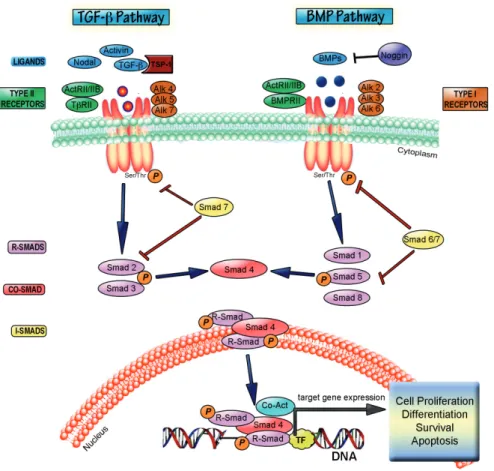
The TGF-β cytokine superfamily is a large group of proteins comprising 33 different members that include: bone morphogenetic proteins (BMPs), growth differentiation factors (GDFs), activins, inhibins, nodal, lefty, mülllerian inhibiting substance (MIS) together with the TGF-β proteins [8, 9]. All members of this cytokine family mediate their effects in a broadly analogous manner, binding specific type I and II transmembrane serine threonine kinase receptors and transducing their signal through similar intracellular Smad proteins [10]. These cytokines are divided into two distinct groups: those of the TGF-β/Activin group which mainly signal through the type I receptors ALK4, -5 and -7 activating Smad2 and -3, and those of the BMP/GDF group [11, 12] which employ ALK1, -2, -3 and -6 to activate Smad1, -5 and -8 [13, 14]. The specificity of Smad activation is therefore mainly determined by the identity of the type I receptor used to transduce the cytokine signal [15] (Figure 1).
TGF-β1, -β2 and -β3 together with some GDFs are unique in that they are synthesized as a large precursor molecule that is cleaved but remains non-covalently linked to its latency associated peptides, in either a small or large complex [18]. The bioavailability of TGF-βs is tightly regulated by the release of active TGF-β from these complexes in the extracellular matrix, so synthesis of TGF-β does not necessarily provide a reliable indication of available cytokine to initiate signaling. Similarly, the bioavailability of BMPs is regulated by binding to secreted extracellular antagonists that prevent BMP (and sometimes Activin) from binding to their receptor [19]. Expression levels of endogenous antagonists, including noggin, chordin, follistatin, gremlin and cerberus, thereby regulate the availability, and therefore, active signaling by their associated ligands [20]. TGF-β signaling is the archetype for signaling by
this cytokine family. TGF-β binds to the constitutively active TGF-β receptor II (TβRII) which can then recruit the type I receptor TGF-β receptor I (TβRI/ALK5). Activation of TβRI by transphosphorylation activates it, initiating downstream signaling [21]. Canonical signaling
Figure 1. TGF-β superfamily signal transduction. TGF-β, nodal or activin ligands bind to Type II receptors, which
then recruit Type I receptors leading to transphosphorylation of type 1 receptors. Activated type I receptors phosphor‐ ylate Smad 2/3 (i.e. R-Smads) which then complex with the co-Smad, Smad4 and translocate to the nucleus to bind DNA at specific DNA motifs. Smad proteins activate or repress transcription through association with various co-activa‐ tor (Co-Act) or co-repressor proteins. This pathway is inhibited by Smad7. BMP signaling operates by a similar para‐ digm. BMP6 and BMP7 bind to their Type II receptor before the complex recruits the Type I receptors, Alk-3 or Alk-6. BMP2 and BMP4, however bind first to their type I receptor before recruiting the type II receptor BMPRII. BMP binding to either receptor can be inhibited by first binding to various extracellular inhibitor proteins, such as noggin. Activa‐ tion of the receptor complex leads to phosphorylation of the receptors and subsequent phosphorylation of Smad1, Smad5, or Smad8, allowing them to form a complex with Smad4. This heteromeric complex translocates to the nu‐ cleus, to target BMP-regulated genes through interaction with co-activators or repressors. Smad 6 and Smad7 may act similarly to inhibit the BMP pathway through interactions with the receptor complex and thus inhibiting R-Smad acti‐ vation. TGF-β and BMP pathways induce the expression of proteins involved in proliferation, differentiation, survival and apoptosis. The diagram is adapted from [16] and [17].
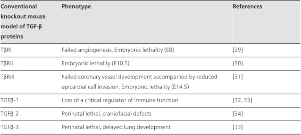
by these cytokines is through the receptor regulated Smads (R-smads). As previously men‐ tioned, TGF-β and activin signal through activation of Smad2 and Smad3, which are phos‐ phorylated by the Type I receptor, and form a heteromeric complex with the common or co-Smad, Smad4 [22]. This Smad complex translocates to the nucleus where it regulates the transcription of numerous genes in cooperation with other transcription factors, coactivators and corepressors. Inhibitory Smads, or I-smads, are Smad-activated proteins that provide negative feedback to the Smad pathway through a variety of mechanisms [16, 23]. BMP signaling is similar in form to TGF-β signaling, although the specifics of individual receptors and R-Smads (1, 5, 8/9) involved vary according to the specific cytokine. For a full review of signaling and receptor nomenclature by this cytokine family please refer to some excellent reviews [14, 24]. The Smad pathway is by no means the only mechanism through which TGF-β cytokine signals are transduced from the receptor to the nucleus. Smad-independent pathways include activation of MAPKs, Ras/ERK, JNK, p38, PI3K-Akt, NF-kappaB, JAK/STAT, PP2A/S6 phosphatases and small Rho-related GTPases (16, 25). Some of the non-Smad kinases can influence Smad directed signaling by complexing with, or modifying the Smad proteins directly [16, 25]. Another level of control was found when it was shown that TGF-β/BMP signaling is both regulated by, and can regulate transcription of miRNAs [26]. Smads can also influence miRNA biogenesis by binding directly to the pri-miRNA to enhance Drosha processing of these molecules to pre-miRNA [27]. An intricate balance between Smad and non-Smad signaling superimposed on cell intrinsic and environmental conditions determines the specificity and the ultimate response of each cell to TGF-β signaling. Thus, there is a complexity to TGF-β superfamily signaling that befits cytokines that signal to multiple different cell types, in context dependent manners to influence many different physiologic processes [28]. Genetic evidence indicates that TGF-β family members regulate embryonic, perinatal or neonatal development of the mouse embryo. Most mice null for one TGF-β superfamily ligand, receptor, protein or signaling protein fail in either gastrulation or mesoderm differentiation. Table 1 lists known phenotypes of mice that are null for specific proteins in the TGF-β superfamily signaling pathways.
Conventional knockout mouse model of TGF-β proteins
Phenotype References
TβRI Failed angiogenesis, Embryonic lethality (E8) [29] TβRII Embryonic lethality (E10.5) [30] TβRIII Failed coronary vessel development accompanied by reduced
epicardial cell invasion. Embryonic lethality (E14.5)
[31] TGFβ-1 Loss of a critical regulator of immune function [32, 33] TGFβ-2 Perinatal lethal, craniofacial defects [34] TGFβ-3 Perinatal lethal, delayed lung development [33]
Conventional knockout mouse model of TGF-β proteins
Phenotype References
Smad1 Embryonic lethality (E10) [35, 36] Smad2 Embryonic lethality (E7.5–E12.5) [37] Smad3 Viable and fertile. Impaired immune function, including defective
neutrophil chemotaxis, and impaired mucosal immunity
[38, 39] Smad4 Increased number of Olig2-expressing progeny [40] Smad5 Embryonic lethality: defective vascular development [41, 42] Smad7 Significantly smaller than wild-type mice, died within a few
days of birth
[43]
Smad8 Viable and fertile [41, 44]
BMPRIA Embryonic lethality (E9.5) [45] BMPRIB Viable and exhibit defects in the appendicular skeleton [46] BMPRII Embryonic lethality (E9.5), arrest at gastrulation [47] BMP2 Embryonic lethality (E7.5-10.5), defective cardiac development
and have defects in cardiac development
[48] BMP3 Increased bone density in adult [49] BMP4 Embryonic lethality (E6.5-E9.5), no mesoderm differentiation
and show little or no mesodermal differentiation
[50] BMP5 Viable, skeletal and cartilage abnormalities [51] BMP6 Viable and fertile; slight delay in ossification. [52] BMP7 Perinatal lethal because of poor kidney development, eye defects
that appear to originate during lens induction.
[53-56] BMP8A Viable: male infertility due to germ cell degeneration [57] BMP8B Viable: male infertility due to germ cell depletion [58] BMP15 Viable: female subfertility [59] Endoglin Embryonic lethality (E11.5) [60, 61] Activin receptor IA
(ALK2)
Embryonic lethality (E9.5) [62] Activin receptor IIB
(ActR2B)
Perinatal lethal [63]
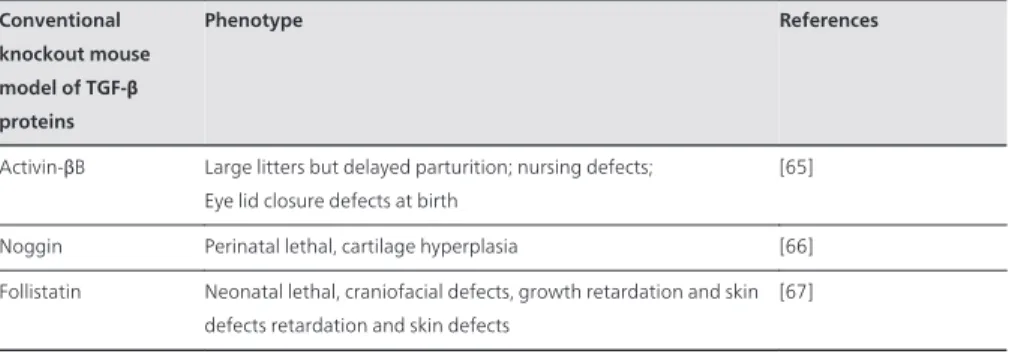
Activin-βA Neonatal lethal, craniofacial defects (cleft palate and loss of whiskers, upper incisors, lower incisors and molars)
Conventional knockout mouse model of TGF-β proteins
Phenotype References
Activin-βB Large litters but delayed parturition; nursing defects; Eye lid closure defects at birth
[65] Noggin Perinatal lethal, cartilage hyperplasia [66] Follistatin Neonatal lethal, craniofacial defects, growth retardation and skin
defects retardation and skin defects
[67]
Table 1. Phenotype of mice that do not express specific TGF-β ligands, receptors or signaling molecules.
3. TGF-β superfamily expression and function in normal adult brain: Role
in neurogenesis
Adult neurogenesis involves proliferation of neural stem cells (NSCs), cell cycle exit, differ‐ entiation, maturation, and integration into the neural circuits, in a process that is involved in learning and memory in the normal adult brain [68]. The neurogenic niche of the adult forebrain subventricular zone (SVZ) is comprised of three major proliferative cell types; A, B and C. Multipotent, self-renewing type B cells occur earliest in the neurogenic lineage of the SVZ and give rise to the rapidly dividing type C cells, or transit amplifying progenitors. Type A cells or neuroblasts differentiate from Type C cells and are migratory neuronal progenitors with proliferative capacity, which migrate to the olfactory bulb where they differentiate into interneurons (reviewed in [69-71]. In the subgranular zone (SGZ) of the hippocampal dentate gyrus (DG), type 1 and type 2 slowly-dividing progenitors give rise to more rapidly dividing intermediate progenitor cells, and these in turn differentiate into immature neuroblasts, which migrate into the granule cell layer, then differentiate into mature neurons and integrate with the existing hippocampal circuitry [71].
Within the CNS, all three isoforms of TGF-β are produced by both glial and neuronal cells [72]. Immunohistochemical studies show widespread expression of TGF-β2 and -β3 in the devel‐ oping CNS, and these proteins play a role in regulation of neuronal migration, glial prolifer‐ ation and differentiation [73-76]. In adult brain, TGF-β receptors are found in all areas of the CNS including the cortex, hippocampus, striatum, brainstem and cerebellum [77, 78]. Immu‐ noreactivity for TβRI and TβRII is detected on neurons, astrocytes and microglia and endo‐ thelial cells located in the cortical gray matter, suggesting that almost every cell type in the CNS is a potential target for TGF-β signaling [79].
The TGF-β superfamily and its downstream targets are capable of controlling proliferation, differentiation, maturation and survival of stem cells and precursors in the neurogenic niches of adult brain [18]. TβRI and TβRII are expressed by Nestin-positive type B and C cells in the SVZ [80, 81]. Our data show mRNA expression of TGF-β1, -β2, and -β3 in both the adult SVZ
and DG [82]. In the adult human brain, TGF-β1 protein expression has been reported in the hippocampus, and the protein levels significantly increased with the age of the individual [83]. As neurogenesis declines with age [84], it has been suggested that TGF-β is a possible regulator of this age-related decline [83]. Signaling by the Smad2/3 pathway is high in the hippocampus and specifically the dentate gyrus, indicating a role for TGF-β and/or activin in regulation of neurogenesis [85, 86]. When TGF-β protein is overexpressed or infused directly into the lateral ventricles of uninjured animals, hippocampal neurogenesis is dramatically inhibited [81, 87]. This may be due to a direct anti-proliferative effect of TGF-β on type 1 and 2 primary NSCs [17]. A direct effect of TGF-β on NSCs is supported by in vitro studies showing that TGF-β1 treatment of cultured adult NSCs induces the cyclin-dependent kinase inhibitor (p21) and leads to cell cycle termination, without altering the differentiation choices of the NSCs [81]. Additionally, overexpression studies lead to increased TGF-β signaling in many different cell types within the neurogenic niche, making the exact contribution of more restricted, endoge‐ nous TGF-β difficult to determine. Recent data have suggested that TGF-β signaling at later stages of neurogenesis is critical for newborn neuron survival and maturation in the DG. Conditional deletion of the TβRI (ALK5) gene specifically in immature and mature neurons, leads to decreased neurogenesis and reduced survival of newborn neurons [85]. Thus, TGF-β potentially has opposing roles at different stages of neurogenesis, providing an additional example of the contextual nature of TGF-β action.
Activin receptors are expressed throughout the brain, with strong expression in the neuronal layers of the hippocampus [88-90]. We have found that mRNA for activin-A and for activin’s endogenous high affinity inhibitor, follistatin, are expressed in both the SVZ and DG of the adult mouse [82] and several recent reports have demonstrated that activin-A modulates adult neurogenesis [88, 91, 92]. Chronic overexpression of follistatin by neurons of the hippocampus almost entirely ablates adult DG neurogenesis, due to drastically lowered survival of adult-generated neurons [91], although short-term infusion of follistatin does not affect neurogenesis in uninjured animals [88]. Infusion of activin to the lateral ventri‐ cle of uninjured mice mildly increases the rate of NSC proliferation and neuron genera‐ tion in the DG, indicating that activin might stimulate division of NSCs. This effect may be indirect as activin has a potent anti-inflammatory effect in the CNS, and may modulate local microglia to stimulate neurogenesis [88]. Smad3 knockout mice have decreased levels of cell proliferation in the SVZ and along the rostral migratory stream, and decreased levels of olfactory bulb neurogenesis [93]. As these mice have defective signaling by both TGF-β and activin, these data suggest that activin signaling in the SVZ may be the predominant Smad3-utilizing cytokine in defining basal levels of neurogenesis. In the DG pSmad2 is normally absent from Sox2-positive type 1 and 2 primary NSCs in the DG of adult mice [17]. However, Smad3 knockout mice also have reduced proliferation in the DG potential‐ ly pointing to a different role for Smad2 and Smad3 in the DG [93].
The BMP family of proteins regulates cell proliferation and fate commitment throughout development and within the adult neurogenic niches [19]. Expression of BMP2, -4 and -7 mRNAs have been reported in neurogenic regions of adult rodent brain [94], and the BMP receptors BMPRIA, -IB and -II are expressed abundantly in neurons, as well as in astrocytes
and ependymal cells [95]. All three of these receptors are expressed in type A cells of the SVZ, while type B and C cells express BMPRIA and BMPRII [96]. In the DG, radial stem cells of the SGZ marked with glial fibrillary acidic protein (GFAP) and Nestin or Sox2 primarily express BMPRIA but not BMPRIB, while mature neurons express only BMPRIB [97]. BMP ligands are also expressed in the adult rat brain [98, 99]. BMP2, -4, -6, and -7 are expressed by cells of the SVZ and DG [96, 97]. In the DG, the BMP signal transducer pSmad1 is strongly expressed in non-dividing primary NSCs and neuroblasts, but is absent in dividing primary NSCs [97], while in the SVZ, pSmad1/5/8 has been reported in primary NSCs and transit amplifying progenitors, but not in DCX-positive neuroblasts [40]. The soluble BMP inhibitor noggin is also expressed by ependymal cells of the SVZ [96] and by cells of the DG [100].
Changing the ratio of BMP to noggin alters the rates of NSC proliferation and neurogenesis in adult animals, indicating that these proteins are primary regulators of basal adult neurogene‐ sis [96, 97, 100]. Administration of exogenous BMP4 or BMP7 potently inhibits the division of NSCs and generation of new neurons in vivo and in vitro [96, 97], as does inhibition of noggin expression [101]. Conversely, infusion of noggin or genetic deletion of the BMPRIA receptor causes an increase in NSC proliferation and generation of NeuN-expressing neurons in the DG [96, 97]. However this increase is transient, there is an eventual depletion of the primary NSC pool and a drastically reduced level of neurogenesis [97]. Decreased BMP signaling in the DG is thought to be responsible for increased neurogenesis driven by exercise [102]. It has been proposed that secretion of noggin from ependymal cells inhibits BMP signaling allowing a low level of basal neurogenesis to occur, while BMP signaling maintains the overall quiescence of the primary NSC pool [96, 97, 100]. Exogenous noggin infusion potentially has a different effect on SVZ NSCs, leaving their proliferation rate unaffected, but causing an increase in the generation of oligodendrocyte precursor cells from primary NSCs at the expense of immature neuro‐ blasts [40]. This noggin infusion phenocopies the effect of conditionally deleting Smad4 in NSCs using GLAST-cre [40] and is in contrast to the pro-neurogenic effects of noggin described by Lim et al [96]. Thus, although there is still some controversy in the field it its clear that the balance between BMP and noggin is critical to proper maintenance of the adult NSC population.
4. Expression of TGF-β related cytokines in the adult rodent brain after
injury
TGF-β family proteins are present in the brain immediately after injury as they are carried into the wound by the blood [103]. Additionally, extracellular TGF-β proteins are activated and released from their latent protein complexes in the brain parenchyma [104]. Local CNS expres‐ sion of TGF-β, activin, and BMP proteins is increased after many different injuries [72, 105, 106]. Following acute brain injury, TGF-β1 levels are elevated in astrocytes, microglia, macrophag‐ es, neurons, ependymal cells and choroid plexus cells with peak expression around 3 days [107-110]. TGF-β2 and -β3 expression has also been found in astrocytes, microglia, endothelial cells and neurons after both ischemic and TBI [111, 112]. We have recently found TGF-β2 expression in oligodendrocytes in the lesioned cortex and corpus callosum [113]. Ischemic lesions as well as TBI show elevated activin-A mRNA as well as mRNA for the BMPRII receptor [90, 94,
114]. Smad proteins are also upregulated after injury and were mainly located in the cerebral cortex, typically in the nucleus and/or in the cytoplasm of astrocytes, oligodendrocytes or neurons [86, 108, 115, 116]. We have summarized many studies that have examined changes in the TGF-β superfamily of cytokines after central nervous system injury in Table 2.
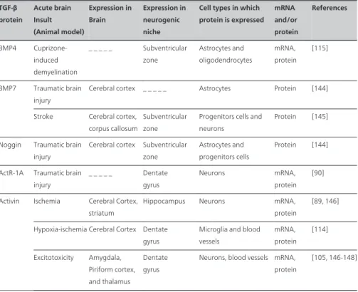
TGF-β protein Acute brain Insult (Animal model) Expression in Brain Expression in neurogenic niche
Cell types in which protein is expressed
mRNA and/or protein
References
TGF-β1 Ischemia Cerebral cortex _ _ _ _ _ Microglia, neurons, oligodendrocytes, endothelial cells, astrocytes, macrophages, and ependymal cells mRNA, protein [107-110] Transient ischemia Cerebellum, Cerebral cortex Hippocampus, Subventricular zone Microglia, T cells, neuroblasts and neurons mRNA, protein [117-120] Permanent ischemia Cerebral cortex, Striatum
_ _ _ _ _ Neurons, neuroblasts mRNA, protein [121-123] Bilateral cerebral ischemia Cerebellum, Cerebral cortex
Dentate gyrus Neurons, vessels Protein [124, 125] Hypoxic-ischemic Cerebral cortex,
Corpus callosum
_ _ _ _ _ Astrocytes, Microglia and blood vessels
Protein [126] Stab wound Cerebral cortex _ _ _ _ _ Neurons Protein [116] Traumatic brain
injury
Cerebral cortex Hippocampus, Subventricular zone Microglia, astrocytes and neurons mRNA, protein [82, 112, 127, 128] Excitotoxic lesion (NMDA) Gray matter surrounding the lesion
_ _ _ _ _ Astrocytes, neurons Protein [129]
Triethyltin exposure
Cerebral cortex Hippocampus Neurons mRNA, protein
[130, 131] Penetrating
brain Injury
Cerebral cortex _ _ _ _ _ Activated glia, meningeal cells, choroid plexus
mRNA, protein
[132]
Excitotoxic Injury _ _ _ _ _ Hippocampus Neurons Protein [133] Irradiation Cerebral cortex _ _ _ _ _ Macrophages and
astrocytes
TGF-β protein Acute brain Insult (Animal model) Expression in Brain Expression in neurogenic niche
Cell types in which protein is expressed mRNA and/or protein References Excitotoxicity with kainic acid
Cerebral cortex Hippocampus Microglia/macrophages, neurons and astrocytes
mRNA, protein
[86, 135-137] Stab wound Cerebral cortex _ _ _ _ _ Astrocytes Protein [138] TGF-β2 Ischemia Cerebral cortex,
cerebellum, striatum
Hippocampus Neurons and endothelial cells, microglia and astrocytes
mRNA, protein
[108, 109, 111] TGF-β3 Ischemia Cerebral cortex Dentate gyrus Neurons mRNA,
protein [111] Traumatic brain
injury
Cerebral cortex Hippocampus Astrocytes Protein [112] TβRI Permanent
ischemia
Cerebral cortex _ _ _ _ _ Astrocytes and neurons mRNA, protein
[122] TβRII Ischemia Cerebral cortex,
midbrain, cerebellum, and brainstem
_ _ _ _ _ Neurons, astrocytes, microglia, endothelial cells, and other non-neuronal cells found in the choroid plexus
mRNA, protein [122, 139, 140] Traumatic brain injury
Cerebral Cortex _ _ _ _ _ Endothelial cells Protein [141] Smad2 Excitotoxicity Cerebral Cortex Hippocampus Neurons, astrocytes and
microglia
Protein [86] pSmad2 Stroke Cerebral Cortex _ _ _ _ _ Astrocytes, activated
microglia Protein [108] pSmad 1,5,8 Cuprizone-induced demyelination _ _ _ _ _ Subventricular zone Oligodendrocytes mRNA, protein [115]
BMPRII Traumatic brain injury _ _ _ _ _ Dentate gyrus Neurons mRNA, protein [90] BMPs and receptors
Ischemia Cerebral Cortex, cerebellum
Hippocampus Neurons mRNA, protein [124, 142, 143] Bilateral cerebral ischemia Cerebral cortex, cerebellum Subventricular zone, dentate gyrus Neurons mRNA, protein [94] Traumatic brain injury
Cerebral cortex Subventricular zone
Astrocytes mRNA, protein
TGF-β protein Acute brain Insult (Animal model) Expression in Brain Expression in neurogenic niche
Cell types in which protein is expressed mRNA and/or protein References BMP4 Cuprizone-induced demyelination _ _ _ _ _ Subventricular zone Astrocytes and oligodendrocytes mRNA, protein [115] BMP7 Traumatic brain injury
Cerebral cortex _ _ _ _ _ Astrocytes Protein [144] Stroke Cerebral cortex,
corpus callosum
Subventricular zone
Progenitors cells and neurons
Protein [145] Noggin Traumatic brain
injury
Cerebral cortex Subventricular zone
Astrocytes and progenitors cells
Protein [144] ActR-1A Traumatic brain
injury _ _ _ _ _ Dentate gyrus Neurons mRNA, protein [90] Activin Ischemia Cerebral Cortex,
striatum
Hippocampus Neurons mRNA, protein
[89, 146] Hypoxia-ischemia Cerebral Cortex Dentate
gyrus
Microglia and blood vessels mRNA, protein [114] Excitotoxicity Amygdala, Piriform cortex, and thalamus Dentate gyrus
Neurons, blood vessels mRNA, protein
[105, 146-148]
Table 2. TGF-β superfamily cytokine and signaling intermediate expression after different forms of injury.
Relatively few studies have examined changes in expression of the TGF-β superfamily of cytokines specifically within the neurogenic regions after brain injury. TGF-β1 expression increases in the SVZ [119] and DG [117, 118, 124] after ischemic injury. Its expression is also induced in neurons of the DG after a demyelinating lesion [131] or after local kainic acid injection [133]. Our group recently found that controlled cortical impact injury increased mRNA expression of many TGF-β cytokines, including TGF-β1 and -β2, activin-A, and BMPs -4, -5, -6, and -7 in the DG and SVZ, demonstrating that a distal injury can alter TGF-β signaling pathways in the neurogenic regions [82]. We have observed upregulation of TGF-β1 and -β3 in GFAP and Nestin positive progenitors in the SVZ and DG after TBI (Figure 2 and unpub‐ lished data). TβRII is expressed in these Nestin positive progenitors in the lateral SVZ (Figure 2d). Phospho-Smad3 (pSmad3) shows strong nuclear localization in these cells as well (Figure 2i and unpublished data) suggesting a role for TGF-β/activin signaling in the regulation of post-injury neurogenesis. In the DG, TβRII is expressed in GFAP-positive precursors with strong pSmad3 nuclear staining (Figure 2m, 2r) suggesting a similar role for TGF-β cytokines in this neurogenic niche.
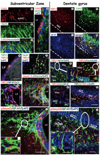
Figure 2. Confocal images of the TGF-β ligands, receptors and signaling proteins in the SVZ and DG in the in‐ jured adult mice brain. Double and triple labelled inmmunofluorescence staining for TGF-β proteins and receptors,
with the following cell-type specific markers: Nestin (for undifferentiated neuronal precursors), NeuN (for mature neu‐ rons), GFAP (for progenitor and astroglial cells), DCX (for neuroblasts). The left column shows coronal sections within
the subventricular zone (SVZ) at 3 (a-g) and 7 (h and i) days after traumatic brain injury (TBI). TβRII (a, red) is expressed in Nestin positive (b, green) neural stem cells (NSCs) in the SVZ, and also in ependymal cells (d), lining the walls of the lateral ventricle (LV). Light TGFβ−1 (green) and predominant TGFβ−3 (red) expression is also found in the walls of the LV where the adult NSCs reside (e). (f) Neurons (NeuN, green) are co-localized with TGFβ−2 (red) in the damaged stria‐ tum. (h) The majority of Smad 1,5,8 proteins (red) are co-expressed with Nestin (green). (i) pSmad3 (red) colocalizes with GFAP (green) in the dorsolateral corner of the SVZ. The right column shows coronal sections within the dentate gyrus (DG) of the hippocampus at 3 (j-q) and 7 (r) days after TBI. (j-m) TGFβ−1 (red, j) and TβRII (green) are colocalized in astrocytes (GFAP, blue) in the hilus and GCL (granule cell layer) of the hippocampus (n) TGFβ−1 (red) is co-localized with astrocytes (GFAP positive cells) located in the subgranular zone (SGZ) of the hippocampus. In (o) TGFβ−2 (red) is co-localized with NeuN (green) positive neurons in the hilus of the dentate gyrus. (p) TGFβ−3 (red) is co-localized with GFAP positive (blue) immature progenitors in the SGZ but not with DCX (green) positive neuroblasts. (q) Immunos‐ taining with TGFβ−1 (green) and TGFβ−3 (red) show they are almost entirely colocalized in the SGZ. (r) pSmad3 stain‐ ing in the nuclei of GFAP positive progenitor cells in the SGZ and hilus of the hippocampus. Scale bars: (c, d, f, (inset in i), m, (inset in n), o, (inset in o), p, (inset in r)) 20 µm; (e, g, h, i, q, r) 50 µm.
Local injury to the hippocampus via saline injection produces a strong induction of activin-βA mRNA in the DG, which can be blocked by inhibiting NMDA receptors [114]. Activin expres‐ sion in the DG is potently induced by seizures, local excitotoxic lesions, hypoxia/ischemia, TBI or permanent MCAO [89, 114, 146, 148, 149]. Cortical weight drop injury also elevates the expression of the activin receptor ActR-I and the BMP receptor BMPRII in the DG [90]. BMPRII expression is also elevated in the DG after global cerebral ischemia [94], and BMP4 levels increase in the SVZ after a demyelinating lesion [115].
The limited studies available indicate that TGF-β, BMP, and activin signaling may all be active in the neurogenic regions after injury. However, it is currently unclear the manner in which they affect the behavior of neural stem cells. Given that these cytokines clearly regulate adult neurogenesis in the uninjured adult, more research in this area is necessary to fully elucidate the effect of brain injury on these signaling pathways, and the mechanisms through which these changes alter post-injury neurogenesis.
5. Injury-induced neurogenesis and its regulation by TGF-β family
proteins
We have described the role of TGF-β proteins in the regulation of neurogenesis under basal conditions. In response to various injuries, the rate of neurogenesis is increased and the fate and migration of the neural progenitors is changed. Cerebral ischemia, excitotoxicity and TBI can all promote neurogenesis in the adult DG and SVZ [88, 150-153]. After injury, the altered environment changes the basic processes of proliferation, differentiation, migration and integration. TGF-β related cytokines have the potential to regulate many of these processes. Alteration in the destination of progenitor cells means that many of the neuroblasts change their usual trajectory and migrate towards and into the lesion [154]. The cell fate of progenitor cells can be altered by the changed environment of the injured brain, in both the neurogenic niche and at the lesion site to which the progenitor cells migrate. The environment around the lesion is now very different than the normal location of these progenitors and thus further differentiation and integration occurs in an entirely unique environment [155]. Additionally, the actions of TGF-β cytokines are highly context dependent, and they can have very different effects in the injured as compared to the uninjured brain.
A major component of the brain post-injury in comparison to the uninjured brain is the inflammatory response, both of local CNS cells and invading macrophages. While the majority of studies have indicated that inflammation is detrimental to neurogenesis, it is now appreciated that the effect of inflammation on neurogenesis is multifaceted [156]. Of particular importance is the response of local microglia and astrocytes in the neurogenic regions. Microglia are potent regulators of neurogenesis, and in certain contexts can powerfully inhibit the process [157]. However microglia have also been shown to pro‐ mote neurogenesis [158, 159], and studies have described differential action of acute vs. chronically activated microglia on NSC division and neurogenesis, as well as for micro‐ glia activated by different mechanisms or by different cytokines [160, 161]. As TGF-β proteins are prominent anti-inflammatory molecules [162], their actions after brain injury can regulate neurogenesis by acting directly on NSCs as well as indirectly through their effects on the glial inflammatory response [163].
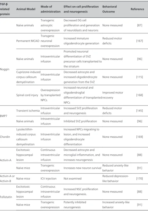
Due to their pleiotropic actions, TGF-β superfamily proteins have been investigated as potential treatments for a variety of CNS injuries, and several studies have demonstrated potential uses for these cytokines as therapeutic molecules (see Table 3). They have also provided insights into the action of these molecules as regulators of neural stem/progenitor cell (NSPC) proliferation and differentiation, with respect to both endogenous and transplant‐ ed stem cell populations.
TGF-β related protein
Animal Model Mode of administration
Effect on cell proliferation and neurogenesis
Behavioral
Outcome Reference
TGFβ-1
Transient ischemiaIntranasal aerosol spray
Decreased NSC proliferation and induce the number of DCX expressing neuronal precursors
Reduced Neurological Severity Score deficits [164]
Adrenalectomy Intraventricular infusion
Decreased the percentage of dividing cells which co-express PSA-NCAM in the DG
None measured [163]
Adrenalectomy Adenoviral overexpression
Increased NSC proliferation
and neurogenesis in the SVZ None measured [165] Prenatal LPS
inflammation
Adult adenoviral overexpression
Inhibited chronic microglial activation and restored neurogenesis
None measured [166]
Naïve animals
Injected into the cerebrospinal fluid
Number of proliferating cells in the hippocampus and in the lateral ventricle wall is substantially reduced, fewer neuronal precursor cells
TGF-β related protein
Animal Model Mode of administration
Effect on cell proliferation and neurogenesis Behavioral Outcome Reference Naïve animals Transgenic astrocytic overexpression Decreased DG cell proliferation and generation of neuroblasts and neurons
None measured [87] Noggin Permanent MCAO Transgenic neuronal overexpression Increased immature oligodendrocyte generation Reduced motor deficits [167]
Naïve animals Intraventricular infusion
Promoted neuronal differentiation of SVZ precursor cells transplanted to the striatum None measured [96] Cuprizone-induced corpus callosum demyelination Intraventricular infusion
Decreased astrocyte and increased oligodendrocyte generation from the SVZ
None measured [115]
Spinal cord injury
Overexpression by transplanted NPCs.
Increased neuronal and oligodendroglial differentiation of transplanted NPCs Improved motor recovery [168] BMP7
Transient ischemiaIntraventricular infusion
Increased SVZ proliferation and neurogenesis
Reduced motor
deficits [145] Naïve animals Intraventricular
infusion Inhibited SVZ proliferation None measured [96]
Chordin Lysolectithin-induced corpus callosum demyelination Intraventricular infusion Increased NPCs migrating to lesion, and increased oligodendrocyte differentiation None measured [169] Activin-A Excitotoxic hippocampal lesion Continuous intraventricular infusion
Decreased astrocyte and microglial inflammation, and increases neurogenesis
None measured [88]
Naïve mice Transgenic
overexpression Increases new neuron survival
Reduced anxiety-like behavior [91] Activin-A or
Activin-B Naïve mice ICV injection Not examined
Reduced depression-like behavior [170] Follistatin Excitotoxic hippocampal lesion Continuous intraventricular infusion Increased NSC proliferation
and neurogenesis. None measured [88] Naïve mice Transgenic
overexpression
Potently inhibited neurogenesis
Increased anxiety-like behavior [91]
5.1. TGF-β1
TGF-β1 treatment improves the outcome in several models of injury as it is strongly neuroprotective [76, 133, 171, 172] and in certain circumstances can promote neurogenesis after injury. After middle cerebral artery occlusion (MCAO) in mice, intranasal treatment with TGF-β1 increases the number of proliferative DCX-positive neural progenitors and the number of new neurons in the SVZ and striatum, while decreasing the fraction of prolifera‐ tive cells that express GFAP [119]. After adrenalectomy, TGF-β also stimulates neurogene‐ sis. TGF-β1 expression is upregulated and is necessary for the increased rates of neurogenesis in the SVZ and DG caused by adrenalectomy [163]. In this model TGF-β mediated downre‐ gulation of microglial activation and proliferation may be partially responsible for the increased neurogenesis [163, 165]. TGF-β1 can also inhibit chronic microglial activation induced by prenatal LPS exposure, and ameliorate the LPS-mediated decrease in neurogen‐ esis [166] suggesting that the anti-inflammatory action of TGF-β participates in its pro-neurogenic effects. Conversely, in naïve animals intracerebroventricular infusion of TGF-β1 lowered the number of DCX-positive neuronal precursors in the neurogenic niches. This reduced level of proliferation in the TGF-β1 infused brains was strongly correlated with an increased accumulation of pSmad2 in Sox2/GFAP expressing cells of the SGZ [81]. Transgen‐ ic overexpression of TGF-β1 in naïve mice also leads to reduce neurogenesis [87]. The opposite effects of TGF-β1 in injured as compared to naïve animals illustrate the difficulty in assigning one specific role to TGF-β1 due to its context-dependent effects. Chronic inflammation, either after lesion or in neurodegenerative disease, provides a different environment for the consequences of TGF-β signaling. The anti-inflammatory actions of TGF-β can have an important role in influencing neurogenic processes, independent of direct effects on neural progenitor cells. Dysregulation of TGF-β signaling is being acknowledged as a potential source for chronic inflammation. Indeed, aberrant TGF-β signaling and consequent accumu‐ lation of activated microglia in the neurogenic regions may play an important role in the progression of Alzheimer’s disease [171, 173].
5.2. Activin
Recent studies have demonstrated a critical role for activin signaling as a modulator of adult neurogenesis [91] in addition to its well-established role as a neuroprotective molecule [174, 175]. After local excitotoxic injury to the hippocampus, ablating activin signaling by infusion of the activin inhibitor follistatin potently inhibits post-injury neurogenesis and exacerbates the inflammatory response of astrocytes and microglia. Conversely, infusion of activin-A facilitates neurogenesis and represses gliosis [88]. Perhaps related to its effects on neurogen‐ esis, activin can also regulate anxiety and depression-like behavior in rodents, and the activin pathway may be a useful therapeutic target for treating depression. Hippocampal infusion of activin-A or activin-B reduces measures of depression in a forced swim test, with a similar efficacy to that of the antidepressant fluoxetine [170]. Further, transgenic mice which overex‐ press activin-A, have decreased anxiety measures in spontaneous place preference tests, while mice which overexpress follistatin, display the reverse [91].
5.3. BMPs
In the naïve rodent, BMPs usually act to suppress neurogenesis in the SVZ and DG whereas the BMP inhibitor noggin promotes it [96]. In contrast, inhibition of BMP signaling by upre‐ gulation of the BMP inhibitor chordin after lysolecithin-induced demyelination of the corpus callosum, led to redirection of SVZ precursors away from a neuronal lineage towards that of oligodendrocytes [169]. This change in differentiation potential was accompanied by a change in the migration pattern of the SVZ precursors, away from the rostral migratory stream, and towards the corpus callosum. Injury-induced changes in expression of regulatory factors often alter the normal pattern of cell differentiation and migration [176, 177]. In a different model of demyelination, cuprizone-induced upregulation of BMP-4 resulted in more SVZ precursors becoming astrocytes, with a concomitant reduction in the number of mature oligodendrocytes [115]. Intraventricular infusion of noggin in this model increased the generation of oligoden‐ drocytes from the SVZ [115] illustrating that inhibition of BMP signaling has the potential to promote remyelination in models of multiple sclerosis. The astrogliogenic potential of BMP has been demonstrated in multiple studies, where various precursors are pushed towards the astrocytic lineage [168, 178]. This is also true with transplanted neural stem cells or mesen‐ chymal stem cells, where BMPs around the implantation site push the transplanted cells towards astrocytes [179]. If these cells are being used to enhance repair after spinal cord or TBI, inhibition of BMP becomes an attractive option to promote neuronal or oligodendrocyte differentiation rather than that of astrocytes. In contrast to all these studies, one group has shown that BMP-7 has neuroprotective properties which may enhance the survival of imma‐ ture neurons [142, 180]. In one study, infusion of BMP-7 into the lateral ventricles of rats 24 hours after transient MCAO led to increased numbers of proliferating NSCs and more mature neurons generated in the SVZ while also facilitating behavioral recovery [145]. However, a different group has shown that transgenic expression of the BMP-inhibitor noggin in neurons after permanent MCAO in the mouse enhances functional recovery [167]. These conflicting data illustrate the sometimes confusing nature of the literature whereby BMP effects, similar to those of TGF-β are extremely contextual and are dependent on the exact model used. Overall, although some BMPs may have neuroprotective properties, the vast majority of the literature supports the view that BMP induction after injury is not beneficial for recovery, and that inhibition of BMP signaling may have therapeutic potential.
6. Future therapeutic strategies
In spite of extensive research in the field of brain injury or stroke, there is little effective treatment for these injuries [182]. Many of the neuroprotective treatments that have been successful in rodents have failed in clinical trials [183]. Harnessing the regenerative capaci‐ ty of the adult brain is one strategy for repairing and replacing injured tissue, together with enhancing neurotrophic support of existing neurons to promote survival [184, 185]. A complementary strategy also under development is transplantation of neural stem cells or committed progenitors into the lesion. However, when multipotent NSCs were implanted
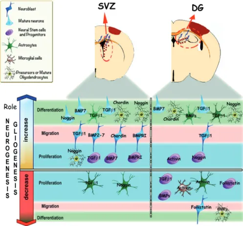
Figure 3. Modulation of neurogenesis and gliogenesis after adult brain injury by members of the TGF-β cyto‐ kine superfamily. In the top panel, the dentate gyrus (DG) in the hippocampus and subventricular zone (SVZ) of the
lateral ventricles are shown after damage to the cerebral cortex. Note the proliferation and migration of cells from the SVZ and DG towards the infarcted area (blue arrows). Red dots represent proliferating and migrating neural stem cells and progenitors cells (NSPCs) located in these neurogenic regions. In the bottom panel, the role of TGF-β proteins at different stages of neurogenesis or gliogenesis after adult brain injury is illustrated. Proliferation, migration or differ‐ entiation are induced or inhibited by growth factors, such as: TGF-β, BMPs proteins, Activin, Follistatin or Noggin. After injury to the brain, TGF-β1 can increase proliferation of NSPCs and induce the differentiation of neuroblasts into neu‐ rons within the SVZ, [119]. BMP7 can induce neural stem cell proliferation, neuronal migration and differentiation [145]; other BMPs proteins (BMP2-7) also can stimulate neuronal migration [94]. The BMP inhibitor proteins noggin and chordin promote NSPC migration and oligodendrocyte proliferation and differentiation, while decreasing astro‐ cyte proliferation [115, 169]. After injury to the brain, within the DG TGF-β1 can reduce the proliferation of immature neurons while increasing neuronal migration and differentiation [165, 166]. BMP7 can enhance NSPC proliferation and neuronal differentiation [96, 145]. Noggin can also increase NSPC proliferation [169]. Generally, BMPs can in‐ crease astroglial differentiation and inhibit oligodendrocyte generation, and the BMP inhibitors Chordin and Noggin can facilitate oligodendrocyte differentiation and proliferation [181]. Activin can induce NSPCs proliferation, and de‐ crease microglial and astroglial proliferation. The activin antagonist, follistatin, reduces proliferating NSPCs and mi‐ grating neuroblasts [88]. In summary, the proliferation, migration and differentiation of cells in the SVZ and the DG may be influenced by the spatial and temporal expression profile of these TGF-β proteins after brain injury.
directly into non-neurogenic regions in the injured brain, such as in the cortex or striatum, they failed to generate neurons but instead generated glial cells [186, 187]. Endogenous neural progenitors also are limited in their differentiation potential, presumably because the post-lesion environment is one that supports glial differentiation in preference to that of neu‐ rons [188]. As TGF-β family members can promote astrogliogenesis [189, 190], it would seem that in some circumstances, inhibition of specific cytokine signals would increase neuronal differentiation. A further consideration for repair and neuronal survival is promotion of oligodendrocyte survival and differentiation, since remyelination is critical to continued survival and function of many neurons. Inhibition of BMP action through infusion of noggin can promote oligodendrocyte differentiation after demyelination [115]. Inflammation after injury is yet one more factor that alters the environment for regeneration. Although often thought of as a short-lived phenomenon, there can be longer lasting inflammatory changes that persist months after injury [191]. One of the major problems with development of members of the TGF-β superfamily or their inhibitors for therapeutic use are the pleiotrop‐ ic nature of their effects. Thus TGF-β1 itself is neuroprotective and anti-inflammatory, which should promote recovery, but it inhibits proliferation of precursors, and also promotes development of the glial scar through upregulation of many extracellular matrix mole‐ cules, and through enhancing the migration of astrocytes [128, 192].
These cytokines act in a context dependent and concentration dependent manner, which adds an additional layer of complexity. To develop better therapeutic strategies we need a deeper understanding of the mechanisms through which the many actions of each cytokine are mediated. We may then be able to target specific molecules in the downstream signaling pathways, to avoid the pleiotropic effects that are emblematic of the activity of this cytokine family.
Acknowledgements
This study was supported by grant from the Center for Neuroscience and Regenerative Medicine (CNRM). SV is supported by a CNRM postdoctoral fellowship. The opinions and assertions contained herein are the private opinions of the authors and are not to be construed as reflecting the views of the Uniformed Services University of the Health Sciences or the US Department of Defense.
Author details
Sonia Villapol, Trevor T. Logan and Aviva J. Symes
Department of Pharmacology and Center for Neuroscience and Regenerative Medicine, Uni‐ formed Services University of the Health Sciences, Bethesda, MD, USA
References
[1] Berciano J, Lafarga M, Berciano M: Santiago Ramon y Cajal. Neurologia 2001, 16(3): 118-121.
[2] Alvarez-Buylla A: Mechanism of neurogenesis in adult avian brain. Experientia 1990, 46(9):948-955.
[3] Parent JM: Injury-induced neurogenesis in the adult mammalian brain. Neuroscientist 2003, 9(4):261-272.
[4] Whitney NP, Eidem TM, Peng H, Huang Y, Zheng JC: Inflammation mediates vary‐ ing effects in neurogenesis: relevance to the pathogenesis of brain injury and neuro‐ degenerative disorders. J Neurochem 2009, 108(6):1343-1359.
[5] Hagg T: Molecular regulation of adult CNS neurogenesis: an integrated view. Trends
Neurosci 2005, 28(11):589-595.
[6] Kronenberg G, Reuter K, Steiner B, Brandt MD, Jessberger S, Yamaguchi M, Kemper‐ mann G: Subpopulations of proliferating cells of the adult hippocampus respond dif‐ ferently to physiologic neurogenic stimuli. J Comp Neurol 2003, 467(4):455-463. [7] Mackowiak M, Chocyk A, Markowicz-Kula K, Wedzony K: Neurogenesis in the
adult brain. Pol J Pharmacol 2004, 56(6):673-687.
[8] Massague J, Cheifetz S, Ignotz RA, Boyd FT: Multiple type-beta transforming growth factors and their receptors. J Cell Physiol Suppl 1987, Suppl 5:43-47.
[9] Massague J: Receptors for the TGF-beta family. Cell 1992, 69(7):1067-1070.
[10] Wrighton KH, Feng XH: To (TGF)beta or not to (TGF)beta: fine-tuning of Smad sig‐ naling via post-translational modifications. Cell Signal 2008, 20(9):1579-1591.
[11] Umulis D, O'Connor MB, Blair SS: The extracellular regulation of bone morphogenet‐ ic protein signaling. Development 2009, 136(22):3715-3728.
[12] Miyazono K, Maeda S, Imamura T: BMP receptor signaling: transcriptional targets, regulation of signals, and signaling cross-talk. Cytokine Growth Factor Rev 2005, 16(3): 251-263.
[13] Ross S, Hill CS: How the Smads regulate transcription. Int J Biochem Cell Biol 2008, 40(3):383-408.
[14] Heldin CH, Moustakas A: Role of Smads in TGFbeta signaling. Cell Tissue Res 2012, 347(1):21-36.
[15] Derynck R, Feng XH: TGF-beta receptor signaling. Biochim Biophys Acta 1997, 1333(2):F105-150.
[16] Derynck R, Zhang YE: Smad-dependent and Smad-independent pathways in TGF-beta family signalling. Nature 2003, 425(6958):577-584.
[17] Kandasamy M, Reilmann R, Winkler J, Bogdahn U, Aigner L: Transforming Growth Factor-Beta Signaling in the Neural Stem Cell Niche: A Therapeutic Target for Hun‐ tington's Disease. Neurol Res Int 2011, 2011:124256.
[18] Moustakas A, Heldin CH: The regulation of TGFbeta signal transduction. Develop‐
ment 2009, 136(22):3699-3714.
[19] Mehler MF, Mabie PC, Zhang D, Kessler JA: Bone morphogenetic proteins in the nervous system. Trends Neurosci 1997, 20(7):309-317.
[20] Walsh DW, Godson C, Brazil DP, Martin F: Extracellular BMP-antagonist regulation in development and disease: tied up in knots. Trends Cell Biol 2010, 20(5):244-256. [21] Kang JS, Liu C, Derynck R: New regulatory mechanisms of TGF-beta receptor func‐
tion. Trends Cell Biol 2009, 19(8):385-394.
[22] Kretzschmar M, Massague J: SMADs: mediators and regulators of TGF-beta signal‐ ing. Curr Opin Genet Dev 1998, 8(1):103-111.
[23] Vogt J, Traynor R, Sapkota GP: The specificities of small molecule inhibitors of the TGFss and BMP pathways. Cell Signal 2011, 23(11):1831-1842.
[24] de Caestecker M: The transforming growth factor-beta superfamily of receptors. Cy‐
tokine Growth Factor Rev 2004, 15(1):1-11.
[25] Zhang YE: Non-Smad pathways in TGF-beta signaling. Cell Res 2009, 19(1):128-139. [26] Saj A, Lai EC: Control of microRNA biogenesis and transcription by cell signaling
pathways. Curr Opin Genet Dev 2011, 21(4):504-510.
[27] Davis BN, Hilyard AC, Nguyen PH, Lagna G, Hata A: Smad proteins bind a con‐ served RNA sequence to promote microRNA maturation by Drosha. Mol Cell 2010, 39(3):373-384.
[28] Wharton K, Derynck R: TGFbeta family signaling: novel insights in development and disease. Development 2009, 136(22):3691-3697.
[29] Larsson J, Goumans MJ, Sjostrand LJ, van Rooijen MA, Ward D, Leveen P, Xu X, ten Dijke P, Mummery CL, Karlsson S: Abnormal angiogenesis but intact hematopoietic potential in TGF-beta type I receptor-deficient mice. Embo J 2001, 20(7):1663-1673. [30] Oshima M, Oshima H, Taketo MM: TGF-beta receptor type II deficiency results in
defects of yolk sac hematopoiesis and vasculogenesis. Dev Biol 1996, 179(1):297-302. [31] Hill CR, Sanchez NS, Love JD, Arrieta JA, Hong CC, Brown CB, Austin AF, Barnett
JV: BMP2 signals loss of epithelial character in epicardial cells but requires the Type III TGFbeta receptor to promote invasion. Cell Signal 2012, 24(5):1012-1022.
[32] Shull MM, Ormsby I, Kier AB, Pawlowski S, Diebold RJ, Yin M, Allen R, Sidman C, Proetzel G, Calvin D et al: Targeted disruption of the mouse transforming growth factor-beta 1 gene results in multifocal inflammatory disease. Nature 1992, 359(6397): 693-699.
[33] Kulkarni AB, Huh CG, Becker D, Geiser A, Lyght M, Flanders KC, Roberts AB, Sporn MB, Ward JM, Karlsson S: Transforming growth factor beta 1 null mutation in mice causes excessive inflammatory response and early death. Proc Natl Acad Sci U S A 1993, 90(2):770-774.
[34] Sanford LP, Ormsby I, Gittenberger-de Groot AC, Sariola H, Friedman R, Boivin GP, Cardell EL, Doetschman T: TGFbeta2 knockout mice have multiple developmental defects that are non-overlapping with other TGFbeta knockout phenotypes. Develop‐
ment 1997, 124(13):2659-2670.
[35] Huang S, Tang B, Usoskin D, Lechleider RJ, Jamin SP, Li C, Anzano MA, Ebendal T, Deng C, Roberts AB: Conditional knockout of the Smad1 gene. Genesis 2002, 32(2): 76-79.
[36] Tremblay KD, Dunn NR, Robertson EJ: Mouse embryos lacking Smad1 signals dis‐ play defects in extra-embryonic tissues and germ cell formation. Development 2001, 128(18):3609-3621.
[37] Heyer J, Escalante-Alcalde D, Lia M, Boettinger E, Edelmann W, Stewart CL, Kucher‐ lapati R: Postgastrulation Smad2-deficient embryos show defects in embryo turning and anterior morphogenesis. Proc Natl Acad Sci U S A 1999, 96(22):12595-12600. [38] Datto MB, Frederick JP, Pan L, Borton AJ, Zhuang Y, Wang XF: Targeted disruption
of Smad3 reveals an essential role in transforming growth factor beta-mediated sig‐ nal transduction. Mol Cell Biol 1999, 19(4):2495-2504.
[39] Yang X, Letterio JJ, Lechleider RJ, Chen L, Hayman R, Gu H, Roberts AB, Deng C: Targeted disruption of SMAD3 results in impaired mucosal immunity and diminish‐ ed T cell responsiveness to TGF-beta. Embo J 1999, 18(5):1280-1291.
[40] Colak D, Mori T, Brill MS, Pfeifer A, Falk S, Deng C, Monteiro R, Mummery C, Sommer L, Gotz M: Adult neurogenesis requires Smad4-mediated bone morphogen‐ ic protein signaling in stem cells. J Neurosci 2008, 28(2):434-446.
[41] Arnold SJ, Maretto S, Islam A, Bikoff EK, Robertson EJ: Dose-dependent Smad1, Smad5 and Smad8 signaling in the early mouse embryo. Dev Biol 2006, 296(1): 104-118.
[42] Chang H, Brown CW, Matzuk MM: Genetic analysis of the mammalian transforming growth factor-beta superfamily. Endocr Rev 2002, 23(6):787-823.
[43] Tojo M, Takebe A, Takahashi S, Tanaka K, Imamura T, Miyazono K, Chiba T: Smad7-deficient mice show growth retardation with reduced viability. J Biochem 2012, 151(6): 621-631.
[44] Huang Z, Wang D, Ihida-Stansbury K, Jones PL, Martin JF: Defective pulmonary vas‐ cular remodeling in Smad8 mutant mice. Hum Mol Genet 2009, 18(15):2791-2801. [45] Mishina Y, Suzuki A, Ueno N, Behringer RR: Bmpr encodes a type I bone morphoge‐
netic protein receptor that is essential for gastrulation during mouse embryogenesis.
Genes Dev 1995, 9(24):3027-3037.
[46] Yi SE, Daluiski A, Pederson R, Rosen V, Lyons KM: The type I BMP receptor BMPRIB is required for chondrogenesis in the mouse limb. Development 2000, 127(3): 621-630.
[47] Beppu H, Kawabata M, Hamamoto T, Chytil A, Minowa O, Noda T, Miyazono K: BMP type II receptor is required for gastrulation and early development of mouse embryos. Dev Biol 2000, 221(1):249-258.
[48] Zhang H, Bradley A: Mice deficient for BMP2 are nonviable and have defects in amn‐ ion/chorion and cardiac development. Development 1996, 122(10):2977-2986.
[49] Daluiski A, Engstrand T, Bahamonde ME, Gamer LW, Agius E, Stevenson SL, Cox K, Rosen V, Lyons KM: Bone morphogenetic protein-3 is a negative regulator of bone density. Nat Genet 2001, 27(1):84-88.
[50] Winnier G, Blessing M, Labosky PA, Hogan BL: Bone morphogenetic protein-4 is re‐ quired for mesoderm formation and patterning in the mouse. Genes Dev 1995, 9(17): 2105-2116.
[51] King JA, Marker PC, Seung KJ, Kingsley DM: BMP5 and the molecular, skeletal, and soft-tissue alterations in short ear mice. Dev Biol 1994, 166(1):112-122.
[52] Solloway MJ, Dudley AT, Bikoff EK, Lyons KM, Hogan BL, Robertson EJ: Mice lack‐ ing Bmp6 function. Dev Genet 1998, 22(4):321-339.
[53] Dupe V, Ghyselinck NB, Thomazy V, Nagy L, Davies PJ, Chambon P, Mark M: Es‐ sential roles of retinoic acid signaling in interdigital apoptosis and control of BMP-7 expression in mouse autopods. Dev Biol 1999, 208(1):30-43.
[54] Wawersik S, Purcell P, Rauchman M, Dudley AT, Robertson EJ, Maas R: BMP7 acts in murine lens placode development. Dev Biol 1999, 207(1):176-188.
[55] Dudley AT, Lyons KM, Robertson EJ: A requirement for bone morphogenetic pro‐ tein-7 during development of the mammalian kidney and eye. Genes Dev 1995, 9(22): 2795-2807.
[56] Luo G, Hofmann C, Bronckers AL, Sohocki M, Bradley A, Karsenty G: BMP-7 is an inducer of nephrogenesis, and is also required for eye development and skeletal pat‐ terning. Genes Dev 1995, 9(22):2808-2820.
[57] Galloway SD, Maughan RJ: The effects of substrate and fluid provision on thermo‐ regulatory and metabolic responses to prolonged exercise in a hot environment. J
[58] Ying Y, Liu XM, Marble A, Lawson KA, Zhao GQ: Requirement of Bmp8b for the generation of primordial germ cells in the mouse. Mol Endocrinol 2000, 14(7): 1053-1063.
[59] Yan C, Wang P, DeMayo J, DeMayo FJ, Elvin JA, Carino C, Prasad SV, Skinner SS, Dunbar BS, Dube JL et al: Synergistic roles of bone morphogenetic protein 15 and growth differentiation factor 9 in ovarian function. Mol Endocrinol 2001, 15(6):854-866. [60] Ray BN, Lee NY, How T, Blobe GC: ALK5 phosphorylation of the endoglin cytoplas‐ mic domain regulates Smad1/5/8 signaling and endothelial cell migration. Carcino‐
genesis 2010, 31(3):435-441.
[61] Li DY, Sorensen LK, Brooke BS, Urness LD, Davis EC, Taylor DG, Boak BB, Wendel DP: Defective angiogenesis in mice lacking endoglin. Science 1999, 284(5419): 1534-1537.
[62] Mishina Y, Crombie R, Bradley A, Behringer RR: Multiple roles for activin-like kin‐ ase-2 signaling during mouse embryogenesis. Dev Biol 1999, 213(2):314-326.
[63] Matzuk MM, Kumar TR, Shou W, Coerver KA, Lau AL, Behringer RR, Finegold MJ: Transgenic models to study the roles of inhibins and activins in reproduction, onco‐ genesis, and development. Recent Prog Horm Res 1996, 51:123-154; discussion 155-127. [64] Ferguson CA, Tucker AS, Christensen L, Lau AL, Matzuk MM, Sharpe PT: Activin is an essential early mesenchymal signal in tooth development that is required for pat‐ terning of the murine dentition. Genes Dev 1998, 12(16):2636-2649.
[65] Vassalli A, Matzuk MM, Gardner HA, Lee KF, Jaenisch R: Activin/inhibin beta B sub‐ unit gene disruption leads to defects in eyelid development and female reproduction.
Genes Dev 1994, 8(4):414-427.
[66] Brunet LJ, McMahon JA, McMahon AP, Harland RM: Noggin, cartilage morphogene‐ sis, and joint formation in the mammalian skeleton. Science 1998, 280(5368):1455-1457. [67] Matzuk MM, Lu N, Vogel H, Sellheyer K, Roop DR, Bradley A: Multiple defects and
perinatal death in mice deficient in follistatin. Nature 1995, 374(6520):360-363. [68] Emsley JG, Mitchell BD, Kempermann G, Macklis JD: Adult neurogenesis and repair
of the adult CNS with neural progenitors, precursors, and stem cells. Prog Neurobiol 2005, 75(5):321-341.
[69] Kazanis I, Lathia J, Moss L, ffrench-Constant C: The neural stem cell microenviron‐ ment. In: StemBook. Cambridge (MA); 2008.
[70] Ihrie RA, Alvarez-Buylla A: Lake-front property: a unique germinal niche by the lat‐ eral ventricles of the adult brain. Neuron 2011, 70(4):674-686.
[71] Ming GL, Song H: Adult neurogenesis in the mammalian brain: significant answers and significant questions. Neuron 2011, 70(4):687-702.
[72] Vivien D, Ali C: Transforming growth factor-beta signalling in brain disorders. Cyto‐
kine Growth Factor Rev 2006, 17(1-2):121-128.
[73] ten Dijke P, Arthur HM: Extracellular control of TGFbeta signalling in vascular de‐ velopment and disease. Nat Rev Mol Cell Biol 2007, 8(11):857-869.
[74] Flanders KC, Ludecke G, Engels S, Cissel DS, Roberts AB, Kondaiah P, Lafyatis R, Sporn MB, Unsicker K: Localization and actions of transforming growth factor-beta s in the embryonic nervous system. Development 1991, 113(1):183-191.
[75] Unsicker K, Meier C, Krieglstein K, Sartor BM, Flanders KC: Expression, localization, and function of transforming growth factor-beta s in embryonic chick spinal cord, hindbrain, and dorsal root ganglia. J Neurobiol 1996, 29(2):262-276.
[76] Dobolyi A, Vincze C, Pal G, Lovas G: The neuroprotective functions of transforming growth factor Beta proteins. Int J Mol Sci 2012, 13(7):8219-8258.
[77] Flanders KC, Ren RF, Lippa CF: Transforming growth factor-betas in neurodegenera‐ tive disease. Prog Neurobiol 1998, 54(1):71-85.
[78] Blottner D, Wolf N, Lachmund A, Flanders KC, Unsicker K: TGF-beta rescues target-deprived preganglionic sympathetic neurons in the spinal cord. Eur J Neurosci 1996, 8(1):202-210.
[79] De Groot CJ, Montagne L, Barten AD, Sminia P, Van Der Valk P: Expression of trans‐ forming growth factor (TGF)-beta1, -beta2, and -beta3 isoforms and TGF-beta type I and type II receptors in multiple sclerosis lesions and human adult astrocyte cul‐ tures. J Neuropathol Exp Neurol 1999, 58(2):174-187.
[80] Miller MW: Expression of transforming growth factor-beta in developing rat cerebral cortex: effects of prenatal exposure to ethanol. J Comp Neurol 2003, 460(3):410-424. [81] Wachs FP, Winner B, Couillard-Despres S, Schiller T, Aigner R, Winkler J, Bogdahn
U, Aigner L: Transforming growth factor-beta1 is a negative modulator of adult neu‐ rogenesis. J Neuropathol Exp Neurol 2006, 65(4):358-370.
[82] Logan V, Symes: TGF-β superfamily gene expression and induction of the Runx1 transcription factor in adult neurogenic regions after brain injury. Submitted 2012. [83] Werry EL, Enjeti S, Halliday GM, Sachdev PS, Double KL: Effect of age on prolifera‐
tion-regulating factors in human adult neurogenic regions. J Neurochem 2010, 115(4): 956-964.
[84] Artegiani B, Calegari F: Age-related cognitive decline: can neural stem cells help us?
Aging (Albany NY) 2012, 4(3):176-186.
[85] He Y, Yung A, Wyss-Coray T: TGF-β signaling in newborn neurons is required for their survival and maturation in the adult dentate gyrus. In: Society for Neuroscience:
![Figure 3. Igf-1 signalling in adult rat hippocampal neurospheres [81].](https://thumb-eu.123doks.com/thumbv2/123doknet/5801894.139796/55.659.258.403.102.385/figure-igf-signalling-in-adult-rat-hippocampal-neurospheres.webp)
