O
pen
A
rchive
T
OULOUSE
A
rchive
O
uverte (
OATAO
)
OATAO is an open access repository that collects the work of Toulouse researchers and makes it freely available over the web where possible.This is an author-deposited version published in : http://oatao.univ-toulouse.fr/ Eprints ID : 16134
To cite this version : Merlo, Adlan and Duru, Paul and Lorthois,
Sylvie Study of the phase separation effect in capillary-size micro-channels. (2016) In: 5th Micro and Nano Flows Conference, 11 September 2016 - 14 September 2016 (Milan, Italy). (Unpublished)
Any correspondence concerning this service should be sent to the repository administrator: staff-oatao@listes-diff.inp-toulouse.fr
Study of the phase separation effect in capillary-size micro-channels
Adlan MERLO 2,1*, Paul DURU 1,2, Sylvie LORTHOIS 2,1* Corresponding author: adlan.merlo@imft.fr
1 Université de Toulouse; INPT, UPS; IMFT (Institut de Mécanique des Fluides de Toulouse), France 2 CNRS; IMFT; Toulouse, France
Abstract It has been known for more than 50 years that the distribution of red blood cells (RBCs) in
microvessel networks is highly heterogeneous. Yet, the phase separation effect (PS), i.e the non-proportional distribution of RBCs between the two daughter branches of a simple divergent bifurcation, is still poorly understood especially when RBC concentration (tube hematocrit) reaches high values (> 20%) and when vessel diameter is close to that of RBCs' (~8µm), as often encountered in microcirculation. The most commonly used PS law has been empirically derived in vivo by Pries et al. [1] but its validity has been recently questioned [2]. Therefore, experimental data obtained in controlled conditions are needed. We have designed squared micro-channel bifurcations, with side ranging from 5 to 20 microns, and adapted the injection geometry. Associated to a new method for the measurement of high tube hematocrit values, this allows for an in vitro systematic investigation of the parameters influencing the PS (tube hematocrit, confinement, bifurcation geometry). Our results are compared with Pries' PS law.
Keywords: Microcirculation, Red Blood Cells, Phase Separation Effect
1. Introduction
RBCs carry oxygen throughout the whole body. Exchanges with the tissues take place mainly in networks of capillary or slightly larger vessels, which diameters are comprised between ~3µm and ~20µm. At rest, RBCs have a biconcave shape of ~8µm in diameter and a thickness of ~2µm. They are able to deform enough to squeeze into the tiniest capillaries. Capillary vessels form dense, complex 3D networks [3]. RBC distribution within these structures exhibits strong spatio-temporal heterogeneities [4,5]. To better understand that behavior, one needs to understand what happens in the simplest component of a network: one simple divergent bifurcation. RBCs split in a non-proportional fashion between the two daughter branches, with respect to total flow fractionation [1]. This phenomenon, called phase separation (PS), is influenced by numerous parameters: RBC to mother branch diameter ratio, daughters to mother diameter ratio, daughters to mother branch flow rate ratio, concentration of RBCs (tube hematocrit) in the mother branch, and the hematocrit profile within this
branch [1]. Yet, no study of the PS has been conducted in such small vessels, in controlled conditions, as pointed out in recent numerical works investigating the distribution of RBCs in realistic or simplified microvascular networks [2,5]. Therefore our aim is to provide reference in vitro experimental data of PS in channels of diameters close to that of the RBCs.
2. Material and Methods
2.1 RBCs suspensions and microfluidic chips
RBCs are centrifugated from whole human blood, washed and suspended in a density-matching solution [6].
Microfluidic chips are fabricated by multiple mask soft lithography, resulting in squared cross-sections of 5, 10 or 20 microns [7] for the channels in the bifurcation, with an improved injection geometry. The chips are composed of a PDMS matrix bounded on a glass slide for microscopic observation and channels enclosure. Different pressure values are imposed at inlet and outlets, thus enabling
to control the flow rate partition between both daughter branches, with a good stability over long periods of time (~45min) and for a wide range of tube hematocrits (~1.5 to ~30%).
2.2 Tube hematocrit and velocity profiles
When a dilute suspension of RBCs flows in a channel, one can count them to obtain a mean hematocrit value. However, this gives no information about the hematocrit profile and it is most often impossible to proceed that way as hematocrit increases. Studies have taken advantage of the Beer-Lambert law [8]. To implement this method, a rigorous and tedious calibration is needed [8]. We have developed a simpler calibration using glass chambers with calibrated thickness of 10 and 20 microns (Leja slides). The idea is to relate the precisely known hematocrit of a prepared RBC suspension to the mean temporal attenuation of light in the depth of the chamber over a group of pixels. For that purpose, a drop of ~2µl is put at the entrance of the chamber which immediately fills by capillarity. After filling, due to lateral migration in the depth of the chamber, RBC distribution is not uniform throughout the chamber. Being of weakly inertial nature, this effect has been counteracted by partially filling its outlet with viscous oil prior to injection. As a consequence, the filling speed was tremendously lowered, allowing to achieve the uniform RBC distribution needed to perform calibration. We found a linear relation between hematocrit and light intensity reduction, up to a plateau value. The linear coefficient is proportional to the chamber depth. Calibration was performed with the same microscope and objectives as the ones used for the PS study in micro-channels.
Velocity profiles have been obtained using the dual slit method [6]. This method, based on temporal correlation between grey levels profiles across the channel, yields the maximal velocity profile of the RBCs, when implemented as shown in [6].
3. Results
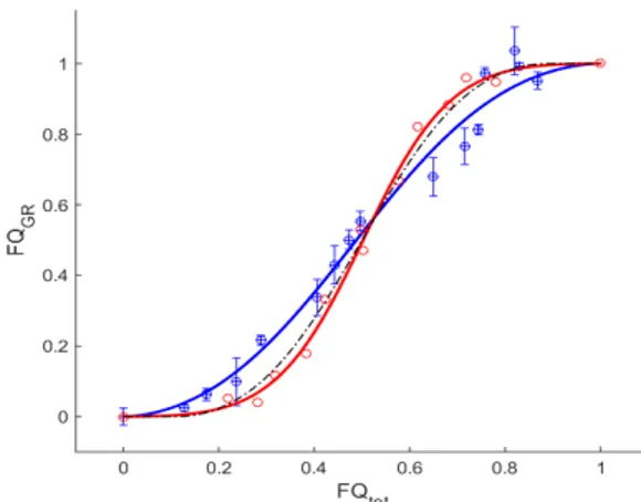
To validate the above methods, we quantified the apparent deviation to mass conservation due to experimental uncertainties and demonstrated values below 15% for all our experimental conditions. Figure 1 displays the results obtained in a symmetric T-shaped bifurcation, with three branches of equal side length (10µm), for dilute (1.5%) and concentrated (25%) suspensions. As in [1], the RBC flow fraction entering one of the daughter branches is plotted against the total blood flow fraction entering that branch. The results obtained are consistent with Pries' PS law when the parameters are rescaled to take into account the difference in size between human and rat RBCs [3]. A systematic analysis of the influence of hematocrit, absolute flow rate in the inlet branch and bifurcation geometry on the parameters of this law is under progress.
References
[1] Pries et al., Microvascular Res. 38, 81-101, 1989 [2] Gould and Linninger, Microcirculation 22, 1-18, 2015 [3] Lorthois et al., Neuroimage 54, 1031-1042, 2011 [4] Desjardins et al., Neurobiol. Aging 37, 1947-1955, 2014 [5] Obrist et al., J. Royal Soc. Interface 368, 2897-2918, 2010 [6] Roman et al., Microvascular Res. 84, 249-261, 2012 [7] Roman et al., Biomicrofluidics 10, 034103, 2016
[8] Pries et al., Am. J. Physiol. 245 (Heart Circ. Physiol. 14), H167-H177, 1983
Fig. 1. PS in a 10µm symmetric T-shaped bifurcation. Mean tube hematocrit in mother branch: ~1.5% (red) ~25% (blue). Error-bars : apparent deviation to mass conservation (not shown in red for clarity). Continuous lines: least-squares fits to Pries' PS law. Discontinuous line : Prediction from Pries PS in 10 µm-diameter vessels in the limit of small hematocrits.
